
Review - UZ Leuven
19
251 ISSN 1479-6678 Future Cardiol. (2012) 8(2), 251–269 Future Cardiology part of 10.2217/FCA.12.10 © 2012 Marc Gewillig Diseases of the aorta predominantly include abnormal narrowing but also dilation. Coarctation of the aorta includes a wide array of anatomic and pathophysiologic variations. It occurs with a frequency of 0.04% of live births and comprises approximately 7% of known congenital heart disease, making it the fourth most common lesion requiring surgical or catheter-based intervention [1] . Indications for treatment Narrowing of the aortic arch predisposes patients to high blood pressure and subsequent carotid intima–media thickness. Long-term follow-up studies even after coarctectomy have shown significant morbidity and mortality beyond the fourth decade, usually secondary to, or associated with, arterial hypertension [2] . This includes premature atherosclerosis, which might lead to myocardial infarction and stroke at younger age. These data suggest that the clini- cian should always aim for an aortic arch free of any gradient in order to avoid arterial hyperten- sion and improve long-term outcome in these patients. Approximately 3–4% of patients with coarctation have an aberrant right subclavian artery arising from the descending aorta distal to the coarctation site, thus necessitating pressure readings in both arms. A blood pressure gra- A blood pressure gra- blood pressure gra- dient, taken at rest, of greater than 20 mmHg between upper and lower extremities generally indicates the need for intervention. Additionally, pressure differences of less than 20 mmHg may warrant intervention if there is hypertension at rest and especially, if there is left ventricular dysfunction or progressive hypertrophy. Lastly, exercise-induced hypertension is increasingly being recognized as another indication, as late outcome is also suboptimal [3] . Mechanism of pressure loss along the aorta Several lesions either isolated or in combination may cause a gradient in the aorta, each with specific opportunities and limitations for trans- catheter treatment: n A discrete narrowing, typically a coarctation; n A tubular hypoplasia: isthmus: distal from left subclavian artery; distal cross: between left carotid artery and left subclavian artery; prox- imal cross: between brachiocephalic trunk and left carotid artery; and lower thoracic or abdominal coarctation; n A steep angulation as seen in a cervical arch (between ascending aorta and proximal cross); n Any tortuous course of the arch; n An atretic segment; when acquired later in life typically the isthmus between the left subclavian artery and the coarctation site; n A postsurgical narrowing, which may consist of residual hypoplasia or stenosis, lack of (catch-up) growth of bordering segments, excessive scar tissue, a circular nonresorbable wire or tube interposition; n A residual, recurrent or growth-induced narrowing after catheter intervention. Treatment of coarctation In 1944 Robert Gross and Clarence Crafoord independently performed the first successful Percutaneous interventions of the aorta Marc Gewillig* 1 , Werner Budts 2 , Derize Boshoff 1 & Geert Maleux 3 1 Pediatric Cardiology, University of Leuven, Belgium 2 Congenital Cardiology, University of Leuven, Belgium 3 Interventional Radiology, University of Leuven, Belgium *Author for correspondence: [email protected] Coarctation of the aorta includes a wide array of anatomical and pathophysiological variations that may cause important long-term morbidity and mortality. Percutaneous techniques, such as balloon dilation and stenting, allow clinicians to safely decrease or abolish most gradients along the aorta, albeit with limitations. Proper patient selection and interventional technique allow clinicians to obtain an adequate stretch or therapeutic tear of the vessel wall, but should avoid complications, such as an excessive tear, dissection, aneurysm formation or rupture. The interventional technique is tailored by patient characteristics such as age, size and growth potential, by characteristics of the lesion such as degree of narrowing, length, angulation(s) and by local regulations. Keywords n aortic arch n balloon angioplasty n covered stent n graft stent n hypertension n stent Review For reprint orders, please contact: [email protected]
Transcript of Review - UZ Leuven

251ISSN 1479-6678Future Cardiol. (2012) 8(2), 251–269
Futu
re C
ard
iolo
gy
part of
10.2217/FCA.12.10 © 2012 Marc Gewillig
Diseases of the aorta predominantly include abnormal narrowing but also dilation. Coarctation of the aorta includes a wide array of anatomic and pathophysiologic variations. It occurs with a frequency of 0.04% of live births and comprises approximately 7% of known congenital heart disease, making it the fourth most common lesion requiring surgical or catheter-based intervention [1].
Indications for treatment Narrowing of the aortic arch predisposes patients to high blood pressure and subsequent carotid intima–media thickness. Long-term follow-up studies even after coarctectomy have shown significant morbidity and mortality beyond the fourth decade, usually secondary to, or associated with, arterial hypertension [2]. This includes premature atherosclerosis, which might lead to myocardial infarction and stroke at younger age. These data suggest that the clini-cian should always aim for an aortic arch free of any gradient in order to avoid arterial hyperten-sion and improve long-term outcome in these patients. Approximately 3–4% of patients with coarctation have an aberrant right subclavian artery arising from the descending aorta distal to the coarctation site, thus necessitating pressure readings in both arms. A blood pressure gra-A blood pressure gra- blood pressure gra-dient, taken at rest, of greater than 20 mmHg between upper and lower extremities generally indicates the need for intervention. Additionally, pressure differences of less than 20 mmHg may warrant intervention if there is hypertension at rest and especially, if there is left ventricular dysfunction or progressive hypertrophy. Lastly, exercise-induced hypertension is increasingly
being recognized as another indication, as late outcome is also suboptimal [3].
Mechanism of pressure loss along the aorta
Several lesions either isolated or in combination may cause a gradient in the aorta, each with specific opportunities and limitations for trans-catheter treatment:
nA discrete narrowing, typically a coarctation;
nA tubular hypoplasia: isthmus: distal from left subclavian artery; distal cross: between left carotid artery and left subclavian artery; prox-imal cross: between brachiocephalic trunk and left carotid artery; and lower thoracic or abdominal coarctation;
nA steep angulation as seen in a cervical arch (between ascending aorta and proximal cross);
nAny tortuous course of the arch;
nAn atretic segment; when acquired later in life typically the isthmus between the left subclavian artery and the coarctation site;
nA postsurgical narrowing, which may consist of residual hypoplasia or stenosis, lack of (catch-up) growth of bordering segments, excessive scar tissue, a circular nonresorbable wire or tube interposition;
nA residual, recurrent or growth-induced narrowing after catheter intervention.
Treatment of coarctation In 1944 Robert Gross and Clarence Crafoord independently performed the first successful
Percutaneous interventions of the aorta
Marc Gewillig*1, Werner Budts2, Derize Boshoff1 & Geert Maleux3
1Pediatric Cardiology, University of Leuven, Belgium2Congenital Cardiology, University of Leuven, Belgium3Interventional Radiology, University of Leuven, Belgium*Author for correspondence: [email protected]
Coarctation of the aorta includes a wide array of anatomical and pathophysiological variations that may cause important long-term morbidity and mortality. Percutaneous techniques, such as balloon dilation and stenting, allow clinicians to safely decrease or abolish most gradients along the aorta, albeit with limitations. Proper patient selection and interventional technique allow clinicians to obtain an adequate stretch or therapeutic tear of the vessel wall, but should avoid complications, such as an excessive tear, dissection, aneurysm formation or rupture. The interventional technique is tailored by patient characteristics such as age, size and growth potential, by characteristics of the lesion such as degree of narrowing, length, angulation(s) and by local regulations.
Keywords
n aortic arch n balloon angioplasty n covered stent n graft stent n hypertension n stent
Revie
wFor reprint orders, please contact: [email protected]
Future Cardiol. (2012) 8(2)252 future science group
surgical repairs of coarctation of the aorta. During the ensuing four decades, surgery remained the only treatment for coarctation. In the late 1970s, percutaneous balloon angioplasty was described as an alternative to surgical repair [4]. Since then, transcatheter interventions have become increasingly popular and in many cases have emerged as the treatment of choice [5,6].
Percutaneous options & mechanism of action
Transcatheter options now include simple angioplasty, angioplasty followed by stenting, primary stenting using bare-metal or covered stents and rescue stenting with covered stent.
By simple angioplasty, the minimum a bal-loon will do is stretch the targeted lesion; however, this will be followed by early and late recoil, making this technique rather inefficient. A lasting result of balloon dilation typically involves a therapeutic tear of the intima and at least partially the media until the adventitia; such a therapeutic tear should be contained to the narrowed zone [7–9]. In order to obtain an adequate tear with a lasting result, some degree of over dilation is frequently required. After dilation, some mechanisms will limit or improve the long-term result: recoil and retraction of nongrowing scar tissue will result in recurrence of the narrowing or stenosis, but catch-up growth and remodeling may occur by release of the stenotic ring and enhanced anterograde flow.
However, such tears predispose for pos-sible complications: dissection, false aneurysm and rupture [10]. A dissection is a tear that extends beyond the coarcted segment in the axial dimension, permitting contrast to track extraluminally in a proximal or distal direc-tion. The false lumen of such dissection may be progressive and cause distal tearing of the vessel wall or occlusion of side vessels. A false aneurysm is a defect in the aortic wall, with contrast beyond the presumed adventitial plane with a discrete length; such a false aneurysm in time may ‘grow’ and eventually tear. An aortic rupture is a frank disruption of the aortic wall, which appears angiographically as extravaza-tion of contrast beyond the confinement of the aorta into the mediastinum or pleural space. A relatively high incidence of aneurysm forma-tion of 2–20% has been reported after balloon dilation [11,12]. Several techniques have been described to improve the hemodynamic result but also limiting the risk for these complica-tions: these techniques involve low-pressure
balloon dilation-interrogation of the stenosed site, progressive and/or stepwise dilation with noncompliant balloons, limiting the size of the balloon based on the narrowing itself or adjacent segments.
Since 1989, bare-metal stents have been used to treat narrowings in the aorta [13–17]. Stents will neutralize many of the shortcomings of simple balloon dilation. A stent will expand and scaffold the targeted region, thereby avoiding recoil and residual or recurrent stenosis. A good result can be obtained by simple stretching of the wall without a tear; there is no need for overdilation. Stenting, therefore, results in less vessel wall complications: less aneurysms, no dissections within the stent as they are auto-matically contained by sealing the intimal flap and less rupture. Where intimal tears occur, the stent provides a surface for formation of neo-intima over the tear and reinforces the weakened areas within the aortic wall, which may later predispose to formation of a false aneurysm. The use of bare-metal stents improved results, further reducing the complication rate of vessel wall trauma to 1–5% [18]. Stents are particularly helpful in some postsurgical patients [19] where a nonresorbable wire was used: balloon dilation will primarily result in tearing the intima from the vessel with major dissection. Stent implan-tation has some shortcomings: the technique is technically more demanding and requires a bigger sheath (more vascular trauma at inser-tion point). The implantation of a foreign body automatically involves other interactions with surrounding tissues, alters local vascular compliance and impedes vessel growth [20].
Initially, stent implantation was only used for cases where surgery and balloon angioplasty had failed. However, as experience increased, stenting gradually became the treatment of choice in selected patients with aortic coarcta-tion [21]. This is especially the case when coarc-tation coexists with hypoplasia of the isthmus or transverse arch, or when balloon dilation tends to have a high failure rate (the stent pro-vides good results with stretching only). These and other morphologic variations such as a tor-tuous coarctation, long segment coarctation, or mild discrete coarctation are now considered candidates for primary stenting. In the adult patients, stenting is now being considered as the treatment of choice in any variant of aortic coarctation. At the other end of the scale, in children less than 10 years of age, it is preferable to avoid stenting, as several redilations may be required until the child is fully grown.
Review Gewillig, Budts, Boshoff & Maleux

www.futuremedicine.com 253future science group
The addition of covered stents has further reduced the incidence of intrastent aneurysm formation and vessel rupture to less than 1%: the covering will seal any tear in the vessel wall [22]. Covered stents also allow clinicians to exclude an unwanted passage to vessels such as an arterial duct or an existing aneurysm [23–25], and allow the creation of a new vessel segment as in aortic arch atresia [26]. Covered stents typi-cally will require larger sheaths (+1 Fr) than bare-metal stents, may unwantedly exclude side-vessels, and if embolized may be more hazardous than bare-metal stents. Currently the discussion is still open as to whether cov-ered stents should be used prophylactically or only in case of a proven complication or specific indication. In countries where covered stents are available (not in the USA due to US FDA restrictions), there is a clear shift towards more prophylactic use of covered stents in order to reduce the early and late complication rates even more [27].
The American Heart Association recently published guidelines for catheter interven-tion for coarctation of the aorta (excluding
indications for covered stents because of FDA restrictions) (see Table 1).
EquipmentBalloons for angioplastyMany balloons are currently available for angio-plasty of the aorta. Relevant differences between balloons are: availability in different sizes and lengths, tapering of balloons (the shorter the better, the nose of the balloon may cause lesions in the arch), compliance, profile upon entrance (determines sheath size for vessel entry), mode of refolding (determines sheath size for exit, dam-age at distal point of sheath), nominal pressure, burst pressure, in- and de-flation time, resis-tance to puncturing (stent or calcium), mode of rupture (punctiform, longitudinal, transverse), coating of balloon and shaft properties (stiff-ness, pushability, stretchability on retrieval). Every balloon manufacturer will make compro-mises on a combination of these features; as we treat different patients with different needs and priorities (predominantly sheath size), it is not uncommon to have different types of balloons available on the shelf at all times.
Table 1. American Heart Association guidelines for catheter intervention for coarctation of the aorta.
Class Recommendations for transcatheter balloon angioplasty of coarctation/recoarctation of the aorta
Recommendations for stent placement in native coarctation and recoarctation of the aorta
I Transcatheter systolic coarctation gradient of >20 mmHg and suitable anatomy, irrespective of patient age (level of evidence: C)
Recurrent coarctation patients of sufficient size for safe stent placement, in whom the stent can be expanded to an adult size, and who have a transcatheter systolic coarctation gradient >20 mmHg (level of evidence: B)Transcatheter systolic coarctation gradient of <20 mmHg in
the presence of significant collateral vessels and suitable angiographic anatomy, irrespective of patient age, as well as in patients with univentricular heart or with significant ventricular dysfunction (level of evidence: C)
IIa As a palliative measure to stabilize a patient when extenuating circumstances are present, such as severely depressed ventricular function, severe mitral regurgitation, low cardiac output or systemic disease affected by the cardiac condition (level of evidence: C)
Transcatheter systolic coarctation gradient of >20 mmHg (level of evidence: B)Transcatheter systolic coarctation gradient of <20 mmHg but with systemic hypertension associated with an anatomic narrowing that explains the hypertension (level of evidence: C)Long-segment coarctation with a transcatheter systolic coarctation gradient >20 mmHg (level of evidence: B)When balloon angioplasty has failed, as long as a stent that can be expanded to an adult size can be implanted (level of evidence: B)
IIb Beyond 4–6 months of age when associated with a transcatheter systolic coarctation gradient >20 mmHg and suitable anatomy (level of evidence: C)
In infants and neonates when complex aortic arch obstruction exists despite surgical or catheter-mediated attempts to relieve this obstruction and when further surgery is regarded as high risk (level of evidence: C)
Native or recurrent coarctation of the aorta in patients with complex coarctation anatomy or systemic conditions such as connective tissue disease or Turner syndrome but should be scrutinized on a case-by-case basis (level of evidence: C)
Transcoarctation gradient of <20 mmHg but with an elevated left ventricular end-diastolic pressure and an anatomic narrowing (level of evidence: C)Transcoarctation gradient of <20 mmHg but in whom significant aortic collaterals exist, which results in an underestimation of the coarctation (level of evidence: C)
Percutaneous interventions of the aorta Review

Future Cardiol. (2012) 8(2)254 future science group
Balloons for stent deliverySimilarly, many balloons are available for stenting of the aorta. The same balloon as for angioplasty may be used, but each technique will determine different ideal properties for the balloon. For stent delivery, additional features, such as the balloon size, material, surface and mode of infla-tion, are important. A shorter balloon will avoid excessive shoulders upon inflation (risk of flar-ing and puncturing the balloon), will enhance stability of the balloon during inflation (less inflation time, less influence of bloodstream), but may make slipping of the stent beyond the effective balloon expansion site more likely. The balloon material should be nonslip and puncture resistant, especially when used with stents and sharp edges; when compacted by the crimped stent, sufficient lumen should remain to allow symmetric inflation of the balloon as asymmet-ric inflation may result in ‘milking-off ’ the stent from the balloon during expansion. Several bal-loons are currently available from different man-ufacturers: Powerflex® and Opta Pro® (Cordis, NJ, USA), Z-Med™ and Balloon in Balloon™ (BIB; NuMED, NY, USA), or Cristal® (Balt, France) and many others.
Initially, all stents were hand-mounted on single large-diameter balloons for expansion and delivery within the coarctation. Large-diameter, single-balloon catheters tend to expand first at their ends, and thereby evert the stent ends such that they protrude radially into the vessel wall, which predisposes for aneurysm or dissection at the end of the stent. An important develop-ment of equipment for delivery of large diam-eter stents has been the BIB catheter. These catheters have an inner balloon and a longer outer balloon that is double the diameter of the inner balloon. The BIB catheters offer the important advantage of opening the stent more uniformly along its length. They do, however, require a larger arterial sheath for introduction: the profile of BIB catheters with outer balloon diameters of 8–14 mm is 9 Fr, 16-mm catheters 10 Fr, 18–20 mm catheters have an 11 Fr pro-file, and 24-mm balloons require a 12 Fr sheath. Thus, while BIB catheters prevent stent flare, cause less stent foreshortening and offer more precise control over stent placement without danger of ‘milking-off ’ the stent during infla-tion, single-balloon catheters are preferable in smaller patients to reduce the risk of injury to the femoral artery at the access site. In order to keep the sheath size as small as possible in smaller children, one may use one low-profile balloon to deliver the stent and anchor it across
the lesion and then further dilate the stent with another high-pressure balloon.
StentsAs for balloons, many stents are available for stenting the aorta. Relevant differences between stents are: crimpability (minimal profile at maxi-mal crimp), distendability (maximal diameter), the pressure required to open and deploy the stent, conformability over the full length, foreshortening and radial strength at different diameters, flairability at the ends, side branch accessibility, sharpness of the edges and the wire within the stent, wire fracture resistance, membrane covering, radiolucency and mag-netic resonance compatibility. Less important stent features for aortic application are flexibility and cell area, which determine tissue prolapse. Depending on local regulations, the following balloon dilatable stents are available to treat congenital lesions: Palmaz XL® 10-series and Genesis XD® (Johnson & Johnson, NJ, USA), Valeo® stent (Bard, GE, USA), Andrastent XL® and XXL® (Andramed, Germany), Intrastent LD Mega® and Max® (ev3, MN, USA), and last but not least the Cheatham-Platinum stent CP® stent (NuMED). Some comparisons are made in Table 2. Covered stents are available as the Covered CP stent (NuMED) and V12 Advanta® stent (Atrium, NH, USA), or can be handmade [28]. Occasionally, self-expanding stents may have some role to play, but these have a lim-ited use in pediatric cardiology practice when treating narrowed segments.
Crimpability of the stent determines the mini-mum sheath size, but this is inversely related to its distendability, foreshortening, radial strength and fracture resistance (Table 1). The Genesis and the (premounted) Valeo stent have a minimal profile of 6 Fr when mounted on a balloon up to 10 mm and can respectively be dilated up to 18–22 mm, however, with significant fore-shortening (>60%) and fracture rate. Most other stents will require a minimal sheath of 9–10 Fr, but depending on which balloon is used (size of an adult aorta varies between 16 and 20 to 22 mm), sheath size may need to go up to 14 Fr. In very premature infants coronary stents can be introduced though a 4 Fr sheath and eventually be dilated up to 5 mm (currently necessitating later surgical removal). The pressure required to open and deploy the stent and the sharpness of the stent wire will determine the required thickness and pressure resistance of the balloon, which in turn will determine the profile of the balloon and thus final profile upon insertion.
Review Gewillig, Budts, Boshoff & Maleux

www.futuremedicine.com 255future science group
Some conformability is desirable to allow the stent to conform to the bend in the arch to pre-vent the stent to damage the vessel wall. When protruding partly over the origin of a neck ves-sel, flaring the end into the side vessel may be desirable, especially when using a covered stent. Stents may have an open or closed cell design, which determines the maximal size of opening to a neck vessel.
A stent ending with a stiff circular wire will offer more grip; this is not required in aortic positions as the stent is usually well fixed by the stenosis, but the wire may cause more ves-sel damage, both at placement and redilation, resulting in dissection or aneurysm formation at the end of the stent.
WiresMost operators find it imperative to use a long, stiff wire with a soft tip, such as a 260 cm Rosen® wire or the Amplatz Super-Stiff® wire. Balloons that track over 0.035” wires are preferable. The wire can be positioned in the ascending aorta or left ventricle; in order to avoid damage to the left ventricular apex, aortic valve or coronary artery, the tip of the wire should be curled. Positioning the wire in the right subclavian artery may offer more stability during stent deployment, but with some risk of dissection of the brachioce-phalic trunk [29]. The left subclavian artery is rarely used due to insufficient space for balloon inflation.
SheathsHand-mounted stents are recommended to be delivered through a long sheath, keeping the stent covered until placed across the stenosis.
There are several appropriate models, the most popular being the straight Cook RB-Mullins® design sheath (COOK, Denmark), which has a competent valve, a radio-opaque tip and a side-arm, allowing hand contrast injections during stent positioning. Some covering or a short, cut-off sheath over the stent is usually required to pass the stent safely through the valve.
Inflation: speed, by hand or by indeflatorInitially it was felt that a balloon should be inflated as fast as possible in order to keep the hemodynamic burden as short as possible. However, this has frequently resulted in asym-metric expansion and migration of the stent dur-ing inflation. Most operators now recommend slow balloon inflation initially, allowing both shoulders to rise around the stent, preventing the stent to be ‘milked’ from the balloon. Further balloon inflation can be done faster. The use of a BIB has nearly abolished the risk of asymmetric stent opening and stent dislodgement.
The balloon can be inflated by hand, allowing the operator to continuously observe the stent and coarctation site, and thus better control the dilation when wrapping the stent around the lesion and stretching and tearing the stenotic region. The operator should know by experi-ence what pressure he can develop with which syringe: the larger the syringe size, the lower the maximal pressure (10 cm3 syringe typically will develop 6–10 standard atmospheres [atm], 20 cm3 syringe 4–6 atm). An indeflator allows a better control of the pressure, but ideally one operator/person should focus on the manom-eter and another operator on the stent expan-sion. Most lesions will open with pressures below
Table 2. Characteristics of currently available stents: crimpability, distentability and foreshortening.
Stent type Minimum inner diameter (Fr)
Stent thickness (Fr)
Minimum outer profile (Fr)
Balloon premounted (Fr)
Stent maximal diameter (mm)
Shrink at 20 mm (%)
Bare-metal stents
Genesis XD® 5 1 6 – 18.5 57
Valeo® P – 6 ≤10 20 50
Andrastent XL® 8 1 9 – 25 25
Andrastent XXL® 9 1 10 – 32 5
Intrastent LD Max® 9 1 11 – 26 3
CP® 7 2 9 – 28 18
Covered stent
CCP® 7 2–3 10 – 26 18
V12 Adventa®† P – 10 ≤16 20(22) 17†Can be dilated up to 22 mm, but will recoil down to 20 mm.Fr: French; P: Premounted.
Percutaneous interventions of the aorta Review

Future Cardiol. (2012) 8(2)256 future science group
6 atm, but when higher pressures are required, an indeflator is necessary.
StabilityAdequate stability is usually offered by the delivery ensemble: stiff wire–stiff balloon shaft–long stiff introducer sheath kept just below the balloon. Wire position will influ-ence stability (ascending aorta versus subcla-vian artery). Sometimes rapid right ventricular (180–240 bpm) pacing has been used with good effect [30]. Rapid pacing may be of importance in adults with a high stroke volume (i.e., aortic regurgitation), or when deploying a stent in the aortic cross closer to the heart. The ideal rate can be determined at the onset by monitoring the ascending aortic pressure whilst pacing the heart at different rates, aiming for a pressure reduction to less than 50 mmHg, obviously at a pacing rate well below the Wenkebach point. In exceptional cases additional stability is needed by snaring and externalizing the distal end of the wire from the right brachial artery to form a very stable ‘railway’ for stent deployment [31].
Technique of balloon angioplasty The procedure is performed under general anes-thesia as it is painful for the patient when stretch-ing or tearing the aorta. Access is obtained via the femoral artery. An aortogram is performed with maximal elongation of the arch: with the catheter in the ascending aorta the frontal plane is placed in right anterior oblique until perfect alignment of the ascending and descending arch is obtained. Subsequently, the lateral plane is rotated in left anterior oblique, exactly 90° to the right anterior oblique plane allowing an aor-togram as perpendicular as possible to the arch. Angiography can be performed using a pigtail catheter with 1-cm radio-opaque markers, allow-ing accurate calibration and exact measurements of the aorta. Alternatively a Multitrack cathe-ter (NuMED, USA) advanced over an 0.035” Amplatz stiff exchange guide wire can be used. The catheter has a 1 cm marker for accurate cali-bration and a monorail system allowing pullback gradients across the coarctation without losing wire position. The systolic diameters of the distal transverse arch (just proximal to the origin of the left subclavian artery), the aortic isthmus (just distal to the origin of the left subclavian artery), the site of coarctation and the descending aorta above the diaphragm are measured.
Different protocols have been reported to select the appropriate balloon size for safe dila-tation of the coarctation. Balloon size is limited
by neighboring segments (the aorta cross, the proximal isthmus, or the thoracic aorta at cross-ing the diaphragm) and/or by the coarcted seg-ment itself (maximum 300% of the minimal diameter, provided that it is well visualized by the aortogram); initial low pressure inflation is recommended to ‘interrogate’ the compliance and narrowing of the vessel; if the stenosis was underestimated, a smaller balloon may be pre-ferred for initial dilation. Progressive dilation can be performed, each time preceded by mea-surement of the residual gradient and an con-trol aortogram to assess the result (Figure 1). As can be expected, a bigger balloon will yield a better gradient relief, however, increasing the complication rate.
For a patient younger than 1 year, balloon angioplasty is typically only palliative: the rate of recoarctation after balloon angioplasty has been reported to be more than 50% [15]. For a patient older than 1 year, balloon angioplasty has a reasonable immediate result, but the rates of recoarctation are still around 26% [16].
Late aneurysms occur in 1.5% after balloon dilatation of a postsurgical coarctation [14], whereas they seem to occur more frequently (8–35%) in patients treated for a native coarc-tation [13]. The lower incidence of aneurysms in postsurgical coarctation patients fits with the idea that the aorta is surrounded by postopera-tive fibrosis and that most of the abnormal aortic wall tissue is removed at the time of surgery.
Technique of stenting The procedure is performed under general anes-thesia and femoral arterial access is obtained in the same manner as for simple balloon dilation. Venous access may be indicated for temporary pacing during stent implantation. Antibiotics are given at the beginning of the procedure. In the presence of a very tight or (nearly) atretic aortic segment, access may be needed via the right or left radial or brachial artery, allowing a catheter (or wire) to be passed from above into the descending aorta (see below). Rarely, carotid arterial access via a surgical arteriotomy is required. A Perclose® (Abbott Vascular) suture can be inserted at this stage to produce fast and lasting hemostasis at the end of the procedure. However, in adolescents and young adults, good hemostasis can be achieved by prolonged manual compression under anesthesia, even after removing a 14 Fr sheath. Heparin at a dose of 50–100 International units/kg is given after access is obtained, the activated clotting time is maintained above 220–250 s throughout
Review Gewillig, Budts, Boshoff & Maleux

www.futuremedicine.com 257future science group
the procedure, particularly due to the risk of long clots developing in the long introducer sheath. A 5 or 6 Fr end hole catheter is passed through the aortic coarctation and positioned in the ascending aorta. In case of a tight or tor-tuous coarctation, it is preferable to cross the lesion with a straight tipped wire from below. Adequate angiography and accurate measure-ments are even more important than for simple balloon dilation. Even small errors can lead to wrong balloon and stent selection and, there-and, there-fore, increase the complication rate significantly. The length of the stent is based on the distance between the left subclavian artery (or the left common carotid artery depending on previ-ous surgical technique and/or site of coarcta-tion) to about 15 mm beyond the site of the coarctation (Figure 2).
To reduce the small risk of vascular complica-tions with bare-metal stents, it is reasonable to test the compliance of the coarctation lesion. A balloon of similar size or even bigger than the intended stent is inflated at low pressure; this maneuver is intended as a diagnostic measure, not as an angioplasty prior to stent placement [32]. If there is a significant residual waist on the balloon, a slightly smaller balloon is chosen for stenting and complete expansion of the stent is postponed until a second catheterization approx-imately 2–6 months later. Test interrogation of the coarctation site is usually not indicated if a covered stent is used.
The maximum balloon diameter on which the stent is mounted is based on either the trans-verse or the distal arch diameter, whichever is the largest, and on occasions 1–2 mm larger. A long Mullins sheath is passed over the 0.035” stiff exchange guide wire. The sheath size ranges between 10 and 14 Fr and is generally 2–3 Fr larger than is required for introduction of the balloon catheter alone. The stent is manually crimped on to the selected balloon, tightly enough to ensure that it does not slip off the balloon. To facilitate introduction through the diaphragm of the sheath, to prevent the stent from slipping off the balloon and to protect the polytetrafluorethene from being torn off in case of a covered stent, a cut-off short sheath of similar size and sufficient length is passed over the stent. Such a short introducer is provided by some stent manufacturers for this purpose.
The stent–balloon assembly is advanced through the long sheath and positioned across the site of the coarctation. Optimal positioning is confirmed by small hand injections of con-trast through the side-arm of the Mullins sheath;
alternatively injections can be made through a second catheter placed in the aorta cross. Whilst maintaining the balloon catheter and wire in position, the Mullins sheath is withdrawn to expose the stent/balloon assembly in position at the site of the coarctation. Keeping the sheath just below the balloon will enhance the stability during inflation. Care must be taken to with-draw the sheath sufficiently below the balloon to allow the balloon to open unrestrained; failure to do so may cause the balloon–stent complex to move during inflation, or milk the stent upon the sheath. Rapid right ventricular pacing is initiated if desired. The balloon is initially inflated slowly to allow the shoulders to form and immobilize the stent on the balloon, then faster until the stent is anchored around the stenosis with both ends open, the stenosis sufficiently relieved, or the maximal balloon pressure reached. Once the stent is deployed, the balloon is deflated and pac-ing is stopped. If a BIB is used, the inner balloon is inflated first, followed by the outer balloon; for fast deflation both balloons can be deflated simultaneously. A bare-metal stent can be expanded to the diameter of the normal vessel at either side of the coarctation, however, in case of a tight coarctation, an undersized balloon should be chosen or the balloon not fully expanded to reduce the likelihood of aortic wall damage. After deflation, the balloon is withdrawn care-fully in order to prevent dislodging the stent. The gradient across the stent is measured. An aortogram is repeated to exclude dissection or aneurysm formation.
Further dilation with a larger balloon is per-formed in some cases until satisfactory relief of the stenotic waist is obtained. Flaring of the ends of a bare-metal stent to achieve contact with the
Figure 1. Balloon dilation of coarctation in a 6-year-old patient with narrow coarctation. (A) Aortogram descending aorta shows narrow discrete coarctation. (B) Through 5 Fr sheath balloon dilation with 8 mm Tyshak® balloon. (C) After control angiogram, dilation with 10 mm Tyshak balloon. (D) Final result: gradient reduction from 30 to 8 mmHg. At last evaluation 2 years later, there was good growth of the distal arch without clinical gradient.
Percutaneous interventions of the aorta Review
Future Cardiol. (2012) 8(2)258 future science group
aortic wall at all points is not performed. By con-trast, flaring of the ends of a covered stent allows adhesion and sealing of the wall, which will bet-ter prevent a tear from creating an aneurysm or extravazation.
Patients are typically discharged the day after the procedure and re-evaluated (clinically and echocardiographically) 4 weeks, 6 months and 1 year after the procedure. Spiral com-puted tomography (CT) scanning is performed 4–6 weeks after the intervention to exclude aneurysm formation, dissection and stent thrombosis.
Anticoagulation: antiaggregation A large stent with high flow is unlikely to throm-bose. We will typically neutralize the heparin
with protamin when removing the sheath, and give no further antithrombotic treatment. Several other regimes have been advocated, but no trial has been reported to prove the need or superiority of any regimen. Some will administer subcutaneous low molecular weight heparin for 24 h. Aspirin may be administered to all the patients the evening prior to the procedure at a dose of 3–5 mg/kg, and be continued for up to 6 months.
Recatheterization is performed only if there is clinical recoarctation, residual arterial hyperten-sion, or CT evidence of aneurysm formation. In patients in whom the stent was initially inten-tionally underinflated, elective recatheterization is performed after 2–6 months to relieve residual stenosis.
I J
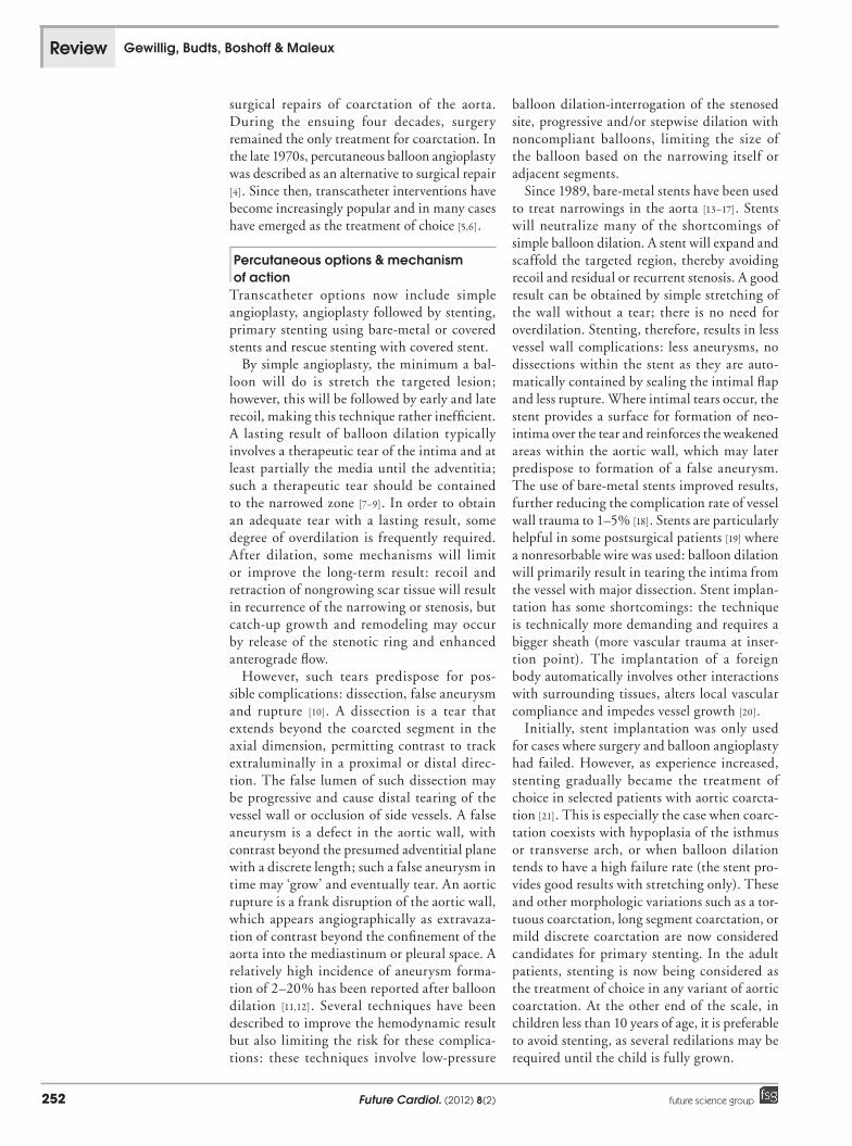
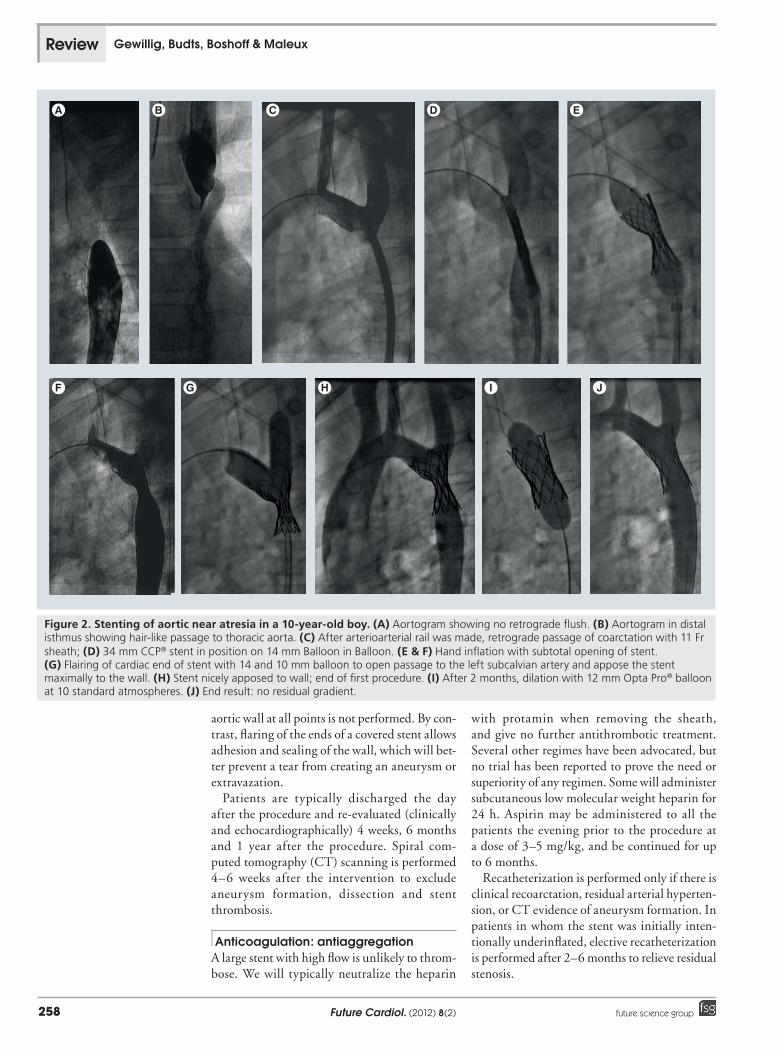
Figure 2. Stenting of aortic near atresia in a 10-year-old boy. (A) Aortogram showing no retrograde flush. (B) Aortogram in distal isthmus showing hair-like passage to thoracic aorta. (C) After arterioarterial rail was made, retrograde passage of coarctation with 11 Fr sheath; (D) 34 mm CCP® stent in position on 14 mm Balloon in Balloon. (E & F) Hand inflation with subtotal opening of stent. (G) Flairing of cardiac end of stent with 14 and 10 mm balloon to open passage to the left subcalvian artery and appose the stent maximally to the wall. (H) Stent nicely apposed to wall; end of first procedure. (I) After 2 months, dilation with 12 mm Opta Pro® balloon at 10 standard atmospheres. (J) End result: no residual gradient.
Review Gewillig, Budts, Boshoff & Maleux

www.futuremedicine.com 259future science group
Bare-metal or covered stentsThe use of covered stents is necessary to safely treat patients with atretic or severly hypoplastic segments, as a transmural tear can be expected. Overall, the procedure with a covered stent is safer than with a bare-metal stent, as an aneu-rysm or wall rupture within the stent is auto-matically contained. This margin of extra safety allows the operator to expand the stent more at the initial procedure and subsequently if indi-cated. Not all problems are neutralized: tears at the edge of the covered stent [33] or at a dis-tance, bleeding from the vessel wall or the side vessels may still occur, but these complications are usually less dramatic. Stenting techniques are slightly different when using covered stents com-pared with bare-metal stents: balloon interroga-tion prior to stenting is rarely performed and the stent is maximally approximated against the wall at deployment in order to obtain maximal sealing around the hypoplastic segment. The combina-tion of a Covered Cheatham Platinum (CCP®) stent with a slightly oversized BIB allows the stent to be neatly wrapped around the coarcta-tion, thereby obtaining maximal approximation to the wall. Progressive dilation of the coarctation site is performed immediately or later, depend-ing on the operator’s estimation on how well the expected tear will be sealed off. The ends of the stent can be flared into the side vessels (usually the subclavian or left carotid artery), if required.
Adapted techniques may be required to avoid excluding side vessels, such as double wire tech-nique [34] and retrograde perforation (from the subclavian artery [35] and theoretically also from the carotid). Experience with stent grafts for tho-racic aortic aneurysms suggests that the origin of the left subclavian artery (although best avoided) can be crossed with a covered stent without any acute effects. Retrograde perforation and opening of the covering allows restoration of antegrade flow.
If sheath size is critical, both CCP and V12 covered stents can be delivered through a 10 Fr sheath, followed by additional dilation as indicated.
In our experience there has been a gradual shift from using bare-metal stent to using covered stents due to the additional therapeu-tic margin and safety issues. Disadvantages of covered stents include the slightly bigger profile (+1 Fr), and more hemodynamic problems if the stent embolizes. Embolization must and can be avoided by a proper technique and great atten-tion to detail. Covered stents may occlude aortic side branches to the spinal cord and therefore
carry a risk of paraplegia. However, the arterial supply to the spinal cord usually originates below the aortic isthmus in the region of T9–T12 vertebrae and should, therefore, not be at risk when stenting a coarctation at the usual location (aortic isthmus).
Measures of successWhile treatment of coarctation of the aorta with balloon expandable endovascular stents is a technically challenging procedure, it is an extremely successful one [36–40]. A multicenter retrospective series of 588 procedures performed between 1989 and 2005 was conducted by the Congenital Cardiovascular Interventional Study Consortium (CCISC) [41]. Of the 588 proce-dures, 580 (98.6%) were successful in reducing the gradient to less than 20 mmHg or increasing the coarctation to descending aortic diameter ratio to at least 0.8. Two patients developed an aortic dissection with rupture: the procedures had to be terminated and emergency surgery undertaken with a bad outcome; in retrospect, such complications are probably avoidable and treatable with the use of covered stents.
In the adult population the initial gradient across the coarctation may not reflect its sever-ity as there may be extensive collateral vessels decompressing the aorta proximal to the steno-sis. Resolution of hypertension cannot necessar-ily be used as a measure of efficacy because the incidence of hypertension may be masked by antihypertensive treatment. Some adult patients without residual stenosis at the coarctation site will continue to be hypertensive with persistent abnormal endothelium [42,43]. However, control of their blood pressure may become easier after stenting. Stenting appears to be effective in reducing resting blood pressure to normal levels in the majority of children and adults.
There is no information on how stenting affects exercise tolerance or how well the stented coarctation segment responds to the increased cardiac output in pregnancy.
Complications Some complications of coarctation stenting have already been discussed. In general, they can be classified into technical, aortic wall or periph-eral vascular complications, or post-procedural hypertension and pain.
Technical complicationsTechnical complications include stent migra-tion of the balloon in the sheath, during deployment of the balloon, dislocation after
Percutaneous interventions of the aorta Review

Future Cardiol. (2012) 8(2)260 future science group
deployment, stent fracture, balloon rupture and overlap of the brachiocephalic vessels [44]. While progressing through the valve or the sheath, the stent may migrate on the balloon; radio-opaque markers on the balloon allow con-firming proper position of the stent on the bal-position of the stent on the bal- on the bal-loon before uncovering the stent in the aorta. If the stent has moved, the stent–balloon–sheath can be exteriorized leaving the wire in place, unloading the stent from the front of the sheath and start all over again.
Stent migration of the balloon during inflation can occur if the balloon is inflated asymmetri-cally. This can be avoided by mildly inflating the balloon before introduction through the sheath, creating small shoulders on both sides of the stent (easy with an indeflator), thereby avoid-ing overcrimping. During stent deployment bal-loon inflation should be started slowly, allowing both shoulders of the balloon to develop and thereby immobilize the stent on the balloon. Stent migration can further be avoided by sim-ply using a BIB: especially when using the bigger sized balloons (>15 mm), the inner balloon is typically within the stent and cannot milk the stent away; the outer balloon will always inflate symmetrically after the inner has been inflated. During or after deployment of the stent, it may migrate more proximal or distal. Frequently the stent can be grabbed with a balloon and reposi-tioned. If the stent cannot be safely repositioned within the coarctation, it should be expanded in the safest location available, away from side branches if possible. If necessary, a second stent can first be placed in the coarctation to allow repositioning of the migrated stent. Balloon rupture can be avoided by using an appropri- can be avoided by using an appropri-ate balloon for a given stent: stents with sharp edges do require thicker, puncture resistant bal-loons. Balloon rupture occurred in 13 out of 588 procedures (2.2%) in the CCISC cohort predominantly when using older stents, such as the abandoned Palmaz 8-series stents; it can result in other complications involving the aortic wall or secondary to embolization of balloon fragments, and if the balloon ruptures prior to full expansion, it will carry a high risk of stent migration.
There remains a debate over whether stent placement over the origin of the brachiocephalic vessels constitutes a complication at all. There have been no demonstrated harmful sequelae from doing so (except at redilation, see below [45]). A late stent fracture occurs typically at the transition of the mobile cross to the fixed retro-pleural thoracic aorta. Currently fractures can
be expected in the stents with minimal metal (Genesis and Valeo) when expanded to larger diameters.
Aortic wall complicationsProcedural complications involving the aorta at or around the site of the coarctation include intimal tears, dissection, aneurysm formation and rupture within the stent, at the edges or at a distance [46–48]. Vascular complications are more prone to develop in patients with connective tis-sue disease, such as Turner syndrome [49]. Most of these complications can be treated, or even better avoided, by using covered stents [50,51]. The general rule that it is easier to stay out of trouble than get out of trouble certainly applies to these situations. For this reason more opera-tors increasingly prefer to use covered stents ‘prophylactically’.
It is important to maintain some large diame-ter covered stents for use in emergency situations. We already mentioned some commercially made covered stents, including the covered CP stent and Atrium balloon expandable stent, but for some emergencies larger self-expanding excluder stents (from Boston Scientific [MA, USA], Gore [AR, USA], Medtronic [MN, USA]) should be available.
Aortic aneurysm is infrequently encountered, but it may be a harbinger of aortic rupture, and is therefore a potentially dangerous complica-tion. It can be seen at the time of the procedure or on interval follow-up. If a large or growing aneurysm is seen to form at the time of the stent placement, it must be excluded with a cov-ered stent to prevent progression and possible rupture [52–54].
Peripheral vascular complicationsPeripheral vascular complications include cerebral vascular accident, peripheral emboli, and injury to access vessels. Neurologic events including cerebral vascular accident occurred in the CCISC group in a total of six out of 588 pro-cedures. Adequate anticoagulation during the procedure is essential as the cranial vessels are crossed with wires for a prolonged time and long sheaths are used where clots may form. Claude Bernard-Horner syndrome was reported follow-ing stenting of a coarctation due to a carotid artery dissection [55].
Significant femoral vessel injury was reported in the CCISC study in a total of 15 out of 588 procedures (2.6%). One patient had place-ment of the arterial sheath above the ingui-nal ligament and developed a retroperitoneal
Review Gewillig, Budts, Boshoff & Maleux

www.futuremedicine.com 261future science group
hematoma. Vessel thrombosis is more frequent in small children. It is current practice, when there is loss of pulse after catheterization, to institute heparin therapy for 24 h. If the pulse has not returned after 24 h, or if the viability of the leg is a concern at any point, throm-bolytic therapy or surgery may be indicated. In our institution, tPA is the agent of choice. There are several reported dosage schemes [56,57], and while tPA can be very effective in restoring femoral artery patency after catheterization, it also carries very significant morbidities [58]. It is still unclear what dosing offers the best balance between safety and efficacy.
Postprocedural hypertensionA significant percentage of predominantly adult patients with coarctation will demonstrate hypertension immediately after the procedure. Patients with systolic blood pressures greater than P99 for age should be monitored carefully, allowing infusions of nitroprusside or esmolol or both. These patients can generally be switched to enteral antihypertensive medications within 24 h after the procedure.
Thoracic pain & abdominal discomfort early after the procedureStenting the aorta may cause thoracic pain for a couple of hours after the procedure; this pain may only become evident when the analgesics from the anesthesia fade away. Thoracic pain remains an alarming symptom, whereby dissec-tion, aneurysm formation, bleeding from aorta or torn intercostal arteries must be excluded (peripheral pulses, echo, CT scan). We have observed such pain on several occasions, and after excluding more malignant causes, con-cluded that the pain was most likely due to stretch of the aorta. This type of pain obviously requires adequate analgesia (opiates) and typi-cally fades away after some hours (overnight), keeping the nervous clinician awake.
Some adult patients will complain of abdom-inal discomfort early after the procedure: it appears that the new pulsatility causes some bowel irritability within the first few hours after the intervention.
Special situations: bail-out stenting in premature neonates
A coarctation in newborns is typically treated by surgery. However, there may be several rea-sons why a clinician may want to defer the sur-gery: extreme prematurity, a critically ill neo-nate with multiorgan failure recovering from
shock, or complex syndromic patients. Stenting a coarcted arch can be performed on short notice and acutely improve the newborn, deferring sur-gery to a safer period with adequate weight or stabilized hemodynamics. This strategy com-pares favorably to current treatment strate-gies when applied in critically ill or vulnerable newborns [59].
The technique in these premature infants is slightly different than previously described. Puncture of the artery is performed with a 21-gauge needle allowing a 0.014” wire to be introduced into the artery. A 4 Fr smooth tapered introducer sheath is placed in the femoral artery. A 4 Fr end hole vertebral catheter is advanced up to the coarctation site where a small (1 cm3) hand injection is made. The coarctation is crossed using an atraumatic 0.014” coronary wire. If the isthmus cannot be entered retrogradely, a transvenous antegrade approach may be used. A new hand injection is made in the cross to delineate the anatomy and the origin of the left subclavian artery. A low-profile, premounted coronary stent (off-label use for any coronary stent) is chosen on the basis of the precath echo-cardiographic measurements and angiography. The stent should ideally cover the arch from just distal of the origin of the left subclavian artery until beyond the coarctation site (length typi-cally 8–12 mm); stent diameter should equal the aortic cross (typically 3–5 mm). Such diameter allows a significant increase in size at the coarcta-tion site; apparently without the risk of vessel tear (‘fetal tissue’ appears to allow significant stretch).
Figure 3. Bail-out stenting in a premature 1500 g infant presenting with critical coarctation. (A) Retrograde aortogram by hand injection of layered contrast-saline through a short 4 Fr sheath; a 4-mm diameter/8-mm length coronary stent is ready for deployment; points of reference are: cranial end just beyond take-off of left subclavian artery, caudal end beyond coarctation site within the thoracic aorta. (B) Inflated balloon has expanded the stent. (C) Mini hand injection through 4 Fr catheter to confirm adequate position of stent.
Percutaneous interventions of the aorta Review

Future Cardiol. (2012) 8(2)262 future science group
The stent is passed ‘unprotected’ through the valve of the sheath. Stent position is controlled with a retrograde aortogram hand injection through the short femoral sheath (sequential gentle aspiration of 5 cm3 saline via a 10-cm3
syringe held vertically, followed by aspiration of 1 cm3 contrast keeping contrast and saline layered and separated; Figure 3a). The stent is deployed using an indeflator at a pressure as rec-ommended by the stent manufacturer (allows opening the stent within a range of 0.6 mm). In premature infants and neonates with primary coarctation, the prostaglandin E1 infusion is stopped immediately after deployment of the stent. If useful and not contraindicated (renal function), stent position is assessed with an aor-togram through the 4 Fr catheter placed just below the stent. Such a stent will typically give a satisfactory result for several weeks to months. In minute premature children below 1000 g, vas-cular complications may be avoided by access-ing the aorta directly during a hybrid procedure: after sternotomy a 4 Fr sheath is inserted in the ascending aorta allowing the stenting procedure to be performed; such an approach also allows for closure of the patent arterial duct [60].
The decision regarding when to surgically remove the stent may vary: after some days when the neonate has sufficiently recovered from the initial cardiogenic shock, after some weeks or months when adequate body weight has been reached, in order to safely perform a coarctectomy, or when additional surgery is planned. If during follow-up more time is required, any coronary stent can be further dilated up to 5 mm; this also reduces the stent length if this were an issue for safe resection.
Staged dilation: redilationStaged dilation, in which stents are expanded to a diameter less than the adjacent aorta and redilated a few months later, may overcome the possible risks of excessive wall damage. A con-trolled injury is allowed time to heal and the arterial wall time to be to restored before full expansion is attempted. However, even with this approach, aneurysm formation may not be avoided.
If stents are implanted in smaller patients, somatic growth of the patient will mean that redilation is required. In this age group, stent-ing should be reserved for exceptional clinical indications rather than routine use because excellent surgical results can be obtained with extended arch repair.
Redilation of a stent is typically a straight-forward procedure as the previous stent is a perfect landmark. However, such procedures may be complicated: an aneurysm may develop within a bare-metal stent, and embolization of peel may occur to side vessels; dissection and aneurysms may develop at the stent ends, but also at a distance: most stents will shorten when dilated to larger diameters, causing longitudi-nal stress on the vessel wall. Theoretically it is safer to dilate gradually in small steps and with short balloons: an excessive shoulder of a big long balloon may tear off the vessel wall from the stent before effective dilation. Such complications have been reported with late (after hours) dissection of the thoracic aorta, with a fatal outcome [61]. A very careful aor-togram should therefore be made, at least at the end of any procedure; if such complication is observed or suspected, implantation of an additional covered stent might be life-saving.
When using covered stents, the operator should aim at the initial procedure for adequate apposition of the stent to the wall, thereby allow-ing maximal adherence of the stent around the narrowed segment. Such safety zones will seal the expected tear at further dilation.
Figure 4. Stenting the transverse arch in a 19-year-old boy with previous stenting of coarctation and residual hypertension. (A) Aortogram shows 12 mm hypoplasia of distal cross between left carotid artery and left subclavian artery, gradient 15 mmHg; a coarctation was previously stented with 20 mm stent. (B) Low pressure balloon inflation of with 20 mm Tyshak® balloon to determine compliance of hypoplastic segment. (C) Deployment of 28 mm bare-metal CP® stent on 20 mm Balloon in Balloon™ at low pressure through 13 Fr sheath. (D) Stent in cross. (E) Flairing of stent into left carotid artery with 16 mm Tyshak balloon. (F) End result: no residual gradient.
Review Gewillig, Budts, Boshoff & Maleux

www.futuremedicine.com 263future science group
Arch atresia A long-standing critical coarctation may occlude, thereby creating an atretic segment by the time treatment is initiated. The prec-atheterization evaluation should try to deter-mine whether there is critical stenosis or atre-sia. Careful Doppler examination may show a minute connection on color flow or a typi-cal saw-tooth pattern. If an atresia or critical stenosis is expected, access through both the brachial/axillary and femoral arteries must be obtained. Angiography is performed proxi-mally and distally from the interrupted arc. In some patients a filiform connection can be found when actively looked for; this then can be crossed with the use of a 0.014” coronary guide wire or an 0.018” wire in a 2 Fr track-ing system [62]. When no connection is found, recanalization may be performed by punctur-ing with a stiff wire (0.035” or the back of an 0.014” wire) or a Brockenbrough® needle, or by radiofrequency Nykanen 0.024” wire through coaxial system (Baylis Medcomp, Montreal, Canada) or a PT2 coronary wire through 2 Fr Progreat™ catheter through 4 Fr cath-eter) [63]. The local anatomy of each patient will determine how the atretic part can best be crossed: usually from the cross to the tho-racic aorta, with an opened lasso at the other end as a target. Once the wire is grasped by the lasso, an arterioarterial rail is made. Some predilation may be required to allow crossing the defect with the delivery sheath. Finally, a covered stent is implanted with sufficient over-lap proximally and distally of the defect. Full stent expansion is performed a few weeks after the initial stenting procedure.
Stenting for transverse aortic archMany patients with a coarctation have an asso-ciated hypoplasia of the transverse arch that leaves a residual gradient. The surgical treatment option for transverse arch hypoplasia is to per-form an extended arch repair, which frequently requires cardiopulmonary bypass and is not risk free, or a bypass graft.
Percutaneous treatment of arch hypoplasia is slightly different from coarctation treatment [64,65]. Balloon dilation only is typically not suc-cessful, stenting is necessary. In this vessel the margin between a therapeutic tear and a cata-strophic rupture is very small: the adventitia is adherent to the media, which is not the case in a typical coarctation, where the adventitia ‘jumps’ from the isthmus over the coarctation to the thoracic aorta. Therefore, the main aim when stenting the transverse arch is to only stretch the wall without creating a tear. In the proximal cross bare-metal stents are almost exclusively used; in the distal cross a covered stent may be used; such stent may be positioned across the subclavian artery and exclude its ori-gin; this can easily be reopened by retrograde perforation of the covering and balloon dilation of the orifice.
An ascending angiogram is performed in a perpendicular projection, with the aim of defin-ing the arch anatomy as well as the origins of the innominate, the left common carotid and the left subclavian arteries. Balloon interroga-tion of the hypoplastic segments of the arch with a thin, low pressure, mildly oversized balloon (Tyshak®, NuMED) is essential: it allows assess-ment of the stretchability of the segments, which determines the balloon size and the desired stent
Figure 5. Exclusion of aneurysm by stent-graft in a 32-year-old woman presenting with an asymptomatic aortic pseudoaneurysm, 28 years after coarctation-related Dacron patch angioplasty. (A) Calibrated flush aortography revealed an aneurysmal dilatation (maximal diameter of 5 cm) of the Dacron patch (arrows) distal to the origin of the left subclavian artery. (B–D) Transfemoral insertion of a Zenith TX2® (COOK) stent-graft (small arrows) loaded in a 20 Fr sheath was performed after carotidosubclavian transposition (large arrow). (E) Control CT scan 6 months after the stent-graft procedure shows the stent-graft (white arrows) completely excluding the pseudoaneurysmal sac.
Percutaneous interventions of the aorta Review

Future Cardiol. (2012) 8(2)264 future science group
conformation during deployment (Figure 4). This technique allows the operator to avoid deploy-ment of an undersized stent, which will anchor itself insufficiently in the wall and embolize, or to tear a narrow, unstretchable segment. The balloon diameter is chosen according to the larg-est stretched diameter of the aortic segment to be stented; the stent needs to be approximately 3–5 mm longer than the distance between the origins of the vessels where the aorta is to be stented. Stents partially protruding over the ori-gin of a carotid artery can be flaired into the ves-sel; struts covering the origin of the left subcla-vian artery are of no concern. During inflation the stent will shorten asymmetrically, typically around the point where the stent touches first and anchors itself in the aortic wall.
Rapid right ventricular pacing may be used during the placement of the stent, as the stent/balloon assembly may be dispaced dis-tally during balloon inflation due to the rela-tively large stroke volume in an adult patient. However, in our experience it is seldom neces-sary due to sufficient stabilization obtained by the wire–shaft–sheath complex.
The stiff Amplatz exchange guide wire is positioned in the ascending aorta or in the left ventricular apex. Once the Mullins sheath is in place across the hypoplastic arch, the stent/bal-loon assembly is passed until it is at the tip of the sheath. Angiograms are performed through the side-arm of the Mullins sheath to check for accu-rate positioning of the stent. The Mullins sheath is withdrawn whilst keeping the stent/balloon assembly in position so as to expose the stent. The stent is inflated manually at low pressure; a BIB allows for the accurate deployment of the stent: the opening will be symmetrical during inflation of the inner balloon, and asymmetric
shortening will only start when the stent anchors itself in the aortic wall. The stent is apposed to the wall under low pressure aiming to stretch and not tear the wall. On balloon inflation, the stent will shorten and so will be clear of the ori-gin of the carotid artery, or the end of the stent can be flaired into it.
What if a residual gradient persists after percutaneous optimalization?
Despite percutaneous optimalization, a sig-nificant residual gradient might persist due to residual hypoplasia that cannot be dilated safely, angulation in a high cervical gothic arch, restric-tive stents or conduits. Surgery in an arch with gradients at different levels can be very prob-lematic, even more so when some segments are stented. Very good gradient relief can then be obtained with an extra-anatomic bypass with very low morbidity [66]. Two approaches can be applied. A repair through a median sternotomy consists of creating a ‘right arch’: insertion of a bypass from the right lateral wall of the ascend-ing aorta, routed around the right margin of the heart, to the supraceliac abdominal aorta. A second approach is through a left thoracotomy with interposition of a graft between the ascend-ing and descending aorta or interposition of a graft between the left subclavian artery and the descending thoracic aorta.
True aneurysmsA true aneurysm consists of dilation of all lay-ers of the vessel wall. Small aneurysms can be treated very effectively with a covered stent as described above. However, several treatment strategies (both surgical and interventional) can later be complicated with large and long thoracic aneurysm formation. Natural evolu-tion of these aneurysms is associated with a high rate of rupture within 15 years of detec-tion [67]. Traditionally, these late postsurgical aneurysms are treated by redo surgery, includ-ing interposition graft placement under cardio-pulmonary bypass, hypothermic circulatory arrest or other methods of distal circulatory support. Postoperative mortality may be as high as 13% and postoperative nonlethal complica-tions are significant and may include paresis of the left recurrent nerve and bleeding requiring rethoracotomy. Consequently, thoracic endovas-cular aneurysm repair (TEVAR) emerged as a minimally invasive alternative for redo surgery after open coarctation repair [68–70]. These tech-niques, however, involve self-expanding systems requiring big sheaths of 22–24 Fr.
Figure 6. Complication of stent-graft: infection in a 43-year-old man with a medical history of a ruptured thoracic pseudoaneurysm, 16 years after redo surgery for a coarctation-related aneurysm, was followed up by CT scan for evaluation of the stent graft. (A) Axial as well as (B) coronal CT images 3 years after stent-graft insertion revealed a clear amount of air bubbles (white arrows) around the stent-graft, suggesting infection. This was confirmed during open repair.
Review Gewillig, Budts, Boshoff & Maleux

www.futuremedicine.com 265future science group
Preinterventional imaging is typically per-formed with magnetic resonance and/or CT imaging, including axial, coronal and angio-graphic 3D reconstructions; catheter angiogra-phy with use of a calibrated pigtail catheter for stent-graft planning and evaluation of aortoiliac diameter and tortuosity. The diameter of a tho-racic aortic stent-graft should be oversized for 10–15% compared with the nominal diameter of the proximal and distal landing zone; the length of the stent-graft should include the length of the aneurysm and the length of the proximal and distal landing zones, which are at least 2 cm each. Typical contraindications for conventional TEVAR are a too small (proximal) luminal diam-eter, too short length of the landing zone and an acute angle of the thoracic arch (‘gothic arch’). These cases should be considered for open or hybrid repair.
The TEVAR procedure should be performed in a sterile environment under optimal image guid-ance using a fixed imaging system (‘hybrid oper-ating room’). After surgical cut down of the groin, the common femoral artery is cannulized with a 7 Fr sheath and a stiff guide wire is placed in the ascending thoracic aorta. A pigtail catheter is nav-igated into the ascending thoracic aorta through percutaneous puncture of the contralateral femo-ral or right brachial artery. The stent-graft is posi-tioned over the aneurysm and carefully deployed under induced hypotension (systolic pressure less than 80 mmHg) and angio graphic (and transesophageal ultrasonographic) imaging con-trol (Figure 5). In general, a tube stent-graft with appropriate diameter and length is used. In case of a small diameter arch and a normal diameter descending thoracic aorta, a reversed and tapered custom-made device is preferred. If not available (i.e., in emergency cases), a telescopic approach with placement of a smaller proximal tube stent-graft and a larger distal tube stent-graft may be an alternative, although this technique is more prone to early or late type III endoleak [71].
Following deployment, the device can be fur-ther conformed and approximated using a large, compliant balloon, therefore obtaining optimal proximal and distal sealing. A final control angio-graphy is performed to evaluate the position of the stent-graft and exclude endoleaks.
On transesophageal ultrasonic monitoring immediately after stent-graft implantation, inten-sifying echodensity will develop around the graft within minutes after aneurysm exclusion due to progressive thrombosis. Biochemically, a transient postimplantation syndrome, including mild leu-kocytosis, elevated levels of C-reactive protein
and moderately elevated body temperature can be observed.
Imaging follow-up can be performed by MRI or CT scanning. Either modality provides ade-quate information, but MRI causes less radia-tion exposure, which is obviously important in a young population.
During the last decade, several small case series (mostly including less than ten patients) have been published, all demonstrating that TEVAR is relatively safe and effective for endovascular repair of aneurysms associated with coarctation sur-gery [72–74]. Major complications seem to be rare, although perioperative and postoperative mor-tality may occur [75,76]. Procedure- and device-related complications encountered after TEVAR for coarctation aneurysms include left-sided arm claudication due to intentional covering of the left subclavian artery, type I and type II endoleak, infolding and collapse of the stent-graft, graft infection (Figure 6), and stent migration (Figure 7).
Hybrid endovascular procedures are now increasingly performed to treat aortic arch aneu-rysms. These procedures are performed using supra-aortic bypass-surgery (i.e., carotid–carotid bypass or even complete rerouting of all
Figure 7. Complication of stent graft: late migration in a 34-year-old man with a previous history of coarctation repair by Dacron patch angioplasty 27 years ago. The patient presented with an asymptomatic pseudoaneurysm extending into the origin of the left subclavian artery (white arrows) as depicted on (A) thoracic angio CT scan and (B) calibrated flush aortography. The patient presented, 2 weeks after uneventful stent-graft (GORE® TAG®) insertion, with major upper thoracic pain and general malaise. (C) CT scan revealed distal migration of the proximal part of the stent-graft (arrows), forming a ‘pseudocoarctation’ and revascularization of the aneurysmal sac. (D) After insertion of a Valiant™ stent-graft (white arrows) through a 22 Fr sheath, complete exclusion of the aneurysmal sac was obtained.
Percutaneous interventions of the aorta Review

Future Cardiol. (2012) 8(2)266 future science group
supra-aortic vessels), with insertion of a stent-graft via transfemoral or transaortic approach as previously described [77]. Fenestrated or scalloped stent-grafts may be an endovascular alternative for some of these hybrid procedures.
Conclusion & recommendationsAlthough treatment of coarctation of the aorta with balloon-expandable endovascular stents is technically challenging, it is a relatively safe and extremely effective treatment modality when used carefully in selected patients. The shift from simple balloon angioplasty, to the implantation of bare-metal stents and, eventually, covered stents, has significantly improved results while decreasing complications. Further research is necessary to determine the incidence of various complications and identify risk factors, allowing refinement of guidelines for even safer and more successful procedures.
With current knowledge and experience our recommendations are as follows:
nIn infants and children less than a year of age, surgery is the treatment of choice for all native coarctation; balloon angioplasty is the treat-ment of choice for most recurrent coarctation; in very premature and critically ill neonates bail-out stenting, followed by later surgical stent removal, may avoid many complications typically observed in this age group;
nBetween the ages of 1 year and the time when the child reaches a weight of 30–35 kg (usually 9–11 years), there is insufficient data to deter-mine whether surgical intervention or balloon angioplasty is preferable for native lesions. Per-cutaneous treatment usually involves several interventions during growth, while surgical results in a single procedure are very good.
Balloon angioplasty is the treatment of choice in this age group for recurrent coarctation;
nIn children weighing more than 35 kg who have not yet reached adult size, it is likely that the treatment of choice for native and recurrent lesions should be endovascular stent placement, as it has been demonstrated that stents can be enlarged safely at a later time to accommodate for somatic growth;
nIn adult-sized adolescents and adult patients, stent placement is the treatment of choice for all lesions, native and recurrent;
nIn many situations the use of covered stents is emerging as the safer option, especially in adults of advanced age, in patients with known vasculitis or other conditions associated with vasculopathy;
nTEVAR is a good option to treat large thoracic aneurysms;
nAll patients require long-term follow-up in order to detect aortic aneurysms or dilation, as well as arterial hypertension, in good time.
Future perspective In the short term we expect the manufacturers to develop better, slimmer and stronger tools. The interventionalists should refine the techniques and guidelines for the different types of lesions, allowing the application of the best technique with the best tool in an individual patient. As interventionalists become more familiar with covered stents, this type of stent will increas-ingly be used to decrease the incidence of early and late complications. Control and regulations are necessary, but requesting in this niche field complex comparative studies without margin for
Executive summary
n Lesions that cause pressure loss in the aorta include a discrete narrowing, a hypoplastic segment, excessive angulation, tortuosity and atresia; such variety requires a tailored strategy.
n Transcatheter options include simple angioplasty and stenting with either bare-metal or covered stents.n The intervention aims to obtain an adequate stretch or a therapeutic tear of the vessel wall, while avoiding complications, such as an
excessive tear, a dissection, an aneurysm or a vessel rupture.n Balloon dilation only is technically easier, but with age-dependent moderate success and relevant vessel wall problems in 2–20%; the
targeted vessel retains growth potential.n Stenting is technically more demanding, with consequently more possible technical complications, but with a higher success rate and
lower incidence of local vessel wall problems (bare-metal stent 1–5%, covered stent <1%); a stent has no growth potential, but can be redilated up to a maximal value.
n When allowed by local regulations, there is a gradual shift from using bare-metal stent to covered stents due to the additional therapeutic margin and safety.
n A covered stent, if not already used for primary stenting, should be readily available for bail-out in case of an excessive vessel wall tear.n Surgery remains, in selected cases, a good if not the better option, especially in infants and small children and in complex arches with
multilevel stenosis in adults.
Review Gewillig, Budts, Boshoff & Maleux

www.futuremedicine.com 267future science group
common sense is not only counterproductive, but even dangerous for the patient.
In the long term, children, including newborns, should be treated with a new generation of stents that can be inserted through small sheaths, give adequate scaffolding for some time allowing the arch to remodel and undergo catch-up growth, and then disappear leaving a vessel wall of normal compliance and normal growth potential.
Financial & competing interests disclosureM Gewillig is proctor for NuMED (USA). The authors have no other relevant affiliations or financial involve-ment with any organization or entity with a financial interest in or financial conflict with the subject matter or materials discussed in the manuscript apart from those disclosed.
No writing assistance was utilized in the production of this manuscript.
ReferencesPapers of special note have been highlighted as:n of interestnn of considerable interest
1. Fyler DC, Buckley LP, Hellenbrand WE et al. Report of the New England regional infant cardiac program. Pediatrics 65, 432–436 (1980).
2. Campbell M. Natural history of coarctation of the aorta. Br. Heart J. 32, 633–640 (1970).
3. Ou P, Celermajer DS, Mousseaux E et al. Exercise-related hypertension predicts resting hypertension, left ventricular and vascular hypertrophy late after coarctation repair. Circulation 114, 504 (2006) (Abstract).
4. Singer MI, Rowen M, Dorsey TJ. Transluminal aortic balloon angioplasty for coarctation of the aorta in the newborn. Am. Heart J. 103, 131–132 (1982).
5. Fletcher SE, Nihill MR, Grifka RG et al. Balloon angioplasty of native coarctation of the aorta: midterm follow-up and prognostic factors. J. Am. Coll. Cardiol. 25, 730–734 (1995).
6. Maheshwari S, Bruckheimer E, Fahey JT, Hellenbrand WE. Balloon angioplasty of postsurgical recoarctation in infants: the risk of restenosis and long term follow-up. J. Am. Coll. Cardiol. 35, 209–213 (2000).
7. Ho SY, Somerville J, Yip WC et al. Transluminal balloon dilation of resected coarcted segments of thoracic aorta: histological study and clinical implications. Int. J. Cardiol. 19, 99–105 (1988).
8. Lock JE, Castaneda-Zuniga WR, Bass JL et al. Balloon dilatation of excised aortic coarctations. Radiology 143, 689–691 (1982).
9. Ino T, Kishiro M, Okubo M et al. Dilatation mechanism of balloon angioplasty in children; assessment by angiography and intravascular ultrasound. Cardiovasc. Interv. Radiol. 21, 102–108 (1998).
10. Qureshi A, McElhinney DB, Lock JE, Landszberg MJ, Lang P, Marshall AC. Acute and intermediate outcomes, and evaluation of injury to the aortic wall, as based on 15 years experience of implanting stents to treat aortic coarctation. Cardiol. Young 17, 307–318 (2007).
n Good overview of algorithms with expected results and complications.
11. Rao PS, Galal O, Smith PA et al. Five- to nine-year follow-up results of balloon angioplasty of native aortic coarctation in infants and children. J. Am. Coll. Cardiol. 27, 462–470 (1996).
12. Fletcher SE, Nihill MR, Grifka RG et al. Balloon angioplasty of native coarctation of the aorta: midterm follow-up and prognostic factors. J. Am. Coll. Cardiol. 25, 730–734 (1995).
13. O’Laughlin MP, Ferry SB, Lock JE et al. Use of endovascular stents in congenital heart disease. Circulation 83, 1923–1939 (1991).
14. Morrow WR, Smith VC, Ehler WJ et al. Balloon angioplasty with stent implantation in experimental coarctation of the aorta. Circulation 89, 2677–2683 (1994).
15. Grifka RG, Vick W 3rd, O Laughlin MP. Balloon expandable intravascular stents: aortic implantation and late further dilation in growing minipigs. Am. Heart J. 126, 979–984 (1993).
16. Suarez de Lezo J, Pan M, Romero M et al. Balloon-expandable stent repair of severe coarctation of aorta. Am. Heart J. 129, 1002–1008 (1995).
17. Magee AG, Brzezinska-Rajszys G, Qureshi SA et al. Stent implantation for aortic coarctation and recoarctation. Heart 82, 600–606 (1999).
18. Bulbul ZR, Bruckheimer E, Love JC et al. Implantation of balloon-expandable stents for coarctation of the aorta: implantation and short-term results. Catheter. Cardiovasc. Diagn. 39, 36–42 (1996).
19. Rosenthal E, Qureshi SA, Tynan M. Stent implantation for aortic recoarctation. Am. Heart J. 129, 1220–1221 (1995).
20. Brili S, Dima I, Ioakeimidis N et al. Evaluation of aortic stiffness and wave reflections in patients after successful coarctation repair. Eur. Heart J. 26, 1394 (2005).
21. Suarez de Lezo J, Pan M, Romera M et al. Immediate and follow-up findings after stent treatment for severe coarctation of aorta. Am. J. Cardiol. 83, 400–406 (1999).
22. Qureshi SA, Zubrzycka M, Brzezinska-Rajszys G et al. Use of covered Cheatham-Platinum stents in aortic coarctation and recoarctation. Cardiol. Young 14, 50–54 (2004).
23. Gunn J, Cleveland T, Gaines P. Covered stent to treat co-existent coarctation and aneursym of the aorta in a young man. Heart 82, 351 (1999).
24. Forbes T, Matisoff D, Dysart J et al. Treatment of coexisting coarctation and aneurysm of the aorta with covered stent in a pediatric patient. Pediatr. Cardiol. 24, 289–291 (2003).
25. Sadiq M, Malick NH, Qureshi SA. Simultaneous treatment of native coarctation of the aorta combined with patent ductus arteriosus using a covered stent. Catheter. Cardiovasc. Interv. 59, 387–390 (2003).
26. Ewert P, Abdul-Khalid H, Peters B et al. Transcatheter therapy of long extreme subaortic aortic coarctation with covered stents. Catheter. Cardiovasc. Interv. 63, 236–239 (2004).
27. Bruckheimer E, Dagan T, Amir G, Birk E. Covered Cheatham-Platinum stents for serial dilation of severe native aortic coarctation. Catheter. Cardiovasc. Interv. 74(1), 117–123 (2009).
28. Kenny D, Cao QL, Kavinsky C, Hijazi ZM. Innovative resource utilization to fashion individualized covered stents in the setting of aortic coarctation. Catheter. Cardiovasc. Interv. 78(3), 413–418 (2011).
29. De Vlieger G, Budts W, Dubois CL. Horner syndrome after stenting of a coarctation of the aorta. Heart 96(9), 714 (2010).
30. Daehnert I, Rotzsch C, Wiener M, Schneider P. Rapid right ventricular pacing is an alternative to adenosine in catheter interventional procedures for congenital heart disease. Heart 90, 1047–1050 (2004).
31. Gillespie MJ, Kreutzer J, Rome JJ. Novel approach to percutaneous stent implantation for coarctation of the aorta: the railway technique. Catheter. Cardiovasc. Interv. 65, 584–587 (2005).
32. Marshall AC, Perry SB, Keane JF et al. Early results and medium- term follow-up of stent
Percutaneous interventions of the aorta Review

Future Cardiol. (2012) 8(2)268 future science group
implantation for mild residual orrecurrent aortic coarctation. Am. Heart J. 139, 1054–1060 (2000).
33. Kenny D, Hijazi ZM. Coarctation of the aorta: from fetal life to adulthood. Cardiol. J. 18, 487–495 (2011).
34. Lampropoulos K, Budts W, Gewillig M. Dual wire technique for aortic coarctation stent placement. Catheter. Cardiovasc. Interv. 78(3), 425–427 (2011).
35. Tsai SF, Hill SL, Cheatham JP. Treatment of aortic arch aneurysm with a NuMED-covered stent and restoration of flow to excluded left subclavian artery: perforation and dilation of e-PTFE can be done! Catheter. Cardiovasc. Interv. 73(3), 385–389 (2009).
36. Suarez de Lezo J, Pan M, Romero M et al. Balloon-expandable stent repair of severe coarctation of the aorta. Am. Heart J. 129, 1002–1008 (1995).
37. Rosenthal E, Qureshi SA, Tynan M. Stent implantation for aortic recoarctation. Am. Heart J. 129, 1220–1221 (1995).
38. Bulbul ZR, Bruckheimer E, Love JC, Fahey JT, Hellenbrand WE. Implantation of balloon-expandable stents for coarctation of the aorta: implantation data and short-term results. Catheter. Cardiovasc. Diagn. 39, 36–42 (1996).
39. Harrison DA, McLaughlin PR, Lazzam C, Connelly M, Benson LN. Endovascular stents in the management of coarctation of the aorta in the adolescent and adult: one year follow up. Heart 85, 561–566 (2001).
40. Hamdan MA, Maheshwari S, Fahey JT, Hellenbrand WE. Endovascular stents for coarctation of the aorta: initial results and intermediate- term follow-up. J. Am. Coll. Cardiol. 38, 1524–1527 (2001).
41. Holzer R, Qureshi S, Ghasemi A et al. Stenting of aortic coarctation: acute, intermediate, and long-term results of a prospective multi-institutional registry – Congenital Cardiovascular Interventional Study Consortium (CCISC). Catheter. Cardiovasc. Interv. 76(4), 553–563 (2010).
nn Very large series reporting the spectrum of success and complications.
42. Kaemmerer H, Oelert F, Bahlmann J et al. Arterial hypertension in adults after surgical treatment of aortic coarctation. Thorac. Cardiovasc. Surg. 46, 121–125 (1998).
43. Gardiner HM, Celermajer DS, Sorensen KE et al. Arterial reactivity is significantly impaired in normotensive young adults after successful repair of aortic coarctation in childhood. Circulation 89, 1745–1750 (1994).
44. van Gameren M, Witsenburg M, Takkenberg JJ et al. Early complications of
stenting in patients with congenital heart disease: a multicentre study. Eur. Heart J. 27(22), 2709–2715 (2006).
45. Boshoff D, Eyskens B, Gewillig M. Late redilation of a stent in the aorta crossing the subclavian artery complicated with a cerebellar infarction. Acta Cardiologia 62(3), 295–297 (2007).
46. Balaji S, Oommen R, Rees PG. Fatal aortic rupture during dilatation of recoarctation. Br. Heart J. 65, 100–101 (1991).
47. Rao PS. Aortic rupture after balloon angioplasty of aortic coarctation. Am. Heart J. 125, 1205–1206 (1993).
48. Fletcher SE, Cheatham JP, Froeming S. Aortic aneurysm following primary balloon angioplasty and secondary endovascular stent placement in the treatment of native coarctation of the aorta. Catheter. Cardiovasc. Diagn. 44, 40–44 (1998).
49. Fejzic Z, van Oort A. Fatal dissection of the descending aorta after implantation of a stent in a 19-year-old female with Turner’s syndrome. Cardiol. Young 15, 529–531 (2005).
50. Kato N, Dake MD, Miller DC et al. Traumatic thoracic aneurysm: treatment with endovascular stent-grafts. Radiology 205, 657–662 (1997).
51. Rousseau H, Soula P, Perreault P et al. Delayed treatment of traumatic rupture of the thoracic aorta with endoluminal covered stent. Circulation 99, 498–504 (1999).
52. Mitchell RS, Miller DC, Dake MD. Stent-graft repair of thoracic aortic aneurysms. Semin. Vasc. Surg. 10, 257–271 (1997).
53. Tan JL, Michael MM. Emergency stent graft deployment for acute aortic rupture following primary stenting for aortic coarctation. Catheter. Cardiovasc. Interv. 65, 306–309 (2005).
54. Varma C, Benson LN, Butany J, McLaughlin PR. Aortic dissection after stent dilatation for coarctation of the aorta: a case report and literature review. Catheter. Cardiovasc. Interv. 59, 528–535 (2003).
55. De Vlieger G, Budts W, Dubois CL. Horner syndrome after stenting of a coarctation of the aorta. Heart 96, 714 (2010).
56. Zenz W, Muntean W, Beitzke A et al. Tissue plasminogen activator for femoral artery thrombosis after arterial cardiac catheterization in infants and children. Br. Heart J. 63, 291–294 (1990).
57. Levy M, Benson LN, Burrows PE et al. Tissue plasminogen activator for the treatment of thromboembolism in infants and children. J. Pediatr. 118, 467–472 (1991).
58. Gupta AA, Leaker M, Andrew M et al. Safety and outcomes of thrombolysis with tissue plasminogen activator for treatment of intravascular thrombosis in children. J. Pediatr. 139, 682–688 (2001).
59. Gorenflo M, Boshoff D, Heying R et al. Bailout stenting for critical coarctation in premature/critical/complex/early recoarcted neonates. Catheter. Cardiovasc. Interv. 75, 553–561 (2010).
n Large series of a two-step procedure to treat small infants with coarctation.
60. Cools B, Meyns B, Gewillig M. Hybrid stenting of aortic coarctation in very low birth weight premature infant. Catheter. Cardiovasc. Interv. (2012) (In Press).
61. Fejzic Z, van Oort A. Fatal dissection of the descending aorta after implantation of a stent in a 19-year-old female with Turner’s syndrome. Cardiol. Young 15, 529–531 (2005).
62. Brown SC, Boshoff DE, Eyskens B, Mertens L, Gewillig M. Use of a microcatheter in a telescopic system to reach difficult targets in complex congenital heart disease. Catheter. Cardiovasc. Interv. 73(5), 676–681 (2009).
63. Butera G, Heles M, Carminati M. Percutaneous treatment of aortic isthmus atresia: use of radiofrequency perforation and covered stents. Catheter. Cardiovasc. Interv. 78(6), 933–939 (2011).
n Large series of treatment of aortic atresia requiring puncture or fulguration.
64. Pihkala J, Pedra CA, Nykanen D et al. Implantation of endovascular stents for hypoplasia of the transverse aortic arch. Cardiol. Young 10, 3–7 (2000).
65. Boshoff D, Budts W, Mertens L et al. Stenting of hypoplastic aortic segments with mild pressure gradients and arterial hypertension. Heart 92(11), 1661–1666 (2006).
n Series focussing on aorta cross hypoplasia, a frequent reason for residual gradient after adequate coarctation repair.
66. Sakellaridis T, Argiriou M, Panagiotakopoulos V et al. Latent congenital defect: interrupted aortic arch in an adult case report and literature review. Vasc. Endovascular Surg. 44(5), 402–406 (2010).
67. Knyshov G, Sitar L, Glagola M, Atamanyuk M. Aortic aneurysms at the site of the repair of coarctation of the aorta: a review of 48 patients. Ann. Thorac. Surg. 61, 935–939 (1996).
68. Ince H, Petzsch M, Rehders T et al. Percutaneous endovascular repair of aneurysm after previous coarctation surgery. Circulation 108, 2967–2970 (2003).
Review Gewillig, Budts, Boshoff & Maleux

www.futuremedicine.com 269future science group
69. Ito T, Kurimoto Y, Kawaharada N et al. Endovascular stent-grafting of anastomotic pseudoaneurysms following thoracic aortic surgery. Gen. Thorac. Cardiovasc. Surg. 57, 528–533 (2009).
70. Smayra T, Otal P, Soula P et al. Pseudoaneurysm and aortobronchial fistula after surgical bypass for aortic coarctation: management with endovascular stent-graft. J. Endovasc. Ther. 8, 422–428 (2001).
71. Midulla M, Dehaene A, Godart F et al. TEVAR in patients with late complications of aortic coarctation repair. J. Endovasc. Ther. 15, 552–557 (2008).
72. Canaud L, Hireche K, Berthet J, Branchereau P, Marty-Ané C, Alric P. Endovascular repair of aortic arch lesions in high-risk patients or after previous aortic surgery: midterm results. J. Thorac. Cardiovasc. Surg. 140, 52–58 (2010).
73. Kutty S, Greenberg RK, Fletcher S, Svensson LG, Latson LA. Endovascular stent grafts for large thoracic aneurysms after coarctation repair. Ann. Thorac. Surg. 85, 1332–1338 (2008).
74. Botta L, Russo V, Oppido G et al. Role of endovascular repair in the management of late pseudo-aneurysms following open surgery for aortic aortctation. Eur. J. Cardiothorac. Surg. 36, 670–674 (2009).
75. Yazar O, Budts W, Maleux G, Daenens K, Houthoofd S, Fourneau I. Thoracic endovascular repair for treatment of late complications after aortic coarctation repair. Ann. Vasc. Surg. 25, 1005–1011 (2011).
76. Zipfel B, Ewert P, Buz S, El Al AA, Hammerschmidt R, Hetzer R. Endovascular stent-graft repair of late pseudoaneurysms after surgery for aortic coarctation. Ann. Thorac. Surg. 91(1), 85–91 (2011).
77. Gottardi R, Funovics M, Eggers N et al. Supra-aortic transposition for combined vascular and endovascular repair of aortic arch pathology. Ann. Thorac. Surg. 86, 1524–1529 (2008).
Percutaneous interventions of the aorta Review