Review Seasonal and post-trauma remodeling in cone ...
16
Review Seasonal and post-trauma remodeling in cone-dominant ground squirrel retina Dana K. Merriman a, * , Benjamin S. Sajdak b , Wei Li c , Bryan W. Jones d a Department of Biology, University of Wisconsin Oshkosh, Oshkosh, WI, USA b Department of Cell Biology, Neurobiology, & Anatomy, Medical College of Wisconsin, Milwaukee, WI, USA c Unit on Retinal Neurobiology, National Eye Institute, Bethesda, MD, USA d Department of Ophthalmology & Visual Science, University of Utah School of Medicine, Salt Lake City, UT, USA article info Article history: Received 29 September 2015 Received in revised form 14 December 2015 Accepted in revised form 14 January 2016 Available online 22 January 2016 Keywords: Cone photoreceptor Microglia Müller cell Reactive gliosis Retinal detachment Retinal pigment epithelium Synaptic plasticity abstract With a photoreceptor mosaic containing ~85% cones, the ground squirrel is one of the richest known mammalian sources of these important retinal cells. It also has a visual ecology much like the human's. While the ground squirrel retina is understandably prominent in the cone biochemistry, physiology, and circuitry literature, far less is known about the remodeling potential of its retinal pigment epithelium, neurons, macroglia, or microglia. This review aims to summarize the data from ground squirrel retina to this point in time, and to relate them to data from other brain areas where appropriate. We begin with a survey of the ground squirrel visual system, making comparisons with traditional rodent models and with human. Because this animal's status as a hibernator often goes unnoticed in the vision literature, we then present a brief primer on hibernation biology. Next we review what is known about ground squirrel retinal remodeling concurrent with deep torpor and with rapid recovery upon re-warming. Notable here is rapidly-reversible, temperature-dependent structural plasticity of cone ribbon synapses, as well as pre- and post-synaptic plasticity throughout diverse brain regions. It is not yet clear if retinal cell types other than cones engage in torpor-associated synaptic remodeling. We end with the small but intriguing literature on the ground squirrel retina's remodeling responses to insult by retinal detachment. Notable for widespread loss of (cone) photoreceptors, there is surprisingly little remodeling of the RPE or Müller cells. Microglial activation appears minimal, and remodeling of surviving second- and third-order neu- rons seems absent, but both require further study. In contrast, traumatic brain injury in the ground squirrel elicits typical macroglial and microglial responses. Overall, the data to date strongly suggest a heretofore unrecognized, natural checkpoint between retinal deafferentiation and RPE and Müller cell remodeling events. As we continue to discover them, the unique ways by which ground squirrel retina responds to hibernation or injury may be adaptable to therapeutic use. © 2016 Elsevier Ltd. All rights reserved. Contents 1. Introduction ....................................................................................................................... 91 2. Ground squirrel visual system ........................................................................................................ 91 2.1. Visual ecology, life history, and genome ............................................... ......................................... 91 2.2. General ocular anatomy ....................................................................................................... 91 2.3. Photoreceptors ............................................................................................................... 92 Abbreviations: AMD, age-related macular degeneration; AOSLO, adaptive optics scanning light ophthalmoscopy; CMP, computational molecular phenotyping; CNS, central nervous system; CRALBP, cellular retinaldehyde binding protein; CRMP-2, collapsin response mediator protein 2; CtBP2, C-terminal binding protein 2; DPYSL2, dihy- dropyrimidinase-like 2; EAAT1, excitatoryamino acid transporter 1; ERG, electroretinogram; GFAP, glial fibrillary acidic protein; GS, ground squirrel; IBA, interbout arousal; IPL, inner plexiform layer; OCT, optical coherence tomography; ONL, outer nuclear layer; OLM, outer limiting membrane; OPL, outer plexiform layer; RBM3, RNA binding motif protein 3; RD, retinal detachment; RP, retinitis pigmentosa; RPE, retinal pigment epithelium; SCN, suprachiasmatic nucleus; SRS, subretinal space; TEM, transmission electron microscopy; 13LGS, thirteen-lined ground squirrel. * Corresponding author. Biology-UW Oshkosh, Oshkosh, WI 54901-8640, USA. E-mail addresses: [email protected] (D.K. Merriman), [email protected] (B.S. Sajdak), [email protected] (W. Li), [email protected] (B.W. Jones). Contents lists available at ScienceDirect Experimental Eye Research journal homepage: www.elsevier.com/locate/yexer http://dx.doi.org/10.1016/j.exer.2016.01.011 0014-4835/© 2016 Elsevier Ltd. All rights reserved. Experimental Eye Research 150 (2016) 90e105
Transcript of Review Seasonal and post-trauma remodeling in cone ...

Review
Seasonal and post-trauma remodeling in cone-dominant groundsquirrel retina
Dana K. Merriman a, *, Benjamin S. Sajdak b, Wei Li c, Bryan W. Jones d
a Department of Biology, University of Wisconsin Oshkosh, Oshkosh, WI, USAb Department of Cell Biology, Neurobiology, & Anatomy, Medical College of Wisconsin, Milwaukee, WI, USAc Unit on Retinal Neurobiology, National Eye Institute, Bethesda, MD, USAd Department of Ophthalmology & Visual Science, University of Utah School of Medicine, Salt Lake City, UT, USA
a r t i c l e i n f o
Article history:Received 29 September 2015Received in revised form14 December 2015Accepted in revised form 14 January 2016Available online 22 January 2016
Keywords:Cone photoreceptorMicrogliaMüller cellReactive gliosisRetinal detachmentRetinal pigment epitheliumSynaptic plasticity
a b s t r a c t
With a photoreceptor mosaic containing ~85% cones, the ground squirrel is one of the richest knownmammalian sources of these important retinal cells. It also has a visual ecology much like the human's.While the ground squirrel retina is understandably prominent in the cone biochemistry, physiology, andcircuitry literature, far less is known about the remodeling potential of its retinal pigment epithelium,neurons, macroglia, or microglia. This review aims to summarize the data from ground squirrel retina tothis point in time, and to relate them to data from other brain areas where appropriate. We begin with asurvey of the ground squirrel visual system, making comparisons with traditional rodent models andwith human. Because this animal's status as a hibernator often goes unnoticed in the vision literature, wethen present a brief primer on hibernation biology. Next we review what is known about ground squirrelretinal remodeling concurrent with deep torpor and with rapid recovery upon re-warming. Notable hereis rapidly-reversible, temperature-dependent structural plasticity of cone ribbon synapses, as well aspre- and post-synaptic plasticity throughout diverse brain regions. It is not yet clear if retinal cell typesother than cones engage in torpor-associated synaptic remodeling. We end with the small but intriguingliterature on the ground squirrel retina's remodeling responses to insult by retinal detachment. Notablefor widespread loss of (cone) photoreceptors, there is surprisingly little remodeling of the RPE or Müllercells. Microglial activation appears minimal, and remodeling of surviving second- and third-order neu-rons seems absent, but both require further study. In contrast, traumatic brain injury in the groundsquirrel elicits typical macroglial and microglial responses. Overall, the data to date strongly suggest aheretofore unrecognized, natural checkpoint between retinal deafferentiation and RPE and Müller cellremodeling events. As we continue to discover them, the unique ways by which ground squirrel retinaresponds to hibernation or injury may be adaptable to therapeutic use.
© 2016 Elsevier Ltd. All rights reserved.
Contents
1. Introduction . . . . . . . . . . . . . . . . . . . . . . . . . . . . . . . . . . . . . . . . . . . . . . . . . . . . . . . . . . . . . . . . . . . . . . . . . . . . . . . . . . . . . . . . . . . . . . . . . . . . . . . . . . . . . . . . . . . . . . . 912. Ground squirrel visual system . . . . . . . . . . . . . . . . . . . . . . . . . . . . . . . . . . . . . . . . . . . . . . . . . . . . . . . . . . . . . . . . . . . . . . . . . . . . . . . . . . . . . . . . . . . . . . . . . . . . . . . . 91
2.1. Visual ecology, life history, and genome . . . . . . . . . . . . . . . . . . . . . . . . . . . . . . . . . . . . . . . . . . . . . . . . . . . . . . . . . . . . . . . . . . . . . . . . . . . . . . . . . . . . . . . . 912.2. General ocular anatomy . . . . . . . . . . . . . . . . . . . . . . . . . . . . . . . . . . . . . . . . . . . . . . . . . . . . . . . . . . . . . . . . . . . . . . . . . . . . . . . . . . . . . . . . . . . . . . . . . . . . . . . 912.3. Photoreceptors . . . . . . . . . . . . . . . . . . . . . . . . . . . . . . . . . . . . . . . . . . . . . . . . . . . . . . . . . . . . . . . . . . . . . . . . . . . . . . . . . . . . . . . . . . . . . . . . . . . . . . . . . . . . . . . 92
Abbreviations: AMD, age-related macular degeneration; AOSLO, adaptive optics scanning light ophthalmoscopy; CMP, computational molecular phenotyping; CNS, centralnervous system; CRALBP, cellular retinaldehyde binding protein; CRMP-2, collapsin response mediator protein 2; CtBP2, C-terminal binding protein 2; DPYSL2, dihy-dropyrimidinase-like 2; EAAT1, excitatory amino acid transporter 1; ERG, electroretinogram; GFAP, glial fibrillary acidic protein; GS, ground squirrel; IBA, interbout arousal;IPL, inner plexiform layer; OCT, optical coherence tomography; ONL, outer nuclear layer; OLM, outer limiting membrane; OPL, outer plexiform layer; RBM3, RNA bindingmotif protein 3; RD, retinal detachment; RP, retinitis pigmentosa; RPE, retinal pigment epithelium; SCN, suprachiasmatic nucleus; SRS, subretinal space; TEM, transmissionelectron microscopy; 13LGS, thirteen-lined ground squirrel.* Corresponding author. Biology-UW Oshkosh, Oshkosh, WI 54901-8640, USA.
E-mail addresses: [email protected] (D.K. Merriman), [email protected] (B.S. Sajdak), [email protected] (W. Li), [email protected] (B.W. Jones).
Contents lists available at ScienceDirect
Experimental Eye Research
journal homepage: www.elsevier .com/locate/yexer
http://dx.doi.org/10.1016/j.exer.2016.01.0110014-4835/© 2016 Elsevier Ltd. All rights reserved.
Experimental Eye Research 150 (2016) 90e105
2.4. Retinal circuitry . . . . . . . . . . . . . . . . . . . . . . . . . . . . . . . . . . . . . . . . . . . . . . . . . . . . . . . . . . . . . . . . . . . . . . . . . . . . . . . . . . . . . . . . . . . . . . . . . . . . . . . . . . . . . . 932.5. Optic nerve and central projections . . . . . . . . . . . . . . . . . . . . . . . . . . . . . . . . . . . . . . . . . . . . . . . . . . . . . . . . . . . . . . . . . . . . . . . . . . . . . . . . . . . . . . . . . . . . 93
3. Ground squirrel hibernation . . . . . . . . . . . . . . . . . . . . . . . . . . . . . . . . . . . . . . . . . . . . . . . . . . . . . . . . . . . . . . . . . . . . . . . . . . . . . . . . . . . . . . . . . . . . . . . . . . . . . . . . . 933.1. Torpor physiology . . . . . . . . . . . . . . . . . . . . . . . . . . . . . . . . . . . . . . . . . . . . . . . . . . . . . . . . . . . . . . . . . . . . . . . . . . . . . . . . . . . . . . . . . . . . . . . . . . . . . . . . . . . . 933.2. Transition season physiology . . . . . . . . . . . . . . . . . . . . . . . . . . . . . . . . . . . . . . . . . . . . . . . . . . . . . . . . . . . . . . . . . . . . . . . . . . . . . . . . . . . . . . . . . . . . . . . . . . . 943.3. Circannual rhythm generation . . . . . . . . . . . . . . . . . . . . . . . . . . . . . . . . . . . . . . . . . . . . . . . . . . . . . . . . . . . . . . . . . . . . . . . . . . . . . . . . . . . . . . . . . . . . . . . . . . 943.4. Retinal remodeling associated with hibernation . . . . . . . . . . . . . . . . . . . . . . . . . . . . . . . . . . . . . . . . . . . . . . . . . . . . . . . . . . . . . . . . . . . . . . . . . . . . . . . . . . 94
3.4.1. Early studies . . . . . . . . . . . . . . . . . . . . . . . . . . . . . . . . . . . . . . . . . . . . . . . . . . . . . . . . . . . . . . . . . . . . . . . . . . . . . . . . . . . . . . . . . . . . . . . . . . . . . . . . . 943.4.2. Contemporary studies . . . . . . . . . . . . . . . . . . . . . . . . . . . . . . . . . . . . . . . . . . . . . . . . . . . . . . . . . . . . . . . . . . . . . . . . . . . . . . . . . . . . . . . . . . . . . . . . . 96
3.5. Brain remodeling associated with hibernation . . . . . . . . . . . . . . . . . . . . . . . . . . . . . . . . . . . . . . . . . . . . . . . . . . . . . . . . . . . . . . . . . . . . . . . . . . . . . . . . . . . 974. Ground squirrel retina after insult . . . . . . . . . . . . . . . . . . . . . . . . . . . . . . . . . . . . . . . . . . . . . . . . . . . . . . . . . . . . . . . . . . . . . . . . . . . . . . . . . . . . . . . . . . . . . . . . . . . . 97
4.1. Early studies . . . . . . . . . . . . . . . . . . . . . . . . . . . . . . . . . . . . . . . . . . . . . . . . . . . . . . . . . . . . . . . . . . . . . . . . . . . . . . . . . . . . . . . . . . . . . . . . . . . . . . . . . . . . . . . . 974.2. Remodeling after retinal detachment . . . . . . . . . . . . . . . . . . . . . . . . . . . . . . . . . . . . . . . . . . . . . . . . . . . . . . . . . . . . . . . . . . . . . . . . . . . . . . . . . . . . . . . . . . . . 97
4.2.1. Photoreceptors and RPE . . . . . . . . . . . . . . . . . . . . . . . . . . . . . . . . . . . . . . . . . . . . . . . . . . . . . . . . . . . . . . . . . . . . . . . . . . . . . . . . . . . . . . . . . . . . . . . 984.2.2. Interneurons . . . . . . . . . . . . . . . . . . . . . . . . . . . . . . . . . . . . . . . . . . . . . . . . . . . . . . . . . . . . . . . . . . . . . . . . . . . . . . . . . . . . . . . . . . . . . . . . . . . . . . . . . . 994.2.3. Macroglia . . . . . . . . . . . . . . . . . . . . . . . . . . . . . . . . . . . . . . . . . . . . . . . . . . . . . . . . . . . . . . . . . . . . . . . . . . . . . . . . . . . . . . . . . . . . . . . . . . . . . . . . . . . . 994.2.4. Microglia . . . . . . . . . . . . . . . . . . . . . . . . . . . . . . . . . . . . . . . . . . . . . . . . . . . . . . . . . . . . . . . . . . . . . . . . . . . . . . . . . . . . . . . . . . . . . . . . . . . . . . . . . . . . 994.2.5. Responses to intervention after detachment . . . . . . . . . . . . . . . . . . . . . . . . . . . . . . . . . . . . . . . . . . . . . . . . . . . . . . . . . . . . . . . . . . . . . . . . . . . . . 99
5. Conclusions and future directions . . . . . . . . . . . . . . . . . . . . . . . . . . . . . . . . . . . . . . . . . . . . . . . . . . . . . . . . . . . . . . . . . . . . . . . . . . . . . . . . . . . . . . . . . . . . . . . . . . . . 995.1. Hibernation repeatedly alters the metabolic background on which GS retina functions . . . . . . . . . . . . . . . . . . . . . . . . . . . . . . . . . . . . . . . . . . . . . . . . 995.2. Hibernation remodels cones, and perhaps more . . . . . . . . . . . . . . . . . . . . . . . . . . . . . . . . . . . . . . . . . . . . . . . . . . . . . . . . . . . . . . . . . . . . . . . . . . . . . . . . . . 1005.3. Retinal detachment is followed by atypical downstream events . . . . . . . . . . . . . . . . . . . . . . . . . . . . . . . . . . . . . . . . . . . . . . . . . . . . . . . . . . . . . . . . . . . 1005.4. Summary . . . . . . . . . . . . . . . . . . . . . . . . . . . . . . . . . . . . . . . . . . . . . . . . . . . . . . . . . . . . . . . . . . . . . . . . . . . . . . . . . . . . . . . . . . . . . . . . . . . . . . . . . . . . . . . . . . 101Acknowledgments . . . . . . . . . . . . . . . . . . . . . . . . . . . . . . . . . . . . . . . . . . . . . . . . . . . . . . . . . . . . . . . . . . . . . . . . . . . . . . . . . . . . . . . . . . . . . . . . . . . . . . . . . . . . . . . . 101References . . . . . . . . . . . . . . . . . . . . . . . . . . . . . . . . . . . . . . . . . . . . . . . . . . . . . . . . . . . . . . . . . . . . . . . . . . . . . . . . . . . . . . . . . . . . . . . . . . . . . . . . . . . . . . . . . . . . . . . . 102
1. Introduction
The ground squirrel (GS) photoreceptor mosaic contains ~85%cones, including a large, nearly pure-cone region near the posteriorpole (Kryger et al., 1998; Long and Fisher, 1983; Sakai et al., 2003).Even those GS species that are smaller than rats have eyes that aresubstantially larger than the rat's. These features combine to makethe GS a rich source of mammalian cones and the circuitry under-lying their function. Hence, GS retina has been invaluable forlandmark cone discoveries including outer segment morphogen-esis (Steinberg et al., 1980), disc shedding (Anderson et al., 1978;Long et al., 1986), retinoid binding proteins (Anderson et al.,1986), glucose metabolism (Winkler et al., 2008), and visualtransduction (Mata et al., 2002; von Schantz et al., 1994; Wang andKefalov, 2011; Weiss et al., 1998).
Since cone damage is catastrophic for human vision, it issomewhat surprising how seldom the GS has been used to modelinjury responses, including retinal remodeling. Some of this islikely due to its status as a wild animal, though captive breeding ofone species is possible (Merriman et al., 2012). As this review willdescribe, what limited information we have suggests that photo-receptor loss from the GS retina results in rather different down-stream responses relative to what has been recorded in otheranimal models and indeed in humans.
We begin this review by briefly reviewing the GS visual system.We then overview GS hibernation and what is currently knownabout retinal remodeling as a seasonal phenomenon. We end byconsidering GS retinal remodeling after experimental insult. Giventhe relative underutilization of this model species, more questionsare raised than answers provided. Where relevant, studies of otherparts of the GS central nervous system are referenced.
2. Ground squirrel visual system
2.1. Visual ecology, life history, and genome
Ground squirrels are strictly diurnal, omnivorous rodents thatroutinely engage in visually-guided predation on fast-moving prey
including insects, other rodents, snakes, and birds. Ground squirrelsalso serve as prey for agile, fast-moving predators. Favoring openshort-grass habitats and bright sunny days, GSs commonly adopt anerect vigilance posture. As such, GSs share much of the human'svisual ecology and thus make useful complements to traditional ratand mouse models of visual function and disease (van Hooser andNelson, 2006).
Unlike rats or mice, GSs do not become reproductive until nearlyone year of age, after their first winter hibernation (section 3.1).Wild GSs experience substantial mortality as juveniles but adultsmay survive 3e4 years (Michener, 1989). In our captive colony of13LGSs, lifespan typically extends to 5e6 years. The GS's longernatural “childhood” and lifespan eases some of the scalabilityproblems presented by traditional rodent models used in CNSdamage research (Agoston, 2015).
Other assets of the GS model include the 13-lined GS genome(13LGS, Ictidomys tridecemlineatus, formerly Spermophilus, formerlyCitellus), which was chosen for sequencing by the National HumanGenome Research Institute in 2005. Currently, over 187,000 13LGSnucleotide sequences are found in GenBank. The 13LGS mito-chondrial genome has also recently been sequenced (Zhang et al.,2015). The markedly slow rate of evolution of GSs makes themuseful for comparative genomics vis-!a-vis rats and mice (reviewedby Rodriguez-Ramos Fernandez and Dubielzig, 2013). A retina-specific RNAseq database with 20,000 genes has also been ac-quired from the 13LGS (Wei Li, unpublished).
2.2. General ocular anatomy
Ocular parameters for several species of GS are shown in Table 1.Ground squirrel eyes are set deeply and laterally within the skull,making enucleation and in situ access to the optic nerve morechallenging than in rats or mice. The entire sclera is darkly pig-mented. Their emmetropic eyes are thought to have low sphericaland chromatic aberration (Sussman et al., 2011; Gur and Sivak,1979). The GS lens is spherical, yellow, and small relative to thesize of the globe (Vaidya, 1965), which facilitates intraocular ma-nipulations of the retina. Its UV filters resemble those of human
D.K. Merriman et al. / Experimental Eye Research 150 (2016) 90e105 91

lens (Hains et al., 2006). These features may contribute to whatmakes the GS retina remarkably amenable to non-invasive retinalimaging by adaptive optics scanning light ophthalmoscopy (AOSLO)(Fig. 1; Sajdak et al., 2016).
The GS optic nerve head is located well above the posterior poleand extends horizontally for several millimeters (Vaidya, 1965);less extensive examples are also seen in rabbit and some cervids (R.Dubielzig, personal communication). Hence, instead of a blind“spot” in its visual field, the GS will have a blind “band”, but itsdorsal position does not interfere with images of the world abovethe ground (Rodriguez-Ramos Fernandez and Dubielzig, 2013).Dorsal from the horizontal nerve head, the outer nuclear layer(ONL) is 1e2 nuclei thick and the photoreceptor:ganglion cell ratiois 4:2, whereas the ventral ONL is 2e3 nuclei thick and the pho-toreceptor:ganglion cell ratio is only 3:2 (Rodriguez-RamosFernandez and Dubielzig, 2013; Vaidya, 1965). This retinal asym-metry has long been ascribed to the “danger above” posed bypredators.
As in human, the GS's retinal vasculature is holangiotic(Rodriguez-Ramos Fernandez and Dubielzig, 2013). Also in com-mon with human (Besharse and Bok, 2011), the GS outer plexiformand photoreceptor layers lack blood capillaries. The GS retinalpigment epithelium (RPE) forms a “particularly robust association”with photoreceptor outer segments (Linberg et al., 2002) thatmakes stripping the neuroretina from the RPE challenging in vitro,but does not impede experimental retinal detachment.
2.3. Photoreceptors
Ground squirrels were once thought to have a pure-cone retina(Hollenberg and Bernstein, 1966), but subsequent microscopicanalysis clearly demonstrated rods (West and Dowling, 1975).Thereafter, electrophysiological and immunocytochemical studiesdifferentiated M-cones (lmax 518 nm), S-cones (lmax 436 nm), androds (lmax 500 nm; reviewed in Jacobs, 1990). Squirrel S-cones arenot UV-sensitive (Peichl, 2005). In California GS, M-cone and rodresponses are mature about two weeks after eyelid opening (P40-45), whereas S-cone responses mature later (P70-80; Jacobs andNeitz, 1984).
Ground squirrel S-cones are distinguished from M-cones bytheir larger ellipsoids (Ahnelt, 1985) and by differential visualpigment immunocytochemistry (Sz"el and R€ohlich, 1988), althoughcertain immunolabeling confounds must be taken into account(Kryger et al., 1998). This is in contrast to bona fide mouse cones(Applebury et al., 2000; Ortin-Martinez et al., 2014), which may co-express two opsins, and to the cone-like photoreceptors of Nrl-/-
mice which may express only S-opsin (Daniele et al., 2005). In arecent paper (Cao et al., 2014), 13LGS cones gave a monophasichyperpolarizing flash response that those authors propose is thenorm for primate, mammalian, and lower vertebrate cones.Notably, compared to primate cones, GS cones appear to have lowerlight sensitivity but faster light responses (Cao et al., 2014; Kraft,1988).
Squirrel rod and cone synapses appear to have similar vesiclerelease kinetics (Li et al., 2010) but scotopic function is notoriouslydifficult to record fromGS eyes (Jacobs, 1990). Interestingly, GS rodsexhibit a sort of hybrid physiology, suggesting special post-receptoral signal processing (Jacobs, 1990). Corbo and Cepko(2005) have attributed this to relaxed selective pressure onscotopic vision in this exclusively diurnal animal. Additionally, theGS's b2 ERG component (ascribed to rod function) dark adapts justas rapidly as its b1 component (ascribed to cone function) (Jacobs,1990), possibly advantageous in a fossorial animal that frequentlytransitions between surface bright light and subterranean darknessin the course of a day.
The only GS species to date in which the photoreceptor mosaichas been mapped is the California GS. First studied by microscopy(Long and Fisher, 1983) and then by immunocytochemistry (Krygeret al., 1998; Sakai et al., 2003), the California GS retina has ~8.75million photoreceptors comprising ~80%M-cones, ~6% S-cones, and~14% rods. The highest photoreceptor density (ca. 45,000/mm2) isin an elliptical region of central retina that contains only ~5% rods.
Fig. 1. In vivo 13LGS photoreceptor mosaic images captured with a custom adaptive optics scanning light ophthalmoscope. Left: confocal image of cone outer segments. Right: non-confocal image of cone inner segments. Scale bar ¼ 20 mm.
Table 1Ocular measurements of ground squirrel eyes. FromaGur and Sivak, 1979 (MexicanGS, 13LGS);bChou & Cullen, 1984 (13LGS);cMcCourt & Jacobs, 1984b (CaliforniaGS);dHughes, 1977 (European GS); eour data (13LGS).
Eye diameter 8.23 mma; 10.5 mmd
Axial length 9.00 mmd; 9.45 mme
Lens thickness 2.91 mma
Anterior chamber depth 1.23 mma
Posterior chamber depth 3.90 mma
Corneal thickness 0.28 mmb
Anterior cornea radiusa 3.27e3.48 mm1
Vitreous humor 9.90 mmb
Focal length 5.00 mmc
Depth of focus ±10.7 Dc
Accommodation range 2-6 Dc
Retinal magnification factor 0.10 mm/degreed
Intraocular pressureb 10.7 mm Hge
Dilated pupil diameterc 4.40 mme
a By ex vivo cross section and in vivo photokeratoscopy, respectively.b Light isoflurane anesthesia.c After 1% tropicamide þ2.5% phenylephrine.
D.K. Merriman et al. / Experimental Eye Research 150 (2016) 90e10592

Photoreceptor density falls by about half at the retinal periphery. Arecent study using AOSLO to image the living 13LGS retina obtainedsimilar results (Sajdak et al., 2016).
Long and Fisher (1983) pointed out that the isodensity lines ofthe California GS's photoreceptor mosaic are intermediate betweenthe very narrow ones of rabbit and the concentric ones of cat.Kryger and colleagues (1998) confirmed this asymmetric distribu-tion of rods and also reported a novel S-cone-rich dorsonasal “rim”
region. This rim's far peripheral placement obviates any role inimage formation, so its functional significance is yet to bediscovered.
The highest proportion of rods within the GS mosaic (~32%) isfound in ventral retina as defined by the unusual placement of theoptic nerve head (Kryger et al., 1998; Long and Fisher, 1983).Notably, GS rods display diurnal nuclear architecture, just as humanrods do (Solovei et al., 2009, 2013). In contrast, rod nuclei ofnocturnal mammals (including mouse and rat) have an invertedchromatin pattern that maximizes photon acquisition (Soloveiet al., 2009). The relevance of this diurnal-nocturnal rod dimor-phism was recently shown to go beyond photon capture in a studycomparing the ability of adult mouse retinal cells to repair double-strand nuclear DNA breaks induced by X-irradiation (Frohns et al.,2014). Of the cell types examined, mouse rods were only half asefficient at repairing DNA as the other cell types e including mousecones. One implication is that rods of diurnal mammals, whichexhibit the normal chromatin pattern, will not suffer the samehandicap at repairing DNA, but to our knowledge this hypothesishas not yet been directly tested. Should this be the case, despitetheir disparate proportions of rods amongst the total photoreceptorpopulation, at least GS and human retinas would share rods that areequally efficient at DNA repair.
2.4. Retinal circuitry
The cone-rich GS retina is especially suitable for studying conesynapses and cone pathways in general. Thus, both the outerplexiform layer (OPL) and inner plexiform layer (IPL) of the GSretina have been productive areas of study. Anatomical studiesusing either 13LGS or California GS have progressively documentedvarious bipolar, amacrine, interplexiform, and ganglion cell types(Abreu et al., 1993; Cuenca et al., 2002, 2003; Li and DeVries, 2006;Linberg et al., 1996; Lugo-Garcia and Blanco, 1993; Lugo-Garcia andKicliter, 1988; Puller et al., 2011; West, 1976).
Notable findings have included a comparison of amacrine cellpopulations in cone-dominant versus rod-dominant species(Cuenca et al., 2002). Using multiple labeling methods, Light andcolleagues (2012) documented a near complete inventory of13LGS bipolar cell types and identified several organizational mo-tifs, which provided a valuable addition to the bipolar classifica-tions of the mammalian retina.
Physiologically, functional gap junctions between mammalianconeswere first characterized in the GS retina (DeVries et al., 2002).Glutamate spillover between neighboring cone pedicles has beenshown to contribute to cone signaling (Szmajda and DeVries, 2011).Temporal filtering of cone signals by postsynaptic glutamate re-ceptors (DeVries and Schwartz, 1999; DeVries, 2000), as well asglutamate microenvironments sampled by bipolar cells (DeVrieset al., 2006), have been demonstrated in the 13LGS retina, whichestablished a foundation for understanding parallel processing ofthe temporal features of the cone signals. In addition, a bipolar celltype that generates both graded and all-or-none action potentialhas been thoroughly characterized in the 13LGS retina (Saszik andDeVries, 2012). Different ganglion cell types, including direction-selective ganglion cells, have been characterized by optic nerve fi-ber recordings (McCourt and Jacobs, 1984a,c).
The cone-rich GS retina, in conjunction with non-overlappingopsin expression in M- and S-cones, render the GS retina particu-larly suitable for color vision study. The developmental course ofspectral mechanisms has been delineated in the California GSretina (Jacobs and Neitz, 1984). It has been shown that, due to thelack of functional gap junctions between S- and M-cones, S-conesare separated from theM-cone network (Li and DeVries, 2004). Thisprovided a foundation for separate S- and M-cone signals that canthen be contrasted in subsequent processing to generate coloropponency. S- and M-cone synaptic connections with differenttypes of bipolar cells have been examined anatomically and func-tionally to outline multiple pathways carrying luminance and colorinformation (Li and DeVries, 2006). An S-cone amacrine cell hasbeen identified to provide S-OFF signals to the downstream coloropponent ganglion cells (Chen and Li, 2012), which have beenrecorded using the multi-electrode array technique (Sher andDeVries, 2012). Together, these two studies identified thepathway that propagates S-cone OFF signals to the color opponentganglion cells in the GS retina (for reviews, see Marshak and Mills,2014; Miyagishima et al., 2014). Spectral responses of California GSganglion cells have also been recorded at optic nerve fibers (Jacobsand Tootell, 1981; Jacobs et al., 1981).
2.5. Optic nerve and central projections
The GS optic nerve carries an estimated 1.2 million axons(Johnson et al., 1998), roughly 25x that of the mouse (Templetonet al., 2014) and 15x that of the rat (Marina et al., 2010), and yetin line with the rhesus macaque (Cull et al., 2003) and the human(Jonas et al., 1990). The optic nerve is somewhat flattened and5e6 mm long (Vaidya, 1965). This is a further indicator of thepertinence of diurnal rodent species to model-based research onhuman visual pathophysiology.
Retinal projections to the lateral geniculate nucleus and else-where exhibit contralateral dominance (Vaidya, 1963) and areconsistent with those of other mammals (Agarwala et al., 1989;Major et al., 2003; Rivera and Lugo, 1998). Compared to the rat ormouse, a greater proportion of dorsolateral cerebral cortex sub-serves vision in the GS brain. Of that visual cortex, a greater pro-portion is devoted to area 17 and 18 than is seen in rats or mice(Campi and Krubitzer, 2010). In common with the visual cortex ofdiurnal primates, GS visual cortex is activated by diffuse illumina-tion (Cooper, 2002). Superior colliculus pathways have also beendescribed (Fredes et al., 2012; Lugo-Garcia and Kicliter, 1987; Majoret al., 2003; Rivera and Lugo, 1998).
3. Ground squirrel hibernation
3.1. Torpor physiology
Ground squirrels meet the many life-threatening challenges ofwinter by fattening in fall and then hibernating winter into springsealed in dark underground chambers. Transcription and trans-lationmostly halt; lipid fuels become preferred over carbohydrates;homeothermy is replaced by heterothermy; andmetabolic rate fallsdramatically (van Breukelen and Martin, 2015). There is substantialrelocation of immune cells and platelets out of the bloodstream(Bouma et al., 2011; de Vrij et al., 2014). Urinary function halts (Janiet al., 2013). Entering hibernation with an empty gut and cachinglittle or no food, torpid squirrels survive on tissue stores for up to 8months in the case of the Arctic GS.
Cortical electrical activity eventually ceases in deep torpor,although hypothalamic functions continue (Bratincsak et al., 2007;Schwartz et al., 2013) and animals respond to even gentle handlingby artificial arousal from torpor (Christian et al., 2014). Spatial
D.K. Merriman et al. / Experimental Eye Research 150 (2016) 90e105 93

memory is somewhat impaired by hibernation (Millesi et al., 2001)whereas social memory, memory of operant conditioning tasks,and ability to acquire mechanosensory habituation are not(Christian et al., 2014; Clemens et al., 2009; Millesi et al., 2001).
During the (summer) active portion of its circannual cycle, GSTbody is ~38 #C. As befits the GS's classification as a “deep” hiber-nator, during (winter) torpor, an active physiological mechanismloosens control of Tbody, which comes to match Tambient. In captiveGS facilities, winter Tambient is typically ~4 #C. In nature, the ArcticGS burrow's Tambient routinely falls well below 0 #C, and has beenexperimentally manipulated to an astonishing $26 #C. Under theseconditions, elevated metabolic rate and thermogenesis defend aminimal Tbody of about$3 #C (Richter et al., 2015). The fall in Tbody isaccompanied by a heart rate only ~5% of the (summer) euthermicrate, and episodic breathing every 30 min or so. Despite this, theextremities of torpid squirrels remain a healthy pink.
Torpor bouts last 1e2 weeks inwinter, interrupted by “interboutarousals” (IBAs) to euthermia lasting 12e24 h. These IBAs occur atremarkably regular intervals (e.g. Fig. 1 of Hindle and Martin, 2013),suggesting that the hibernation season is under a high degree ofcontrol. Re-warming begins in the head and spreads posteriorlyover the body. Recent evidence suggests that 13LGS brain neuronsthemselves are thermogenic (Laursen et al., 2014). Most bodyfunctions resume, though the animals do not eat or drink. DuringIBAs, GSs may reposition themselves, but have been shown tomostly sleep (Larkin et al., 2002; Walker et al., 1977). The stagger-ingly high metabolic costs of IBAs raise the question of their pur-pose, if any. Evidence that extracellular mouse brain metabolitesare far more efficiently flushed by glymphatic flow during sleepthan waking (Xie et al., 2013) suggests an attractive hypothesis forIBAs that has yet to be tested.
3.2. Transition season physiology
Spring and fall represent transition phases of the circannualcycle, known as “emergence” and “immergence” respectively. Theexact timing of spring emergence varies depending on sex, soiltemperature, snow cover, and the circannual clock. The process ofre-establishing homeothermy, full organ function, and conscious-ness is termed “arousal”.
Female GSs are fertile within 1e2 days of arousal because theyenter winter hibernation in a sort of paused estrus. In contrast,male GS gonads regress in late spring after a mere 4 weeks or so ofspermatogenesis, and so require a few days of early springeuthermia to regain reproductive capacity. Curiously, male ArcticGSs seem to maintain a 12 day head start on female emergenceregardless of female timing, which is in turn closely tied to thevariable of snow cover (Sheriff et al., 2013). How this staggeredtiming is accurately signaled is unknown.
In our captive colony, synchronous spring emergence is inducedon a calendar that obeys the “males first” rule seen in thewild. Afterremoval to awarm, lit room, torpid 13LGs are euthermic andmobilewithin an hour. Handling triggers and even accelerates arousalfrom torpor (Christian et al., 2014), evidence of a rapid, extremephysiological transition. Melatonin signaling has recently beenshown to play a role in brain neuroprotection during arousal, byinhibiting caspase activity and optimizing mitochondrial function(Schwartz et al., 2015).
In late summer and early fall, GSs increase body mass (captiveadult 13LGSs to ~300 g) as they acquire extra white adipose tissue.This visible fattening is but one example of the alterations inphysiology that precede, i.e. anticipate, entry into actual torpor. Ithas been estimated that hibernation cuts metabolic costs to 6% ofeuthermic values (Ruf and Geiser, 2015). The observed savingscannot be ascribed to lower Tbody alone. This underscores the
importance of ascertaining a GS's physiological state, particularly inlate summer and fall.
Landmark events of the immergence transition are sponta-neous, brief test bouts of torpor (Russell et al., 2010). Test bouts arerecognized by an animal that is cool to the touch, is difficult to“waken”, and staggers if it moves. There is also anorexia accom-panied by lack of urination and defecation. Those individuals whotend to immerge earliest are male and/or older. In captive adult13LGSs, we have observed test bouts as early as late June, thoughAugust is more typical. We have been unable to prevent test boutsin captivity despite the continued imposition of summertime zeit-gebers (e.g. 16:8 LD photoperiod), more evidence that the immer-gence transition is strongly tied to a circannual clock that has longbeen of interest to hibernation physiologists.
3.3. Circannual rhythm generation
Circannual rhythms have evolved in many organisms, evensimple ones like protists (for an excellent review, see Helms et al.,2013). Notably, the GS's months-long hibernation in constantdarkness deprives its circannual clock of an important zeitgeber foran extended period. Another challenge is the GS's profound Tbodycycling from 0 #C to 38#. Whereas an extensive literature existsdescribing transition season physiological adjustments, less isknown for certain about the light- and temperature-independentclock mechanism that controls them. Reflecting the uncertainty atpresent, Drew and colleagues (2007) have explored routes of GSCNS regulation that reveal candidate central locations of the cir-cannual clock. More recently, Hut et al. (2014) have proposed a newmodel placing the circannual clock in the pars tuberalis of thepituitary.
The circannual rhythm does not appear to be coupled to thesuprachiasmatic nucleus (SCN) circadian clock system. Studies us-ing the European hamster, a deep hibernator with a winter cyclesimilar to that of the GS, have demonstrated that SCN clock genesremain transcriptionally active throughout winter, but lose theircircadian rhythm of output signals (Revel et al., 2007). This hasbeen interpreted as the cessation of circadian function for abouthalf of every year of the animal's life. A study of European GSsrevealed disrupted circadian patterns of activity and Tbody thatlasted up to 2 weeks after the animals had resumed euthermia (Hutet al., 2002), suggesting that GS circadian clock function is re-entrained by zeitgebers upon emergence from hibernation. Withrespect to the retina, one wonders if photoreceptor disc sheddingmight also be dysregulated in the first days aboveground in spring,an idea which to our knowledge has not been examined.
3.4. Retinal remodeling associated with hibernation
3.4.1. Early studiesKuwabara (1975) was the first to examine the GS retina during
hibernation. His primary interest was the retinal pigmentepithelium-cone outer segment (RPE-COS) interface. His trans-mission electron microscopy (TEM) study included the 13LGS, frog,and bat. Squirrels were experimentally induced to hibernate in acold room during two successive falls. Hibernation was ended aftertwo months by removing the 13LGSs to a warm, lit room andreinstating food and water. Three time points were examined usinglight and electron microscopy: before hibernation (presumably, aeuthermic animal inwhat we understand today to have been in thepre-immergence transition state); during hibernation (presumably,a hypothermic animal in full torpor; interbout arousals are notmentioned); and after hibernation (a euthermic animal 1e2 weeksafter removal from the cold room).
Kuwabara (1975) reported the progressive loss of synaptic
D.K. Merriman et al. / Experimental Eye Research 150 (2016) 90e10594

vesicles and ribbons from the cone pedicles of torpid animals,alongside a marked shortening of COSs but without any increase inRPE phagosomes. Time of day of collection was not mentioned, andthe dark period timing of squirrel COS shedding would not bepublished for several years (Long et al., 1986). Indeed, by the end oftwo months' hibernation, COSs were “almost absent in severalanimals” (Kuwabara, 1975). No changes were reported in rod OS
length nor in the inner segment mitochondria of rods or cones.Partial recovery to normal COS length and synaptic morphologywas observed one week after arousal, and was complete anotherweek after that.
Overall, Kuwabara's study appeared to demonstrate progressive,seasonal, reversible, cone-specific degeneration. Complicating thisinterpretation is that one eye from each GS was enucleated before,
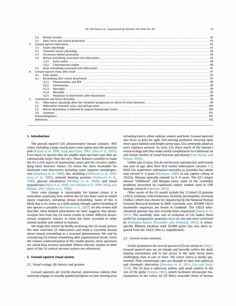
Fig. 2. AeD: Transmission electron microscope (TEM) images showing similar cone synapse ultrastructure of the torpid 13LGS and the Tg P347L rabbit. The latter is a model ofretinitis pigmentosa. EeF: Computational molecular phenotyping (CMP) of retina from a 13LGS recently emerged from torpor. Small molecule signals have been superimposed ontoTEM structural data, quantitatively informing as to cell type and metabolic state. A. Torpid 13LGS cone terminal with an aggregate of degenerate ribbon structures and synapticvesicles that has moved ~500 nm away from the remnant active zone ribbon. Scale bar, 500 nm. B: Tg P347L rabbit cone terminal showing degenerate ribbon structures resemblinghibernating squirrel and human RP. Scale bar, 500 nm. C: Higher magnification view of torpid 13LGS cone terminal (inset box from A) demonstrating an ectopic synaptic cloud(asterisk) and clear vesicles from fragmented Golgi apparatus (white arrow) along with the remnant synaptic ribbon (black arrow). Scale bar, 500 nm. D: Higher magnification viewTg P347L rabbit cone terminal (inset box from B), demonstrating an ectopic synaptic cloud (asterisk) as well as truncated or remnant synaptic ribbon (black arrow) and the clearfragmented Golgi vesicles (white arrow). Scale bar, 500 nm. E: CMP overlay mapping of GABA (red), glycine (green), and glutamate (blue) signals. Both excitatory (photoreceptor,bipolar and ganglion cells) and inhibitory retinal neurons (amacrine cells) can be visualized. Scale bar, 30 mm. F: CMP overlay mapping of taurine (red), glutamine (green), andglutamate (blue) signals. Alternate mapping of the same specimen in E demonstrates Müller cells alongside excitatory and inhibitory cell classes. Scale bar, 30 mm.
D.K. Merriman et al. / Experimental Eye Research 150 (2016) 90e105 95

during, or after hibernation. The now-monocular GS was allowed tosurvive until the next time point, at which time its remaining eyewas collected. Time post-enucleationwas not provided for any datashown. This protocol thus added variables of surgical stress, woundhealing, and chronology to the underlying hibernation physiology.
A second TEM study of the hibernating 13LGS retina appearedtwo years later (Rem"e and Young, 1977). These authors monitoredtheir GSs daily, keeping track of interbout arousals. Retinascollected in July, August, and September served as controls for thetorpid condition but, based on reported body masses, some controlanimals were well into the immergence transition stage. Hiberna-tion was initiated by placing all GSs in a cold, dark room on thesame fall day. Lights were on occasionally for “a few hours”, acondition that would never occur in nature. Retinas from both eyesof torpid animals were collected after 3e10 weeks in the cold room.To assess the retina's recovery from hibernation, one eye wasenucleated from a small number of torpid GSs that aroused andsurvived for 3e9 days in the warm room with photoperiodiclighting. Hence, as in the Kuwabara (1975) study, recovery fromtorpor was also on a background of surgical stress and woundhealing.
Rem"e and Young (1977) found little evidence for COS sheddingeven in (summer) controls, possibly because euthanasia occurred indaytime. As did Kuwabara (1975), they recorded the loss of synapticvesicles and most ribbons from cone pedicle active sites. However,while shortened COSs were observed in torpid animals, it was a farsubtler effect than Kuwabara (1975) reported. Also in contrast tothe earlier study, the size and number of ellipsoid mitochondriawere markedly reduced in torpid GS cones. Upon arousal, mito-chondria and synaptic ribbons recovered more quickly than didCOS length (~3 days versus ~7 days). Overall, the second studyfound little evidence that cone degeneration was progressive,suggesting instead a new steady state during torpor that rapidlyrecovered upon arousal.
3.4.2. Contemporary studiesAbout ten years ago, we began to re-examine retinal remodeling
during hibernation using TEM plus a variety of methods notavailable to the hibernation pioneers. These methods includeimmunoconfocal microscopy and computational molecular phe-notyping (CMP), which is further explained below. Some of our datahave been published only in abstract form.
Using standard immunocytochemistry, we observed limitedCOS shortening but significantly reduced immunostaining for cy-tochrome oxidase in cone ellipsoids of 13LGS during torpor (Gruberet al., 2006), in agreement with Rem"e and Young (1977). Our sub-sequent study of ellipsoid mitochondria showed evidence of eitherincreased fission or decreased fusion during torpor, and under-scored the notion that mitochondrial activity is regulated by theamount of mitochondrial protein (Kaden et al., 2013). We have alsoused optical coherence tomography (OCT) imaging to examine the13LGS retina during torpor, in comparison with arousal (Li et al.,2014). Signal amplitude and contrast are quite reduced duringtorpor, but alterations in cone ellipsoids are most evident.
Our TEM data also confirmed shorter ribbons at the cone pedicleactive sites and aggregates of ribbon material some distance awayin retinas collected from torpid 13LGS (Fig. 2AC). We showed this tobe a homogeneous feature across the torpid GS retina using anti-CtBP2/RIBEYE to immunostain synaptic ribbons (Mehta et al.,2013; Qiao et al., 2013; Vaughan et al., 2007). In hibernating GS,RIBEYE aggregate formation is strongly temperature-dependent,since it can be induced by cold treatment of 13LGS retinal tissuescollected from euthermic (summer) animals (Wei Li, unpublished).Most RIBEYE aggregates disappeared within 8 h of rewarming toeuthermia, apparently re-deployed to the cone pedicle active sites.
Interestingly, TEM images of cone synapses from torpid 13LGSare strikingly similar to those of degenerative cone synapses fromthe Tg 347L rabbit model of retinitis pigmentosa (Fig. 2BD; Joneset al., 2011). Cone degeneration in the rabbit model is, however,progressive and irreversible, in contrast to what seems to be thealternative steady state in hibernation.
Subsequently, our anti-BASSOON immunolabeling showed thatBASSOON largely remains at the cone active site during torpor, evenwhile RIBEYE aggregates detach from it (Qiao et al., 2013). In viewof the fact that torpor occurs in continuous darkness, we note that(in mice) light exposure contributes to BASSOON's ability to anchorribbons at the rod spherule's active zone (Spiwoks-Becker et al.,2013). Curiously, whereas 13LGS brain transcriptomics demon-strate upregulation of BASSOON transcripts during fall pre-
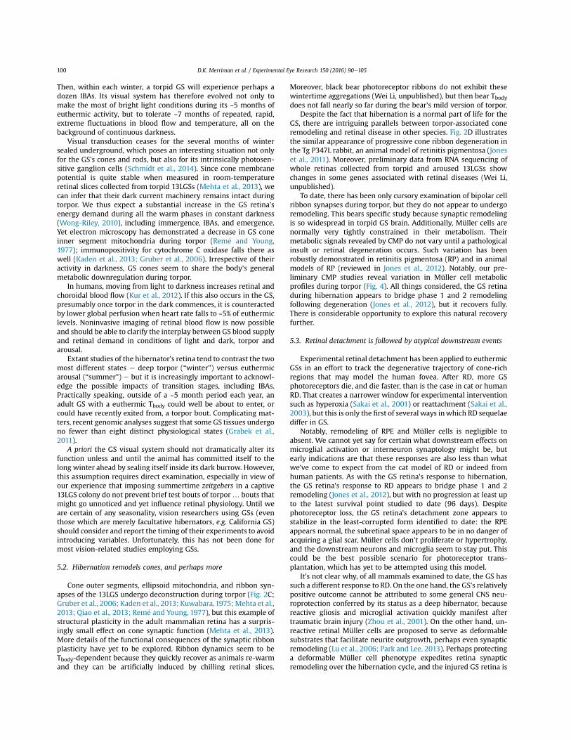
Fig. 3. CMP mapping of retina from a recently-emerged 13LGS. A: GABA (g), glycine(G), and glutamate (B) signals are shown with gGE / RGB mapping. ON cone bipolarcells are revealed by glycine content derived from coupling with AII amacrine cells. Asurprisingly large number of total bipolar cells (>90%) exhibit a glycinergic signature.B: Taurine (T), glutamine (Q), and glutamate (E) signals are shown with TQE/ RGB.Müller cells appear uniformly yellow/olive across the retina.
Fig. 4. CMP overlay mapping of retina collected from a torpid 13LGS. Taurine (T),glutamine (Q), and glutamate (E) signals are shown with TQE/ RGB demonstratingMüller cell metabolic signatures. Note the variation in the glutamine signal of indi-vidual Müller cell endfeet (arrows).
D.K. Merriman et al. / Experimental Eye Research 150 (2016) 90e10596

immergence, winter torpor, and winter IBAs (Schwartz et al., 2013),this is not seen in 13LGS retinal transcriptomes collected at similarhibernation stages (Wei Li, unpublished).
A follow-up tested the hypothesis that cone ribbon disruptionduring torpor would disrupt postsynaptic signaling (Mehta et al.,2013). Cone resting membrane potential, and the placement andamount of glutamate receptor immunopositivity, were unchangedduring torpor, and cone calcium currents were not significantlydifferent. However, there was a significant reduction in size andnumber of the miniature-like excitatory post-synaptic currents(mlEPSC) reflecting reduced vesicle release, particularly multi-vesicular release. With cone depolarization, mlEPSC frequency andamplitude did increase in retinas of torpid GSs, but never to the levelseen in retinas of aroused GSs. The detailed molecular mechanismsand physiological consequences associated with GS ribbon synapseplasticity during hibernation remain to be illustrated.
We have only just begun to use CMP to explore the GS retina'shistology, ultrastructure, metabolism and plasticity over the hi-bernation cycle. In CMP, small molecule signals are superimposedonto TEM structural data, quantitatively informing as to cell typeandmetabolic state (Jones andMarc, 2007). This approach has beenvery useful in addressing phenotype reprogramming and syn-aptology shifts that occur during retinal degenerations (Jones et al.,2011, 2012; Marc et al., 2007), i.e. during changes in physiologicalstate. Here we present only a sampling of our preliminary data,which does not yet encompass all of the GS's circannual cycle.
As shown in Fig. 2E, CMP mapping of GABA, glycine, and gluta-mate signals readily reveals excitatory and inhibitory neurons of the13LGS retina, which will allow us to appropriately identify any al-terations in synaptic relationships going forward. Moreover, alter-nate mapping of taurine, glutamine, and glutamate signals in thesame retinal sample provides valuable metabolic status informationabout Müller cells (Fig. 2F) and how this might change over time.
We have been particularly interested in applying CMP to GS bi-polar cells because, by standard TEM, their synaptic ribbons appearunaffected by hibernation. In a typical (rod-dominated) mammalianretina, total bipolar cells comprise ON, OFF, and rod types in roughlyequal proportions (e.g. mouse, W€assle et al., 2009). It is to be ex-pected that, in the cone-dominant GS retina (only 15% rods), theproportion of rod bipolar cells would be smaller. Using CMP, ON conebipolar cells are readily demonstrated by their glycine signals, whichthey acquire through gap junction coupling with AII amacrine cells.Indeed, in 13LGS retina just after emergence from torpor, glycinergicsignatures clearly predominate (Fig. 3A). One interpretation is that,as 13LGS arouse from torpor, there are alterations in bipolar cellcoupling patterns with amacrine cells. If confirmed in our ongoingwork, this is the first evidence of hibernation-associated synapticremodeling outside of cone pedicles.
In the healthy retina of every species examined up till now, CMPmapping has revealed homogeneous small molecular signalswithin all Müller cells, representing tightly controlled metabolicregulation (Fig. 3B). Only in retinas undergoing retinal degenera-tion have Müller cells within the same region exhibited dissimilarCMP signals (reviewed in Jones et al., 2012). We therefore note that,during torpor, individual 13LGS Müller cells do in fact exhibit dis-similar metabolic signals, particularly in glutamine and glutathione(Fig. 4). This variation is once again reminiscent of retinal degen-eration, but in GS represents a readily-reversible physiological statein a healthy animal. Overall, CMP has great potential to reveal thepart that retinal macroglia play in modulating retinal metabolismover the hibernation cycle.
3.5. Brain remodeling associated with hibernation
A growing literature on hibernating GS brain increasingly
documents physiological adaptations that may well extend toretina. Recent reviews address the GS's intrinsic tolerance of brainhypoxia (Garbarino et al., 2015; Larson et al., 2014) and brainischemia (Lee and Hallenbeck, 2013), supporting the notion thathibernation is a neuroprotected state (Dave et al., 2012). Moreover,presynaptic ribbon dynamics similar to that seen in cones have alsobeen reported in GS pinealocytes (McNulty et al., 1990). Otherstudies report dendrite withdrawal in hibernating GS frontal cortex(Ruediger et al., 2007), cerebellum (Popov and Bocharova, 1992),thalamus (von der Ohe et al., 2007), and hippocampus (Popov et al.,2007; Sallmen et al., 2003; von der Ohe et al., 2007). All findingspoint to reversible, temperature-driven changes in the location of,and association between, pre- and postsynaptic proteins. Thus,synaptic recovery on rewarming relies upon a reservoir of nearbyproteins, not on new protein synthesis (von der Ohe et al., 2007).
An elegant study has compared the proteomes of 13LGS fore-brains collected during multiple physiological states across thehibernation year (Hindle and Martin, 2013). Of the more than 3000forebrain proteins surveyed, fewer than 3% showed significantdifferences, and there was surprisingly little change in metabolicenzyme content. Most of the altered proteins were related to thecytoskeleton, cytoskeletal regulation, and Caþ2 regulation, with themajority differing based on Tbody not time of year. The relevance ofthese findings to neuroplasticity is obvious.
While hibernating brain neuron remodeling studies vastlyoutnumber retinal neuron remodeling studies at this time, theknowledge base for GS retinal circuitry may recommend it as auseful tissue to explore this phenomenon in detail.
4. Ground squirrel retina after insult
4.1. Early studies
Relatively few studies have examined GS retinal responses tophysical, nutritional, and pharmacotoxic insults. Vaidya (1965)used retinal cautery solely to elucidate 13LGS central visual path-ways. Berson (1973) fed 13LGSs a Vitamin A-deficient diet andmaintained them in either dim or moderate cyclic light, prelimi-narily reporting pathology only in those animals maintained inmoderate light. Farber and colleagues (1983, 1981) inducedphotoreceptor degeneration in 13LGS retinas using intracardiacinjections of iodoacetate, which inhibits glycolysis. Anderson andcolleagues (1988) induced (cone) photoreceptor degeneration inCalifornia GS retinas using intraocular injections of tunicamycin,which inhibits protein glycosylation.
While ground-breaking in their ownways, these early GS papersdid not directly address neuronal or glial remodeling responses.That changed about 15 years ago with a group of experimentalretinal detachment studies using the California GS.
4.2. Remodeling after retinal detachment
In retinal detachment (RD), physical distancing of the neuro-retina from the choriocapillaris separates photoreceptors fromtheir lifeline as well as from their companion RPE. The result in thedetachment zone is the deconstruction of all photoreceptor outerand inner segments and the retraction of rod spherules. Over time,many photoreceptors die by apoptosis (Fisher et al., 2005, 2007;Mervin et al., 1999). Impact on human vision is worst with macu-lar detachment. Examination of rare human RD specimens longafter initial injury demonstrates not only photoreceptor loss, butalso widespread remodeling of the RPE, surviving photoreceptors,second and third order neurons, Müller cells, and nerve fiber layerastrocytes (Sethi et al., 2005).
It bears mention that, alone among retinal cell types and in all
D.K. Merriman et al. / Experimental Eye Research 150 (2016) 90e105 97

mammalian species examined to date, cones within detachmentzones rapidly downregulate their expression of cone-specific pro-teins, making it impossible to detect them using immunocyto-chemistry. This is true of cones that survive RD and remaindeconstructed for months or years (Fisher et al., 2005). Upon suc-cessful reattachment to the RPE, deconstructed cones quicklyresume cone-specific protein expression, becoming identifiable byimmunolabeling once more. This unlucky phenomenon, plus thenumeric rod dominance of most mammals (including humans),explains why rhodopsin immunolabeling is routinely used to assessphotoreceptor responses to detachment, even in overwhelminglycone-dominant mammals like the GS. The assessment of cone re-sponses to RD thus depends on low-throughput methods like TEMthat do not depend on protein expression.
Rescue of photoreceptor structure and function by reattachmentto the RPE is possible so long as subretinal scars due to Müller cellgliosis have not formed. Reattachment does not reverse allabnormal changes (Fig. 5A) and indeed seems to trigger additionalremodeling (Lewis et al., 2003), including the expansion of Müllercell endfoot processes into the vitreous where they contribute tofibrocontractile complications such as proliferative vitreoretinop-athy (Fisher et al., 2005).
Human RD sequelae are recapitulated in animal models, withthe distinct advantage of monitoring events chronologically bothpost-RD and post-reattachment as a way to assess interventionopportunities (Cuenca et al., 2014; Jones et al., 2012). The questionof how well any animal RD model stacks up to human RD cannottruly be answered at this time, because human data are sporadicwith many patient variables. Particularly lacking are ultrastructuraland histochemical studies of human foveal detachment that couldbe compared to existing animal data. The animal data themselveshave employed different times post-RD and different types ofanalysis have been applied to these samples. Studies on non-human primates, which arguably have the most relevance, havesmall sample sizes due to the expense and logistics of primate usein research.
Outside of non-human primates, the cat has proven the bestmatch overall (Fig. 5B; Fisher et al., 2007, 2005), clearly demon-strating not only reactive gliosis but also extensive, aberrant
neuritogenesis by primary, secondary, and tertiary retinal neurons(Coblentz et al., 2003; Sakai et al., 2014; Lewis et al., 1998; Linberget al., 2006, 2009). Post-RD remodeling of remnant neuropil likelyexplains why physical reattachment fails to coincide with func-tional recovery (Jones et al., 2012), especially with macular reat-tachment (Ross, 2002). Aberrant neuritogenesis is also a feature ofhuman AMD (Sullivan et al., 2007) and of epiretinal membranesfrom multiple causes (Lesnik-Oberstein et al., 2011).
Experimental RD was originally applied to the California GSbecause of its numeric cone dominance and its central retina'sunique potential to model foveal RD in a sub-primate (Jacobs et al.,2002). As in cat, sterile injections of sodium hyaluronate are used tocreate detachments of controlled height and diameter as desired,aided by the GS's small lens. The following summary derives from aquartet of GS studies (Lewis et al., 2005; Linberg et al., 2002; Sakaiet al., 2001, 2003) and an authoritative review of experimental RD(Fisher et al., 2005). Time points post-RD ranged from 10 h to 28days; a reattachment study followed GSs out to 96 days of recovery.All data derive from euthermic, aroused California GSs whose hi-bernation stage was otherwise undetermined. It is debatedwhether California GSs hibernate in the southern part of their rangewhere these studies were conducted, but they do so in the northernpart.
4.2.1. Photoreceptors and RPEAfter detachment, GS rods and M-cones deconstructed as
described in other mammalian species. Outer segments weredisorganized and lost, cones stopped expressing cone-specificproteins, and rod visual pigment redistributed over most of thecell (Fig. 5C). As GS photoreceptor OSs degenerated, the RPEcontinued to phagocytose them, at least in the early stages. Ulti-mately, GS photoreceptors died by apoptosis and were cleared fromthe subretinal space by cells identified as macrophages, withpossible contributions from microglia and Müller cells.
The detachment zone in GS retina was notable for how many ofits photoreceptors died and how quickly (Fig. 5C), something it hasin commonwith rabbit, but not with cat or humanwhere up to 50%of photoreceptors may survive for over a year in their decon-structed state even when reattachment is not attempted (Fig. 5AB).
Fig. 5. Immunoconfocal microscopy of detached human, cat, and California GS retina co-labeled with antibodies to rhodopsin and GFAP, plus a lectin that identifies microglia, bloodvessels, and epiretinal membranes. Cones are not shown because they cease to express cone markers after RD. In all three images, the ILM is at the bottom and the SRS is at top. Thiswork was originally published in Lewis et al., 2005. Microglial cell activation following retinal detachment: a comparison between species. Mol Vis 11, 491-500. Reprinted courtesyof the Authors and Molecular Vision. Copyright by the Authors (2005). A, Human, >30 days post-RD, had undergone reattachment prior to its excision. Note surviving butdeconstructed rods (red), upregulated GFAP (blue), scattered microglia (green), and epiretinal membrane at ILM (green, bottom). B, Cat, 28 days post-RD (no reattachment). Notesurviving but deconstructed rods (red), upregulated GFAP including a glial scar in the SRS (blue, top), and scattered microglia (green). C, California GS, 7 days post-RD (no reat-tachment). Note surviving but deconstructed rods (red), normal expression of GFAP (blue), and normal position of microglia (green; large green objects near ILM are blood vessels).
D.K. Merriman et al. / Experimental Eye Research 150 (2016) 90e10598

Despite enhanced photoreceptor death, the squirrel OPL appearedunaffected, other than the obvious loss of cone pedicles and rodspherules. The outer limiting membrane (OLM) appeared intact,and a flat outer retinal surface in the detachment zone (formed bybranchedMüller cell processes) was the result. Remarkably, despitenear-complete photoreceptor loss, squirrel RPE did not proliferateor dedifferentiate to any appreciable degree.
4.2.2. InterneuronsStudied only with structural means to date, there were no
apparent changes in the GS inner nuclear or ganglion cell layers,and the plexiform layers appeared unaffected other than the loss ofphotoreceptor terminals from the OPL. This finding probablyshould be considered preliminary until it is confirmed usingimmunocytochemical probes that identify inner retinal neurons.
4.2.3. MacrogliaOutside of the GS visual system papers reviewed here, we have
found only one paper that has examined reactive gliosis in any GS:Zhou and colleagues (2001) documented local astrocytosis ineuthermic Arctic GS brain three days after damage commenced.Thus, we know that the euthermic GS central nervous system iscapable of reactive gliosis.
With this positive control in mind, it is remarkable that no ev-idence of Müller cell reactivity was found in detached (euthermic)California GS retina. Instead, Müller cell processes neatly filled onlythe empty space left by dead photoreceptors, preserving the gen-eral structure of what were once the OLM and ONL, save for anoccasional presumed horizontal cell. There was no evidence ofMüller cell proliferation, migration, or remodeling associated withscarring (Fig. 5C). We have confirmed this finding in detachedretina of a second GS species, the 13LGS (Fig. 6).
In fact, after detachment, California GS Müller cells actuallyincreased expression of normal function proteins, includingCRALBP, glutamine synthetase, and EAAT1, responses proposed toimprove the metabolic health of surviving (cone) photoreceptors.Occasional nerve fiber layer (NFL) astrocytes extended persistentGFAP-immunopositive processes into the outer retina, more oftenin reattached GS retina. This evidence suggests a natural inhibition,in at least two GS species, of what we think of as normal macroglialresponses to photoreceptor degeneration.
4.2.4. MicrogliaOutside of the GS visual system papers reviewed here, we have
found only one paper that has examined microglial activation inany GS: Zhou and colleagues (2001) documented local microglialactivation in euthermic Arctic GS brain three days after damagecommenced. Thus, we know that the euthermic GS central nervoussystem is capable of responding to insult in this way.
With this positive control in mind, California GS microglia didnot mount the response to cone death after RD that we have cometo expect from other mammalian species (Fig. 5C). A caveat here,fully acknowledged by the study authors, is that the probes used atthe time (Griffonia and Ricinis lectins and antibody to CD11b)cannot always distinguish between microglia and macrophages.Microglial response to detachment should be re-examined in GSwith additional probes that differentiate activated from restingstates (reviewed in Karlstetter et al., 2015).
4.2.5. Responses to intervention after detachmentAs in cat, GS cone apoptosis was reduced when squirrels were
continuously exposed to 70% O2 immediately after experimental RDup to 3 days later. Hyperoxia also lessened (but did not eliminate)other signs of cone deconstruction. Additionally, reattachment ofGS retina 24 h after detachment dramatically boosted the numberof viable (cone) photoreceptors, with concomitant recovery of thecone ERG contrast gain. The overall picture from California GS afterreattachment is a slow recovery over ~3 months, similar to whatlittle is known about cone recovery in human patients after reat-tachment. Thus, GS cones perish quickly after detachment, andrecover slowly after reattachment.
5. Conclusions and future directions
5.1. Hibernation repeatedly alters the metabolic background onwhich GS retina functions
Most GS species, including all of those employed in visionresearch to date, are either facultative or obligate hibernators, so itis fair to generalize that hibernation physiology is part of the GSmodel. Hibernation (along with estivation) is evolution's solutionto seasonal problems of physiological stress, but it is a solution thatbrought its own set of challenges. Hibernation's challenges havebeen met not only by GSs but by a group of lower primates, thenocturnal dwarf lemurs of Madagascar, including one who burrowsunderground (Blanco et al., 2013).
Ground squirrels survive several years in the wild (Michener,1989), experiencing multiple hibernation cycles in a lifetime.
Fig. 6. Immunoconfocal images of control and detached 13LGS retina co-labeled with antibodies to nestin (red), GFAP (blue), and vimentin (green). Brackets show the ONL, ar-rowheads the ILM. A, Normal control retina. B, Retina 4 days after detachment. As in California GS, detached 13LGS retina remains nestin-immunonegative, and vimentin and GFAPlabeling remain unchanged, indications of quiescent macroglia despite widespread cone death.
D.K. Merriman et al. / Experimental Eye Research 150 (2016) 90e105 99
Then, within each winter, a torpid GS will experience perhaps adozen IBAs. Its visual system has therefore evolved not only tomake the most of bright light conditions during its ~5 months ofeuthermic activity, but to tolerate ~7 months of repeated, rapid,extreme fluctuations in blood flow and temperature, all on thebackground of continuous darkness.
Visual transduction ceases for the several months of wintersealed underground, which poses an interesting situation not onlyfor the GS's cones and rods, but also for its intrinsically photosen-sitive ganglion cells (Schmidt et al., 2014). Since cone membranepotential is quite stable when measured in room-temperatureretinal slices collected from torpid 13LGSs (Mehta et al., 2013), wecan infer that their dark current machinery remains intact duringtorpor. We thus expect a substantial increase in the GS retina'senergy demand during all the warm phases in constant darkness(Wong-Riley, 2010), including immergence, IBAs, and emergence.Yet electron microscopy has demonstrated a decrease in GS coneinner segment mitochondria during torpor (Rem"e and Young,1977); immunopositivity for cytochrome C oxidase falls there aswell (Kaden et al., 2013; Gruber et al., 2006). Irrespective of theiractivity in darkness, GS cones seem to share the body's generalmetabolic downregulation during torpor.
In humans, moving from light to darkness increases retinal andchoroidal blood flow (Kur et al., 2012). If this also occurs in the GS,presumably once torpor in the dark commences, it is counteractedby lower global perfusion when heart rate falls to ~5% of euthermiclevels. Noninvasive imaging of retinal blood flow is now possibleand should be able to clarify the interplay between GS blood supplyand retinal demand in conditions of light and dark, torpor andarousal.
Extant studies of the hibernator's retina tend to contrast the twomost different states e deep torpor (“winter”) versus euthermicarousal (“summer”) e but it is increasingly important to acknowl-edge the possible impacts of transition stages, including IBAs.Practically speaking, outside of a ~5 month period each year, anadult GS with a euthermic Tbody could well be about to enter, orcould have recently exited from, a torpor bout. Complicating mat-ters, recent genomic analyses suggest that some GS tissues undergono fewer than eight distinct physiological states (Grabek et al.,2011).
A priori the GS visual system should not dramatically alter itsfunction unless and until the animal has committed itself to thelong winter ahead by sealing itself inside its dark burrow. However,this assumption requires direct examination, especially in view ofour experience that imposing summertime zeitgebers in a captive13LGS colony do not prevent brief test bouts of torpor… bouts thatmight go unnoticed and yet influence retinal physiology. Until weare certain of any seasonality, vision researchers using GSs (eventhose which are merely facultative hibernators, e.g. California GS)should consider and report the timing of their experiments to avoidintroducing variables. Unfortunately, this has not been done formost vision-related studies employing GSs.
5.2. Hibernation remodels cones, and perhaps more
Cone outer segments, ellipsoid mitochondria, and ribbon syn-apses of the 13LGS undergo deconstruction during torpor (Fig. 2C;Gruber et al., 2006; Kaden et al., 2013; Kuwabara,1975;Mehta et al.,2013; Qiao et al., 2013; Rem"e and Young, 1977), but this example ofstructural plasticity in the adult mammalian retina has a surpris-ingly small effect on cone synaptic function (Mehta et al., 2013).More details of the functional consequences of the synaptic ribbonplasticity have yet to be explored. Ribbon dynamics seem to beTbody-dependent because they quickly recover as animals re-warmand they can be artificially induced by chilling retinal slices.
Moreover, black bear photoreceptor ribbons do not exhibit thesewintertime aggregations (Wei Li, unpublished), but then bear Tbodydoes not fall nearly so far during the bear's mild version of torpor.
Despite the fact that hibernation is a normal part of life for theGS, there are intriguing parallels between torpor-associated coneremodeling and retinal disease in other species. Fig. 2D illustratesthe similar appearance of progressive cone ribbon degeneration inthe Tg P347L rabbit, an animal model of retinitis pigmentosa (Joneset al., 2011). Moreover, preliminary data from RNA sequencing ofwhole retinas collected from torpid and aroused 13LGSs showchanges in some genes associated with retinal diseases (Wei Li,unpublished).
To date, there has been only cursory examination of bipolar cellribbon synapses during torpor, but they do not appear to undergoremodeling. This bears specific study because synaptic remodelingis so widespread in torpid GS brain. Additionally, Müller cells arenormally very tightly constrained in their metabolism. Theirmetabolic signals revealed by CMP do not vary until a pathologicalinsult or retinal degeneration occurs. Such variation has beenrobustly demonstrated in retinitis pigmentosa (RP) and in animalmodels of RP (reviewed in Jones et al., 2012). Notably, our pre-liminary CMP studies reveal variation in Müller cell metabolicprofiles during torpor (Fig. 4). All things considered, the GS retinaduring hibernation appears to bridge phase 1 and 2 remodelingfollowing degeneration (Jones et al., 2012), but it recovers fully.There is considerable opportunity to explore this natural recoveryfurther.
5.3. Retinal detachment is followed by atypical downstream events
Experimental retinal detachment has been applied to euthermicGSs in an effort to track the degenerative trajectory of cone-richregions that may model the human fovea. After RD, more GSphotoreceptors die, and die faster, than is the case in cat or humanRD. That creates a narrower window for experimental interventionsuch as hyperoxia (Sakai et al., 2001) or reattachment (Sakai et al.,2003), but this is only the first of several ways inwhich RD sequelaediffer in GS.
Notably, remodeling of RPE and Müller cells is negligible toabsent. We cannot yet say for certain what downstream effects onmicroglial activation or interneuron synaptology might be, butearly indications are that these responses are also less than whatwe've come to expect from the cat model of RD or indeed fromhuman patients. As with the GS retina's response to hibernation,the GS retina's response to RD appears to bridge phase 1 and 2remodeling (Jones et al., 2012), but with no progression at least upto the latest survival point studied to date (96 days). Despitephotoreceptor loss, the GS retina's detachment zone appears tostabilize in the least-corrupted form identified to date: the RPEappears normal, the subretinal space appears to be in no danger ofacquiring a glial scar, Müller cells don't proliferate or hypertrophy,and the downstream neurons and microglia seem to stay put. Thiscould be the best possible scenario for photoreceptor trans-plantation, which has yet to be attempted using this model.
It's not clear why, of all mammals examined to date, the GS hassuch a different response to RD. On the one hand, the GS's relativelypositive outcome cannot be attributed to some general CNS neu-roprotection conferred by its status as a deep hibernator, becausereactive gliosis and microglial activation quickly manifest aftertraumatic brain injury (Zhou et al., 2001). On the other hand, un-reactive retinal Müller cells are proposed to serve as deformablesubstrates that facilitate neurite outgrowth, perhaps even synapticremodeling (Lu et al., 2006; Park and Lee, 2013). Perhaps protectinga deformable Müller cell phenotype expedites retina synapticremodeling over the hibernation cycle, and the injured GS retina is
D.K. Merriman et al. / Experimental Eye Research 150 (2016) 90e105100

simply the “unintended” beneficiary.Another contributing factor could be the GS retina's pronounced
cone dominance (85%), a feature that cannot be separated from itshibernator status. Indeed, Lewis & Fisher (2000) have suggestedthat gliotic Müller cell outgrowth through the rod-dominant catretina's OLM during subretinal scar formation is especially associ-ated with cones. If that holds true generally in mammals, OLMdisruption might be greatly exaggerated in damaged cone-dominant retinas, potentially enacting a powerful selective pres-sure against it. In this regard, it would be useful to assess RDsequelae in non-hibernating, diurnal, cone-dominant rodent spe-cies (e.g. the Nile rat, 33% cones; the degu, 33% cones; the sand rat,44% cones). Interestingly, the Nile rat requires unprecedenteddosages of intense light and the DNA-disruptor MNU to attain(cone) photoreceptor degeneration, and even then there appears tobe a muted Müller cell response (Boudard et al., 2011).
Taking this a step further, it would be instructive to examine RDin one of the diurnal species of tree shrew because they have evenmore cones than do GSs (95%; Müller and Peichl, 1993), a tropicalanimal that does not hibernate. While a favorite model of experi-mental myopia, the tree shrew doesn't seem to have ever been usedin experimental RD. Should the tree shrew exhibit muted Müllercell reactivity much as does the GS, we would conclude that hi-bernation physiology has little relationship with the attenuation.Instead, some aspect of the cone-rich retinal microenvironmentmight play a role, a finding that could possibly have relevance forthe human fovea, albeit on a much smaller scale.
Strikingly, Müller cells of GFAP-/-Vimentin-/- mice also fail tomount a gliotic response after experimental RD. However, lack ofthese intermediate filaments renders the retinas of these knockoutmice unusually vulnerable to shear stress (Verardo et al., 2008), andtheir inner retinal cells are unusually vulnerable to ischemia-reperfusion injury (Wunderlich et al., 2015). Both consequenceswould be terrible disadvantages in a wild-living hibernator.
Cebulla and colleagues (2012) have referred to the GS's lack ofpost-RD reactive gliosis as a “research liability”. In truth, all animalmodels carry some sort of liability when it comes to modelinghuman vision. The mouse has certainly received its fair share ofcriticism (Baker, 2013). We take the alternate position that eachmodel species is a vital natural experiment, in the spirit of AugustKrogh's famous dictum, with something to contribute. It might beappropriate to view the GS at the far end of a gliosis continuum,with a checkpoint thanks to natural selection (Weil et al., 2008).After all, just within the mouse, Müller cell reactivity variesconsiderably depending on the inherited degeneration (Hippertet al., 2015). Moreover, different strains of mice subjected toexperimental RD react differently to RD (Matsumoto et al., 2014).For all we know, this could be true in humans, too.
Debate continues whether typical retinal macro- and microglialresponses to deafferentiation are helpful or harmful to survivingneurons. Strong arguments have beenmade in favor of limiting glialremodeling because it impedes clinical interventions (Jones et al.,2012; Marc et al., 2007; Pearson, 2014) and may exacerbate coneloss. The evidence to date in GS is that its limited glial response toRD does not attenuate photoreceptor loss, but does coincide withapparent stability of inner retinal neurons. Perhaps the two are co-regulated somehow, in a way that photoreceptor death and glialreactivity are not (Fisher et al., 2005). The knockout mouse litera-ture continues to produce candidate control mechanisms thatcould be assessed in GS retina (Cuenca et al., 2014; Karlstetter et al.,2015). Some intriguing findings regarding Müller cell glucocorti-coid receptor activation are being produced with a chick model ofRD (Gallina et al., 2015). Post-injury, these receptors upregulatebriefly and, when an agonist is applied, Müller cell proliferation andmicroglial reactivity are both reduced. If further examination of GS
retina confirms minimal microglial reaction post-RD, it would bepossible to determine if co-inhibition of negative remodeling(Wang and Wong, 2014) in the natural world occurs by a similarmechanism.
In addition, Müller cell reprogramming for the purposes ofretinal regeneration has been proposed, with experiments done inmice (Goldman, 2014). To borrow a term from that author, withtheir uniquely stable properties in the face of severe retinal insult,how easily might GS Müller cells be “persuaded” in this regard?
Lastly, the GS's lack of gliofibrotic scarring after RD is curiouslyechoed in its skeletal muscle. When muscle's damaged state isprolonged, its eventual repair includes fibrosis, another version ofscarring (Mann et al., 2011). Andres-Mateos and colleagues (2012)modeled focal muscle damage in 13LGS using a toxin injection.When damage occurred during torpor, muscle repair was inhibited,prolonging the damage state for weeks. When those animals wereallowed to emerge naturally in spring, complete muscle repairresulted but, unexpectedly, without any fibrosis (Andres-Mateoset al., 2012).
5.4. Summary
Ground squirrel retinas contain a disproportionate number ofcones, making them a worthy model of cone-based visual function.They share with human a diurnal habit that includes specific ar-chitecture of rod nuclei and attendant DNA repair ability. It isimportant to remain cognizant of seasonal hibernation physiologywhen using this animal model. Concurrent with low Tbody duringhibernation torpor, GS cones exhibit deconstructive remodeling oftheir OS, ellipsoid mitochondria, and ribbon synapses. These fea-tures of the torpid GS retina resemble the early stages of retinaldegeneration in non-hibernators, but do not progress and arerapidly reversed upon arousal from torpor. Additional study maywell reveal more features of hibernation physiology in additionalcell types, along with mechanisms underlying natural recovery.
Widespread cone death by experimental RD in the euthermic(“summer”) California GS and 13LGS has revealed the strongestdisconnect yet between deafferentiation and remodeling of Müllercells. Interneuron remodeling and microglial reactivity also appearreduced or absent, but more investigation of th is needed. Groundsquirrel evolution seems to have imposed a checkpoint in theretinal degenerative trajectory that may create a favorable envi-ronment for therapeutic approaches to photoreceptor loss.
Understanding the GS's evolved solutions d- to many of thesame problems faced by patientsd- may provide insights for noveltherapies (Buffenstein et al., 2014; van Breukelen and Martin, 2015;Weil et al., 2008). Hibernation's relevance to neuroprotectionafforded by ischemic preconditioning has not gone unnoticed (Daveet al., 2012). Moreover, the 13LGS's suitability for noninvasive im-aging technologies such as OCT and AOSLO, and its growing use incomparative genomics, epigenomics, transcriptomics, and prote-omics, position this animal model well to facilitate translationalhypotheses in the retina and more generally in the central nervoussystem (Alvarado et al., 2015; Biggar and Storey, 2014; Grabek et al.,2015; Schwartz et al., 2013; Xu et al., 2013).
Acknowledgments
We thank T.B. Connor MD (Medical College of Wisconsin) forcarrying out the experimental retinal detachments on 13LGS, andG.P. Lewis and G. Luna (University of California Santa Barbara) forproviding the corresponding microscopy (Fig. 3).
We thank Elsevier and G.P. Lewis and colleagues (University ofCalifornia Santa Barbara) for permitting the reprint of the imagesshown in Fig. 5.
D.K. Merriman et al. / Experimental Eye Research 150 (2016) 90e105 101

We thank S.K. Fisher (University of California Santa Barbara) forhelpful discussion during the preparation of this manuscript.
This work was funded by the University of Wisconsin OshkoshFaculty Development and Student Collaborative Grant Programsand the AxleTech International Endowed Professorship (D.K.M.),NEI Core Grant P30EY001931 (B.S.S.), the NEI Intramural ResearchProgram (W.L.), and NIH EY015128, EY02576, EY014800 VisionCore, and an unrestricted grant from Research to Prevent Blindnessto the Moran Eye Center (B.W.J.).
References
Abreu, M., Kicliter, E., Lugo-Garcia, N., 1993. Displaced amacrine cells in the ganglioncell layer of the ground squirrel retina. P. R. Health Sci. J. 12, 137e141.
Agarwala, S., Petry, H.M., May 3rd, J.G., 1989. Retinal projections in the groundsquirrel (Citellus tridecemlineatus). Vis. Neurosci. 3, 537e549.
Agoston, D., 2015. Bench-to-bedside and bedside back to the bench; seeking abetter understanding of the acute pathophysiological process in severe trau-matic brain injury. Front. Neurol. 6, 47. http://dx.doi.org/10.3389/fneur.2015.00047.
Ahnelt, P.K., 1985. Characterization of the color related receptor mosaic in theground squirrel retina. Vis. Res. 25, 1557e1567.
Alvarado, S., Mak, T., Liu, S., Storey, K.B., Szyf, M., 2015. Dynamic changes in globaland gene-specific DNA methylation during hibernation in adult thirteen-linedground squirrels, Ictidomys tridecemlineatus. J. Exp. Biol. 218, 1787e1795.http://dx.doi.org/10.1242/jeb.116046.
Anderson, D.H., Fisher, S.K., Steinberg, R.H., 1978. Mammalian cones: disc shedding,phagocytosis, and renewal. Invest. Ophthalmol. Vis. Sci. 17, 117e133.
Anderson, D.H., Neitz, J., Saari, J.C., Kaska, D.D., Fenwick, J., Jacobs, G.H., Fisher, S.K.,1986. Retinoid-binding proteins in cone-dominant retinas. Invest. Ophthalmol.Vis. Sci. 27, 1015e1026.
Anderson, D.H., Williams, D.S., Neitz, J., Fariss, R.N., Fliesler, S.J., 1988. Tunicamycin-induced degeneration in cone photoreceptors. Vis. Neurosci. 1, 153e158.
Andres-Mateos, E., Mejias, R., Soleimani, A., Lin, B.M., Burks, T.N., Marx, R., Lin, B.,Zellars, R.C., Zhang, Y., Huso, D.L., Marr, T.G., Leinwand, L.A., Merriman, D.K.,Cohn, R.D., 2012. Impaired skeletal muscle regeneration in the absence offibrosis during hibernation in 13-lined ground squirrels. PLoS ONE 7, e48884.http://dx.doi.org/10.1371/journal.pone.0048884.
Applebury, M.L., Antoch, M.P., Baxter, L.C., Chun, L.L., Falk, J.D., Farhangfar, F.,Kage, K., Krzystolik, M.G., Lyass, L.A., Robbins, J.T., 2000. The murine conephotoreceptor: a single cone type expresses both S and M opsins with retinalspatial patterning. Neuron 27, 513e523.
Baker, M., 2013. Through the eyes of a mouse. Nature 502, 156e158.Berson, E.L., 1973. Experimental and therapeutic aspects of photic damage to the
retina. Invest. Ophthalmol. 12, 35e44.Besharse, J.C., Bok, D., 2011. The Retina and its Disorders. Academic Press.Biggar, K.K., Storey, K.B., 2014. New approaches to comparative and animal stress
biology research in the post-genomic era: a contextual overview. Comput.Struct. Biotechnol. J. 11, 138e146. http://dx.doi.org/10.1016/j.csbj.2014.09.006.
Blanco, M.B., Dausmann, K.H., Ranaivoarisoa, J.F., Yoder, A.D., 2013. Undergroundhibernation in a primate. Sci. Rep. 3, 1768. http://dx.doi.org/10.1038/srep01768.
Boudard, D.L., Acar, N., Bretillon, L., Hicks, D., 2011. Retinas of the diurnal rodentArvicanthis ansorgei are highly resistant to experimentally induced stress anddegeneration. Invest. Ophthalmol. Vis. Sci. 52, 8686e8700. http://dx.doi.org/10.1167/iovs.11-8162.
Bouma, H.R., Kroese, F.G., Kok, J.W., Talaei, F., Boerema, A.S., Herwig, A.,Draghiciu, O., van Buiten, A., Epema, A.H., van Dam, A., Strijkstra, A.M.,Henning, R.H., 2011. Low body temperature governs the decline of circulatinglymphocytes during hibernation through sphingosine-1-phosphate. Proc. Natl.Acad. Sci. U. S. A. 108, 2052e2057. http://dx.doi.org/10.1073/pnas.1008823108.
Bratincsak, A., McMuller, D., Miyake, S., Toth, Z.E., Hallenbeck, J.M., Palovits, M.,2007. Spatial and temporal activation of brain regions in hibernation: c-fosexpression during the hibernation bout in thirteen-lined ground squirrel.J. Comp. Neurol. 505, 443e458. http://dx.doi.org/10.1002/cne.21507.
Buffenstein, R., Nelson, O.L., Corbit, K.C., 2014. Questioning the preclinical paradigm:natural, extreme biology as an alternative discovery platform. Aging 6,913e920.
Campi, K.L., Krubitzer, L., 2010. Comparative studies of diurnal and nocturnal ro-dents: differences in lifestyle result in alterations in cortical field size andnumber. J. Comp. Neurol. 518, 4491e4512. http://dx.doi.org/10.1002/cne.22466.
Cao, L.H., Luo, D.G., Yau, K.W., 2014. Light responses of primate and othermammalian cones. Proc. Natl. Acad. Sci. U. S. A. 111, 2752e2757. http://dx.doi.org/10.1073/pnas.1400268111.
Cebulla, C.M., Zelinka, C.P., Scott, M.A., Lubox, M., Bingham, A., Rasiah, S.,Mahmoud, A.M., Fischer, A.J., 2012. A chick model of retinal detachment: conerich and novel. PLoS One 7, e44257. http://dx.doi.org/101371/journal.pone.0044257.
Chen, S., Li, W., 2012. A color-coding amacrine cell may provide a blue-off signal in amammalian retina. Nat. Neurosci. 15, 954e956. http://dx.doi.org/10.1038/nn.3128.
Christian, S.L., Rasley, B.T., Roe, T., Moore, J.T., Harris, M.B., Drew, K.L., 2014.
Habituation of Arctic ground squirrels (Urocitellus parryii) to handling andmovement during torpor to prevent artificial arousal. Front. Physiol. 5, 174.http://dx.doi.org/10.3389/fphys.2014.00174.
Chou, B.R., Cullen, A.P., 1984. Spectral transmittance of the ocular media of thethirteen-lined ground squirrel. Spermophilus tridecemlineatus. Can. J. Zool 62,825e830.
Clemens, L.E., Heldmaier, G., Exner, C., 2009. Keep cool: memory is retained duringhibernation in Alpine marmots. Physiol. Behav. 98, 78e84. http://dx.doi.org/10.1016/j.physbeh.2009.04.013.
Coblentz, F.E., Radeke, M.J., Lewis, G.P., Fisher, S.K., 2003. Evidence that ganglioncells react to retinal detachment. Exp. Eye Res. 76, 333e342.
Cooper, R.M., 2002. Diffuse light increases metabolic activity in the lateral genic-ulate nucleus, visual cortex, and superior colliculus of the cone-dominatedground squirrel visual system. Vis. Res. 42, 2899e2907.
Corbo, J.C., Cepko, C.L., 2005. A hybrid photoreceptor expressing both rod and conegenes in a mouse model of enhanced S-cone syndrome. PLoS Genet. 1, e11.http://dx.doi.org/10.1371/journal.pgen.0010011.
Cuenca, N., Deng, P., Linberg, K.A., Fisher, S.K., Kolb, H., 2003. Choline acetyl-transferase is expressed by non-starburst amacrine cells in the ground squirrelretina. Brain Res. 964, 21e30.
Cuenca, N., Deng, P., Linberg, K.A., Lewis, G.P., Fisher, S.K., Kolb, H., 2002. The neu-rons of the ground squirrel retina as revealed by immunostains for calciumbinding proteins and neurotransmitters. J. Neurocytol. 31, 649e666.
Cuenca, N., Fernandez-Sanchez, L., Campello, L., Maneu, V., De la Villa, P., Lax, P.,Pinilla, I., 2014. Cellular responses following retinal injuries and therapeuticapproaches for neurodegenerative diseases. Prog. Retinal Eye Res. 43, 17e75.http://dx.doi.org/10.1016/j.preteyeres.2014.07.001.
Cull, G., Cioffi, G.A., Dong, J., Homer, L., Wang, L., 2003. Estimating normal opticnerve axon numbers in non-human primate eyes. J. Glaucoma 12, 301e306.
Daniele, L.L., Lillo, C., Lyubarsky, A.L., Nikonov, S.S., Philp, N., Mears, A.J., Swaroop, A.,Williams, D.S., Pugh Jr., E.N., 2005. Cone-like morphological, molecular, andelectrophysiological features of the photoreceptors of the Nrl knockout mouse.Invest. Ophthalmol. Vis. Sci. 46, 2156e2167. http://dx.doi.org/10.1167/iovs.04-1427.
Dave, K.R., Christian, S.L., Perez-Pinzon, M.A., Drew, K.L., 2012. Neuroprotection:lessons from hibernators. Comp. Biol. Physiol. 162, 1e9. http://dx.doi.org/10.1016/j.cbpb.2012.01.008.
de Vrij, E.L., Vogelaar, P.C., Goris, M., Houwertjes, M.C., Herwig, A., Dugbartey, G.J.,Boerema, A.S., Strijkstra, A.M., Bouma, H.R., Henning, R.H., 2014. Platelet dy-namics during natural and pharmacologically induced torpor and forced hy-pothermia. PLoS ONE 9, e93218. http://dx.doi.org/10.1371/journal.pone.0093218.
DeVries, S.H., 2000. Bipolar cells use kainate and AMPA receptors to filter visualinformation into separate channels. Neuron 28, 847e856.
DeVries, S.H., Li, W., Saszik, S., 2006. Parallel processing in two transmitter micro-environments at the cone photoreceptor synapse. Neuron 50, 735e748. http://dx.doi.org/10.1016/j.neuron.2006.04.034.
DeVries, S.H., Qi, X., Smith, R., Makous, W., Sterling, P., 2002. Electrical couplingbetween mammalian cones. Curr. Biol. 12, 1900e1907.
DeVries, S.H., Schwartz, E.A., 1999. Kainate receptors mediate synaptic transmissionbetween cones and 'Off' bipolar cells in a mammalian retina. Nature 397,157e160. http://dx.doi.org/10.1038/16462.
Drew, K.L., Buck, C.L., Barnes, B.M., Christian, S.L., Rasley, B.T., Harris, M.B., 2007.Central nervous system regulation of mammalian hibernation: implications formetabolic suppression and ischemia tolerance. J. Neurochem. 102, 1713e1726.
Farber, D.B., Souza, D.W., Chase, D.G., 1983. Cone visual cell degeneration in groundsquirrel retina: disruption of morphology and cyclic nucleotide metabolism byiodoacetic acid. Invest. Ophthalmol. Vis. Sci. 24, 1236e1249.
Farber, D.B., Souza, D.W., Chase, D.G., Lolley, R.N., 1981. Cyclic nucleotides of cone-dominant retinas. Reduction of cyclic AMP levels by light and by cone degen-eration. Invest. Ophthalmol. Vis. Sci. 20, 24e31.
Fisher, S.K., Lewis, G.P., Linberg, K.A., Verardo, M.R., 2005. Cellular remodeling inmammalian retina: results from studies of experimental retinal detachment.Prog. Retinal Eye Res. 24, 395e431.
Fisher, S.K., Lewis, G.P., Linberg, K.A., Barawid, E., Verardo, M.R., 2007. Cellularremodeling in mammalian retina induced by retinal detachment. In: Kolb, H.,Fernandez, E., Nelson, R. (Eds.), Webvision: The Organization of the Retina andVisual System. University of Utah Health Sciences Center, Salt Lake City, UT.
Fredes, F., Vega-Zuniga, T., Karten, H., Mpodozis, J., 2012. Bilateral and ipsilateralascending tectopulvinar pathways in mammals: a study in the squirrel (Sper-mophilus beecheyi). J Comp. Neurol. 520, 1800e1818. http://dx.doi.org/10.1002/cne.23014.
Frohns, A., Frohns, F., Naumann, S.C., Layer, P.G., Lobrich, M., 2014. Inefficientdouble-strand break repair in murine rod photoreceptors with inverted het-erochromatin organization. Curr. Biol. 24, 1080e1090. http://dx.doi.org/10.1016/j.cub.2014.03.061.
Gallina, D., Zelinka, C.P., Cebulla, C.M., Fischer, A.J., 2015. Activation of glucocorticoidreceptors in Müller glia is protective to retinal neurons and suppressesmicroglial reactivity. Exp. Neurol. 273, 114e125. http://dx.doi.org/10.1016/j.expneurol.2015.08.007.
Garbarino, V.R., Orr, M.E., Rodriguez, K.A., Buffenstein, R., 2015. Mechanisms ofoxidative stress resistance in the brain: lessons learned from hypoxia tolerantextremophilic vertebrates. Arch. Biochem. Biophys. 576, 8e16. http://dx.doi.org/10.1016/j.abb.2015.01.029.
Goldman, D., 2014. Müller glial cell reprogramming and retinal regeneration. Nat.
D.K. Merriman et al. / Experimental Eye Research 150 (2016) 90e105102

Rev. Neurosci 15, 431e442. http://dx.doi.org/10.1038/nrn3723.Grabek, K.R., Karimpour-Fard, A., Epperson, L.E., Hindle, A., Hunter, L.E., Martin, S.L.,
2011. Multistate proteomics analysis reveals novel strategies used by a hiber-nator to precondition the heart and conserve ATP for winter heterothermy.Physiol. Genomics 43, 1263e1275.
Grabek, K.R., Martin, S.L., Hindle, A.G., 2015. Proteomics approaches shed new lighton hibernation physiology. J. Comp. Physiol. B 185, 607e627. http://dx.doi.org/10.1007/s00360-015-0905-9.
Gruber, A.R., Betts, K.E., Lewis, G.P., Fisher, S.K., Vaughan, D.K., 2006. Photoreceptorantigens are altered during season hibernation of a cone-dominant rodent.Invest. Ophthalmol. Vis. Sci. 47, a2845.
Gur, M., Sivak, J.G., 1979. Refractive state of the eye of a small diurnal mammal: theground squirrel. Am. J. Optom. Physiol. Opt. 56, 689e695.
Hains, P.G., Simpanya, M.F., Giblin, F., Truscott, R.J., 2006. UV filters in the lens of thethirteen lined ground squirrel (Spermophilus tridecemlineatus). Exp. Eye Res. 82,730e737. http://dx.doi.org/10.1016/j.exer.2005.09.014.
Helm, B., Ben-Shlomo, R., Sheriff, M.J., Hut, R.A., Foster, R., Barnes, B.M.,Dominoni, D., 2013. Annual rhythms that underlie phenology: biological time-keeping meets environmental change. Proc. Biol. Sci. 280, 20130016. http://dx.doi.org/10.1098/rspb.2013.0016.
Hindle, A.G., Martin, S.L., 2013. Cytoskeletal regulation dominates temperature-sensitive proteomic changes of hibernation in forebrain of 13-lined groundsquirrels. PLoS ONE 8, e71627. http://dx.doi.org/10.1371/journal.pone.0071627.
Hippert, C., Graca, A.B., Barber, A.C., West, E.L., Smith, A.J., Ali, R.R., Pearson, R.A.,2015. Muller glia activation in response to inherited retinal degeneration ishighly varied and disease-specific. PLoS ONE 10, e0120415. http://dx.doi.org/10.1371/journal.pone.0120415.
Hollenberg, M.J., Bernstein, M.H., 1966. Fine structure of the photoreceptor cells ofthe ground squirrel (Citellus tridecemlineatus tridecemlineatus). Am. J. Anat. 118,359e373. http://dx.doi.org/10.1002/aja.1001180204.
Hughes, A., 1977. The topography of vision in mammals of contrasting life stylecomparative optics and retinal organization. In: Crescitelli, F. (Ed.), Handbook ofSensory Physiology, VII/5: The Visual System in Vertebrates. Springer-Verlag,Berlin, pp. 613e656.
Hut, R.A., Dardente, H., Riede, S.J., 2014. Seasonal timing: how does a hibernatorknow when to stop hibernating? Curr. Biol. 24, R602eR605. http://dx.doi.org/10.1016/j.cub.2014.05.061.
Hut, R., Van der Zee, E., Jansen, K., Gerkema, M., Daan, S., 2002. Gradual reap-pearance of post-hibernation circadian rhythmicity correlates with numbers ofvasopressin-containing neurons in the suprachiasmatic nuclei of Europeanground squirrels. J. Comp. Physiol. B 172, 59e70.
Jacobs, G.H., 1990. Duplicity theory and ground squirrels: linkages between pho-toreceptors and visual function. Vis. Neurosci. 5, 311e318.
Jacobs, G.H., Blakeslee, B., Tootell, R.B., 1981. Color-discrimination tests on fibers inground squirrel optic nerve. J. Neurophys. 45, 903e914.
Jacobs, G.H., Calderone, J.B., Sakai, T., Lewis, G.P., Fisher, S.K., 2002. An animal modelfor studying cone function in retinal detachment. Doc. Ophthalmol. 104,119e132.
Jacobs, G.H., Neitz, J., 1984. Development of spectral mechanisms in the groundsquirrel retina following lid opening. Exp. Brain Res. 55, 507e514.
Jacobs, G.H., Tootell, R.B., 1981. Spectral-response properties of optic-nerve fibers inthe ground squirrel. J. Neurophys. 45, 891e902.
Jani, A., Martin, S.L., Jain, S., Keys, D., Edelstein, C.L., 2013. Renal adaptation duringhibernation. Am. J. Physiol. Ren. Physiol. 305, F1521eF1532. http://dx.doi.org/10.1152/ajprenal.00675.2012.
Johnson, P.T., Geller, S.F., Reese, B.E., 1998. Distribution, size and number of axons inthe optic pathway of ground squirrels. Exp. Brain Res. 118, 93e104.
Jonas, J.B., Muller-Bergh, J.A., Schlotzer-Schrehardt, U.M., Naumann, G.O., 1990.Histomorphometry of the human optic nerve. Invest. Ophthalmol. Vis. Sci. 31,736e744.
Jones, B.W., Kondo, M., Terasaki, H., Lin, Y., McCall, M., Marc, R.E., 2012. Retinalremodeling. Jpn. J. Ophthalmol. 56, 289e306. http://dx.doi.org/10.1007/s10384-012-0147-2.
Jones, B.W., Kondo, M., Terasaki, H., Watt, C.B., Rapp, K., Anderson, J., Lin, Y.,Shaw, M.V., Yang, J.H., Marc, R.E., 2011. Retinal remodeling in the Tg P347Lrabbit, a large-eye model of retinal degeneration. J. Comp. Neurol. 519,2713e2733. http://dx.doi.org/10.1002/cne.22703.
Jones, B.W., Marc, R.E., 2007. Visualizing the metabolome. Microsc. Microanal. 13(Suppl. 2) http://dx.doi.org/10.1017/S1431927607072601.
Kaden, T., Du, J., Goel, A., Li, W., 2013. Structural and functional properties ofphotoreceptor mitochondria in awake and hibernation ground squirrels. Invest.Ophthalmol. Vis. Sci. 54, a6082.
Karlstetter, M., Scholz, R., Rutar, M., Wong, W.T., Provis, J.M., Langmann, T., 2015.Retinal microglia: just bystander or target for therapy? Prog. Retinal Eye Res. 45,30e57. http://dx.doi.org/10.1016/j.preteyeres.2014.11.004.
Kraft, T.W., 1988. Photocurrents of cone photoreceptors of the golden-mantledground squirrel. J. Physiol. 404, 199e213.
Kryger, Z., Galli-Resta, L., Jacobs, G.H., Reese, B.E., 1998. The topography of rod andcone photoreceptors in the retina of the ground squirrel. Vis. Neurosci. 15,685e691.
Kuwabara, T., 1975. Cytologic changes of the retina and pigment epithelium duringhibernation. Invest. Ophthalmol. 14, 457e467.
Kur, J., Newman, E.A., Chan-Ling, T., 2012. Cellular and physiological mechanismsunderlying blood flow regulation in the retina and choroid in health and dis-ease. Prog. Retin Eye Res. 31, 377e406. http://dx.doi.org/10.1016/
j.preteyeres.2012.04.004.Larkin, J.E., Franken, P., Heller, H.C., 2002. Loss of circadian organization of sleep and
wakefulness during hibernation. Am. J. Physiol. Reg. I 282, R1086eR1095.http://dx.doi.org/10.1152/ajpregu.00771.2000.
Larson, J., Drew, K.L., Folkow, L.P., Milton, S.L., Park, T.J., 2014. No oxygen? Noproblem! intrinsic brain tolerance to hypoxia in vertebrates. J. Exp. Biol. 217,1024e1039. http://dx.doi.org/10.1242/jeb.085381.
Laursen, W.J., Funk, O., Goodman, J., Merriman, D.K., Ingolia, N.T., Bagriantsev, S.N.,Gracheva, E.O., 2014. Molecular adaptations to extreme thermogenesis inmammalian hibernators. Biophys. J. 106, 337a.
Lee, Y.J., Hallenbeck, J.M., 2013. SUMO and ischemic tolerance. Neuromol. Med. 15,771e781. http://dx.doi.org/10.1007/s12017-013-8239-9.
Lesnik Oberstein, S.Y., Lewis, G.P., Dutra, T., Fisher, S.K., 2011. Evidence that neuritesin human epiretinal membranes express melanopsin, calretinin, rod opsin andneurofilament protein. Br. J Ophthalmol. 95, 266e272. http://dx.doi.org/10.1136/bjo.2010.180679.
Lewis, G.P., Fisher, S.K., 2000. Muller cell outgrowth after retinal detachment: as-sociation with cone photoreceptors. Invest. Ophthalmol. Vis. Sci. 41, 1542e1545.
Lewis, G.P., Linberg, K.A., Fisher, S.K., 1998. Neurite outgrowth from bipolar andhorizontal cells after experimental retinal detachment. Invest. Ophthalmol. Vis.Sci. 39, 424e434.
Lewis, G.P., Sethi, C.S., Carter, K.M., Charteris, D.G., Fisher, S.K., 2005. Microglial cellactivation following retinal detachment: a comparison between species. Mol.Vis 11, 491e500.
Lewis, G.P., Sethi, C.S., Linberg, K.A., Charteris, D.G., Fisher, S.K., 2003. Experimentalretinal reattachment: a new perspective. Mol. Neurobiol. 28, 159e175.
Li, W., Chen, S., DeVries, S.H., 2010. A fast rod photoreceptor signaling pathway inthe mammalian retina. Nat. Neurosci. 13, 414e416. http://dx.doi.org/10.1038/nn.2507.
Li, W., DeVries, S.H., 2004. Separate blue and green cone networks in themammalian retina. Nat. Neurosci. 7, 751e756. http://dx.doi.org/10.1038/nn1275.
Li, W., DeVries, S.H., 2006. Bipolar cell pathways for color and luminance vision in adichromatic mammalian retina. Nat. Neurosci. 9, 669e675. http://dx.doi.org/10.1038/nn1686.
Li, W., Li, Y., Qiao, F., Hollyfield, J.G., Quian, H., Bell, B.A., 2014. Retinal changesassociated with hibernation revealed by OCT. Invest. Ophthalmol. Vis. Sci. 55,a2174.
Light, A.C., Zhu, Y., Shi, J., Saszik, S., Lindstrom, S., Davidson, L., Li, X., Chiodo, V.A.,Hauswirth, W.W., Li, W., DeVries, S.H., 2012. Organizational motifs for groundsquirrel cone bipolar cells. J. Comp. Neurol. 520, 2864e2887. http://dx.doi.org/10.1002/cne.23068.
Linberg, K.A., Lewis, G.P., Fisher, S.K., 2009. Retraction and remodeling of rodspherules are early events following experimental retinal detachment: an ul-trastructural study using serial sections. Mol. Vis. 15, 10e25.
Linberg, K.A., Lewis, G.P., Matsumoto, B., Fisher, S.K., 2006. Immunocytochemicalevidence that rod-connected horizontal cell axon terminals remodel inresponse to experimental retinal detachment in the cat. Mol. Vis. 12,1674e1686.
Linberg, K.A., Sakai, T., Lewis, G.P., Fisher, S.K., 2002. Experimental retinal detach-ment in the cone-dominant ground squirrel retina: morphology and basicimmunocytochemistry. Vis. Neurosci. 19, 603e619.
Linberg, K.A., Suemune, S., Fisher, S.K., 1996. Retinal neurons of the Californiaground squirrel, Spermophilus beecheyi: a golgi study. J. Comp. Neurol. 365,173e216. http://dx.doi.org/10.1002/(SICI)1096-9861(19960205)365:2<173::AID-CNE1>3.0.CO;2e2.
Long, K.O., Fisher, S.K., 1983. The distributions of photoreceptors and ganglion cellsin the California ground squirrel, Spermophilus beecheyi. J. Comp. Neurol. 221,329e340. http://dx.doi.org/10.1002/cne.902210308.
Long, K.O., Fisher, S.K., Fariss, R.N., Anderson, D.H., 1986. Disc shedding and auto-phagy in the cone-dominant ground squirrel retina. Exp. Eye Res. 43, 193e205.
Lu, Y.B., Franze, K., Seifert, G., Steinhauser, C., Kirchhoff, F., Wolburg, H., Guck, J.,Janmey, P., Wei, E.Q., Kas, J., Reichenbach, A., 2006. Viscoelastic properties ofindividual glial cells and neurons in the CNS. Proc. Natl. Acad. Sci. U. S. A. 103,17759e17764. http://dx.doi.org/10.1073/pnas.0606150103.
Lugo-Garcia, N., Blanco, R.E., 1993. Morphology and distribution of dopaminergicneurons in the ground squirrel retina. P. R. Health Sci. J. 12, 143e146.
Lugo-Garcia, N., Kicliter, E., 1987. Superior colliculus efferents to five subcorticalvisual system structures in the ground squirrel. Brain Res. 426, 131e141.
Lugo-Garcia, N., Kicliter, E., 1988. Morphology of ganglion cells which project to thedorsal lateral geniculate and superior colliculus in the ground squirrel. BrainRes. 454, 67e77.
Major, D.E., Rodman, H.R., Libedinsky, C., Karten, H.J., 2003. Pattern of retinal pro-jections in the California ground squirrel (Spermophilus beecheyi): anterogradetracing study using cholera toxin. J. Comp. Neurol. 463, 317e340. http://dx.doi.org/10.1002/cne.10764.
Mann, C.J., Perdiguero, E., Kharraz, Y., Aguilar, S., Pessina, P., Serrano, A.L., Munoz-Canoves, P., 2011. Aberrant repair and fibrosis development in skeletal muscle.Skelet Muscle 1, 21. http://dx.doi.org/10.1186/2044-5040-1-21.
Marc, R.E., Jones, B.W., Anderson, J.R., Kinard, K., Marshak, D.W., Wilson, J.H.,Wensel, T., Lucas, R.J., 2007. Neural reprogramming in retinal degeneration.Invest. Ophthalmol. Vis. Sci. 48, 3364e3371. http://dx.doi.org/10.1167/iovs.07-0032.
Marina, N., Bull, N.D., Martin, K.R., 2010. A semiautomated targeted samplingmethod to assess optic nerve axonal loss in a rat model of glaucoma. Nat.
D.K. Merriman et al. / Experimental Eye Research 150 (2016) 90e105 103

Protoc. 5, 1642e1651. http://dx.doi.org/10.1038/nprot.2010.128.Marshak, D.W., Mills, S.L., 2014. Short-wavelength cone-opponent retinal ganglion
cells in mammals. Vis. Neurosci. 31, 165e175. http://dx.doi.org/10.1017/s095252381300031x.
Mata, N.L., Radu, R.A., Clemmons, R.C., Travis, G.H., 2002. Isomerization andoxidation of vitamin a in cone-dominant retinas: a novel pathway for visual-pigment regeneration in daylight. Neuron 36, 69e80.
Matsumoto, H., Kataoka, K., Tsoka, P., Connor, K.M., Miller, J.W., Vavvas, D.G., 2014.Strain difference in photoreceptor cell death after retinal detachment in mice.Invest. Ophthalmol. Vis. Sci. 55, 4165e4174. http://dx.doi.org/10.1167/iovs.14-14238.
McCourt, M.E., Jacobs, G.H., 1984a. Directional filter characteristics of optic nervefibers in California ground squirrel (Spermophilus beecheyi). J. Neurophys. 52,1200e1212.
McCourt, M.E., Jacobs, G.H., 1984b. Refractive state, depth of focus and accommo-dation of the eye of the California ground squirrel (Spermophilus beecheyi). Vis.Res. 24, 1261e1266.
McCourt, M.E., Jacobs, G.H., 1984c. Spatial filter characteristics of optic nerve fibersin California ground squirrel (Spermophilus beecheyi). J. Neurophys. 52,1181e1199.
McNulty, J.A., Fox, L.M., Spurrier, W.A., 1990. A circannual cycle in pinealocytesynaptic ribbons in the hibernating and seasonally reproductive 13-linedground squirrel (Spermophilus tridecemlineatus). Neurosci. Lett. 119, 237e240.
Mehta, B., Snellman, J., Chen, S., Li, W., Zenisek, D., 2013. Synaptic ribbons influencethe size and frequency of miniature-like evoked postsynaptic currents. Neuron77, 516e527. http://dx.doi.org/10.1016/j.neuron.2012.11.024.
Merriman, D.K., Lahvis, G., Jooss, M., Gesicki, J.A., Schill, K., 2012. Current practices ina captive breeding colony of 13-lined ground squirrels (Ictidomys tridecemli-neatus). Lab. Anim. N. Y. 41, 315e325. http://dx.doi.org/10.1038/laban.150.
Mervin, K., Valter, K., Maslim, J., Lewis, G., Fisher, S., Stone, J., 1999. Limitingphotoreceptor death and deconstruction during experimental retinal detach-ment: the value of oxygen supplementation. Am. J. Ophthalmol. 128, 155e164.
Michener, G.R., 1989. Sexual differences in interyear survival and life-span ofRichardson's ground squirrels. Can. J. Zool. 67, 1827e1831. http://dx.doi.org/10.1139/z89-260.
Millesi, E., Prossinger, H., Dittami, J.P., Fieder, M., 2001. Hibernation effects onmemory in European ground squirrels (Spermophilus citellus). J. Biol. Rhythm.16, 264e271.
Miyagishima, K.J., Grünert, U., Li, W., 2014. Processing of S-cone signals in the innerplexiform layer of the mammalian retina. Vis. Neurosci. 31, 153e163.
Müller, B., Peichl, L., 1993. Horizontal cells in the cone-dominated tree shrew retina:morphology, photoreceptor contacts, and topographical distribution.J. Neurosci. 13, 3628e3646.
Ortin-Martinez, A., Nadal-Nicolas, F.M., Jimenez-Lopez, M., Alburquerque-Bejar, J.J.,Nieto-Lopez, L., Garcia-Ayuso, D., Villegas-Perez, M.P., Vidal-Sanz, M., Agudo-Barriuso, M., 2014. Number and distribution of mouse retinal cone photore-ceptors: differences between an albino (Swiss) and a pigmented (C57/BL6)strain. PLoS ONE 9, e102392. http://dx.doi.org/10.1371/journal.pone.0102392.
Park, S., Lee, Y.J., 2013. Nano-mechanical compliance of Müller cells investigated byatomic force microscopy. Int. J. Biol. Sci. 9, 702e706. http://dx.doi.org/10.7150/ijbs.6473.
Pearson, R.A., 2014. Advances in repairing the degenerate retina by rod photore-ceptor transplantation. Biotechnol. Adv. 32, 485e491. http://dx.doi.org/10.1016/j.biotechadv.2014.01.001.
Peichl, L., 2005. Diversity of mammalian photoreceptor properties: adaptations tohabitat and lifestyle? Anat. Rec. A 287, 1001e1012. http://dx.doi.org/10.1002/ar.a.20262.
Popov, V.I., Bocharova, L.S., 1992. Hibernation-induced structural changes in syn-aptic contacts between mossy fibres and hippocampal pyramidal neurons.Neuroscience 48, 53e62.
Popov, V.I., Medvedev, N.I., Patrushev, I.V., Ignat'ev, D.A., Morenkov, E.D.,Stewart, M.G., 2007. Reversible reduction in dendritic spines in CA1 of rat andground squirrel subjected to hypothermia-normothermia in vivo: a three-dimensional electron microscope study. Neuroscience 149, 549e560. http://dx.doi.org/10.1016/j.neuroscience.2007.07.059.
Puller, C., Ondreka, K., Haverkamp, S., 2011. Bipolar cells of the ground squirrelretina. J Comp. Neurol. 519, 759e774. http://dx.doi.org/10.1002/cne.22546.
Qiao, F., Shen, C., Merriman, D.K., Li, W., 2013. Mechanisms of photoreceptor syn-aptic ribbon plasticity in the hibernating ground squirrel retina. Invest. Oph-thalmol. Vis. Sci. 54, a2488.
Rem"e, C.E., Young, R.W., 1977. The effects of hibernation on cone visual cells in theground squirrel. Invest. Ophthalmol. Vis. Sci. 16, 815e840.
Revel, F.G., Herwig, A., Garidou, M.L., Dardente, H., Menet, J.S., Masson-P"evet, M.,Simonneaux, V., Saboureau, M., P"evet, P., 2007. The circadian clock stops tickingduring deep hibernation in the European hamster. Proc. Natl. Acad. Sci. U.S.A.104, 13816e13820.
Richter, M.M., Williams, C.T., Lee, T.N., Toien, O., Florant, G.L., Barnes, B.M., Buck, C.L.,2015. Thermogenic capacity at subzero temperatures: how low can a hibernatorgo? Physiol. Biochem. Zool. PBZ 88, 81e89. http://dx.doi.org/10.1086/679591.
Rivera, N., Lugo, N., 1998. Four retinal ganglion cell types that project to the superiorcolliculus in the thirteen-lined ground squirrel (Spermophilus tridecemlineatus).J. Comp. Neurol. 396, 105e120.
Rodriguez-Ramos Fernandez, J., Dubielzig, R.R., 2013. Ocular comparative anatomyof the family Rodentia. Vet. Ophthalmol. 16 (Suppl. 1), 94e99. http://dx.doi.org/10.1111/vop.12070.
Ross, W.H., 2002. Visual recovery after macula-off retinal detachment. Eye 16,440e446. http://dx.doi.org/10.1038/sj.eye.6700192.
Ruediger, J., Van der Zee, E.A., Strijkstra, A.M., Aschoff, A., Daan, S., Hut, R.A., 2007.Dynamics in the ultrastructure of asymmetric axospinous synapses in thefrontal cortex of hibernating European ground squirrels (Spermophilus citellus).Synapse 61, 343e352. http://dx.doi.org/10.1002/syn.20380.
Ruf, T., Geiser, F., 2015. Daily torpor and hibernation in birds and mammals. Biol.Rev. Camb. Philos. Soc. 90, 891e926. http://dx.doi.org/10.1111/brv.12137.
Russell, R.L., O'Neill, P.H., Epperson, L.E., Martin, S.L., 2010. Extensive use of torpor in13-lined ground squirrels in the fall prior to cold exposure. J. Comp. Physiol. B180, 1165e1172. http://dx.doi.org/10.1007/s00360-010-0484-8.
Sajdak, B.S., Sulai, Y.N., Langlo, C.L., Luna, G., Fisher, S.K., Merriman, D.K., Dubra, A.,2016. Noninvasive imaging of the thirteen-lined ground squirrel photoreceptormosaic. Vis. Neurosci. 33, E003. http://dx.doi.org/10.1017/S0952523815000346.
Sakai, T., Calderone, J.B., Lewis, G.P., Linberg, K.A., Fisher, S.K., Jacobs, G.H., 2003.Cone photoreceptor recovery after experimental detachment and reattach-ment: an immunocytochemical, morphological, and electrophysiological study.Invest. Ophthalmol. Vis. Sci. 44, 416e425.
Sakai, T., Lewis, G.P., Linberg, K.A., Fisher, S.K., 2001. The ability of hyperoxia to limitthe effects of experimental detachment in cone-dominated retina. Invest.Ophthalmol. Vis. Sci. 42, 3264e3273.
Sakai, T., Tsuneoka, H., Lewis, G.P., Fisher, S.K., 2014. Remodelling of retinal on- andoff-bipolar cells following experimental retinal detachment. Clin. Exp. Oph-thalmol. 42, 480e485. http://dx.doi.org/10.1111/ceo.12246.
Sallmen, T., Lozada, A.F., Anichtchik, O.V., Beckman, A.L., Panula, P., 2003. Increasedbrain histamine H3 receptor expression during hibernation in golden-mantledground squirrels. BMC Neurosci. 4, 24. http://dx.doi.org/10.1186/1471-2202-4-24.
Saszik, S., DeVries, S.H., 2012. A mammalian retinal bipolar cell uses both gradedchanges in membrane voltage and all-or-nothing Naþ spikes to encode light.J. Neurosci. 32, 297e307. http://dx.doi.org/10.1523/jneurosci.2739-08.2012.
Schmidt, T.M., Alam, N.M., Chen, S., Kofuji, P., Li, W., Prusky, G.T., Hattar, S., 2014.A role for melanopsin in alpha retinal ganglion cells and contrast detection.Neuron 82, 181e188. http://dx.doi.org/10.1016/j.neuron.2014.03.022.
Schwartz, C., Ballinger, M.A., Andrews, M.T., 2015. Melatonin receptor signalingcontributes to neuroprotection upon arousal from torpor in thirteen-linedground squirrels. Am. J. Physiol. Regul. Integr. Comp. Physiol. 309,R1292eR1300. http://dx.doi.org/10.1152/ajpregul.00292.2015.
Schwartz, C., Hampton, M., Andrews, M.T., 2013. Seasonal and regional differencesin gene expression in the brain of a hibernating mammal. PLoS ONE 8, e58427.http://dx.doi.org/10.1371/journal.pone.0058427.
Sethi, C.S., Lewis, G.P., Fisher, S.K., Leitner, W.P., Mann, D.L., Luthert, P.J.,Charteris, D.G., 2005. Glial remodeling and neural plasticity in human retinaldetachment with proliferative vitreoretinopathy. Invest. Ophthalmol. Vis. Sci.46, 329e342. http://dx.doi.org/10.1167/iovs.03-0518.
Sher, A., DeVries, S.H., 2012. A non-canonical pathway for mammalian blue-greencolor vision. Nat. Neurosci. 15, 952e953. http://dx.doi.org/10.1038/nn.3127.
Sheriff, M.J., Richter, M.M., Buck, C.L., Barnes, B.M., 2013. Changing seasonality andphenological responses of free-living male arctic ground squirrels: the impor-tance of sex. Philos. Trans. Roy. Soc. Lond. B 368, 20120480. http://dx.doi.org/10.1098/rstb.2012.0480.
Solovei, I., Kreysing, M., Lanctot, C., Kosem, S., Peichl, L., Cremer, T., Guck, J., Joffe, B.,2009. Nuclear architecture of rod photoreceptor cells adapts to vision inmammalian evolution. Cell 137, 356e368. http://dx.doi.org/10.1016/j.cell.2009.01.052.
Solovei, I., Wang, A.S., Thanisch, K., Schmidt, C.S., Krebs, S., Zwerger, M., Cohen, T.V.,Devys, D., Foisner, R., Peichl, L., Herrmann, H., Blum, H., Engelkamp, D.,Stewart, C.L., Leonhardt, H., Joffe, B., 2013. LBR and lamin A/C sequentially tetherperipheral heterochromatin and inversely regulate differentiation. Cell 152,584e598. http://dx.doi.org/10.1016/j.cell.2013.01.009.
Spiwoks-Becker, I., Lamberti, R., Tom Dieck, S., Spessert, R., 2013. Evidence forsynergistic and complementary roles of Bassoon and darkness in organizing theribbon synapse. Neuroscience 236, 149e159. http://dx.doi.org/10.1016/j.neuroscience.2012.12.031.
Steinberg, R.H., Fisher, S.K., Anderson, D.H., 1980. Disc morphogenesis in vertebratephotoreceptors. J. Comp. Neurol. 190, 501e508. http://dx.doi.org/10.1002/cne.901900307.
Sullivan, R.K.P., WoldeMussie, E., Pow, D.V., 2007. Dendritic and synaptic plasticityof neurons in the human age-related macular degeneration retina. Invest.Ophthalmol. Vis. Sci. 48, 2782e2791. http://dx.doi.org/10.1167/iovs.06-1283.
Sussman, D., Chou, B.R., Lakshminarayanan, V., 2011. Eye model for the groundsquirrel. J. Mod. Opt. 58, 1889e1896. http://dx.doi.org/10.1080/09500340.2010.548608.
Szel, A., Rohlich, P., 1988. Four photoreceptor types in the ground squirrel retina asevidenced by immunocytochemistry. Vis. Res. 28, 1297e1302.
Szmajda, B.A., Devries, S.H., 2011. Glutamate spillover between mammalian conephotoreceptors. J. Neurosci. 31, 13431e13441. http://dx.doi.org/10.1523/jneur-osci.2105-11.2011.
Templeton, J.P., Struebing, F.L., Lemmon, A., Geisert, E.E., 2014. ImagePAD, a novelcounting application for the Apple iPad, used to quantify axons in the mouseoptic nerve. Exp. Eye Res. 128, 102e108. http://dx.doi.org/10.1016/j.exer.2014.10.001.
Vaidya, P.G., 1963. A study of the lateral geniculate body in the diurnal groundsquirrel, Citellus tridecemlineatus and in the nocturnal guinea pig. Anat. Rec. 147,499e505.
D.K. Merriman et al. / Experimental Eye Research 150 (2016) 90e105104
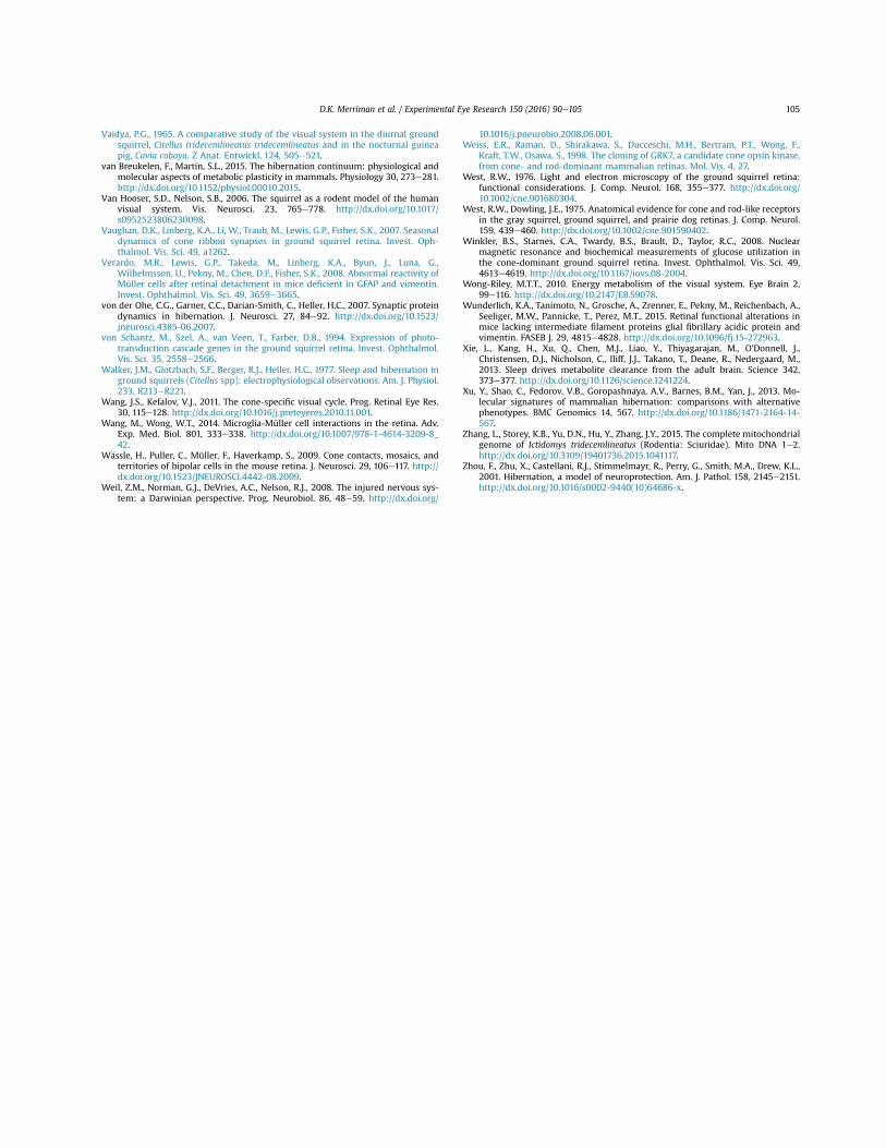
Vaidya, P.G., 1965. A comparative study of the visual system in the diurnal groundsquirrel, Citellus tridecemlineatus tridecemlineatus and in the nocturnal guineapig, Cavia cobaya. Z Anat. Entwickl. 124, 505e521.
van Breukelen, F., Martin, S.L., 2015. The hibernation continuum: physiological andmolecular aspects of metabolic plasticity in mammals. Physiology 30, 273e281.http://dx.doi.org/10.1152/physiol.00010.2015.
Van Hooser, S.D., Nelson, S.B., 2006. The squirrel as a rodent model of the humanvisual system. Vis. Neurosci. 23, 765e778. http://dx.doi.org/10.1017/s0952523806230098.
Vaughan, D.K., Linberg, K.A., Li, W., Traub, M., Lewis, G.P., Fisher, S.K., 2007. Seasonaldynamics of cone ribbon synapses in ground squirrel retina. Invest. Oph-thalmol. Vis. Sci. 49, a1262.
Verardo, M.R., Lewis, G.P., Takeda, M., Linberg, K.A., Byun, J., Luna, G.,Wilhelmsson, U., Pekny, M., Chen, D.F., Fisher, S.K., 2008. Abnormal reactivity ofMüller cells after retinal detachment in mice deficient in GFAP and vimentin.Invest. Ophthalmol. Vis. Sci. 49, 3659e3665.
von der Ohe, C.G., Garner, C.C., Darian-Smith, C., Heller, H.C., 2007. Synaptic proteindynamics in hibernation. J. Neurosci. 27, 84e92. http://dx.doi.org/10.1523/jneurosci.4385-06.2007.
von Schantz, M., Szel, A., van Veen, T., Farber, D.B., 1994. Expression of photo-transduction cascade genes in the ground squirrel retina. Invest. Ophthalmol.Vis. Sci. 35, 2558e2566.
Walker, J.M., Glotzbach, S.F., Berger, R.J., Heller, H.C., 1977. Sleep and hibernation inground squirrels (Citellus spp): electrophysiological observations. Am. J. Physiol.233, R213eR221.
Wang, J.S., Kefalov, V.J., 2011. The cone-specific visual cycle. Prog. Retinal Eye Res.30, 115e128. http://dx.doi.org/10.1016/j.preteyeres.2010.11.001.
Wang, M., Wong, W.T., 2014. Microglia-Müller cell interactions in the retina. Adv.Exp. Med. Biol. 801, 333e338. http://dx.doi.org/10.1007/978-1-4614-3209-8_42.
W€assle, H., Puller, C., Müller, F., Haverkamp, S., 2009. Cone contacts, mosaics, andterritories of bipolar cells in the mouse retina. J. Neurosci. 29, 106e117. http://dx.doi.org/10.1523/JNEUROSCI.4442-08.2009.
Weil, Z.M., Norman, G.J., DeVries, A.C., Nelson, R.J., 2008. The injured nervous sys-tem: a Darwinian perspective. Prog. Neurobiol. 86, 48e59. http://dx.doi.org/
10.1016/j.pneurobio.2008.06.001.Weiss, E.R., Raman, D., Shirakawa, S., Ducceschi, M.H., Bertram, P.T., Wong, F.,
Kraft, T.W., Osawa, S., 1998. The cloning of GRK7, a candidate cone opsin kinase,from cone- and rod-dominant mammalian retinas. Mol. Vis. 4, 27.
West, R.W., 1976. Light and electron microscopy of the ground squirrel retina:functional considerations. J. Comp. Neurol. 168, 355e377. http://dx.doi.org/10.1002/cne.901680304.
West, R.W., Dowling, J.E., 1975. Anatomical evidence for cone and rod-like receptorsin the gray squirrel, ground squirrel, and prairie dog retinas. J. Comp. Neurol.159, 439e460. http://dx.doi.org/10.1002/cne.901590402.
Winkler, B.S., Starnes, C.A., Twardy, B.S., Brault, D., Taylor, R.C., 2008. Nuclearmagnetic resonance and biochemical measurements of glucose utilization inthe cone-dominant ground squirrel retina. Invest. Ophthalmol. Vis. Sci. 49,4613e4619. http://dx.doi.org/10.1167/iovs.08-2004.
Wong-Riley, M.T.T., 2010. Energy metabolism of the visual system. Eye Brain 2,99e116. http://dx.doi.org/10.2147/EB.S9078.
Wunderlich, K.A., Tanimoto, N., Grosche, A., Zrenner, E., Pekny, M., Reichenbach, A.,Seeliger, M.W., Pannicke, T., Perez, M.T., 2015. Retinal functional alterations inmice lacking intermediate filament proteins glial fibrillary acidic protein andvimentin. FASEB J. 29, 4815e4828. http://dx.doi.org/10.1096/fj.15-272963.
Xie, L., Kang, H., Xu, Q., Chen, M.J., Liao, Y., Thiyagarajan, M., O'Donnell, J.,Christensen, D.J., Nicholson, C., Iliff, J.J., Takano, T., Deane, R., Nedergaard, M.,2013. Sleep drives metabolite clearance from the adult brain. Science 342,373e377. http://dx.doi.org/10.1126/science.1241224.
Xu, Y., Shao, C., Fedorov, V.B., Goropashnaya, A.V., Barnes, B.M., Yan, J., 2013. Mo-lecular signatures of mammalian hibernation: comparisons with alternativephenotypes. BMC Genomics 14, 567. http://dx.doi.org/10.1186/1471-2164-14-567.
Zhang, L., Storey, K.B., Yu, D.N., Hu, Y., Zhang, J.Y., 2015. The complete mitochondrialgenome of Ictidomys tridecemlineatus (Rodentia: Sciuridae). Mito DNA 1e2.http://dx.doi.org/10.3109/19401736.2015.1041117.
Zhou, F., Zhu, X., Castellani, R.J., Stimmelmayr, R., Perry, G., Smith, M.A., Drew, K.L.,2001. Hibernation, a model of neuroprotection. Am. J. Pathol. 158, 2145e2151.http://dx.doi.org/10.1016/s0002-9440(10)64686-x.
D.K. Merriman et al. / Experimental Eye Research 150 (2016) 90e105 105