
Review Review UNDERSTANDING CELLULITIS OF THE … · Review Review Patients with leg ulceration are...
8
Review Patients with leg ulceration are at risk of developing cellulitis, a potentially life-threatening bacterial infection of the skin. It is vital that the clinician can recognise the symptoms and differentiate them from other conditions of the lower limb. An understanding of when to initiate appropriate treatment promptly is essential to minimise distress and pain and improve long-term outcomes for the patient. UNDERSTANDING CELLULITIS OF THE LOWER LIMB Cellulitis arises following bacterial infection of the skin. It presents as a swollen, red area that feels hot and tender, and which may spread rapidly. If the patient develops any complications or the condition becomes more serious, if left untreated, the spreading bacterial infection may quickly become life- threatening. Therefore, it is important that the healthcare professional can recognise the signs and symptoms of cellulitis and identify the point at which appropriate treatment should be initiated or immediate medical attention sought. Patients wilth leg ulceration and associated skin conditions are at risk of cellulitis. This article will outline how to recognise cellulitis in this patient group and will provide tips on its management, particularly focusing upon when transfer between primary and secondary care is necessary. What is cellulitis? Cellulitis is a frequently diagnosed bacterial infection of the skin that usually presents initially as inflammation. Inflammation Mary Eagle is Independent Tissue Viability Adviser, Farnborough, Hampshire is a normal body response to trauma and may cause swelling, redness (erythema), pain, or warmth. However, when cellulitis- associated inflammation occurs, it is potentially serious. It may affect only the skin’s surface or it can be more severe, affecting both the skin and the subcutaneous tissues beneath, and it can spread to the lymph nodes and bloodstream. Cellulitis can occur anywhere on the body, but is most commonly encountered on the lower legs, especially near the shins and ankles (Cox and Lawence, 1998). People at risk of cellulitis include those with trauma or disrupted areas of the skin or other medical conditions (Table 1). All chronic leg ulcers contain bacteria (Gilcrist, 1999) and Cox and Lawence (1998) suggested that cellulitis secondary to leg ulceration may be caused by a large variety of organisms which are known to colonise chronic wounds, including streptococci, staphylococci, Pseudomonas spp., Bacteriodes spp. (Cox and Lawence, 1998), and other lesser common varieties. How does cellulitis occur? Under normal circumstances, the skin provides an effective barrier against invasion by micro-organisms that live on the skin or that are present in the environment. It is a first- line defence that stops micro- Figure 1. Extensive, spreading wet and dry cellulitis. 34 Wound Essentials • Volume 2 • 2007
Transcript of Review Review UNDERSTANDING CELLULITIS OF THE … · Review Review Patients with leg ulceration are...
ReviewReview ReviewReview
Patients with leg ulceration are at risk of developing cellulitis, a potentially life-threatening bacterial infection of the skin. It is vital that the clinician can recognise the symptoms and differentiate them from other conditions of the lower limb. An understanding of when to initiate appropriate treatment promptly is essential to minimise distress and pain and improve long-term outcomes for the patient.
UNDERSTANDING CELLULITISOF THE LOWER LIMB
Cellulitis arises following bacterial infection of the skin. It presents as a swollen, red area that feels hot and tender, and which may spread rapidly. If the patient develops any complications or the condition becomes more serious, if left untreated, the spreading bacterial infection may quickly become life-threatening. Therefore, it is important that the healthcare professional can recognise the signs and symptoms of cellulitis and identify the point at which appropriate treatment should be initiated or immediate medical attention sought.
Patients wilth leg ulceration and associated skin conditions are at risk of cellulitis. This article will outline how to recognise cellulitis in this patient group and will provide tips on its management, particularly focusing upon when transfer between primary and secondary care is necessary.
What is cellulitis?Cellulitis is a frequently diagnosed bacterial infection of the skin that usually presents initially as infl ammation. Infl ammation
Mary Eagle is Independent Tissue Viability Adviser, Farnborough, Hampshire
is a normal body response to trauma and may cause swelling, redness (erythema), pain, or warmth. However, when cellulitis-associated infl ammation occurs, it is potentially serious. It may affect only the skin’s surface or it can be more severe, affecting both the skin and the subcutaneous tissues beneath, and it can spread to the lymph nodes and bloodstream.
Cellulitis can occur anywhere on the body, but is most commonly encountered on the lower legs, especially near the shins and ankles (Cox and Lawence, 1998).
People at risk of cellulitis include those with trauma or disrupted areas of the skin or other medical
conditions (Table 1). All chronic leg ulcers contain bacteria (Gilcrist, 1999) and Cox and Lawence (1998) suggested that cellulitis secondary to leg ulceration may be caused by a large variety of organisms which are known to colonise chronic wounds, including streptococci, staphylococci, Pseudomonas spp., Bacteriodes spp. (Cox and Lawence, 1998), and other lesser common varieties.
How does cellulitis occur?Under normal circumstances, the skin provides an effective barrier against invasion by micro-organisms that live on the skin or that are present in the environment. It is a fi rst-line defence that stops micro-
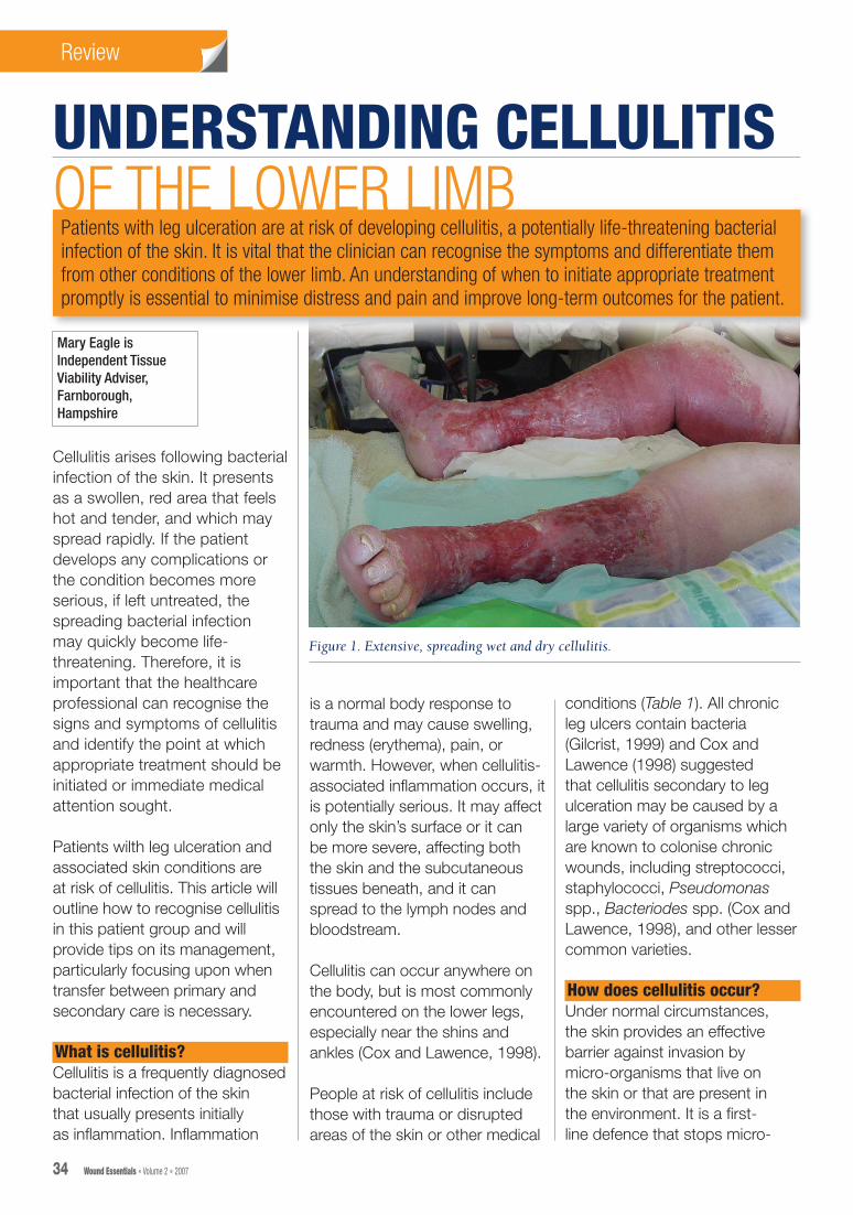
Figure 1. Extensive, spreading wet and dry cellulitis.
34 Wound Essentials • Volume 2 • 2007
34-44Cellulitis.indd 2 4/6/07 8:46:18 am
ReviewReview ReviewReview
36 Wound Essentials • Volume 2 • 2007
organisms from entering the body and multiplying.
A bacterial infection occurs when bacteria successfully invade the soft tissues through small wounds/abrasions on the skin surface or through existing conditions, e.g. leg ulceration or tinea pedis (athlete’s foot).
Usually, the immune system kills any invading bacteria, but in at-risk patients (Table 1) the bacteria are more likely to successfully multiply and cause an infection. This triggers an inflammatory response from the immune system, resulting in lower limb cellulitis.
Clinical symptoms of cellulitis of the lower limbThe common symptoms of cellulitis, which may result in skin changes affecting its colour, sensation and temperature are:8Redness of the skin: presents
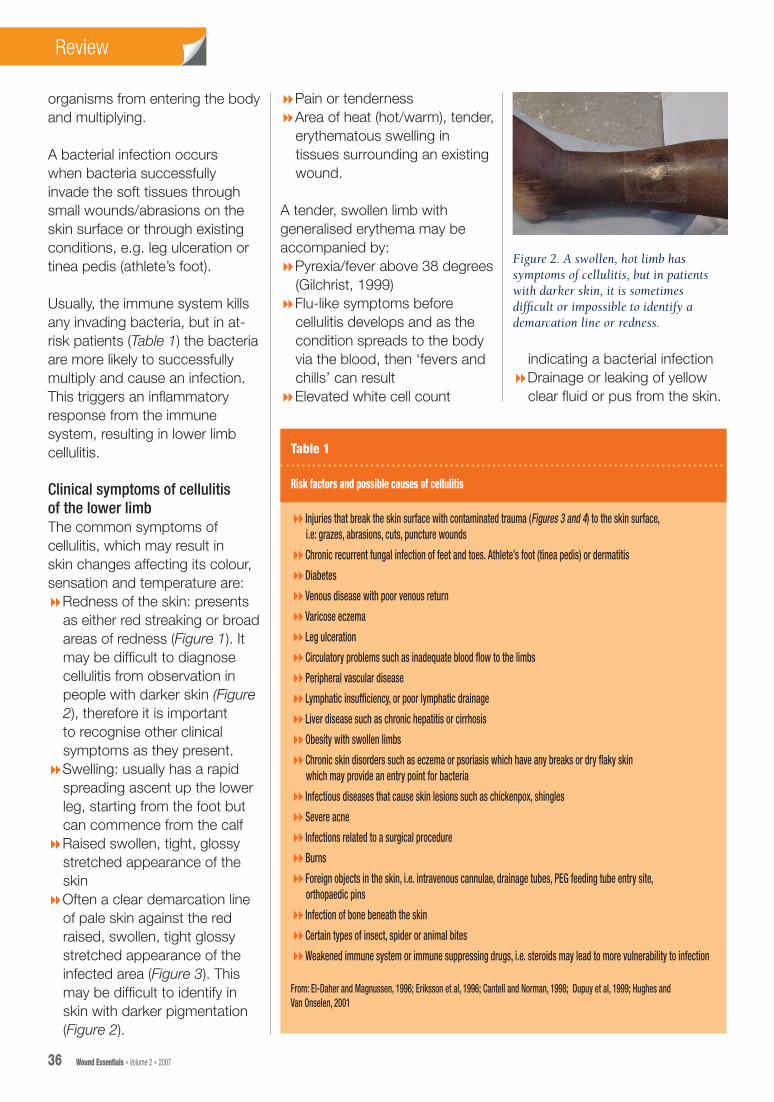
as either red streaking or broad areas of redness (Figure 1). It may be difficult to diagnose cellulitis from observation in people with darker skin (Figure 2), therefore it is important to recognise other clinical symptoms as they present.
8Swelling: usually has a rapid spreading ascent up the lower leg, starting from the foot but can commence from the calf
8Raised swollen, tight, glossy stretched appearance of the skin
8Often a clear demarcation line of pale skin against the red raised, swollen, tight glossy stretched appearance of the infected area (Figure 3). This may be difficult to identify in skin with darker pigmentation (Figure 2).
8Pain or tenderness8Area of heat (hot/warm), tender,
erythematous swelling in tissues surrounding an existing wound.
A tender, swollen limb with generalised erythema may be accompanied by: 8Pyrexia/fever above 38 degrees
(Gilchrist, 1999)8Flu-like symptoms before
cellulitis develops and as the condition spreads to the body via the blood, then ‘fevers and chills’ can result
8Elevated white cell count
indicating a bacterial infection 8Drainage or leaking of yellow
clear fluid or pus from the skin.
Table 1
Risk factors and possible causes of cellulitis
8Injuries that break the skin surface with contaminated trauma (Figures 3 and 4) to the skin surface, i.e: grazes, abrasions, cuts, puncture wounds
8Chronic recurrent fungal infection of feet and toes. Athlete’s foot (tinea pedis) or dermatitis
8Diabetes
8Venous disease with poor venous return
8Varicose eczema
8Leg ulceration
8Circulatory problems such as inadequate blood flow to the limbs
8Peripheral vascular disease
8Lymphatic insufficiency, or poor lymphatic drainage
8Liver disease such as chronic hepatitis or cirrhosis
8Obesity with swollen limbs
8Chronic skin disorders such as eczema or psoriasis which have any breaks or dry flaky skin which may provide an entry point for bacteria
8Infectious diseases that cause skin lesions such as chickenpox, shingles
8Severe acne
8Infections related to a surgical procedure
8Burns
8Foreign objects in the skin, i.e. intravenous cannulae, drainage tubes, PEG feeding tube entry site, orthopaedic pins
8Infection of bone beneath the skin
8Certain types of insect, spider or animal bites
8Weakened immune system or immune suppressing drugs, i.e. steroids may lead to more vulnerability to infection
From: El-Daher and Magnussen, 1996; Eriksson et al, 1996; Cantell and Norman, 1998; Dupuy et al, 1999; Hughes and Van Onselen, 2001
Figure 2. A swollen, hot limb has symptoms of cellulitis, but in patients with darker skin, it is sometimes difficult or impossible to identify a demarcation line or redness.
34-44Cellulitis.indd 4 4/6/07 8:46:19 am
ReviewReview ReviewReview
Figure 7. Contact dermatitis with clear line from allergy to bandaging.
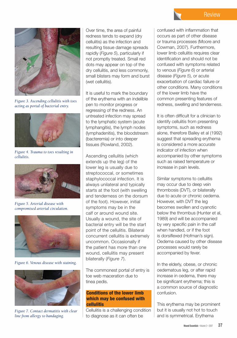
Figure 4. Trauma to toes resulting in cellulitis.
Figure 5. Arterial disease with compromised arterial circulation.
Figure 6. Venous disease with staining.
Wound Essentials • Volume 2 • 2007 37
Over time, the area of painful redness tends to expand (dry cellulitis) as the infection and resulting tissue damage spreads rapidly (Figure 5), particularly if not promptly treated. Small red dots may appear on top of the dry cellulitis, and less commonly, small blisters may form and burst (wet cellulitis).
It is useful to mark the boundary of the erythema with an indelible pen to monitor progress or regressing of the redness. An untreated infection may spread to the lymphatic system (acute lymphangitis), the lymph nodes (lymphadenitis), the bloodstream (bacteremia) or into deeper tissues (Rowland, 2002).
Ascending cellulitis (which extends up the leg) of the lower leg is usually due to streptococcal, or sometimes staphylococcal infection. It is always unilateral and typically starts at the foot (with swelling and tenderness on the dorsum of the foot). However, initial symptoms may be in the calf or around wound site. Usually a wound, the site of bacterial entry will be the start point of the cellulitis. Bilateral concurrent cellulitis is extremely uncommon. Occasionally if the patient has more than one wound, cellulitis may present bilaterally (Figure 7).
The commonest portal of entry is toe web maceration due to tinea pedis.
Conditions of the lower limb which may be confused withcellulitis Cellulitis is a challenging condition to diagnose as it can often be
confused with inflammation that occurs as part of other disease or trauma processes (Moore and Cowman, 2007). Furthermore, lower limb cellulitis requires clear identification and should not be confused with symptoms related to venous (Figure 6) or arterial disease (Figure 5), or acute exacerbation of cardiac failure or other conditions. Many conditions of the lower limb have the common presenting features of redness, swelling and tenderness.
It is often difficult for a clinician to identify cellulitis from presenting symptoms, such as redness alone, therefore Bailey et al (1992) suggest that spreading erythema is considered a more accurate indicator of infection when accompanied by other symptoms such as raised temperature or increase in pain levels.
Similar symptoms to cellulitis may occur due to deep vein thrombosis (DVT), or bilaterally due to acute or chronic oedema. However, with DVT the leg becomes swollen and cyanotic below the thrombus (Hunter et al, 1989) and will be accompanied by very specific pain in the calf when handled, or if the foot is dorsiflexed (Hofman’s sign).Oedema caused by other disease processes would rarely be accompanied by fever.
In the elderly, obese, or chronic oedematous leg, or after rapid increase in oedema, there may be significant erythema; this is a common source of diagnostic confusion.
This erythema may be prominent but it is usually not hot to touch and is symmetrical. Erythema
Figure 3. Ascending cellulitis with toes acting as portal of bacterial entry.
34-44Cellulitis.indd 5 4/6/07 8:46:26 am
ReviewReview
38 Wound Essentials • Volume 2 • 2007
resulting from cellulitis is accompanied by local tenderness and malaise/fever. Table 2 enables a comparison of lower leg cellulitis with other causes of leg swelling.
Erysipelas is a superficial tissue infection, often only affecting the dermal tissue, causing large areas of painful blistering and it is frequently confused with cellulitis (Beldon and Burton, 2005).
Cellulitis secondary to preceding venous eczema should be differentiated, as the commonest infection in such cases is staphylococcal. Similarly, cellulitis secondary to leg ulceration may be caused by a large variety of bacterial organisms. Clinical presentation of allergic contact dermatitis of the lower leg may look similar to cellulitis. Allergic contact dermatitis in leg ulcer patients is often caused by the application of dressing, creams
Table 2
Lower leg cellulitis and other causes of leg swelling
Disorder Lower leg cellulitis Other causes of leg swelling
Age Any adult, but most frequently the elderly
Gender Minor female predominance Either
Family history May be predominance Non-specific
Sites Rapid ascent up lower leg, usually initially with swelling and tenderness of dorsum of the foot; may commence in calf; may commence around a wound site
Usually first apparent at ankle
Symptoms Local tenderness, systemic fever and malaise
None, sensation of tightness or pain (depending on cause and rate of evolution)
Signs common to both Swelling and erythema of lower leg
Rapid ascent of erythema up the leg, usually confluent but sometimes with spared ’skip’ lesions. Unilateral. Commonly with tinea pedis also present. Fever and malaise are typical; lymphangitis and tender lymph glands are also common
Simple oedema is not usually erythematous. In some individuals with obese or lymphoedematous legs, erythema may be prominent but is usually symmetrical
Associated features Chronic lymphoedema is both a predisposing factor and a consequence of recurrent episodes. Ulceration may occur
Depends on cause. May be heart failure, pulmonary emboli, etc
Important tests Bacteriology swabs if any blister or port of entry, to identify bacterial type. Mycology samples from tinea pedis
Depends on likely cause. May require ECG, chest x-ray, Doppler ultrasound, and venography
Adapted from: Cox and Lawence (1998)
and lotions containing allergens. All areas of the skin in contact with the allergen will react (Figure 7). Clinical presentation of allergic contact dermatitis may look similar to cellulitis. Removing exposure to the allergen and taking active steps to avoid allergens completely will reduce symptoms, but referral to a specialist is appropriate if symptoms do not rapidly respond.
These and the other differential diagnoses (Table 3) require
different treatments and have different long-term significances, so distinction between them and cellulitis is important. Practitioners are advised to refer for expert opinion if there is any doubt relating to the diagnosis of cellulitis.
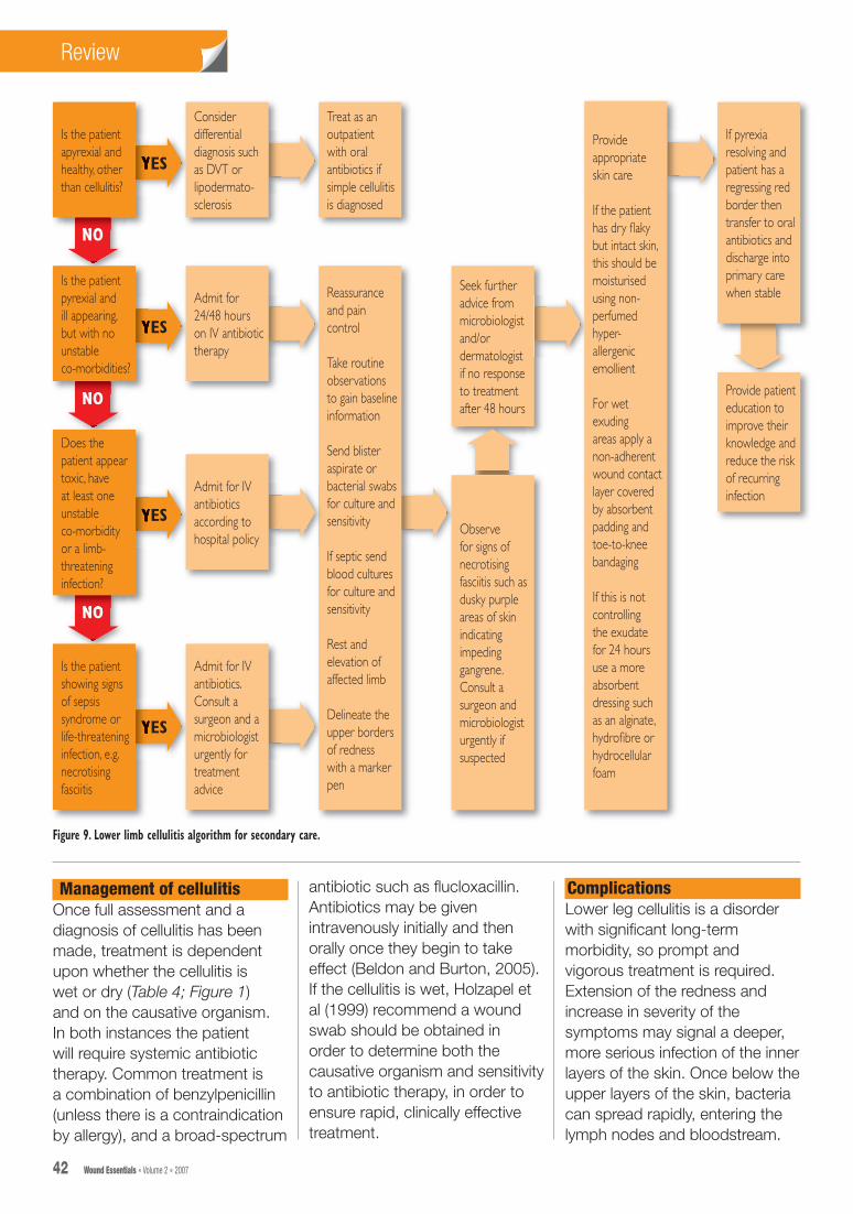
Lower limb algorithmsIn all cases, whatever the cause of cellulitis, referral to algorithms (Figures 8 and 9) for primary and secondary care treatment regimes will guide the practitioner to the correct course
Table 3
Other differential diagnosis which may cause hot swollen legs
8Deep vein thrombosis
8Acute oedema due to heart failure, hypoprotein-aemia
8Lymphoedema
8Varicose eczema
8Tibial compartment syndrome
8Gout
8Acute muscular conditions8Early herpes zoster8Erythema nodulosum8Allergic contact dermatitis (Figure 7)8Pelvic surgery8Pelvic irradiationFrom: Chmel and Hamdy, 1984; Dankert and Bouma, 1987; Bouma and Dankert, 1988; Gooptu and Powell, 1999; Tavadia et al, 2003; Baker et al, 1991
34-44Cellulitis.indd 6 4/6/07 8:46:27 am

ReviewReview
YES
YES
YES
NO
NO
NO
YES
NO
YESYES
YES YES
NONO
NONO
YESYES
NO
YES
Is the patient known to have venous insuffi ciency?
YESAre both legs symptomatic?
YES
NOHas the patient been wearing compression?
Perform leg ulcer assessment, including Doppler ultrasound. Apply compression therapy if appropriate, if patient has complicating factors, i.e. arterial disease or diabetes; refer to specialist leg ulcer clinic
Ensure hosiery:Correct size and strength. Still functioning — may need renewal
YES
NO
Is the patient’s blood pressure outside normal range?
YES
NO
On auscultation is the patient having multiple coupled beats?
Inform GPElevate limbs and encourage pedal exercises
NO
YES
Is the patient dyspnoeic and known to have cardiac failure?
NO
YESHas the patient taken his/her medication?
Ensure patient has medication.Elevate limbs and encourage pedal exercises.Inform GP and review in 24 hours.
YES
NOIs the patient pyrexial?
YES
NOIs there general infl ammation present?
Local infl ammation may be due to wound healing. Review regularly
Oral antibiotics – if no response in 24 hours, i.e. demarcation line extends, then admit to hospital.Rest and elevation
YES
Is the patient feverish and generally symptomatic with either wet or dry infl amed legs?OR has widespread wet infl ammation?
Patient requires hospitalisation for :Intravenous antibioticsRestElevation Intensive skin/wound care
Figure 8. Lower limb cellulitis algorithm for primary care.
40 Wound Essentials • Volume 2 • 2007
of action. These may include identifi cation of the caustive organism via the microbiology department. Analysis of bacterial wound swabs and blister
aspirate to determine bacterial culture and sensitivity can guide treatment. If the patient is showing signs of systemic sepsis, blood cultures should be
sent for culture and sensitivity (Beldon and Burton, 2005). If treatment is in doubt, immediate action should be taken to seek specialist advice.
34-44Cellulitis.indd 8 4/6/07 8:46:33 am
YES
YES
YES
42 Wound Essentials • Volume 2 • 2007
ReviewReview
Figure 9. Lower limb cellulitis algorithm for secondary care.
NO
YES
NO
YES
Is the patient apyrexial and healthy, other than cellulitis?
Consider differential diagnosis such as DVT or lipodermato-sclerosis
Treat as an outpatientwith oral antibiotics if simple cellulitis is diagnosed
NO
YES
NO
Is the patient pyrexial and ill appearing, but with no unstableco-morbidities?
Admit for 24/48 hours on IV antibiotic therapy
Reassurance and pain control
Take routine observations to gain baseline information Send blister aspirate or bacterial swabs for culture and sensitivity
If septic send blood cultures for culture and sensitivity Rest and elevation of affected limb
Delineate the upper borders of redness with a marker pen
Seek further advice from microbiologist and/or dermatologist if no response to treatment after 48 hours
Provide appropriate skin care
If the patient has dry fl aky but intact skin, this should be moisturised using non-perfumed hyper-allergenic emollient
For wet exuding areas apply a non-adherent wound contact layer covered by absorbent padding and toe-to-knee bandaging
If this is not controlling the exudate for 24 hours use a more absorbent dressing such as an alginate, hydrofi bre or hydrocellular foam
If pyrexia resolving and patient has a regressing red border then transfer to oral antibiotics and discharge into primary care when stable
NO
YES
NO
Does the patient appear toxic, have at least one unstable co-morbidity or a limb-threatening infection?
Admit for IV antibiotics according to hospital policy
YES
Is the patient showing signs of sepsis syndrome or life-threatening infection, e.g. necrotising fasciitis
Admit for IV antibiotics. Consult a surgeon and a microbiologist urgently for treatment advice
Provide patient education to improve their knowledge and reduce the risk of recurring infection
Observe for signs of necrotising fasciitis such as dusky purple areas of skin indicating impeding gangrene. Consult a surgeon and microbiologist urgently if suspected
Management of cellulitisOnce full assessment and a diagnosis of cellulitis has been made, treatment is dependent upon whether the cellulitis is wet or dry (Table 4; Figure 1) and on the causative organism. In both instances the patient will require systemic antibiotic therapy. Common treatment is a combination of benzylpenicillin (unless there is a contraindication by allergy), and a broad-spectrum
antibiotic such as fl ucloxacillin. Antibiotics may be given intravenously initially and then orally once they begin to take effect (Beldon and Burton, 2005). If the cellulitis is wet, Holzapel et al (1999) recommend a wound swab should be obtained in order to determine both the causative organism and sensitivity to antibiotic therapy, in order to ensure rapid, clinically effective treatment.
ComplicationsLower leg cellulitis is a disorder with signifi cant long-term morbidity, so prompt and vigorous treatment is required. Extension of the redness and increase in severity of the symptoms may signal a deeper, more serious infection of the inner layers of the skin. Once below the upper layers of the skin, bacteria can spread rapidly, entering the lymph nodes and bloodstream.
34-44Cellulitis.indd 10 4/6/07 8:46:38 am
ReviewReview
Wound Essentials • Volume 2 • 2007 43
for this condition which, at first, resembles a dusky cellulitis, but this quickly turns into an extending necrosis of the skin and subcutaneous tissues. The prognosis is often poor despite early surgical debridement and systemic antibiotics (Hunter et al, 1989).
Recurring cellulitis may cause lymphangitis which with time, and if untreated, can progress to lymphoedema. Early treatment of these symptoms is crucial. If cellulitis recurs, long-term treatment with penicillin should be considered (Hunter et al, 1989).
Long-term treatment and prevention of cellulitisIn order for the skin to return to its normal elasticity, moisturising with an emollient is required for several weeks. The patient should be encouraged to maintain good skin care as a matter of course to minimise risk. Scarring usually fades. Exposure to sun or trauma to the area should be avoided if possible as the new skin remains vulnerable for several weeks (Baxter and McGregor, 2001). If cellulitis is secondary to venous disease and ulceration, the patient may need to be referred to receive compression therapy (following a full assessment to exclude arterial disease). The main treatment for venous disease and venous ulceration, remains: 8Full holistic assessment: an
appropriate dressing should be applied according to the current needs of the wound; this may include a dressing impregnated with an anti-microbial agent
In rare cases, streptococcal infection can spread to the deep layers of the fascial lining around muscles, resulting in necrotising fasciitis. This represents an
extreme emergency requiring urgent hospital treatment (Rowland, 2002). A mixture of pathogens including streptococci and anaerobes are responsible
Table 4
Management of cellulitis
Antibiotic regimen Combination of antibiotics sensitive to streptococci and staphylococci bacteria. May be intravenous or oral
Dry skin Keep dry. As oedema resolves skin wrinkles and may slough away. 50/50 soft white paraffin in liquid paraffin will help re-establish skin integrity
Wet skin Control exudate with absorbent dressing (foam, alginate, hydrofibre) to reduce maceration of surrounding skin
Oedema Elevate limb, rest with foot of bed elevated
Pain Regular analgesia according to patient need
Inflammation Monitor area, mark with skin marker pen to monitor whether cellulites is resolving or advancing
Exercise Dorsiflexion and plantaflexion foot exercise will aid drainage of oedema (Hofman, 1998)
Van Onselen, 2001
Table 5
Advice for the patient at risk of lower limb cellulitis
Avoid situations which may damage the skin, particularly if there are problems with circulation or swelling
Practice good skin and nail care
Moisturise skin regularly after bathing
Trim toenails carefully
Wear well fitting shoes/slippers which do not rub
Check feet for cuts or grazes especially if you have loss of sensation in your legs
Avoid walking bare foot
Watch for signs of infection and get any inflammation, redness or swelling checked by GP or nurse
Talk to your GP if you have any swelling which does not go away
Check any broken areas of skin to make sure they get no bigger
Certain injuries may be at greater risk of infection than others. Antibiotics may be needed for: 8Animal or human bites8Puncture injuries deeper than 1cm8Crushed tissues that bleed8Burns that blister8Deep injuries with dirt in them8Injuries in contact with sea or dirty water8FrostbitePromptly treat any superficial infection or break in the skin surface, such as athletes foot, as broken skin will be a portal of entry for bacteria, don’t wait until symptoms of cellulitis appear
34-44Cellulitis.indd 11 4/6/07 8:46:39 am
44 Wound Essentials • Volume 2 • 2007
ReviewReview
8Compression therapy, either bandaging or hosiery
8Calf muscle pump and pedal exercise
8Elevation of the lower limb, when at rest
8Patient education and concordance with an agreed treatment regime
8Adequate nutrition8Early recognition of the
symptoms of cellulitis.
The patient should be educated about the longer-term problems associated with recurring cellulitis and the steps they can take (Table 5) to reduce their risk of recurrence.
The patient or carer can learn to recognise signs of early cellulitis and should discuss with their GP if it is appropriate for them to have antibiotics readily available to take at the onset of symptoms. If the patient has diagnosed venous disease and ulceration, concordance with their compression therapy regime is important, as this aids venous return and helps to prevent further swelling.
ConclusionIt is important that clinicians recognise the symptoms of cellulitis and are able to differentiate from other causes of swelling and redness in the lower limb. Cellulitis must be identified by a knowledgeable competent practitioner who can process the correct treatment pathway, as prompt action will minimise distress and pain for the patient. Patients should be educated to take responsibility for recognising the potential recurrence of cellulitis and when to seek help.
Bailey IS, Karran SE, Toyn K, Brough P, Ranaboldo C, Karran SJ (1992) Community surveillance of complications after hernia repair. Br Med J 304(6825): 469–71
Baker SR, Stacey MC, Joop-McKay AG, Hospkin SE, Thompson PJ (1991) Epidemiology of chronic venous ulcers. Br J Surg 78: 864–7
Baxter H, McGregor F (2001) Understanding and managing cellulitis. Nurs Stand 15(44): 50–6
Beldon P, Burton F (2005) Management guidelines for lower limb cellulites. Wounds UK 1(3): 16–22
Bouma J, Dankert J (1988) Recurrent acute leg cellulitis in patients after radical vulvectomy. Gynaecol Oncol 29(1): 50–7
Cantell M, Norman DC (1998) Skin and soft tissue infections in the elderly. Baillière’s Clinical Infectious Diseases 5(1): 71–81
Chmel H, Hamdy M (1984) Recurrent streptococcal cellulites complicating radical hysterectomy and radiation therapy. Obstet Gynaecol 63(6): 862–4
Cox N, Lawence CM (1998) Diagnostic Problems in Dermatology. Mosby, London: 146–7
Cutting KF, White RJ, Mahoney P, Harding KG (2005) Clinical identification of wound infection a Delphi approach. In: European Wound Management Association Position Document. Identifying Criteria for Wound Infection. MEP, London: 6–9
Dankert J, Bouma J (1987) Recurrent acute leg cellulites after hysterectomy with pelvic lymphadenectomy. Br J Obstet Gynaecol 94: 788–90
Dupuy A, Benchikhi R, Roujeau J et al (1999) Risk factors for erysipelas of the leg (cellulites): case control study. Br Med J 318: 1591–4
El-Daher N, Magnussen CR (1996)
Skin and soft tissue Infections: Outpatient Management and Indications for Hospitalisation. Consultant 36(12): 2563–6
Eriksson B, Jorup-Romstrom C, Karkkonen K, Sjoblom AC, Holm SE (1996) Erysipelas: clinical and bacterial spectrum and serological aspects. Clinical infectious Disease 23(5): 1091–8
Gilchrist B (1999) Wound infection. In: Wound Management Theory and Practice. Eds, Millar M, Glover D. Nursing Times Books. Emap Healthcare Ltd, Dorchester
Gooptu C, Powell SM (1999) The problem of rubber sensitivity (types I & IV) in chronic leg ulcer and stasis eczema patients. Contact Dermatitis 41: 89–93
Hughes E, Van Onselen J, eds (2001) Dermatology Nursing: A Practical Guide. Churchill Livingstone, London: 207
Hunter J, Savin J, Dahl M (1989) Clinical Dermatology 140. Blackwell Scientific Publications, London
Hofman D (1998) Oedema and the management of venous leg ulcers. J Wound Care 7(7): 345–8
Holzapel L, Jacquet-Francillon T, Rahmani J et al (1999) Microbiological evaluation of infected wounds of the extremities in 214 adults. J Accident Emergency Med 16: 32–4
Jones J, Hunter D (1995) Consensus methods for medical and heath services research. Br Med J 311(7001): 376–80
Moore Z, Cowman S (2007) Effective wound management: identifying criteria for infection. Nurs Stand 21(24): 68–76
Rowland B (2002) Gale Encyclopedia of Medicine. The Gale Group Inc Gale, Detroit, USA
Tavadia S, Bianchi J, Dawe RS et al (2003) Allergic contact dermatitis in venous leg ulcer patients. Contact Dermatitis 48(5): 261–5WE
34-44Cellulitis.indd 12 4/6/07 8:46:39 am


![Patient education for preventing diabetic foot ulceration … · [Intervention Review] Patient education for preventing diabetic foot ulceration Johannes AN Dorresteijn 1, Didi MW](https://static.fdocuments.us/doc/165x107/5ecb99029d0d2c34515c80a4/patient-education-for-preventing-diabetic-foot-intervention-review-patient.jpg)