Review PKA: a portrait of protein kinase...
11
Review PKA: a portrait of protein kinase dynamics S.S. Taylor * , J. Yang, J. Wu, N.M. Haste, E. Radzio-Andzelm, G. Anand Howard Hughes Medical Institute, Bethesda, MD, USA Department of Chemistry and Biochemistry, University of California, San Diego, La Jolla, CA 92014-0654, USA Received 6 November 2003; accepted 12 November 2003 Abstract Protein kinases play a critical role in the integration of signaling networks in eukaryotic cells. cAMP-dependent protein kinase (PKA) serves as a prototype for this large and highly diverse enzyme family. The catalytic subunit of PKA provides the best example of how a protein kinase recognizes its substrates, as well as inhibitors, and also show how the enzyme moves through the steps of catalysis. Many of the relevant conformational states associated with the catalytic cycle which have been captured in a crystal lattice are summarized here. From these structures, we can begin to appreciate the molecular events of catalysis as well as the intricate orchestration of critical residues in the catalytic subunit that contribute to catalysis. The entire molecule participates. To fully understand signaling by PKA, however, requires an understanding of a large set of related proteins, not just the catalytic subunit. This includes the regulatory subunits that serve as receptors for cAMP and the A kinase anchoring proteins (AKAPs) that serve as scaffolds for PKA. The AKAPs localize PKA to specific sites in the cell by docking to the N-terminus of the regulatory subunits, thus creating microenvironments for PKA signaling. To fully appreciate the diversity and integration of these molecules, one needs not only high-resolution structures but also an appreciation of how these molecules behave in solution. Thus, in addition to obtaining high-resolution structures by X-ray crystallography and NMR, we have used fluorescent tools and also hydrogen/deuterium exchange coupled with mass spectrometry to probe the dynamic properties of these proteins and how they interact with one another. The molecular features of these molecules are described. Finally, we describe a new recombinantly expressed PKA reporter that allows us to monitor PKA activity in living cells. D 2004 Elsevier B.V. All rights reserved. Keywords: PKA; Dynamic; Kinase inhibitor; Balanol; Catalysis; AKAP 1. Introduction Since approximately 2% of mammalian genomes encode for protein kinases [1], while 4% of plant genomes code for protein kinases [2], these enzymes constitute one of the largest gene families. The protein kinases regulate a myriad of cellular processes during growth and development, they are an integral part of the machinery that is activated in response to stress, they are essential for memory, and they are directly involved in orchestrating cell death. Because of their widespread involvement in the regulation of cellular events and because defects in protein kinase function are associated directly with so many diseases, these enzymes are primary targets for therapeutic intervention. The devel- opment of Gleevec, an adenosine triphosphate (ATP) analog with specificity for the Abl oncogene, the cause of myelog- enous leukemia, demonstrated the remarkable specificity that such drugs are capable of displaying in spite of the highly conserved fold of the protein kinase core with its conserved ATP binding pocket [3]. One of the simplest members of the protein kinase family is cAMP-dependent protein kinase (PKA). Its kinetic prop- erties have been well-defined, its crystal structure in the presence of nucleotide and peptide substrate, as well as the recently solved structures of the apoenzyme [4] and of an aluminum fluoride complex containing adenosine diphos- phate (ADP) and substrate peptide [5], provide a detailed description of intermediates in the reaction pathway. These structures also allow us to appreciate the malleability of the protein kinase core as it binds its substrates and prepares to transfer the g-phosphate of ATP to its protein substrate. This 1570-9639/$ - see front matter D 2004 Elsevier B.V. All rights reserved. doi:10.1016/j.bbapap.2003.11.029 Abbreviations: cAMP, cyclic-3V,5V-adenosine monophosphate; PKA, cAMP-dependent protein kinase; PKG, cGMP-dependent protein kinase; ATP, adenosinetriphosphate; ADP, adenosine diphosphate; PKI, heat stable protein kinase A inhibitor; D/D domain, dimerization/docking domain; AKAP, A-kinase anchoring proteins * Corresponding author. Department of Chemistry and Biochemistry, University of California, San Diego, La Jolla, CA 92014-0654, USA. Tel.: +1-858-534-3677; fax: +1-858-534-8193. E-mail address: [email protected] (S.S. Taylor). www.bba-direct.com Biochimica et Biophysica Acta 1697 (2004) 259 – 269
Transcript of Review PKA: a portrait of protein kinase...

www.bba-direct.com
Biochimica et Biophysica Acta 1697 (2004) 259–269
Review
PKA: a portrait of protein kinase dynamics
S.S. Taylor*, J. Yang, J. Wu, N.M. Haste, E. Radzio-Andzelm, G. Anand
Howard Hughes Medical Institute, Bethesda, MD, USA
Department of Chemistry and Biochemistry, University of California, San Diego, La Jolla, CA 92014-0654, USA
Received 6 November 2003; accepted 12 November 2003
Abstract
Protein kinases play a critical role in the integration of signaling networks in eukaryotic cells. cAMP-dependent protein kinase (PKA)
serves as a prototype for this large and highly diverse enzyme family. The catalytic subunit of PKA provides the best example of how a
protein kinase recognizes its substrates, as well as inhibitors, and also show how the enzyme moves through the steps of catalysis. Many of
the relevant conformational states associated with the catalytic cycle which have been captured in a crystal lattice are summarized here. From
these structures, we can begin to appreciate the molecular events of catalysis as well as the intricate orchestration of critical residues in the
catalytic subunit that contribute to catalysis. The entire molecule participates. To fully understand signaling by PKA, however, requires an
understanding of a large set of related proteins, not just the catalytic subunit. This includes the regulatory subunits that serve as receptors for
cAMP and the A kinase anchoring proteins (AKAPs) that serve as scaffolds for PKA. The AKAPs localize PKA to specific sites in the cell by
docking to the N-terminus of the regulatory subunits, thus creating microenvironments for PKA signaling. To fully appreciate the diversity
and integration of these molecules, one needs not only high-resolution structures but also an appreciation of how these molecules behave in
solution. Thus, in addition to obtaining high-resolution structures by X-ray crystallography and NMR, we have used fluorescent tools and
also hydrogen/deuterium exchange coupled with mass spectrometry to probe the dynamic properties of these proteins and how they interact
with one another. The molecular features of these molecules are described. Finally, we describe a new recombinantly expressed PKA reporter
that allows us to monitor PKA activity in living cells.
D 2004 Elsevier B.V. All rights reserved.
Keywords: PKA; Dynamic; Kinase inhibitor; Balanol; Catalysis; AKAP
1. Introduction
Since approximately 2% of mammalian genomes encode
for protein kinases [1], while 4% of plant genomes code for
protein kinases [2], these enzymes constitute one of the
largest gene families. The protein kinases regulate a myriad
of cellular processes during growth and development, they
are an integral part of the machinery that is activated in
response to stress, they are essential for memory, and they
are directly involved in orchestrating cell death. Because of
their widespread involvement in the regulation of cellular
1570-9639/$ - see front matter D 2004 Elsevier B.V. All rights reserved.
doi:10.1016/j.bbapap.2003.11.029
Abbreviations: cAMP, cyclic-3V,5V-adenosine monophosphate; PKA,
cAMP-dependent protein kinase; PKG, cGMP-dependent protein kinase;
ATP, adenosine triphosphate; ADP, adenosine diphosphate; PKI, heat stable
protein kinase A inhibitor; D/D domain, dimerization/docking domain;
AKAP, A-kinase anchoring proteins
* Corresponding author. Department of Chemistry and Biochemistry,
University of California, San Diego, La Jolla, CA 92014-0654, USA.
Tel.: +1-858-534-3677; fax: +1-858-534-8193.
E-mail address: [email protected] (S.S. Taylor).
events and because defects in protein kinase function are
associated directly with so many diseases, these enzymes
are primary targets for therapeutic intervention. The devel-
opment of Gleevec, an adenosine triphosphate (ATP) analog
with specificity for the Abl oncogene, the cause of myelog-
enous leukemia, demonstrated the remarkable specificity
that such drugs are capable of displaying in spite of the
highly conserved fold of the protein kinase core with its
conserved ATP binding pocket [3].
One of the simplest members of the protein kinase family
is cAMP-dependent protein kinase (PKA). Its kinetic prop-
erties have been well-defined, its crystal structure in the
presence of nucleotide and peptide substrate, as well as the
recently solved structures of the apoenzyme [4] and of an
aluminum fluoride complex containing adenosine diphos-
phate (ADP) and substrate peptide [5], provide a detailed
description of intermediates in the reaction pathway. These
structures also allow us to appreciate the malleability of the
protein kinase core as it binds its substrates and prepares to
transfer the g-phosphate of ATP to its protein substrate. This

S.S. Taylor et al. / Biochimica et Biophysica Acta 1697 (2004) 259–269260
malleability is likely a conserved feature of all protein
kinases with PKA merely serving as a prototype.
The activity of the catalytic (C) subunit is regulated by a
set of four different regulatory (R) subunit isoforms. Al-
though each regulatory subunit has a conserved organization
of subdomains with a dimerization/docking (D/D) domain at
the amino terminus and two tandem cAMP binding domains
at the carboxyl terminus, the R subunits play distinct roles
and are not functionally redundant [6]. In addition to the
regulatory subunits, specificity is also achieved by the
scaffold proteins, the A kinase anchoring proteins (AKAPs),
which target PKA through the regulatory subunits to differ-
ent sites within the cell and in close proximity to specific
substrates. All of these proteins contribute to the PKA
signaling networks that permeate every mammalian cell.
Each also contributes in novel ways to the dynamic features
of this network.
To fully describe the molecular function of a protein
kinase as well as the function of that kinase within the
context of a cell requires a variety of techniques. In addition
to crystallography which provides static high-resolution
structures, it is necessary to use methods that allow us to
appreciate the dynamics of these proteins in solution. In
parallel, fluorescent techniques are being developed that
allow us to follow kinase activation and localization in real
time in living cells [7]. The malleability of the protein
kinase provides for a wide range of conformational states
that can be targeted for therapeutic intervention. In addition
to the active site cleft, there are also multiple surface sites
that can be targeted that will disrupt normal cellular function
as well as disruption from the scaffolds. The static crystal
structures provide clues as to the conformational versatility
of the molecules; however, solution methods are required if
we are to appreciate the dynamic behavior of these mole-
cules as they move through the catalytic cycle and as they
move between substrates, inhibitors, and scaffold proteins.
To complement crystallographic structures, we have used
fluorescence anisotropy [8,9], NMR [10], and, most recent-
ly, hydrogen/deuterium (H/D) exchange coupled with mass
spectrometry [11,12] to probe the dynamical behavior of
PKA and its scaffold proteins. H/D exchange, in particular,
can be used to map domain boundaries, to map protein:-
protein interaction sites, and to map ligand-induced confor-
mational changes. With these multiple techniques, we are
beginning to assemble a description of the structure, func-
tion, and dynamical properties of PKA.
2. The catalytic subunit
2.1. Kinetic pathway for phosphoryl transfer
The classic work of Cook et al. [13] and Adams and
Taylor [14] have described many features of the reaction
pathway for the catalytic subunit. There is no obligatory
order for binding of substrates; however, given the high
concentration of ATP in the cell, it is assumed that ATP
typically binds first. This preferred order pathway for
binding of peptide substrate and ATP was first described
by Walsh and Ashby [15]. The pre-steady state kinetics later
revealed a very rapid (>500 s� 1) phosphoryl transfer step
(k3) with a rate-limiting step (20 s� 1) that corresponds to
release of the ADP product [16].
As summarized in Fig. 1, we have tried to capture these
various steps along the reaction pathway in a crystal lattice
and in so doing have defined a set of open and closed
conformations. Parallel fluorescence anisotropy studies have
provided a dynamic profile of these various states where the
enzyme toggles between an ensemble of many conforma-
tional states [9]. In an effort to trap a transition state
complex, we have also crystallized an ADP, substrate
peptide complex with aluminum fluoride [5].
2.2. Open and closed conformations are essential parts of
the catalytic cycle
Like all members of the protein kinase family, the
catalytic subunit has a small amino terminal domain that
is associated mostly with ATP binding and a larger carboxyl
terminal domain that serves as a docking site for the protein
substrate and also contains the conserved residues that direct
the transfer of the phosphate. While the small lobe is
dominated by h strands, the large lobe is mostly helical
with two of the helices, the E and F helices, spanning the
width of the enzyme and serving as the hydrophobic core of
the large lobe. In addition to the conserved core that is
shared by all members of the protein kinase family (residues
40–300 of the PKA catalytic subunit), the catalytic subunit
has 40 additional residues, mostly a single helix, at the
amino terminus and 50 residues at the carboxyl terminus
that wind around the surface of the large and small lobes and
then docks to a hydrophobic pocket on the surface of the
small lobe.
The open and closed conformations described here most
likely represent some of the physiological conformations of
the active enzyme in solution as all of these proteins are
fully phosphorylated on Thr197 in the activation loop and
on Ser338. Overall, they reflect a highly dynamic protein
that is a moving target for capture by substrates, by
physiological inhibitors, and artificially by small molecule
inhibitors. While the open and intermediate conformations
are quite dynamic [17], we also have described two very
stable conformational states. One is the inhibited state where
the catalytic subunit is bound to ATP and a peptide derived
from the heat-stable protein kinase inhibitor (PKI) (5–24)
[18]. Unlike the transition state mimic with aluminum
fluoride, the PKI:ATP complex is trapped in a transition
state-like complex with no available acceptor for the g-
phosphate. A second very stable conformational state, dis-
cussed in more detail later, was observed when the catalytic
subunit was crystallized with a natural product inhibitor,
balanol [19].

Fig. 1. Conformational states associated with catalysis. The catalytic subunit is capable of assuming an ensemble of open and closed conformational states as it
goes through the various stages of catalysis. (A) Some of these conformational states have been captured in a crystal lattice while fluorescence anisotropy has
demonstrated some of the dynamical properties of the enzyme as it toggles between these different states. The apoenzyme (PDB ID 1J3H), the adenosine binary
complex (1BKX), the binary complex with a substrate peptide (1JLU), and a transition state complex with aluminum fluoride:ADP, and a substrate peptide
(1L3R) are shown. The apoenzyme is the most open conformation while the two binary complexes represent intermediate stages of closing. The aluminum
fluoride complex represents a fully closed conformation. Phosphorylation sites are shown as blue spheres, nucleotide is shown as red sticks and peptide is
shown as yellow ribbon. The glycine-rich loop is highlighted in turquoise. (B) Rotation of the ternary complex containing ATP and PKI (5–24) shows how the
N- and C-termini flank the core. Phosphorylation sites are red and the C helix is highlighted in turquoise.
S.S. Taylor et al. / Biochimica et Biophysica Acta 1697 (2004) 259–269 261
2.3. Apoenzyme reveals a ‘‘preformed’’ active site
The structure of the apoenzyme revealed an open con-
formation where the small lobe is displaced due to both a
hinging motion and a sliding motion of the small lobe
relative to the large lobe [4]. Several features of this enzyme
had not been fully appreciated previously. For example, it is
in the apoenzyme structure that one can begin to fully
appreciate the different dynamic properties of the two lobes.
Most of the small lobe, in the absence of ligands, appears to
be quite dynamic based on the temperature factors and the
difficulty in tracing the chain fully in regions such as the tip
of the glycine-rich loop. The portion of the carboxyl
terminal tail which eventually will clamp down onto the
small lobe when ATP and peptide are bound is also quite
disordered.
In contrast to the small lobe, the large lobe is quite stable.
Indeed most of the active site where phosphoryl transfer
takes place is already formed in the apoenzyme [4]. There
does not appear to be a requirement for significant induced

S.S. Taylor et al. / Biochimica et Biophysica Acta 1697 (2004) 259–269262
fit to accommodate binding of substrate. The stability of the
large lobe is due to the solid hydrophobic core which
anchors the catalytic loop and the magnesium-positioning
loop at the active site cleft so that they are poised for action.
Although these two loops, each containing essential resi-
dues, are positioned between h strands 6 and 7 and 8 and 9,
respectively, each loop is anchored firmly through hydro-
phobic side chains to the hydrophobic core of the domain.
This extended hydrophobic core accounts for the remark-
able stability of this domain. The only portion of the large
lobe at the active site that is not firmly anchored by
hydrophobic interactions to the core is the activation loop.
In contrast, this loop is oriented in its active conformation
by the phosphorylation of Thr197. Through multiple hydro-
Fig. 2. The catalytic loop is docked by hydrophobic interactions to the large lobe a
structure of the apoenzyme revealed that the large lobe, including the catalytic loop a
of ATP and substrates. It is anchored to the large lobe primarily by hydrophobic inter
buried in the large lobe. The hydrophilic residues of the catalytic loop face into th
intervening hydrophobic residues point inward to the hydrophobic surface of the F h
the entire molecule through the phosphorylation of Thr197 and its interaction with A
on the surface at opposite ends of the large lobe. Glu230 contributes directly to the P-
C-terminus of the F helix and to Glu170 in the catalytic loop. The other end of the F
chain of Asp220 helps to anchor the catalytic loop by hydrogen bonding to the back
loop. The side chain of Tyr164 also anchors the catalytic loop to the magnesium-po
gen bonding and ionic interactions, nucleated by this single
phosphate, the enzyme is locked into a stable conforma-
tional state and ensures that the active site is optimally
configured for catalysis [20,21].
In Fig. 2, one can better appreciate how the catalytic loop
is specifically anchored to the F helix. This is one of two
helices that permeate the core, even though, in general, it is
quite unusual to have hydrophobic helices like this. Typi-
cally, helices in proteins are amphipathic with the hydro-
phobic surface facing the core and the hydrophilic surface
facing the solvent. The hydrophobic properties of this helix
are a conserved feature of all protein kinases. The two ends
of the F helix are hydrophilic and also carry out specific
conserved roles. At the amino terminus is Asp220, con-
nd is mostly preformed prior to the binding of nucleotide and substrates. The
nd the magnesium-positioning loop, is mostly preformed prior to the binding
actions shown here in panel A. The F helix is a very hydrophobic helix that is
e active site cleft where they are poised to participate in catalysis while the
elix. Panel B shows how the activation segment, residues 184–208, integrates
rg165. The two ends of the F helix (Panel C) are hydrophilic and are exposed
2 Arginine recognition site of the peptide. Thus the P-2 Arg is anchored to the
helix is Asp220, one of the invariant residues in all protein kinases. The side
bone amides of 17 Tyr164 and Arg165 that immediately precede the catalytic
sitioning loop by interacting with the backbone carbonyl oxygen of Asp184.

S.S. Taylor et al. / Biochimica et Biophysica Acta 1697 (2004) 259–269 263
served in all protein kinases. It serves to anchor the catalytic
loop through interactions of its side chain with the backbone
carbonyl moieties of Tyr164 and Arg165 which just precede
the catalytic loop. Arg165 is an essential anchor to the
phosphate moiety on Thr197, and Asp166 is the catalytic
base that starts the catalytic loop. At the carboxyl terminus
of the F helix is Glu230 which contributes to recognition of
the P-2 arginine in the peptide substrate. This P-2 arginine is
anchored on the other side by Glu170, which is also part of
the catalytic loop. Just beyond the F helix are residues 235–
239 that form the hydrophobic pocket where the P-11 side
chain of PKI (5–24) docks. As seen in Fig. 2B, the catalytic
loop, the magnesium positioning loop, and the F helix are
all integrated by phosphorylation of Thr197 in the activation
loop through its interaction with Arg165. As shown in Fig.
2C, the end of the activation segment, Asp208 is anchored
to the C helix through its interactions with Trp222 and
Arg280. Fig. 2 allows us to appreciate the extensive
interactions that permeate the entire molecule.
As seen in Fig. 1, and also in Fig. 3, the apoenzyme
structure represents one of the most open conformations of
the catalytic subunit that we have observed so far. It
probably reflects the major conformation state in solution
that the enzyme adopts prior to any ligand binding. In this
state, the small lobe rotates and slides away from the large
lobe, the glycine-rich loop is highly dynamic and is away
from the active site. The C-terminal segment (318–326),
Fig. 3. Opening and closing of the glycine-rich loop at the active site cleft.
The catalytic loop is locked into a stable conformation even when nothing is
bound to the active site cleft, whereas the glycine-rich loop is positioned for
catalysis only when the enzyme assumes a fully closed conformation. It is the
most mobile element in the conserved catalytic core. The position of the
glycine-rich loop relative to the catalytic loop is shown here for the
conformational states represented in Fig. 1. The a-carbon of Ser53 is
rendered as a sphere.
described previously as a ‘‘gate’’ controlling the access to
the nucleotide binding site by covering the front of the
active site cleft, is largely disordered [4]. The position of the
glycine-rich loop relative to the catalytic loop is an indicator
of the ‘‘openness’’ of the conformation, and in this apoen-
zyme, the tip of the glycine-rich loop is far from the
catalytic loop (Figs. 1 and 3). Fluorescence anisotropy
where selected cysteines were labeled with a fluorescent
probe also confirmed that the conformation of the apoen-
zyme was likely to be very open and highly dynamic [9].
All these features ensure the active site of the apoenzyme
has maximum accessibility to the nucleotide substrate.
2.4. Binary complexes reflect an intermediate conforma-
tional state
In the presence of either an ATP analog or a peptide
substrate or inhibitor the enzyme assumes a more closed
conformation, but the enzyme remains quite dynamic. As
seen in Figs. 1 and 3, the glycine-rich loop is closer to the
catalytic loop but the tip of the loop is still quite dynamic
and is not anchored firmly. The fluorescence anisotropy
studies are consistent with this model of a highly dynamical
state that is poised for transfer of the g-phosphate. Until the
tip of the loop, specifically the backbone amide of Ser53, is
bound to the g-phosphate of ATP, the enzyme is in a
dynamical state. When these structures are compared to
the structure of the apoenzyme, it is found that a few
additional residues, most importantly Lys72, become ori-
ented in their catalytically competent state as a consequence
of binding the nucleotide (described below). The critical
step for catalysis is engaging the tip of the glycine-rich loop,
and neither of these structures is capable of doing that.
Comparing the adenosine binary structure [22] with the
apo form, we see that upon binding of adenosine the C-
terminal ‘‘gate’’ is now traceable, albeit still quite dynamic.
Tyr330 in this segment will become part of the P-3 peptide
recognition site. Lys72 is also ordered even though the
triphosphate moiety of ATP is not present. Although the
substrate binding site on the large lobe is largely preformed,
binding of nucleotide also seems to further engage or orient
some of the residues involved in peptide binding. This
communication from the nucleotide binding at the active
site to the distal peptide binding on the large lobe is
probably mediated through the hydrophobic core [4]. The
intermediate conformation captured by the binary complex
is very likely predominant when ATP binds to the active site
at the initial stage of the catalytic cycle.
2.5. Aluminum fluoride:ADP:substrate complex reflects a
transition state complex
In an effort to capture the catalytic subunit in a state that
accurately reflects the transition state, the catalytic subunit
was co-crystallized with ATP, a substrate peptide, and
aluminum fluoride (Fig. 4). The peptide docks onto the

Fig. 4. An aluminum fluoride complex mimics docking of ATP and peptide
to the active site cleft. The ATP:PKI (5–24) ternary complex resembles a
transition state complex but is missing an acceptor moiety for the
phosphate. The ternary complex with ATP and PKI (5–24) is shown here
superimposed with the structure of the AlF3:ADP:PKS (5–24) complex.
Shown in panel A is the position of the catalytic loop and the glycine-rich
loop (Ser53) in their putative active and fully closed conformations. In
panel B, the position of the ATP is shown with the g-phosphate moiety
locked between the catalytic loop and the glycine-rich loop through its
interactions with Lys168 and the backbone amide of Ser53. The position of
the ADP:AlF3 is superimposed. In panel C, the positioning of the peptide is
shown with the P site residue poised for catalysis. Thr201 from the P + 1
loop which plays a critical role in positioning both the Lys168 and Asp166
in the catalytic loop is also shown.
S.S. Taylor et al. / Biochimica et Biophysica Acta 1697 (2004) 259–269264
large lobe and makes further interactions with the small
lobe, including the glycine-rich loop, to bring it even closer
to the active site. The planar aluminum fluoride lies equi-
distant between the h–g bridging oxygen of ADP and the
phosphoacceptor hydroxyl. The resulting structure does
indeed appear to resemble a transition state that is poised
to transfer the g-phosphate of ATP. In this structure, as in the
adenosine binary complex, the adenine binding pocket is
occupied. What is essential, however, is for the glycine-rich
loop to grab onto the g-phosphate of ATP and orient it for
transfer to the protein substrate. This is mediated by the
backbone amide of Ser53 hydrogen bonding to the g-
phosphate. In this structure the g-phosphate is clamped
between the side chain of Lys168 in the catalytic loop and
the backbone amide of Ser53. The distance between the
aluminum and the g-phosphorus of ADP and the oxygen at
the P site of the substrate peptide is equidistant (approxi-
mately 2.3 A). It is poised for a direct in-line transfer as
predicted earlier [5].
One can see that the active site takes conformations
from open to intermediate to closed, as the catalysis
proceeds. The open conformation allows maximum access
of ATP to the active site. Binding of nucleotide syner-
gistically orchestrates the binding of peptide. The closed
conformation excludes water from the active site, medi-
ated by the side chains of Phe54 and Phe187, and brings
the phosphate donor and acceptor close together, to
ensure that phosphoryl transfer takes place efficiently.
The open and closing of the active site provide the
structural environment for the catalysis to proceed and
indeed constitutes an integral part of the catalysis (Fig. 4).
A recent quantum mechanical calculation of catalysis
using the essential atoms poised in the transition state
indicate that catalysis proceeds rapidly [23]. The energy
barrier is low, approximately 11 kcal. This calculation
also reveals that Asp166, positioned to serve as a catalytic
base, actually functions as a proton trap by accepting the
proton from the peptide substrate after phosphoryl transfer
has occurred.
2.6. The ternary complex containing ATP:PKI represents a
closed conformation and resembles a transition state
complex
When the structure of the aluminum fluoride complex
is compared to the ATP:PKI (5–24) complex, the simi-
larities are remarkable (Fig. 4). The side chains of the
conserved catalytic residues in the catalytic loop (Fig. 4A)
are positioned to accept ATP or in the case of the ATP
mimic, ADP:AlF3. Upon binding the nucleotide, the g-
phosphate of ATP is clamped between Lys168 in the
catalytic loop and the backbone amide of Ser53 in the
glycine-rich loop (Fig. 4B). The addition of peptide (Fig.
4C) completes the assembly of substrates at the active site
cleft. PKI (5–24) is an inhibitory peptide derived from
the heat-stable PKI [15]. The single missing feature,
compared to the AlF3 complex which has a substrate
peptide, is that there is no acceptor for the phosphate. The
peptide, PKI (5–24), is a pseudosubstrate where the P
site Ser is replaced with an Ala; thus there is no acceptor
for the phosphate. In both structures, the aromatic side
chains of Phe54 and Phe187 very effectively shield the

S.S. Taylor et al. / Biochimica et Biophysica Acta 1697 (2004) 259–269 265
active site from the water solvent. Both the inhibitor
peptide and ATP bind to the catalytic subunit with high
affinity (2 nM for PKI (5–24) and 60 nM for ATP). This
high affinity binding is synergistic [24]. It is not clear
whether this mechanism is unique for PKA or whether
other inhibitor proteins are dependent on the presence of
ATP to achieve tight binding. In the case of PKA, this
inhibitor complex does appear to be a good mimic of the
transition state.
2.7. ATP inhibitors
To date, most small molecule kinase inhibitors are
competitive inhibitors of ATP. These analogs that are
Fig. 5. Balanol locks the catalytic subunit into a stable but open
conformation. (A) Structure of balanol (ball and stick), superimposed with
ATP (black stick). (B) Balanol is anchored to the active site cleft by
hydrophobic and hydrophilic interactions with conserved residues. The
entire glycine rich loop (residues 47–38), is engaged with the inhibitor. (C)
Superimposition of glycine rich loop when balanol (grey ribbon) and ATP
(ball and stick) are bound to the catalytic subunit. ATP is taken from the
ternary complex (1ATP).
mimics of ATP can lock the enzyme into either an open
or closed conformation. Because of the inherent malleability
of the protein kinase core, in principle a small molecule
inhibitor could select any number of conformational states
to bind. Most small molecule inhibitors are competitive
inhibitors of ATP and dock to the adenine binding pocket in
a way that mimics ATP. Typically, these inhibitors form at
least one hydrogen bond to the backbone of the linker
segment that joins the small and larger lobe (residues
120–127 in PKA). Although this adenine binding pocket
is quite conserved, the ATP analog inhibitors have been able
to achieve remarkable specificity. Identification of a ‘‘gate-
keeper’’ residue at the adenine binding pocket provides
some explanation for the selectivity of these analogs
[25,26].
Several inhibitors have been co-crystallized with the
catalytic subunit including H-89 series [27] and stauro-
sporin [28]. One of the high affinity inhibitors that has
equivalent 10 nM affinity for cGMP-dependent protein
kinase (PKG) and PKC is Balanol, a natural product
inhibitor synthesized by the fungus Verticillium bal-
anoides [29]. Balanol contains four rings, and one of
these, the A ring, fits into the adenine binding pocket
(Fig. 5A,B). The other three rings complement the ribose
binding pocket (B ring) and the glycine-rich loop (rings C
and D) with multiple contact sites. The balanol:catalytic
subunit complex is also quite thermostable. Surprisingly,
the position of the glycine-rich loop in the balanol
complex is quite open (Fig. 5C). Instead of bringing the
tip of the glycine-rich loop into close proximity with the
catalytic loop, it is pointing away from the active site
cleft. Nevertheless, this open conformation is quite stable
based on crystallographic temperature factors and on
thermostability [19,25].
3. The regulatory subunits
3.1. Organization and isoform diversity
The regulatory subunits are modular, highly dynamic
proteins that have multiple functions. Not only do they
capture the catalytic subunit and inhibit its catalytic activ-
ity, they also serve to target the holoenzyme to the AKAP
scaffold proteins. They are also the primary receptor in
eukaryotic cells for cAMP. cAMP is an ancient signaling
molecule, and the cAMP binding module found in the
regulatory subunits is an ancient signaling motif that
allows cells to translate an extracellular signal into a
biological response. Docking to the AKAPs is mediated
by the stable D/D domain that lies at the amino terminus
while the cAMP binding modules and the regions that bind
to the catalytic subunit are localized to the linker region
and to the two tandem cAMP binding domains at the
carboxyl terminus (Fig. 5). The stable domains are linked
by a highly mobile and variable linker segment. The
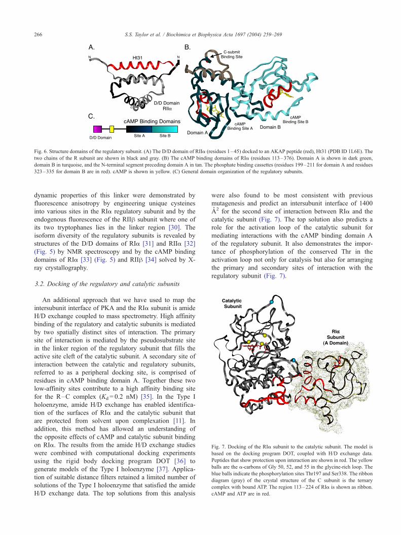
Fig. 7. Docking of the RIa subunit to the catalytic subunit. The model is
based on the docking program DOT, coupled with H/D exchange data.
Peptides that show protection upon interaction are shown in red. The yellow
balls are the a-carbons of Gly 50, 52, and 55 in the glycine-rich loop. The
blue balls indicate the phosphorylation sites Thr197 and Ser338. The ribbon
diagram (gray) of the crystal structure of the C subunit is the ternary
complex with bound ATP. The region 113–224 of RIa is shown as ribbon.
cAMP and ATP are in red.
Fig. 6. Structure domains of the regulatory subunit. (A) The D/D domain of RIIa (residues 1–45) docked to an AKAP peptide (red), Ht31 (PDB ID 1L6E). The
two chains of the R subunit are shown in black and gray. (B) The cAMP binding domains of RIa (residues 113–376). Domain A is shown in dark green,
domain B in turquoise, and the N-terminal segment preceding domain A in tan. The phosphate binding cassettes (residues 199–211 for domain A and residues
323–335 for domain B are in red). cAMP is shown in yellow. (C) General domain organization of the regulatory subunits.
S.S. Taylor et al. / Biochimica et Biophysica Acta 1697 (2004) 259–269266
dynamic properties of this linker were demonstrated by
fluorescence anisotropy by engineering unique cysteines
into various sites in the RIa regulatory subunit and by the
endogenous fluorescence of the RIIh subunit where one of
its two tryptophanes lies in the linker region [30]. The
isoform diversity of the regulatory subunits is revealed by
structures of the D/D domains of RIa [31] and RIIa [32]
(Fig. 5) by NMR spectroscopy and by the cAMP binding
domains of RIa [33] (Fig. 5) and RIIh [34] solved by X-
ray crystallography.
3.2. Docking of the regulatory and catalytic subunits
An additional approach that we have used to map the
intersubunit interface of PKA and the RIa subunit is amide
H/D exchange coupled to mass spectrometry. High affinity
binding of the regulatory and catalytic subunits is mediated
by two spatially distinct sites of interaction. The primary
site of interaction is mediated by the pseudosubstrate site
in the linker region of the regulatory subunit that fills the
active site cleft of the catalytic subunit. A secondary site of
interaction between the catalytic and regulatory subunits,
referred to as a peripheral docking site, is comprised of
residues in cAMP binding domain A. Together these two
low-affinity sites contribute to a high affinity binding site
for the R–C complex (Kd = 0.2 nM) [35]. In the Type I
holoenzyme, amide H/D exchange has enabled identifica-
tion of the surfaces of RIa and the catalytic subunit that
are protected from solvent upon complexation [11]. In
addition, this method has allowed an understanding of
the opposite effects of cAMP and catalytic subunit binding
on RIa. The results from the amide H/D exchange studies
were combined with computational docking experiments
using the rigid body docking program DOT [36] to
generate models of the Type I holoenzyme [37]. Applica-
tion of suitable distance filters retained a limited number of
solutions of the Type I holoenzyme that satisfied the amide
H/D exchange data. The top solutions from this analysis
were also found to be most consistent with previous
mutagenesis and predict an intersubunit interface of 1400
A2 for the second site of interaction between RIa and the
catalytic subunit (Fig. 7). The top solution also predicts a
role for the activation loop of the catalytic subunit for
mediating interactions with the cAMP binding domain A
of the regulatory subunit. It also demonstrates the impor-
tance of phosphorylation of the conserved Thr in the
activation loop not only for catalysis but also for arranging
the primary and secondary sites of interaction with the
regulatory subunit (Fig. 7).

S.S. Taylor et al. / Biochimica et Biophysica Acta 1697 (2004) 259–269 267
4. A kinase anchoring proteins
4.1. A kinase anchoring proteins contribute to localization
In addition to the catalytic and regulatory subunits, PKA
is targeted to specific sites in the cell by AKAPs. These
multidomain proteins are capable of sequestering a number
of signaling molecules such as phosphatases, kinases, and
phosphodiesterases in close proximity to target substrates
[38,39]. This sequestration provides a mechanism for gen-
erating microdomains for localized signaling. This adds
further to the complexity of the signaling networks and
opens up the possibility that signaling by cAMP could take
place in one location and might not require the diffusion of
cAMP across the cell. Indeed there is significant data in the
literature to suggest that such sequestration must occur [40].
The AKAPs provide a mechanism for achieving this.
4.2. Molecular basis for AKAP binding to PKA
The AKAPs bind to the regulatory subunits through an
amphipathic helix [41]. The RII subunits typically bind to
AKAPs in the low nM range [42]. Recently, RIa has also
been shown to bind to AKAPs although usually the
affinity is less (50–100 nM). Several dual specific AKAPs
that bind to both RI and RII have now been identified
[43,44]. Amphipathic helices with selectivity for both RI
and RII have also now been identified using spot array
analysis [41,45]. The amphipathic helix in the AKAP
binds to the amino terminal D/D domain of the regulatory
subunit. With the structures of the RIa and RIIa D/D
domains, recently solved by NMR spectroscopy [10], and
the structure of RIIa [32] docked to an AKAP peptide,
Ht31 (see Fig. 6A) [46], we are beginning to unravel the
molecular basis for targeting of PKA to AKAPs. This
small module, shown in Fig. 8, leads to the generation of
an extended network that brings the kinase close to the
substrates that it modulates.
The AKAPs themselves have been less well studied. In
an effort to determine whether the AKAPs contain func-
tional domains as well as significant regions of disorder,
DAKAP2, a dual specific AKAP was characterized using H/
D exchange coupled with mass spectrometry. DAKAP2
Fig. 8. Design of a recombinant AKAR. The recombinant AKAR shown
here provides a FRET response when the PKA-specific peptide is
phosphorylated in cells. CFP and YFP refer to cyan fluorescent protein
and yellow fluorescent protein, respectively. The peptide linking the 14–
3–3 domain and the YPF contains the PKA-specific sequence underlined
where the arrow indicates the site of phosphorylation.
contains near its amino terminus a putative RGS domain
and at is carboxyl terminal end the A kinase binding (AKB)
domain. The linker region contains a putative PKA phos-
phorylation site, but no known domain maps to this region.
Based on the exchangeability of the backbone amides to
deuterium, the RGS domain appeared to be folded in a
manner that was consistent with it having a conformation
that resembles other RGS domains [12]. The carboxyl
terminal helix was also shielded due to interaction with
another part of the molecule or possibly to dimerization. In
contrast, the linker region appeared to be quite unstructured.
Within 10 s, all of the backbone amides in the linker region
had fully exchanged. This initial profile suggests that the
AKAPs will also contribute significantly to the dynamic
properties of this signaling complex.
The carboxyl terminal 40 residues contain the AKB
motif and the docking of this motif to the RIa and RIIa
regulatory subunits has been mapped by H/D exchange
(manuscript submitted). In addition, the three terminal
residues are predicted to be a PDZ binding motif. Recently
a binding partner was identified for this motif. Gisler et al.
[47] identified PDZ-KI and PDZ-K2 as PDZ binding
proteins in kidney proximal tubules that bind to the
carboxyl terminus of DAKAP2. PDZ-K1 also binds to
the Na+ phosphate exchanger that mediates uptake of
phosphate. This process of phosphate uptake is regulated
by PKA through a mechanism that involves internalization
of the exchanger. This poses a novel PKA signaling
complex where the details of this process are being
unraveled.
5. Monitoring of PKA activity in living cells
While we can monitor kinase activity and measure
kinetic properties in vitro, it is ultimately the activity of
the enzyme in cells that is important to monitor. A recent
advance in the development of a recombinant probe, A
Kinase Activity Reporter (AKAR), to measure PKA activity
has made this possible [7]. AKAR was engineered by fusing
a cyan fluorescent protein (CFP) to a peptide that could be
phosphorylated by PKA (Fig. 8). This was followed by a
phosphate binding protein, 14–3–3, and then a yellow
fluorescent protein (YFP). Following phosphorylation, this
construct shows enhanced fluorescence resonance transfer
(FRET) as measured by emission at 527 nm. The response is
seen both in vitro and in vivo. In addition, Zhang et al. [7]
was able to show delayed response to forskolin stimulation
when the AKAR was localized to the nucleus. She also
demonstrated that the signal was more rapid when the
kinase was directly associated with the AKAR, either by
fusing an AKAP peptide to AKAR and thereby recruiting
endogenous PKA or by fusing a regulatory subunit directly
to the YFP. The AKAR can now be used to more compre-
hensively evaluate the importance of targeting for PKA
signaling in cells.

S.S. Taylor et al. / Biochimica et Biophysica Acta 1697 (2004) 259–269268
6. Conclusions and perspective
The integration of signaling by PKA is complex and
involves a variety of primary and auxiliary positions. While
the primary signaling molecules for PKA are the regulatory
and catalytic subunits, there are also auxiliary proteins that
contribute to building an extended network that brings PKA
in close proximity to its substrates. In addition, there are
multiple isoforms of the regulatory and catalytic subunits
and numerous splice variants of the catalytic subunit as well
as splice variants of the AKAPs that compound the com-
plexity of these signaling networks. It is this entire assembly
that constitutes the physiological signaling complex, and it
is capable of creating microdomains within a single cell.
Thus one can disrupt the network not only by generating
inhibitors to the active catalytic subunit, but also by stabi-
lizing the inhibited complex and also by disrupting target-
ing. Each strategy is capable of disrupting function.
As indicated in this review, in order to fully appreciate
the complexity of protein kinase structure and function
requires a variety of different techniques. It requires not
only high-resolution structures of the participating mole-
cules but also complementary solution methods that allow
us to appreciate the dynamics of the molecules in a more
physiological environment. Ultimately, we must be able to
monitor the interactions and functioning of these molecules
in living cells. This demands a broad spectrum of scientific
expertise. Our challenge is to build interdisciplinary teams
that can address such questions. To meet this challenge, we
have used a variety of approaches, in addition to crystal-
lography and NMR, to probe the structure and function of
PKA. We have used fluorescence anisotropy to probe local
motions and FRET to monitor PKA activity in living cells.
We also have used H/D exchange coupled with mass
spectrometry to probe domain organization, ligand- and
protein-induced conformational changes, and to define pro-
tein:protein interfaces. In this way, we are beginning to
better understand the molecular features and complexity of
these signaling networks.
Acknowledgements
This research was supported by grants from the national
Institutes of Health to SST (GM19301, GM34921, and
DK54441).
References
[1] G. Manning, D. Whyte, R. Martinez, T. Hunter, S. Sudarsanam, The
protein kinase complement of the human genome, Science 298 (2002)
1912–1934.
[2] J.H. Tchieu, F. Fana, J.L. Fink, J. Harper, T.M. Nair, R.H. Niedner,
D.W. Smith, K. Steube, T.M. Tam, S. Veretnik, D. Wang, M. Grib-
skov, The PlantsP and PlantsT functional genomics databases, Nu-
cleic Acids Res. 31 (2003) 342–344.
[3] T. Schindler, W. Bornmann, P. Pellicena, W.T. Miller, B. Clarkson, J.
Kuriyan, Structural mechanism for STI-571 inhibition of abelson ty-
rosine kinase, Science 289 (2000) 1938–1942.
[4] P. Akamine, P. Madhusudan, J. Wu, N.H Xuong, L.F. Ten Eyck, S.S.
Taylor, Dynamic features of cAMP-dependent protein kinase revealed
by apoenzyme crystal structure, J. Mol. Biol. 327 (2003) 159–171.
[5] Madhusudan, P. Akamine, N.H. Xuong, S.S. Taylor, Crystal struc-
ture of a transition state mimic of the catalytic subunit of cAMP-
dependent protein kinase, Nat. Struct. Biol. 9 (2002) 273–277.
[6] P. Amieux, G.S. McKnight, The essential role of RI alpha in the
maintenance of regulated PKA activity, Ann. N.Y. Acad. Sci. 968
(2002) 75–95.
[7] J. Zhang, Y. Ma, S.S. Taylor, R.Y. Tsien, Genetically encoded report-
ers of protein kinase A activity reveal impact of substrate tethering,
Proc. Natl. Acad. Sci. U. S. A. 98 (2001) 14997–15002.
[8] F. Li, M. Gangal, J.M. Jones, J. Deich, K. Lovett, S.S. Taylor, D.A.
Johnson, Consequence of cAMP and catalytic-subunit binding on the
flexibility of the A-kinase regulatory subunit, Biochemist 39 (2000)
15626–15632.
[9] F. Li, M. Gangal, C. Juliano, E. Gorfain, S.S. Taylor, D.A. Johnson,
Evidence for an internal entropy contribution to phosphoryl transfer:
a study of domain closure, backbone flexibility, and the catalytic
cycle of cAMP-dependent protein kinase, J. Mol. Biol. 315 (2002)
459–469.
[10] P. Banky, M. Roy, M.G. Newlon, D. Morikis, N.M. Haste, S.S. Tay-
lor, P.A. Jennings, Related protein – protein interaction modules
present drastically different surface topographies despite a conserved
helical platform, J. Mol. Biol. 330 (2003) 1117–1129.
[11] G.S. Anand, C.A. Hughes, J.M. Jones, S.S. Taylor, E.A. Komives,
Amide H/2H exchange reveals communication between the cAMP
and catalytic subunit-binding sites in the R(I)alpha subunit of protein
kinase A, J. Mol. Biol. 323 (2002) 377–386.
[12] Y. Hamuro, L. Burns, J. Canaves, R. Hoffman, S. Taylor, V. Woods,
Domain organization of D-AKAP2 revealed by enhanced deuterium
exchange-mass spectrometry (DXMS), J. Mol. Biol. 321 (2002)
703–714.
[13] P.F. Cook, M.E. Neville, K.E. Vrana, F.T. Hartl, R. Roskoski Jr.,
Adenosine cyclic 3V,5V-monophosphate dependent protein kinase:
kinetic mechanism for the bovine skeletal muscle catalytic subunit,
Biochemistry 21 (1982) 5794–5799.
[14] J.A. Adams, S.S. Taylor, Energetic limits of phosphotransfer in the
catalytic subunit of cAMP-dependent protein kinase as measured by
viscosity experiments, Biochemist 31 (1992) 8516–8522.
[15] D.A. Walsh, C.D. Ashby, Protein kinases: aspects of their regulation
and diversity, Recent Prog. Horm. Res. 29 (1973) 329.
[16] J.A. Adams, Participation of ADP dissociation in the rate-determining
step in cAMP-dependent protein kinase, Biochemist 36 (1997)
15733–15738.
[17] D.A. Johnson, P. Akamine, E. Radzio-Andzelm, Madhusudan, S.S.
Taylor, Dynamics of cAMP-dependent protein kinase, Am. Chem.
Soc. Monogr. 101 (2001) 2243–2270.
[18] J. Zheng, D.R. Knighton, L.F. Ten Eyck, R. Karlsson, N.-h. Xuong,
S.S. Taylor, J.M. Sowadski, Crystal structure of the catalytic subunit
of cAMP-dependent protein kinase complexed with MgATP and pep-
tide inhibitor, Biochemistry 32 (1993) 2154–2161.
[19] N. Narayana, T.C. Diller, K. Koide, M.E. Bunnage, K.C. Nicolaou,
L.L. Brunton, N.H. Xuong, L.F. Ten Eyck, S.S. Taylor, Crystal struc-
ture of the potent natural product inhibitor balanol in complex with
the catalytic subunit of cAMP-dependent protein kinase, Biochemis-
try 38 (1999) 2367–2376.
[20] C.M. Smith, E. Radzio-Andzelm, Madhusudan, P. Akamine, S.S.
Taylor, The catalytic subunit of cAMP-dependent protein kinase: pro-
totype for an extended network of communication, Prog. Biophys.
Mol. Biol. 71 (1999) 313–341.
[21] S.S. Taylor, E. Radzio-Andzelm, Three protein kinase structures de-
fine a common motif, Structure 2 (1994) 345–355.
[22] N. Narayana, S. Cox, N.-h. Xuong, L.F. Ten Eyck, S.S. Taylor, A

S.S. Taylor et al. / Biochimica et Biophysica Acta 1697 (2004) 259–269 269
binary complex of the catalytic subunit of cAMP-dependent protein
kinase and adenosine further defines conformational flexibility, Struc-
ture 5 (1997) 921–935.
[23] M. Valiev, R. Kawai, J.A. Adams, J.H. Weare, The role of the puta-
tive catalytic base in the phosphoryl transfer reaction in a protein
kinase: first-principles calculations, J. Am. Chem. Soc. 125 (2003)
9926–9927.
[24] J. Lew, N. Coruh, I. Tsigelny, S. Garrod, S.S. Taylor, Synergistic
binding of nucleotides and inhibitors to cAMP-dependent protein
kinase examined by acrylodan fluorescence spectroscopy, J. Biol.
Chem. 272 (1997) 1507–1513.
[25] P. Akamine, P. Madhusudan, L.L. Brunton, H.D. Ou, J.M. Canaves,
N.H. Xuong, S.S. Taylor, Balanol analogs probe specificity determi-
nants and conformational malleability of cAMP-dependent protein
kinase catalytic subunit, J. Mol. Biol. 327 (2003) 159–171.
[26] K.M. Specht, K.M. Shokat, The emerging power of chemical genet-
ics, Curr. Opin. Cell Biol. 14 (2002) 155–159.
[27] R.A. Engh, A. Girod, V. Kinzel, R. Huber, D. Bossemeyer, Crystal
structures of catalytic subunit of cAMP-dependent protein kinase in
complex with isoquinolinesulfonyl protein kinase inhibitors H7, H8,
and H89, J. Biol. Chem. 271 (1996) 26157–26164.
[28] L. Prade, R.A. Engh, A. Girod, V. Kinzel, R. Huber, D. Bossemeyer,
Staurosporine-induced conformational changes of cAMP-dependent
protein kinase catalytic subunit explain inhibitory potential, Structure
5 (1997) 1627–1637.
[29] P. Kulanthaivel, Y.F. Hallock, C. Boros, J.S.M.S. Hamilton, L.M.
Ballas, C.R. Loomis, J.B. Jiang, B. Katz, J.R. Steiner, J. Clardy,
Balanol: a novel and potent inhibitor of protein kinase C from the
fungus Verticillium balanoides, J. Am. Chem. Soc. 115 (1993)
6452–6453.
[30] K.M. Zawadzki, C.P. Pan, M.D. Barkley, D. Johnson, S.S. Taylor,
Endogenous tryptophan residues of cAPK regulatory subunit type
IIbeta reveal local variations in environments and dynamics, Proteins
51 (2003) 552–561.
[31] P. Banky, L.J. Huang, S.S. Taylor, Dimerization/docking domain of
the type Ia regulatory subunit of cAMP-dependent protein kinase:
requirements for dimerization and docking are distinct but overlap-
ping, J. Biol. Chem. 273 (1998) 35048–35055.
[32] M.G. Newlon, M. Roy, D. Morikis, Z.E. Hauseken, V. Coghlan, J.D.
Scott, P.A. Jennings, The molecular basis for protein kinase A anchor-
ing revealed by solution NMR, Nat. Struct. Biol. 6 (1999) 222–227.
[33] Y. Su, W.R.G. Dostmann, F.W. Herberg, K. Durick, N.-h. Xuong, L.F.
Ten Eyck, S.S. Taylor, K.I. Varughese, Regulatory (RIa) subunit of
protein kinase a: structure of deletion mutant with cAMP binding
domains, Science 269 (1995) 807–819.
[34] T.C. Diller, Madhusudan, N.-h. Xuong, S.S. Taylor, Molecular basis
for regulatory subunit diversity in cAMP-dependent protein kinase:
crystal structure of the type IIh regulatory subunit, Structure 9 (2001)
73–82.
[35] F.W. Herberg, W.R. Dostmann, M. Zorn, S.J. Davis, S.S. Taylor,
Crosstalk between domains in the regulatory subunit of cAMP-de-
pendent protein kinase: influence of amino terminus on cAMP bind-
ing and holoenzyme formation, Biochemistry 33 (1994) 7485–7494.
[36] J.G. Mandell, V.A. Roberts, M.E. Pique, V. Kotlovyi, J.C. Mitchell,
E. Nelson, I. Tsigelny, L.F. Ten Eyck, Protein docking using a
continuum electrostatics and geometric fit, Protein Eng. 14 (2001)
105–113.
[37] G.S. Anand, D. Law, J.G. Mandell, A.N. Snead, I. Tsigelny, S.S.
Taylor, L.F. Ten Eyck, E.A. Komives, Identification of the protein
kinase A regulatory RIa-catalytic subunit by amid hydrogen/deute-
rium exchange and protein docking, PNAS 100 (2003) 13264–
13269.
[38] T. Pawson, J.D. Scott, Signaling through scaffold anchoring, and
adaptor proteins, Science 278 (1997) 2075–2080.
[39] K.L. Dodge, S. Khouangsathiene, M.S. Kapiloff, R. Mouton, E.V.
Hill, M.D. Houslay, L.K. Langeberg, J.D. Scott, mAKAP assembles
a protein kinase A/PDE4 phosphodiesterase cAMP signaling module,
EMBO J. 20 (2001) 1921–1930.
[40] S.F. Steinberg, L.L. Brunton, Compartmentation of G protein-coupled
signaling pathways in cardiac myocytes, Annu. Rev. Pharmacol. Tox-
icol. 41 (2001) 751–773.
[41] L.L. Burns-Hamuro, Y. Ma, S. Kammerer, U. Reineke, C. Self, C.
Cook, G.L. Olson, C.R. Cantor, A. Braun, S.S. Taylor, Designing
isoform-specific peptide disruptors of protein kinase a localization,
Proc. Natl. Acad. Sci. U. S. A. 100 (2003) 4072–4077.
[42] F.W. Herberg, A. Maleszka, T. Eide, L. Vossebein, K. Tasken, Anal-
ysis of A-kinase anchoring protein (AKAP) interaction with protein
kinase A (PKA) regulatory subunits: PKA isoform specificity in
AKAP binding, J. Mol. Biol. 298 (2000) 329–339.
[43] L.J. Huang, K. Durick, J.A. Weiner, J. Chun, S.S. Taylor, Identifi-
cation of a novel protein kinase A anchoring protein that binds both
type I and type II regulatory subunits, J. Biol. Chem. 272 (1997)
8057–8064.
[44] L.J. Huang, L. Wang, Y. Ma, K. Durick, G. Perkins, T.J. Deerinck,
M.H. Ellisman, S.S. Taylor, NH2-terminal targeting motifs direct
dual specificity A-kinase anchoring protein 1(D-AKAP1) to either
mitochondria or endoplasmic reticulum, J. Cell Biol. 145 (1999)
951–959.
[45] L.L. Burns, J.M. Canaves, J.K. Pennypacker, D.K. Blumenthal, S.S.
Taylor, Isoform specific differences in binding of a dual-specificity A-
kinase anchoring protein to type I and type II regulatory subunits of
PKA, Biochemistry 42 (2003) 5754–5763.
[46] M.G. Newlon, M. Roy, D. Morikis, D.W. Carr, R. Westphal, J.D.
Scott, P.A. Jennings, A novel mechanism of PKA anchoring revealed
by solution structures of anchoring complexes, EMBO J. 7 (2001)
1651–1662.
[47] S.M. Gisler, C. Madjdpour, D. Bacic, S. Pribanic, S.S. Taylor, J.
Biber, H. Murer, PDZK1: II. An anchoring site for the PKA-binding
protein D-AKAP2 in renal proximal tubular cells, Kidney Int. 64
(2003) 1746–1754.