![Laparoscopic and Thoracoscopic Surgerynebraskasurgicalresearch.com/s NSR/chapter... · Laparoscopic and thoracoscopic surgery / [edited by] Constantine T. Frantzides. cm. Includes](https://static.fdocuments.us/doc/165x107/5fc981c7613d2d6d1d5b3041/laparoscopic-and-thoracoscopic-surgerynebr-nsrchapter-laparoscopic-and-thoracoscopic.jpg)
REVIEW Open Access Thoracoscopic removal of dental ...
4
REVIEW Open Access Thoracoscopic removal of dental prosthesis impacted in the upper thoracic esophagus Luigi Bonavina * , Alberto Aiolfi, Stefano Siboni and Emanuele Rausa Abstract Dental appliances are the most common cause of accidental foreign body esophageal impaction, especially in the elderly population with decreased oral sensory perception. A 47-year-old man with history of oligophrenia and recurrent epileptic seizures was referred to our hospital following dislocation and ingestion of his upper dental prosthesis. Endoscopic removal and clipping of an esophageal tear had been unsuccessfully attempted. A chest CT scan confirmed entrapment of the dental prosthesis in the upper thoracic esophagus, the presence of pneumomediastinum, and the close proximity of one of the metal clasps of the prosthesis to the left subclavian artery. A video-assisted right thoracoscopy in the left lateral decubitus position was performed and the foreign body was successfully removed. The patient was then allowed to wear the retrieved prosthesis after dentistry consultation and repair of the wire clasps by a dental technician. At the 6-month follow-up visit the patient was doing very well without any trouble in swallowing. Keywords: Esophagus, Esophageal perforation, Dental prosthesis, Thoracoscopy Introduction Accidental ingestion of foreign bodies is frequent in adult individuals with mental retardation or psychiatric disorders. Most of the little ingested foreign bodies pass the gastrointestinal tract without consequences. How- ever, 10-20% of the patients may require endoscopic removal, and 1% or less may require surgery due to en- trapment of the foreign body in the cervical (57%), thoracic (26%), or distal (17%) esophagus [1]. Dental ap- pliances are the most common cause of accidental for- eign body esophageal impaction, especially in the elderly population with decreased oral sensory perception [2]. The large size, sharp edges, and metal clasps of dental prostheses make endoscopic removal unsafe and carry a high risk of perforation in such circumstances. We present a case of successful thoracoscopic removal of dental pros- thesis impacted in the upper thoracic esophagus. Case report A 47-year-old man with history of oligophrenia and re- current epileptic seizures was referred to our hospital 3 days after dislocation and ingestion of his upper dental prosthesis. Before patient’ s referral, multiple flexible endoscopic attempts had been unsuccessfully performed, the last one leading to an intramural perforation partially repaired with endoclips. The patient’ s main complaints were dysphagia, odynophagia, and hypersalivation. He was afebrile, with normal leucocyte count, and slight ele- vation of C-reactive protein. Broad-spectrum antibiotic therapy (piperacillin + tazobactam) was started upon hospital admission. The physical examination did not re- veal subcutaneous emphysema. A gastrografin swallow study showed extravasation of contrast at the level of the upper thoracic esophagus; a chest CT scan confirmed the presence of pneumomediastinum and the close proximity of one of the metal clasps of the prosthesis to the left subclavian artery (Figure 1A-B). A video-assisted right thoracoscopy in the left lateral decubitus position was performed to remove the foreign body. Three ports were used: 10 mm optical port in the 6 th intercostal space, 10 mm port in the 5 th intercostal space, and 5 mm port in the 4 th intercostal space, and. The exploration of the right chest showed a bulging of the upper mediastinal compartment above the confluence of the azygos vein into the superior vena cava (Figure 1C). There was no pleural contamination. After incision of the * Correspondence: [email protected] Department of Biomedical Sciences for Health, Division of General Surgery, University of Milano Medical School, IRCCS Policlinico San Donato, Via Morandi 30, 20097, San Donato Milanese, (Milano), Italy WORLD JOURNAL OF EMERGENCY SURGERY © 2014 Bonavina et al.; licensee BioMed Central Ltd. This is an Open Access article distributed under the terms of the Creative Commons Attribution License (http://creativecommons.org/licenses/by/2.0), which permits unrestricted use, distribution, and reproduction in any medium, provided the original work is properly cited. The Creative Commons Public Domain Dedication waiver (http://creativecommons.org/publicdomain/zero/1.0/) applies to the data made available in this article, unless otherwise stated. Bonavina et al. World Journal of Emergency Surgery 2014, 9:5 http://www.wjes.org/content/9/1/5
Transcript of REVIEW Open Access Thoracoscopic removal of dental ...
WORLD JOURNAL OF EMERGENCY SURGERY
Bonavina et al. World Journal of Emergency Surgery 2014, 9:5http://www.wjes.org/content/9/1/5
REVIEW Open Access
Thoracoscopic removal of dental prosthesisimpacted in the upper thoracic esophagusLuigi Bonavina*, Alberto Aiolfi, Stefano Siboni and Emanuele Rausa
Abstract
Dental appliances are the most common cause of accidental foreign body esophageal impaction, especially in theelderly population with decreased oral sensory perception. A 47-year-old man with history of oligophrenia andrecurrent epileptic seizures was referred to our hospital following dislocation and ingestion of his upper dentalprosthesis. Endoscopic removal and clipping of an esophageal tear had been unsuccessfully attempted. A chestCT scan confirmed entrapment of the dental prosthesis in the upper thoracic esophagus, the presence ofpneumomediastinum, and the close proximity of one of the metal clasps of the prosthesis to the left subclavianartery. A video-assisted right thoracoscopy in the left lateral decubitus position was performed and the foreignbody was successfully removed. The patient was then allowed to wear the retrieved prosthesis after dentistryconsultation and repair of the wire clasps by a dental technician. At the 6-month follow-up visit the patient wasdoing very well without any trouble in swallowing.
Keywords: Esophagus, Esophageal perforation, Dental prosthesis, Thoracoscopy
IntroductionAccidental ingestion of foreign bodies is frequent inadult individuals with mental retardation or psychiatricdisorders. Most of the little ingested foreign bodies passthe gastrointestinal tract without consequences. How-ever, 10-20% of the patients may require endoscopicremoval, and 1% or less may require surgery due to en-trapment of the foreign body in the cervical (57%),thoracic (26%), or distal (17%) esophagus [1]. Dental ap-pliances are the most common cause of accidental for-eign body esophageal impaction, especially in the elderlypopulation with decreased oral sensory perception [2].The large size, sharp edges, and metal clasps of dentalprostheses make endoscopic removal unsafe and carry ahigh risk of perforation in such circumstances. We presenta case of successful thoracoscopic removal of dental pros-thesis impacted in the upper thoracic esophagus.
Case reportA 47-year-old man with history of oligophrenia and re-current epileptic seizures was referred to our hospital 3
* Correspondence: [email protected] of Biomedical Sciences for Health, Division of General Surgery,University of Milano Medical School, IRCCS Policlinico San Donato, ViaMorandi 30, 20097, San Donato Milanese, (Milano), Italy
© 2014 Bonavina et al.; licensee BioMed CentrCommons Attribution License (http://creativecreproduction in any medium, provided the orwaiver (http://creativecommons.org/publicdomstated.
days after dislocation and ingestion of his upper dentalprosthesis. Before patient’s referral, multiple flexibleendoscopic attempts had been unsuccessfully performed,the last one leading to an intramural perforation partiallyrepaired with endoclips. The patient’s main complaintswere dysphagia, odynophagia, and hypersalivation. Hewas afebrile, with normal leucocyte count, and slight ele-vation of C-reactive protein. Broad-spectrum antibiotictherapy (piperacillin + tazobactam) was started uponhospital admission. The physical examination did not re-veal subcutaneous emphysema. A gastrografin swallowstudy showed extravasation of contrast at the level of theupper thoracic esophagus; a chest CT scan confirmedthe presence of pneumomediastinum and the closeproximity of one of the metal clasps of the prosthesis tothe left subclavian artery (Figure 1A-B).A video-assisted right thoracoscopy in the left lateral
decubitus position was performed to remove the foreignbody. Three ports were used: 10 mm optical port in the6th intercostal space, 10 mm port in the 5th intercostalspace, and 5 mm port in the 4th intercostal space, and.The exploration of the right chest showed a bulging of theupper mediastinal compartment above the confluence ofthe azygos vein into the superior vena cava (Figure 1C).There was no pleural contamination. After incision of the
al Ltd. This is an Open Access article distributed under the terms of the Creativeommons.org/licenses/by/2.0), which permits unrestricted use, distribution, andiginal work is properly cited. The Creative Commons Public Domain Dedicationain/zero/1.0/) applies to the data made available in this article, unless otherwise
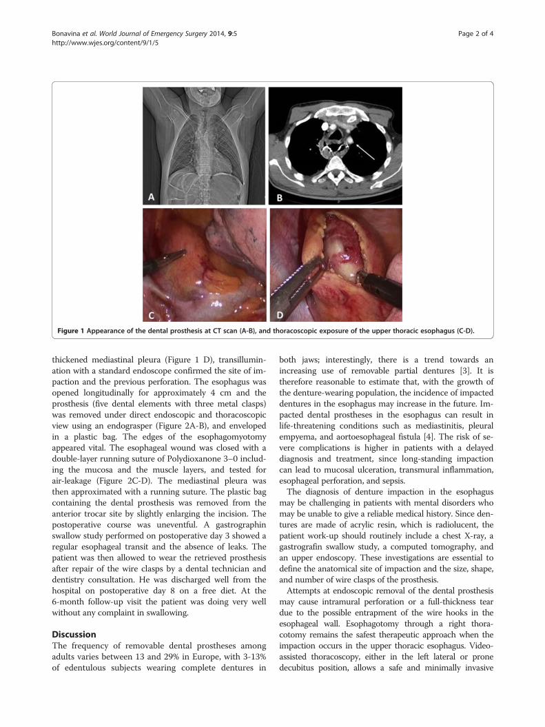
Figure 1 Appearance of the dental prosthesis at CT scan (A-B), and thoracoscopic exposure of the upper thoracic esophagus (C-D).
Bonavina et al. World Journal of Emergency Surgery 2014, 9:5 Page 2 of 4http://www.wjes.org/content/9/1/5
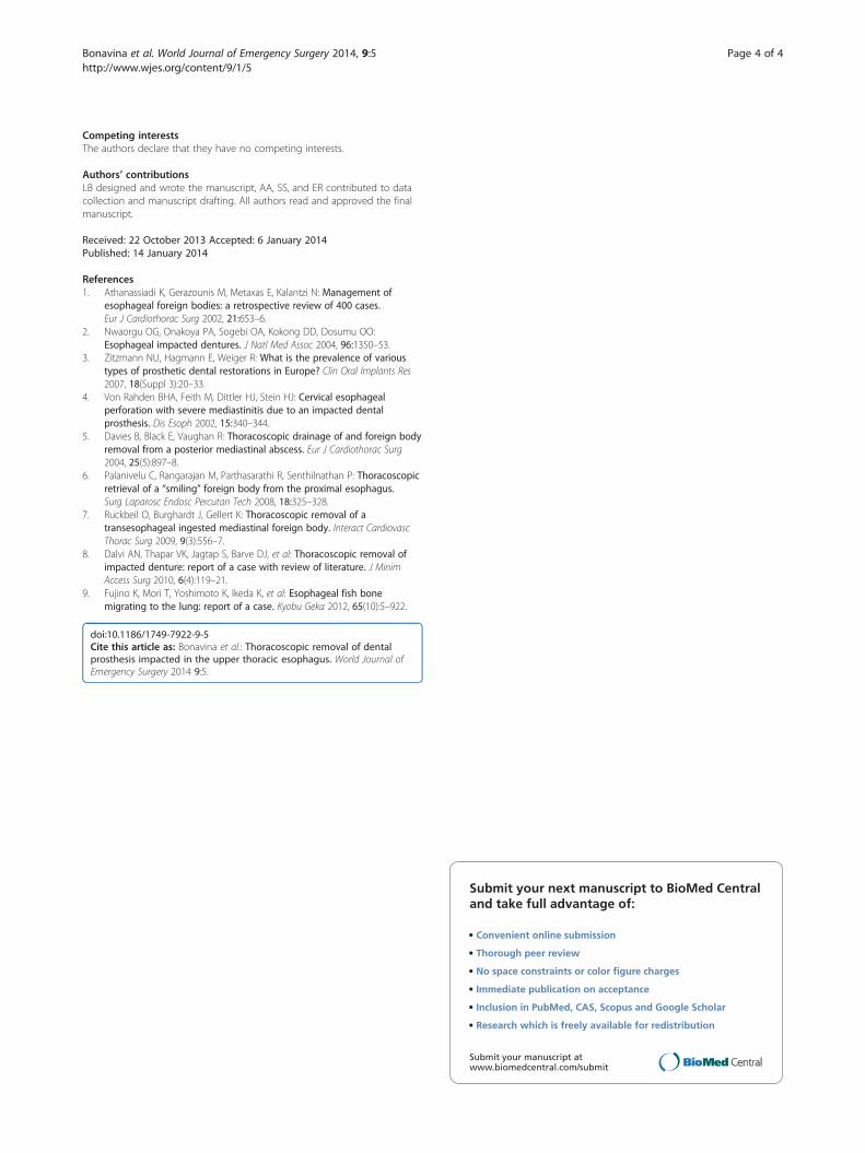
thickened mediastinal pleura (Figure 1 D), transillumin-ation with a standard endoscope confirmed the site of im-paction and the previous perforation. The esophagus wasopened longitudinally for approximately 4 cm and theprosthesis (five dental elements with three metal clasps)was removed under direct endoscopic and thoracoscopicview using an endograsper (Figure 2A-B), and envelopedin a plastic bag. The edges of the esophagomyotomyappeared vital. The esophageal wound was closed with adouble-layer running suture of Polydioxanone 3–0 includ-ing the mucosa and the muscle layers, and tested forair-leakage (Figure 2C-D). The mediastinal pleura wasthen approximated with a running suture. The plastic bagcontaining the dental prosthesis was removed from theanterior trocar site by slightly enlarging the incision. Thepostoperative course was uneventful. A gastrographinswallow study performed on postoperative day 3 showed aregular esophageal transit and the absence of leaks. Thepatient was then allowed to wear the retrieved prosthesisafter repair of the wire clasps by a dental technician anddentistry consultation. He was discharged well from thehospital on postoperative day 8 on a free diet. At the6-month follow-up visit the patient was doing very wellwithout any complaint in swallowing.
DiscussionThe frequency of removable dental prostheses amongadults varies between 13 and 29% in Europe, with 3-13%of edentulous subjects wearing complete dentures in
both jaws; interestingly, there is a trend towards anincreasing use of removable partial dentures [3]. It istherefore reasonable to estimate that, with the growth ofthe denture-wearing population, the incidence of impacteddentures in the esophagus may increase in the future. Im-pacted dental prostheses in the esophagus can result inlife-threatening conditions such as mediastinitis, pleuralempyema, and aortoesophageal fistula [4]. The risk of se-vere complications is higher in patients with a delayeddiagnosis and treatment, since long-standing impactioncan lead to mucosal ulceration, transmural inflammation,esophageal perforation, and sepsis.The diagnosis of denture impaction in the esophagus
may be challenging in patients with mental disorders whomay be unable to give a reliable medical history. Since den-tures are made of acrylic resin, which is radiolucent, thepatient work-up should routinely include a chest X-ray, agastrografin swallow study, a computed tomography, andan upper endoscopy. These investigations are essential todefine the anatomical site of impaction and the size, shape,and number of wire clasps of the prosthesis.Attempts at endoscopic removal of the dental prosthesis
may cause intramural perforation or a full-thickness teardue to the possible entrapment of the wire hooks in theesophageal wall. Esophagotomy through a right thora-cotomy remains the safest therapeutic approach when theimpaction occurs in the upper thoracic esophagus. Video-assisted thoracoscopy, either in the left lateral or pronedecubitus position, allows a safe and minimally invasive

Figure 2 Esophagotomy (A), removal of the dental prosthesis (B), and suture of the esophageal wall and mediastinal pleura (C-D).
Bonavina et al. World Journal of Emergency Surgery 2014, 9:5 Page 3 of 4http://www.wjes.org/content/9/1/5
retrieval of the dental prosthesis followed by primaryesophageal suture when there is no major pleural contam-ination and the edges of the esophagomyotomy appearvital. In the literature, a few cases of thoracoscopic removalof ingested foreign bodies have been reported; three of the6 patients required an esophagotomy due to an impacteddenture (Table 1). In our patient, thoracoscopic removalwas successfully performed after previous failed endoscopicprocedures complicated by intramural perforation. Expos-ure of the upper thoracic esophagus was possible withoutthe need to divide the arch of the azygos vein.
Table 1 Thoracoscopic management of ingested esophageal f
Author Year Description Surgical app
Davies B. [5] 2004 China cup fragment migrated inthe mediastinum, with abscess
Right-side thora(3-port acc
Palanivelu C. [6] 2008 Impacted denture Right-side thora(3-port acc
Rückbeil O. [7] 2009 Metallic needle migratedin the mediastinum
Right-side thora(3- port acc
Dalvi AN. [8] 2010 Impacted denture Right-side thora(4-port acc
Fujino K. [9] 2012 Fish bone migrated to lung Right-side thora(NS)
Present case 2013 Impacted denture Right-side thora(3-port acc
(NS: non specified).
Based on our experience and the available literaturewe conclude that thoracoscopic esophagotomy repre-sents a safe and effective treatment for patients withimpacted dentures in the esophagus. Multiple attemptsat flexible and rigid esophagoscopy should definitely beabandoned in such patients, especially when a dentalprosthesis has passed the cricophageal sphincter. Edu-cation and close follow-up of patients wearing remov-able dental prostheses is critical to prevent accidentalimpaction in the esophagus and the dangerous sequelaeof esophageal perforation.
oreign bodies in adults: literature review
roach Operative decubitus Treatment Outcome
coscopyess)
NS Foreign body removaland abscess drainage
Good
coscopyess)
Prone Esophagotomy, foreignbody removal and suture
Good
coscopyess)
Left lateral Foreign body removal Good
coscopyess)
Left lateral Esophagotomy, foreignbody removal and suture
Good
coscopy NS Foreign body removal Good
coscopyess)
Left lateral Esophagotomy, foreignbody removal and suture
Good
Bonavina et al. World Journal of Emergency Surgery 2014, 9:5 Page 4 of 4http://www.wjes.org/content/9/1/5
Competing interestsThe authors declare that they have no competing interests.
Authors’ contributionsLB designed and wrote the manuscript, AA, SS, and ER contributed to datacollection and manuscript drafting. All authors read and approved the finalmanuscript.
Received: 22 October 2013 Accepted: 6 January 2014Published: 14 January 2014
References1. Athanassiadi K, Gerazounis M, Metaxas E, Kalantzi N: Management of
esophageal foreign bodies: a retrospective review of 400 cases.Eur J Cardiothorac Surg 2002, 21:653–6.
2. Nwaorgu OG, Onakoya PA, Sogebi OA, Kokong DD, Dosumu OO:Esophageal impacted dentures. J Natl Med Assoc 2004, 96:1350–53.
3. Zitzmann NU, Hagmann E, Weiger R: What is the prevalence of varioustypes of prosthetic dental restorations in Europe? Clin Oral Implants Res2007, 18(Suppl 3):20–33.
4. Von Rahden BHA, Feith M, Dittler HJ, Stein HJ: Cervical esophagealperforation with severe mediastinitis due to an impacted dentalprosthesis. Dis Esoph 2002, 15:340–344.
5. Davies B, Black E, Vaughan R: Thoracoscopic drainage of and foreign bodyremoval from a posterior mediastinal abscess. Eur J Cardiothorac Surg2004, 25(5):897–8.
6. Palanivelu C, Rangarajan M, Parthasarathi R, Senthilnathan P: Thoracoscopicretrieval of a “smiling” foreign body from the proximal esophagus.Surg Laparosc Endosc Percutan Tech 2008, 18:325–328.
7. Ruckbeil O, Burghardt J, Gellert K: Thoracoscopic removal of atransesophageal ingested mediastinal foreign body. Interact CardiovascThorac Surg 2009, 9(3):556–7.
8. Dalvi AN, Thapar VK, Jagtap S, Barve DJ, et al: Thoracoscopic removal ofimpacted denture: report of a case with review of literature. J MinimAccess Surg 2010, 6(4):119–21.
9. Fujino K, Mori T, Yoshimoto K, Ikeda K, et al: Esophageal fish bonemigrating to the lung: report of a case. Kyobu Geka 2012, 65(10):5–922.
doi:10.1186/1749-7922-9-5Cite this article as: Bonavina et al.: Thoracoscopic removal of dentalprosthesis impacted in the upper thoracic esophagus. World Journal ofEmergency Surgery 2014 9:5.
Submit your next manuscript to BioMed Centraland take full advantage of:
• Convenient online submission
• Thorough peer review
• No space constraints or color figure charges
• Immediate publication on acceptance
• Inclusion in PubMed, CAS, Scopus and Google Scholar
• Research which is freely available for redistribution
Submit your manuscript at www.biomedcentral.com/submit