REVIEW Open Access Patent foramen ovale and scuba … · Patent foramen ovale and scuba diving: a...
7
REVIEW Open Access Patent foramen ovale and scuba diving: a practical guide for physicians on when to refer for screening Oliver Sykes 1* and James E Clark 2 Abstract Divers are taught some basic physiology during their training. There is therefore some underlying knowledge and understandable concern in the diving community about the presence of a patent foramen ovale (PFO) as a cause of decompression illness (DCI). There is an agreement that PFO screening should not be done routinely on all divers; however, when to screen selected divers is not clear. We present the basic physiology and current existing guidelines for doctors, advice on the management and identify which groups of divers should be referred for consideration of PFO screening. Venous bubbles after diving and right to left shunts are common, but DCI is rare. Why this is the case is not clear, but the divers look to doctors for guidance on PFO screening and closure; both of which are not without risks. Ideally, we should advise and apply guidelines that are consistent and based on best available evidence. We hope this guideline and flow chart helps address these issues with regard to PFOs and diving. Keywords: Patent foramen ovale, Decompression illness, Arterial gas embolism, Screening Review Introduction Decompression illness (DCI) encompasses decompres- sion sickness (DCS) and arterial gas embolism (AGE). The differentiation of the pathological processes in prac- tice can be difficult, but the treatment is similar; hence, both are given the modern overarching term of decom- pression illness. DCS occurs as a result of venous bub- bles forming in the tissues and vessels, which can cause mechanical, embolic and biochemical effects with mani- festations ranging from trivial to fatal [1]. AGE is caused by arterial bubbles as a result of ruptured lung alveoli from gas trapping in the lungs or blood shunting from the venous right atrial side to the arterial left atrial side of the heart. This is known as a right to left shunt. Symptoms usually appear shortly after, or within 30 min of surfacing, but can have delayed onset. These symp- toms are frequently neurological in nature [2-4] and can be profound. The lungs are an effective filter, and a right to left shunt, such as a patent foramen ovale (PFO), is therefore a route for bubbles to avoid this filter and enter the arterial system. This is known as a paradoxical embolism and is depicted in Figure 1. Usually, the blood pressure on the arterial left side is higher than the ve- nous right, which prevents right to left flow. However, this pressure differential is reversed on releasing a Valsalva manoeuvre, causing the right atrium to fill before the left atrium. Unfortunately, 30 to 60 min post-dive is the peak time for bubble liberation [5], which coincides with divers climbing into boats, lifting heavy kit, straining and uncon- sciously performing Valsalva manoeuvres. In the fetus, the foramen ovale is vital to allow blood to bypass the lungs, which are not in use. On breathing at birth, there is a flap valve effect [6] (Figure 2), and the negative intra-thoracic pressure helps closes this route. While in about 30% there remains a leak, DCI is still a rare event [1,7]. This suggests that not all divers with a PFO are at increased risk of DCI [8]. However, those that are susceptible appear to get the more serious neurological symptoms. Moon examined 91 patients with a two-dimensional echocardiogram, who were eva- luated and/or treated for DCS at Duke University * Correspondence: [email protected] 1 London Hyperbaric Medicine, Whipp's Cross University Hospital, London E11 1NR, UK Full list of author information is available at the end of the article © 2013 Sykes and Clark; licensee BioMed Central Ltd. This is an Open Access article distributed under the terms of the Creative Commons Attribution License (http://creativecommons.org/licenses/by/2.0), which permits unrestricted use, distribution, and reproduction in any medium, provided the original work is properly cited. Sykes and Clark Extreme Physiology & Medicine 2013, 2:10 http://www.extremephysiolmed.com/content/2/1/10
Transcript of REVIEW Open Access Patent foramen ovale and scuba … · Patent foramen ovale and scuba diving: a...
Sykes and Clark Extreme Physiology & Medicine 2013, 2:10http://www.extremephysiolmed.com/content/2/1/10
REVIEW Open Access
Patent foramen ovale and scuba diving: apractical guide for physicians on when to referfor screeningOliver Sykes1* and James E Clark2
Abstract
Divers are taught some basic physiology during their training. There is therefore some underlying knowledge andunderstandable concern in the diving community about the presence of a patent foramen ovale (PFO) as a causeof decompression illness (DCI). There is an agreement that PFO screening should not be done routinely on alldivers; however, when to screen selected divers is not clear. We present the basic physiology and current existingguidelines for doctors, advice on the management and identify which groups of divers should be referred forconsideration of PFO screening. Venous bubbles after diving and right to left shunts are common, but DCI is rare.Why this is the case is not clear, but the divers look to doctors for guidance on PFO screening and closure; bothof which are not without risks. Ideally, we should advise and apply guidelines that are consistent and based onbest available evidence. We hope this guideline and flow chart helps address these issues with regard to PFOsand diving.
Keywords: Patent foramen ovale, Decompression illness, Arterial gas embolism, Screening
ReviewIntroductionDecompression illness (DCI) encompasses decompres-sion sickness (DCS) and arterial gas embolism (AGE).The differentiation of the pathological processes in prac-tice can be difficult, but the treatment is similar; hence,both are given the modern overarching term of decom-pression illness. DCS occurs as a result of venous bub-bles forming in the tissues and vessels, which can causemechanical, embolic and biochemical effects with mani-festations ranging from trivial to fatal [1]. AGE is causedby arterial bubbles as a result of ruptured lung alveolifrom gas trapping in the lungs or blood shunting fromthe venous right atrial side to the arterial left atrial sideof the heart. This is known as a right to left shunt.Symptoms usually appear shortly after, or within 30 minof surfacing, but can have delayed onset. These symp-toms are frequently neurological in nature [2-4] and canbe profound. The lungs are an effective filter, and a right
* Correspondence: [email protected] Hyperbaric Medicine, Whipp's Cross University Hospital, London E111NR, UKFull list of author information is available at the end of the article
© 2013 Sykes and Clark; licensee BioMed CentCommons Attribution License (http://creativecreproduction in any medium, provided the or
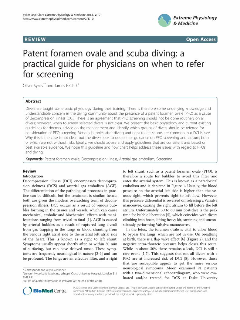
to left shunt, such as a patent foramen ovale (PFO), istherefore a route for bubbles to avoid this filter andenter the arterial system. This is known as a paradoxicalembolism and is depicted in Figure 1. Usually, the bloodpressure on the arterial left side is higher than the ve-nous right, which prevents right to left flow. However,this pressure differential is reversed on releasing a Valsalvamanoeuvre, causing the right atrium to fill before the leftatrium. Unfortunately, 30 to 60 min post-dive is the peaktime for bubble liberation [5], which coincides with diversclimbing into boats, lifting heavy kit, straining and uncon-sciously performing Valsalva manoeuvres.In the fetus, the foramen ovale is vital to allow blood
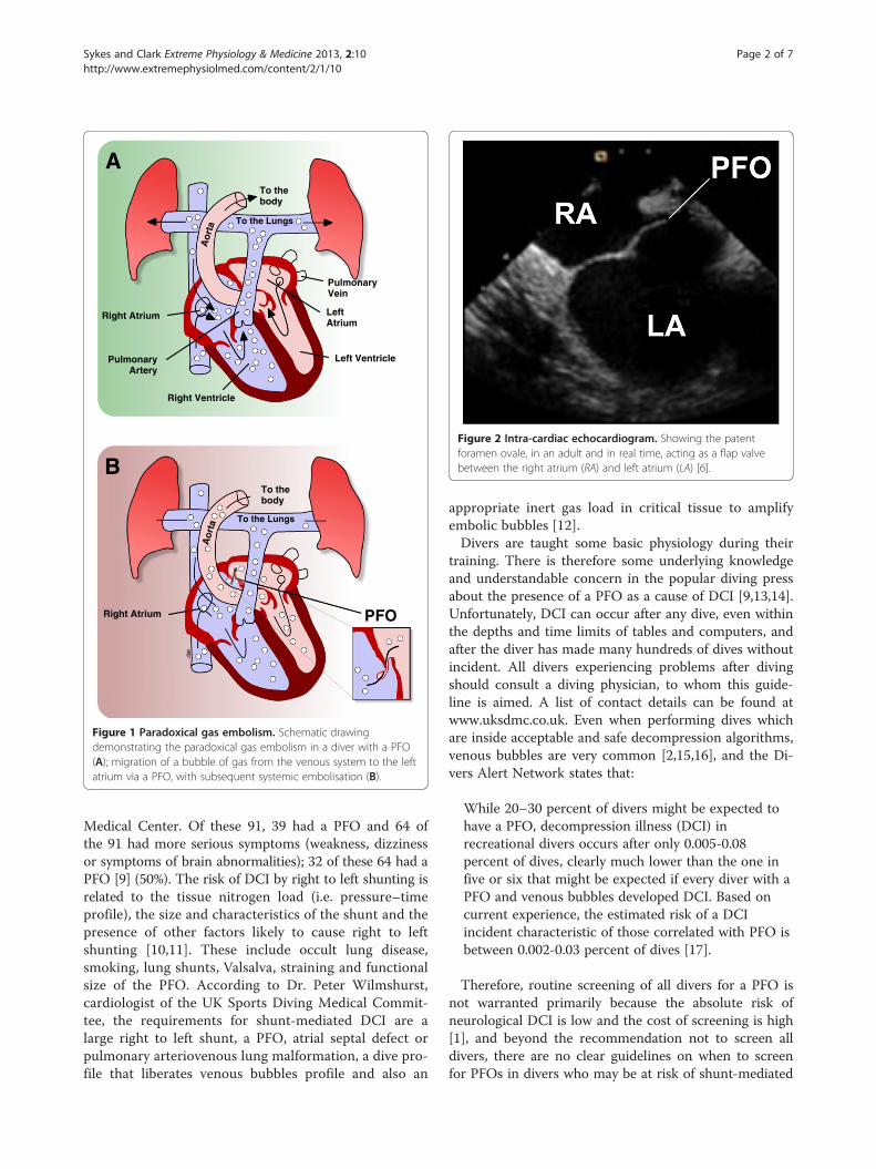
to bypass the lungs, which are not in use. On breathingat birth, there is a flap valve effect [6] (Figure 2), and thenegative intra-thoracic pressure helps closes this route.While in about 30% there remains a leak, DCI is still arare event [1,7]. This suggests that not all divers with aPFO are at increased risk of DCI [8]. However, thosethat are susceptible appear to get the more seriousneurological symptoms. Moon examined 91 patientswith a two-dimensional echocardiogram, who were eva-luated and/or treated for DCS at Duke University
ral Ltd. This is an Open Access article distributed under the terms of the Creativeommons.org/licenses/by/2.0), which permits unrestricted use, distribution, andiginal work is properly cited.
Pulmonary Vein
Left Atrium
Left Ventricle
Right Ventricle
Pulmonary Artery
Right Atrium
To the body
To the Lungs
Aor
ta
Right Atrium
To the body
To the Lungs
Aor
ta
PFO
A
B
J.Clark
Figure 1 Paradoxical gas embolism. Schematic drawingdemonstrating the paradoxical gas embolism in a diver with a PFO(A); migration of a bubble of gas from the venous system to the leftatrium via a PFO, with subsequent systemic embolisation (B).
Figure 2 Intra-cardiac echocardiogram. Showing the patentforamen ovale, in an adult and in real time, acting as a flap valvebetween the right atrium (RA) and left atrium (LA) [6].
Sykes and Clark Extreme Physiology & Medicine 2013, 2:10 Page 2 of 7http://www.extremephysiolmed.com/content/2/1/10
Medical Center. Of these 91, 39 had a PFO and 64 ofthe 91 had more serious symptoms (weakness, dizzinessor symptoms of brain abnormalities); 32 of these 64 had aPFO [9] (50%). The risk of DCI by right to left shunting isrelated to the tissue nitrogen load (i.e. pressure–timeprofile), the size and characteristics of the shunt and thepresence of other factors likely to cause right to leftshunting [10,11]. These include occult lung disease,smoking, lung shunts, Valsalva, straining and functionalsize of the PFO. According to Dr. Peter Wilmshurst,cardiologist of the UK Sports Diving Medical Commit-tee, the requirements for shunt-mediated DCI are alarge right to left shunt, a PFO, atrial septal defect orpulmonary arteriovenous lung malformation, a dive pro-file that liberates venous bubbles profile and also an
appropriate inert gas load in critical tissue to amplifyembolic bubbles [12].Divers are taught some basic physiology during their
training. There is therefore some underlying knowledgeand understandable concern in the popular diving pressabout the presence of a PFO as a cause of DCI [9,13,14].Unfortunately, DCI can occur after any dive, even withinthe depths and time limits of tables and computers, andafter the diver has made many hundreds of dives withoutincident. All divers experiencing problems after divingshould consult a diving physician, to whom this guide-line is aimed. A list of contact details can be found atwww.uksdmc.co.uk. Even when performing dives whichare inside acceptable and safe decompression algorithms,venous bubbles are very common [2,15,16], and the Di-vers Alert Network states that:
While 20–30 percent of divers might be expected tohave a PFO, decompression illness (DCI) inrecreational divers occurs after only 0.005-0.08percent of dives, clearly much lower than the one infive or six that might be expected if every diver with aPFO and venous bubbles developed DCI. Based oncurrent experience, the estimated risk of a DCIincident characteristic of those correlated with PFO isbetween 0.002-0.03 percent of dives [17].
Therefore, routine screening of all divers for a PFO isnot warranted primarily because the absolute risk ofneurological DCI is low and the cost of screening is high[1], and beyond the recommendation not to screen alldivers, there are no clear guidelines on when to screenfor PFOs in divers who may be at risk of shunt-mediated
Sykes and Clark Extreme Physiology & Medicine 2013, 2:10 Page 3 of 7http://www.extremephysiolmed.com/content/2/1/10
DCI. Here, we present a practical approach to a com-mon problem of what to do with a diver who may war-rant or request a referral for a PFO check. These areguidelines for doctors treating divers and should not beused in place of diver training.
Current guidelinesAccording to the UK Sports Diving Medical Committee [18]:
Approximately one quarter of the population have apatent foramen ovale or a small atrial septal defect, butthe risk of paradoxical embolism is much greater inthose with large shunts [10,19]. Decompression illnessis very unusual in sport divers after dives to less than 20metres and we have not observed neurologicaldecompression illness that appears to be the result ofparadoxical embolism in sport divers after dives to thatdepth. We have observed neurological decompressionillness associated with a large shunt in a professionaldiver who did a working dive at 18 m, which requiredin-water stops that were performed correctly. Ittherefore seems reasonable that sport divers known tohave intra-cardiac shunts should be allowed to diveshallower than 15 m, provided no other cardiac contraindications exists. If a diver with a shunt wishes to godeeper than 15 m the options include use of nitrox withan air decompression table (to reduce bubble liberationand tissue nitrogen load) and the use of a table such asthe DCIEM (Defence and Civil Institute ofEnvironmental Medicine) table which is believed toresult in little or no bubble nucleation. It will also bepossible for some individuals to return to unrestricteddiving after trans-catheter closure of the defect.
For commercial divers, the Health and Safety Execu-tive (HSE) state that [20]:
Examination for the presence of an intra-cardiacshunt is not a requirement for either the initial or theannual examination. However, examination for patentforamen ovale should be performed in a diver whohas suffered neurological, cutaneous or cardio-respiratory decompression illness, particularly wherethere is a history of migraine with aura or where thedive profile was not obviously contributory, since itmay contribute to an assessment of the overall risk tothe diver of continuing to dive. A positive finding isnot necessarily a reason for a finding of unfitness.However, the opinion of a cardiologist with an interestin diving medicine is recommended.
The National Institute of Clinical Excellence (NICE)has produced guidelines on the closure of PFOs in di-vers, [21] which also emphasises the importance of
involving a cardiologist knowledgeable in diving medi-cine. The assessment of the presence and size of a PFOcan be poor and can therefore lead to people getting in-appropriate advice and being put at risk. The Underseaand Hyperbaric Medical Society (UHMS) Best PractiseGuidelines [22] state that PFO testing may be consideredafter severe or repetitive neurological DCS and may helpin advising divers to modify their dive profiles. CarlEdmond's Diving Medicine [23] agrees that the risk froma PFO is not great enough for it to be appropriate to testall divers, and repair of the hole is probably more dan-gerous than diving with it.
When to referThere should probably be different advices for differentdivers, and we will cover the following categories, basedon the current standard operating procedure at LondonHyperbaric Medicine: (a) no DCI, (b) one episode ofDCI, (c) more than one episode of DCI, (d) migrainesand (e) commercial divers.
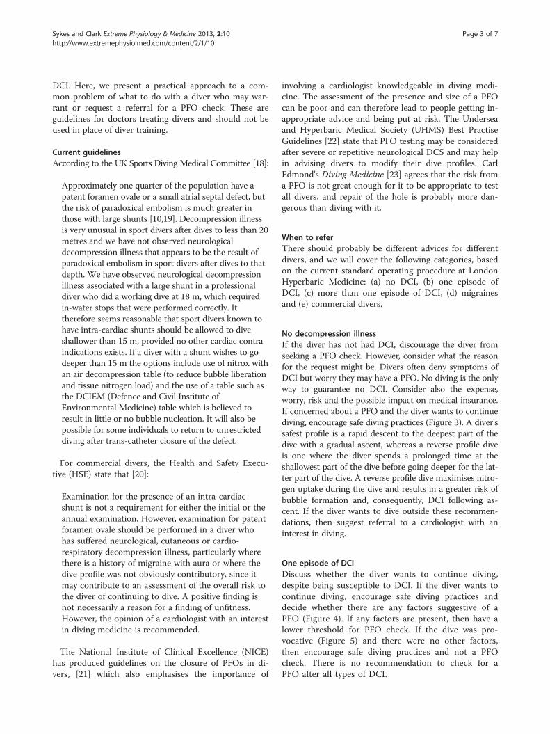
No decompression illnessIf the diver has not had DCI, discourage the diver fromseeking a PFO check. However, consider what the reasonfor the request might be. Divers often deny symptoms ofDCI but worry they may have a PFO. No diving is the onlyway to guarantee no DCI. Consider also the expense,worry, risk and the possible impact on medical insurance.If concerned about a PFO and the diver wants to continuediving, encourage safe diving practices (Figure 3). A diver'ssafest profile is a rapid descent to the deepest part of thedive with a gradual ascent, whereas a reverse profile diveis one where the diver spends a prolonged time at theshallowest part of the dive before going deeper for the lat-ter part of the dive. A reverse profile dive maximises nitro-gen uptake during the dive and results in a greater risk ofbubble formation and, consequently, DCI following as-cent. If the diver wants to dive outside these recommen-dations, then suggest referral to a cardiologist with aninterest in diving.
One episode of DCIDiscuss whether the diver wants to continue diving,despite being susceptible to DCI. If the diver wants tocontinue diving, encourage safe diving practices anddecide whether there are any factors suggestive of aPFO (Figure 4). If any factors are present, then have alower threshold for PFO check. If the dive was pro-vocative (Figure 5) and there were no other factors,then encourage safe diving practices and not a PFOcheck. There is no recommendation to check for aPFO after all types of DCI.
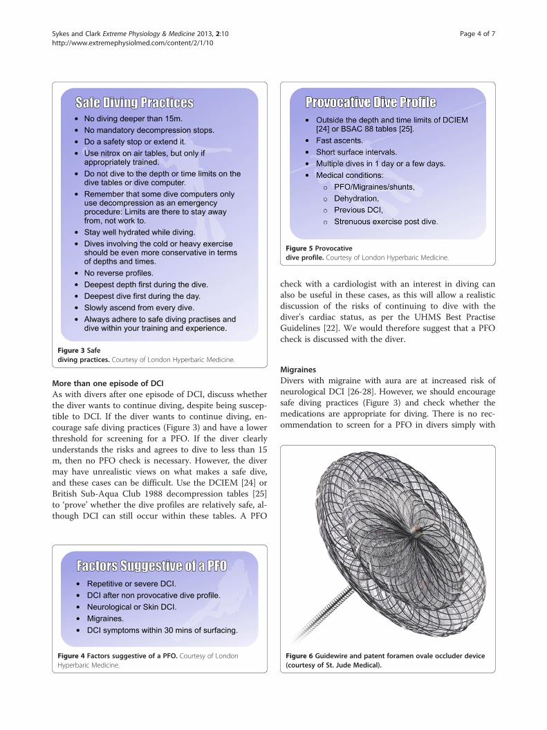
Figure 3 Safediving practices. Courtesy of London Hyperbaric Medicine.
Figure 5 Provocativedive profile. Courtesy of London Hyperbaric Medicine.
Sykes and Clark Extreme Physiology & Medicine 2013, 2:10 Page 4 of 7http://www.extremephysiolmed.com/content/2/1/10
More than one episode of DCIAs with divers after one episode of DCI, discuss whetherthe diver wants to continue diving, despite being suscep-tible to DCI. If the diver wants to continue diving, en-courage safe diving practices (Figure 3) and have a lowerthreshold for screening for a PFO. If the diver clearlyunderstands the risks and agrees to dive to less than 15m, then no PFO check is necessary. However, the divermay have unrealistic views on what makes a safe dive,and these cases can be difficult. Use the DCIEM [24] orBritish Sub-Aqua Club 1988 decompression tables [25]to ‘prove’ whether the dive profiles are relatively safe, al-though DCI can still occur within these tables. A PFO
Figure 4 Factors suggestive of a PFO. Courtesy of LondonHyperbaric Medicine.
check with a cardiologist with an interest in diving canalso be useful in these cases, as this will allow a realisticdiscussion of the risks of continuing to dive with thediver's cardiac status, as per the UHMS Best PractiseGuidelines [22]. We would therefore suggest that a PFOcheck is discussed with the diver.
MigrainesDivers with migraine with aura are at increased risk ofneurological DCI [26-28]. However, we should encouragesafe diving practices (Figure 3) and check whether themedications are appropriate for diving. There is no rec-ommendation to screen for a PFO in divers simply with
Figure 6 Guidewire and patent foramen ovale occluder device(courtesy of St. Jude Medical).
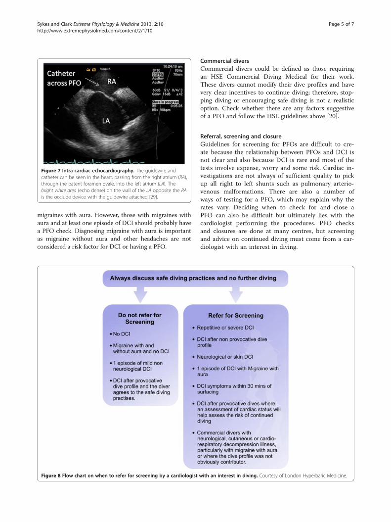
Figure 7 Intra-cardiac echocardiography. The guidewire andcatheter can be seen in the heart, passing from the right atrium (RA),through the patent foramen ovale, into the left atrium (LA). Thebright white area (echo dense) on the wall of the LA opposite the RAis the occlude device with the guidewire attached [29].
Sykes and Clark Extreme Physiology & Medicine 2013, 2:10 Page 5 of 7http://www.extremephysiolmed.com/content/2/1/10
migraines with aura. However, those with migraines withaura and at least one episode of DCI should probably havea PFO check. Diagnosing migraine with aura is importantas migraine without aura and other headaches are notconsidered a risk factor for DCI or having a PFO.
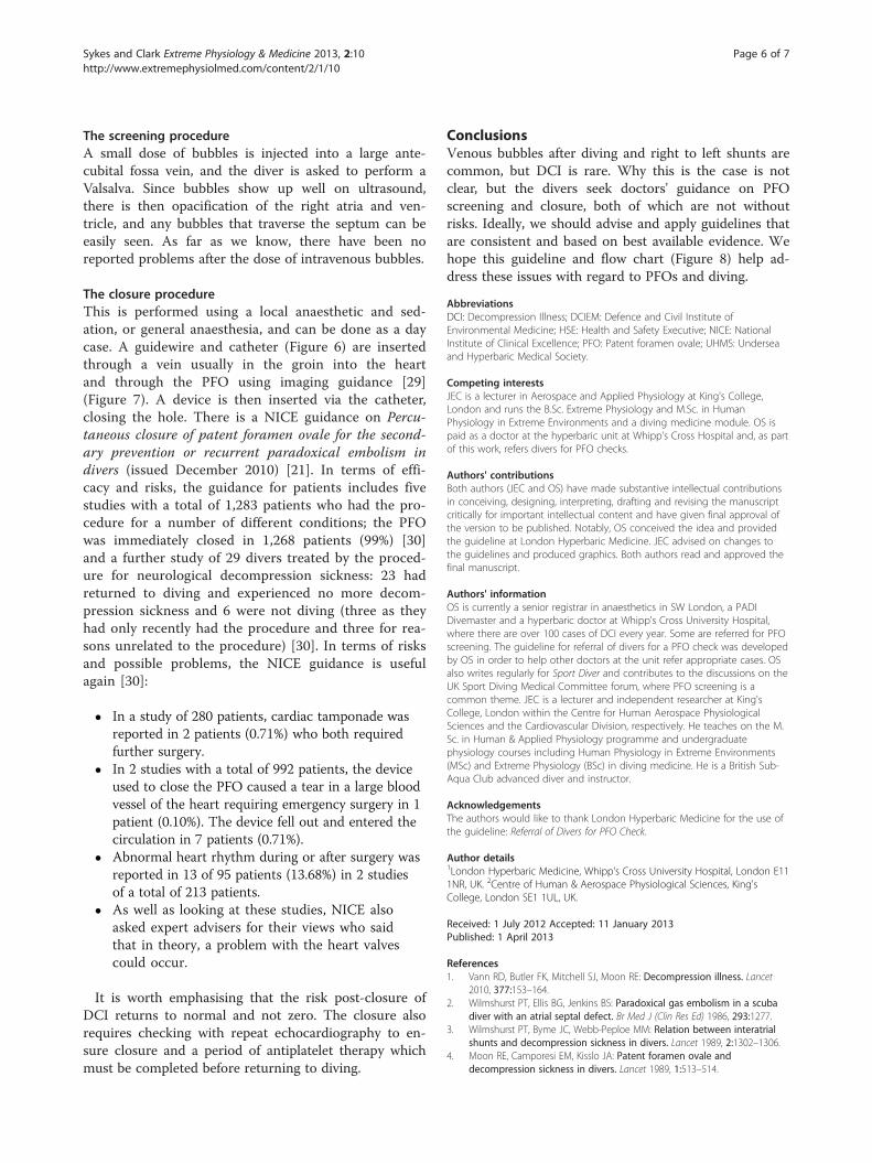
Figure 8 Flow chart on when to refer for screening by a cardiologist
Commercial diversCommercial divers could be defined as those requiringan HSE Commercial Diving Medical for their work.These divers cannot modify their dive profiles and havevery clear incentives to continue diving; therefore, stop-ping diving or encouraging safe diving is not a realisticoption. Check whether there are any factors suggestiveof a PFO and follow the HSE guidelines above [20].
Referral, screening and closureGuidelines for screening for PFOs are difficult to cre-ate because the relationship between PFOs and DCI isnot clear and also because DCI is rare and most of thetests involve expense, worry and some risk. Cardiac in-vestigations are not always of sufficient quality to pickup all right to left shunts such as pulmonary arterio-venous malformations. There are also a number ofways of testing for a PFO, which may explain why therates vary. Deciding when to check for and close aPFO can also be difficult but ultimately lies with thecardiologist performing the procedures. PFO checksand closures are done at many centres, but screeningand advice on continued diving must come from a car-diologist with an interest in diving.
with an interest in diving. Courtesy of London Hyperbaric Medicine.
Sykes and Clark Extreme Physiology & Medicine 2013, 2:10 Page 6 of 7http://www.extremephysiolmed.com/content/2/1/10
The screening procedureA small dose of bubbles is injected into a large ante-cubital fossa vein, and the diver is asked to perform aValsalva. Since bubbles show up well on ultrasound,there is then opacification of the right atria and ven-tricle, and any bubbles that traverse the septum can beeasily seen. As far as we know, there have been noreported problems after the dose of intravenous bubbles.
The closure procedureThis is performed using a local anaesthetic and sed-ation, or general anaesthesia, and can be done as a daycase. A guidewire and catheter (Figure 6) are insertedthrough a vein usually in the groin into the heartand through the PFO using imaging guidance [29](Figure 7). A device is then inserted via the catheter,closing the hole. There is a NICE guidance on Percu-taneous closure of patent foramen ovale for the second-ary prevention or recurrent paradoxical embolism indivers (issued December 2010) [21]. In terms of effi-cacy and risks, the guidance for patients includes fivestudies with a total of 1,283 patients who had the pro-cedure for a number of different conditions; the PFOwas immediately closed in 1,268 patients (99%) [30]and a further study of 29 divers treated by the proced-ure for neurological decompression sickness: 23 hadreturned to diving and experienced no more decom-pression sickness and 6 were not diving (three as theyhad only recently had the procedure and three for rea-sons unrelated to the procedure) [30]. In terms of risksand possible problems, the NICE guidance is usefulagain [30]:
� In a study of 280 patients, cardiac tamponade wasreported in 2 patients (0.71%) who both requiredfurther surgery.
� In 2 studies with a total of 992 patients, the deviceused to close the PFO caused a tear in a large bloodvessel of the heart requiring emergency surgery in 1patient (0.10%). The device fell out and entered thecirculation in 7 patients (0.71%).
� Abnormal heart rhythm during or after surgery wasreported in 13 of 95 patients (13.68%) in 2 studiesof a total of 213 patients.
� As well as looking at these studies, NICE alsoasked expert advisers for their views who saidthat in theory, a problem with the heart valvescould occur.
It is worth emphasising that the risk post-closure ofDCI returns to normal and not zero. The closure alsorequires checking with repeat echocardiography to en-sure closure and a period of antiplatelet therapy whichmust be completed before returning to diving.
ConclusionsVenous bubbles after diving and right to left shunts arecommon, but DCI is rare. Why this is the case is notclear, but the divers seek doctors' guidance on PFOscreening and closure, both of which are not withoutrisks. Ideally, we should advise and apply guidelines thatare consistent and based on best available evidence. Wehope this guideline and flow chart (Figure 8) help ad-dress these issues with regard to PFOs and diving.
AbbreviationsDCI: Decompression Illness; DCIEM: Defence and Civil Institute ofEnvironmental Medicine; HSE: Health and Safety Executive; NICE: NationalInstitute of Clinical Excellence; PFO: Patent foramen ovale; UHMS: Underseaand Hyperbaric Medical Society.
Competing interestsJEC is a lecturer in Aerospace and Applied Physiology at King's College,London and runs the B.Sc. Extreme Physiology and M.Sc. in HumanPhysiology in Extreme Environments and a diving medicine module. OS ispaid as a doctor at the hyperbaric unit at Whipp's Cross Hospital and, as partof this work, refers divers for PFO checks.
Authors' contributionsBoth authors (JEC and OS) have made substantive intellectual contributionsin conceiving, designing, interpreting, drafting and revising the manuscriptcritically for important intellectual content and have given final approval ofthe version to be published. Notably, OS conceived the idea and providedthe guideline at London Hyperbaric Medicine. JEC advised on changes tothe guidelines and produced graphics. Both authors read and approved thefinal manuscript.
Authors' informationOS is currently a senior registrar in anaesthetics in SW London, a PADIDivemaster and a hyperbaric doctor at Whipp's Cross University Hospital,where there are over 100 cases of DCI every year. Some are referred for PFOscreening. The guideline for referral of divers for a PFO check was developedby OS in order to help other doctors at the unit refer appropriate cases. OSalso writes regularly for Sport Diver and contributes to the discussions on theUK Sport Diving Medical Committee forum, where PFO screening is acommon theme. JEC is a lecturer and independent researcher at King'sCollege, London within the Centre for Human Aerospace PhysiologicalSciences and the Cardiovascular Division, respectively. He teaches on the M.Sc. in Human & Applied Physiology programme and undergraduatephysiology courses including Human Physiology in Extreme Environments(MSc) and Extreme Physiology (BSc) in diving medicine. He is a British Sub-Aqua Club advanced diver and instructor.
AcknowledgementsThe authors would like to thank London Hyperbaric Medicine for the use ofthe guideline: Referral of Divers for PFO Check.
Author details1London Hyperbaric Medicine, Whipp's Cross University Hospital, London E111NR, UK. 2Centre of Human & Aerospace Physiological Sciences, King'sCollege, London SE1 1UL, UK.
Received: 1 July 2012 Accepted: 11 January 2013Published: 1 April 2013
References1. Vann RD, Butler FK, Mitchell SJ, Moon RE: Decompression illness. Lancet
2010, 377:153–164.2. Wilmshurst PT, Ellis BG, Jenkins BS: Paradoxical gas embolism in a scuba
diver with an atrial septal defect. Br Med J (Clin Res Ed) 1986, 293:1277.3. Wilmshurst PT, Byme JC, Webb-Peploe MM: Relation between interatrial
shunts and decompression sickness in divers. Lancet 1989, 2:1302–1306.4. Moon RE, Camporesi EM, Kisslo JA: Patent foramen ovale and
decompression sickness in divers. Lancet 1989, 1:513–514.
Sykes and Clark Extreme Physiology & Medicine 2013, 2:10 Page 7 of 7http://www.extremephysiolmed.com/content/2/1/10
5. Nishi RY, Brubakk AO, Eftedal OS: Bubble detection. In Bennett and Elliott'sPhysiology and Medicine of Diving. 5th edition. Edited by Brubakk AO,Neuman TS. London: Saunders; 2003:504–505.
6. Patent foramen ovale acting as a flap valve: http://www.imagingeconomics.com/all-news/18357-advances-in-echocardiography.
7. Laden GD: Patent foramen ovale and decompression illness in divers.Lancet 1997, 349:288.
8. Cross SJ, Evan SA, Thomson LF, Lee HS, Jennings KP, Shields TG: Safety ofsubaqua diving with a patent foramen ovale. BMJ 1992, 304:481–482.
9. Moon RE, Rorem L: Patent foramen ovale. What is it, and what are theimplications for divers? http://www.diversalertnetwork.org/medical/articles/Patent_Foramen_Ovale.
10. Wilmshurst PT, Davidson C, O'Connell G, Byme C: Role of cardiorespiratoryabnormalities, smoking and dive characteristics in the manifestations ofneurological decompression illness. Clin Sci 1994, 86:297–303.
11. Wilmshurst PT, Treacher DF, Crowther A, Smith SE: Effects of patentforamen ovale on arterial saturation during exercise and oncardiovascular responses to deep breathing, valsalva manoeuvre, andpassive tilt: relation relation to history of decompression illness in divers.Br Heart J 1994, 71:229–231.
12. Wilmshurst P: The PFO story: from divers and astronauts to stroke andmigraine. http://www.advancedecho.co.uk/The%20PFO%20story.pdf.
13. DeNoble P: PFO and decompression illness in recreational divers. http://www.alertdiver.com/294.
14. Sykes O: Patent Foramen Ovale. Winter Park: Sport Diver; 2012.15. Powell MR, Spencer MP, Von Ramm OT: Ultrasonic surveillance of
decompression. In Bennett and Elliott's Physiology and Medicine of Diving.3rd edition. Edited by Bennett PB, Elliott DH. London: Bailliere Tindall;1982:404–434.
16. Vik A, Jenssen BM, Brubakk AO: Arterial gas bubbles after decompressionin pigs with patent foramen ovale. Undersea Hyperb Med 1993,20:121–131.
17. Bové AA, Moon RE: Patent foramen ovale - is it important to divers?http://www.diversalertnetwork.org/medical/articles/Patent_Foramen_Ovale_Is_It_Important_to_Divers.
18. Wilmshurst PT, Walsh K, Morrison L: Transcatheter occlusion of foramenovale with a button device after neurological decompression illness inprofessional divers. Lancet 1996, 348:752–753.
19. Cartoni D, De Castro S, Valente G, Costanzo C, Pelliccia A, Beni S, DiAngelantonio E, Papetti F, Vitali Serdoz L, Fedele F: Identification ofprofessional scuba divers with patent foramen ovale at risk ofdecompression illness. Am J Cardiol 2004, 94:270–273.
20. The medical examination and assessment of divers (MA1). http://www.hse.gov.uk/diving/ma1.pdf.
21. NHS: IPG371 Percutaneous closure of patent foramen ovale for the secondaryprevention of recurrent paradoxical embolism in divers: guidance. http://guidance.nice.org.uk/IPG371/Guidance/pdf/English.
22. UHMS best practice guidelines: prevention and treatment of decompressionsickness and arterial gas embolism. http://membership.uhms.org/resource/resmgr/dcsandage_prevandmgt_uhms-fi.pdf.
23. Decompression physiology and susceptibility. http://www.divingmedicine.info/Ch%2013%20SM10c.pdf.
24. Defence and Civil Institute of Environmental Medicine: DCIEM DivingManual: Air Decompression Procedures and Tables. Richmond: Universal DiveTechtronics Inc.; 1992.
25. BSAC 88 decompression tables. http://www.bsac.com/shop.asp?section=1362§ionTitle=Decompression+Tables&itemid=1677.
26. Engel GL, Webb JP, Ferris EB, Romano J, Ryder H, Blankenhorn MA: Amigraine-like syndrome complicating decompression sickness.Scintillating scotomas, focal neurologic signs and headache: clinical andelectroencephalographic observations. War Med 1944, 5:304–314.
27. Wilmshurst P, Nightingale S: Relationship between migraine and cardiacand pulmonary right-to-left shunts. Clin Sci 2001, 100:215–220.
28. Wilmshurst P, Pearson M, Nightingale S: Re-evaluation of the relationshipbetween migraine and persistent foramen ovale and other right-to-leftshunts. Clin Sci 2005, 108:365–367.
29. Guidewire and catheter in the heart. http://eso-cdn.bestpractice.bmj.com/best-practice/images/bp/en-gb/951-3_default.jpg.
30. NHS: Percutaneous closure of patent foramen ovale for the secondaryprevention of recurrent paradoxical embolism in divers. Patient version:understanding NICE guidance. http://guidance.nice.org.uk/IPG371/PublicInfo/pdf/English.
doi:10.1186/2046-7648-2-10Cite this article as: Sykes and Clark: Patent foramen ovale and scubadiving: a practical guide for physicians on when to refer for screening.Extreme Physiology & Medicine 2013 2:10.
Submit your next manuscript to BioMed Centraland take full advantage of:
• Convenient online submission
• Thorough peer review
• No space constraints or color figure charges
• Immediate publication on acceptance
• Inclusion in PubMed, CAS, Scopus and Google Scholar
• Research which is freely available for redistribution
Submit your manuscript at www.biomedcentral.com/submit