REVIEW Open Access Catching the engram: strategies to ...
12
REVIEW Open Access Catching the engram: strategies to examine the memory trace Masanori Sakaguchi 1 and Yasunori Hayashi 1,2* Abstract Memories are stored within neuronal ensembles in the brain. Modern genetic techniques can be used to not only visualize specific neuronal ensembles that encode memories (e.g., fear, craving) but also to selectively manipulate those neurons. These techniques are now being expanded for the study of various types of memory. In this review, we will summarize the genetic methods used to visualize and manipulate neurons involved in the representation of memory engrams. The methods will help clarify how memory is encoded, stored and processed in the brain. Furthermore, these approaches may contribute to our understanding of the pathological mechanisms associated with human memory disorders and, ultimately, may aid the development of therapeutic strategies to ameliorate these diseases. Introduction One of the major aims of modern memory research is to locate the physical substrate of memory (also referred to as ‘memory trace’ or ‘neural substrates of memory’) in the brain. At the beginning of the 20 th century, Richard Semon introduced the word, ‘engram’ , to describe the memory trace, ‘Its result, namely, the enduring though primarily latent modification in the irritable substance produced by stimulus, I have called an Engram, ....’ [1]. Later, Karl Lashley, a pioneer in the field of behavioral neuroscience, attempted to identify the location of the engram in rodents using a maze task [2]. He systematic- ally lesioned different parts of the brain and examined the behavioral consequences. Although he failed to lo- cate a specific brain region where the memory trace exists, this approach has been and still is one of the most commonly used methods to understand the role of selected brain regions in memory. Clinical studies of an epilepsy patient, known by the initials H.M., led to the accidental discovery of a mile- stone in memory research. To provide therapeutic relief for the epileptic seizures that H.M was experiencing, the hippocampi and amygdalae were surgically removed. Fortunately, with this treatment, H.M.’ s epilepsy was cured. However, surgical intervention also produced severe anterograde and temporally-graded retrograde amnesia of autobiographical memory [3]. Subsequent studies of H.M. revolutionized our view of memory by suggesting that there are particular locations in the brain that play an essential part in memory. The association of elementary events has been pro- posed to play a central role in memory [4]. In line with this notion, recent developments in memory research have focused on associative learning and memory [5]. Pavlov was well known for implementing an experi- mental paradigm to quantify associative memory, using a so-called classical conditioning paradigm. In classical conditioning, subjects associate two different sensory stimuli, defined as the conditioned stimulus (CS) and the unconditioned stimulus (US) [6]. The CS is a cue that is neutral but salient enough to be recognized by the subject, whereas the US is a cue that evokes an in- nate response in the subject, leading to an uncondi- tioned response (UR). When the subject learns the association between the CS and the US, the subject may display a response similar to the UR upon exposure to the CS alone. This response is called a conditioned re- sponse (CR). Many modern memory researchers have not only re- confirmed the earlier findings of Pavlov using different variants of classical conditioning paradigms, but these subsequent studies have provided many further insights into the neurobiology associated with memory (reviewed in [7]). Philips and LeDoux showed that distinct brain * Correspondence: [email protected] 1 Brain Science Institute, RIKEN, Wako, Saitama 351-0198, Japan 2 Saitama University Brain Science Institute, Saitama University, Saitama 338-8570, Japan © 2012 Sakaguchi and Hayashi.; licensee BioMed Central Ltd. This is an Open Access article distributed under the terms of the Creative Commons Attribution License (http://creativecommons.org/licenses/by/2.0), which permits unrestricted use, distribution, and reproduction in any medium, provided the original work is properly cited. Sakaguchi and Hayashi Molecular Brain 2012, 5:32 http://www.molecularbrain.com/content/5/1/32
Transcript of REVIEW Open Access Catching the engram: strategies to ...

Sakaguchi and Hayashi Molecular Brain 2012, 5:32http://www.molecularbrain.com/content/5/1/32
REVIEW Open Access
Catching the engram: strategies to examine thememory traceMasanori Sakaguchi1 and Yasunori Hayashi1,2*
Abstract
Memories are stored within neuronal ensembles in the brain. Modern genetic techniques can be used to not onlyvisualize specific neuronal ensembles that encode memories (e.g., fear, craving) but also to selectively manipulatethose neurons. These techniques are now being expanded for the study of various types of memory. In this review,we will summarize the genetic methods used to visualize and manipulate neurons involved in the representation ofmemory engrams. The methods will help clarify how memory is encoded, stored and processed in the brain.Furthermore, these approaches may contribute to our understanding of the pathological mechanisms associatedwith human memory disorders and, ultimately, may aid the development of therapeutic strategies to amelioratethese diseases.
IntroductionOne of the major aims of modern memory research is tolocate the physical substrate of memory (also referred toas ‘memory trace’ or ‘neural substrates of memory’) inthe brain. At the beginning of the 20th century, RichardSemon introduced the word, ‘engram’, to describe thememory trace, ‘Its result, namely, the enduring thoughprimarily latent modification in the irritable substanceproduced by stimulus, I have called an Engram, . . ..’ [1].Later, Karl Lashley, a pioneer in the field of behavioralneuroscience, attempted to identify the location of theengram in rodents using a maze task [2]. He systematic-ally lesioned different parts of the brain and examinedthe behavioral consequences. Although he failed to lo-cate a specific brain region where the memory traceexists, this approach has been and still is one of the mostcommonly used methods to understand the role ofselected brain regions in memory.Clinical studies of an epilepsy patient, known by the
initials H.M., led to the accidental discovery of a mile-stone in memory research. To provide therapeutic relieffor the epileptic seizures that H.M was experiencing, thehippocampi and amygdalae were surgically removed.Fortunately, with this treatment, H.M.’s epilepsy wascured. However, surgical intervention also produced
* Correspondence: [email protected] Science Institute, RIKEN, Wako, Saitama 351-0198, Japan2Saitama University Brain Science Institute, Saitama University, Saitama338-8570, Japan
© 2012 Sakaguchi and Hayashi.; licensee BioMCreative Commons Attribution License (http:/distribution, and reproduction in any medium
severe anterograde and temporally-graded retrogradeamnesia of autobiographical memory [3]. Subsequentstudies of H.M. revolutionized our view of memory bysuggesting that there are particular locations in the brainthat play an essential part in memory.The association of elementary events has been pro-
posed to play a central role in memory [4]. In line withthis notion, recent developments in memory researchhave focused on associative learning and memory [5].Pavlov was well known for implementing an experi-mental paradigm to quantify associative memory, usinga so-called classical conditioning paradigm. In classicalconditioning, subjects associate two different sensorystimuli, defined as the conditioned stimulus (CS) andthe unconditioned stimulus (US) [6]. The CS is a cuethat is neutral but salient enough to be recognized bythe subject, whereas the US is a cue that evokes an in-nate response in the subject, leading to an uncondi-tioned response (UR). When the subject learns theassociation between the CS and the US, the subjectmay display a response similar to the UR upon exposureto the CS alone. This response is called a conditioned re-sponse (CR).Many modern memory researchers have not only re-
confirmed the earlier findings of Pavlov using differentvariants of classical conditioning paradigms, but thesesubsequent studies have provided many further insightsinto the neurobiology associated with memory (reviewedin [7]). Philips and LeDoux showed that distinct brain
ed Central Ltd. This is an Open Access article distributed under the terms of the/creativecommons.org/licenses/by/2.0), which permits unrestricted use,, provided the original work is properly cited.

Sakaguchi and Hayashi Molecular Brain 2012, 5:32 Page 2 of 12http://www.molecularbrain.com/content/5/1/32
regions have different contributions to memory forma-tion [8]. Using a contextual fear conditioning paradigm,they showed that the hippocampus, in cooperation withthe amygdala, plays an essential role in associative learn-ing of a specific context (CS) and a foot-shock (US). Thisfinding is in contrast to tone-fear conditioning, wheresubjects learn that a neutral tone (CS) can act as a pre-dictor of a foot-shock (US). In this paradigm the amyg-dala played an essential role but the hippocampus wasdispensable. While this study highlighted an importantfunctional distinction between the hippocampus andamygdala in fear memory, it was still unclear at whichstage of memory (e.g., acquisition, retention, recall) eachbrain region was important.Kim and Fanselow tried to examine the temporal win-
dow when the hippocampus was important for memory,by applying electrolytic lesions to the hippocampus atvarious time points after contextual fear conditioning[9]. Rats that were lesioned 1 day after training did notretain the contextual fear memory, whereas animalsthat received the lesion 28 days later retained the mem-ory at the same level as sham operated rats. These find-ings were supported by numerous other studies (e.g.the case study of H.M. and other studies using pharma-cological/genetic inhibition approaches or electrolytic/surgical lesions) and led to the proposal that certaintypes of memory are initially encoded by the hippocam-pus as recent memory and then gradually transferredfrom the hippocampus to other brain regions as remotememory [9-14]. The process of memory transfer is com-monly referred to as the systems consolidation theory(Figure 1). However, whether the hippocampus is still
Figure 1 Systems memory consolidation theory. Autobiographical mema certain period of time, neural ensembles outside of the hippocampus bewide change is called systems memory consolidation.
engaged in remote memory and whether the transferredmemory is the same as the original memory is stillunder much debate [15-21].The obvious shortfall in the lesion studies is that
surgical lesions not only destroy all locally existingstructures including neuronal and glial cells but alsoincoming, outgoing, and even passing fibers. Also, theirreversibility of the procedure makes it difficult to de-termine the exact role of the lesioned brain regions atdifferent stages of memory. Using drugs that selectivelyinactivate neuronal activity has some notable advan-tages over physical lesions and can help elucidatethe relationship between biological phenomenon andmemory. For example, Kandel et al. elucidated themolecular mechanisms of memory related behavior byapplying drugs (e.g., cAMP or CRE oligonucleotides)onto target neurons of Aplysia [22]. Morris showed alink between long term potentiation and spatial mem-ory by applying a pharmacological reagent that blocksLTP (AP5, an antagonist of NMDA type glutamatereceptors) directly into the brains of rats [23]. Recentadvances in genetic techniques (e.g. optogenetics) haveenabled the manipulation of neural activity at an evenhigher level of sophistication and temporal control (e.g.,inactivating targeted neurons in reversible manner atspecific points in time [24,25]). Using these techni-ques, it is now possible to visualize and control specificneuronal circuits that encode associative memory[26,27].Given these advances, in this review, we will provide
an overview of recent studies that aim to allocate thememory engram at the circuit, cellular, and synaptic
ory is initially encoded in neural ensembles in the hippocampus. Aftercome responsible for the retention of memory. This dynamic system

Sakaguchi and Hayashi Molecular Brain 2012, 5:32 Page 3 of 12http://www.molecularbrain.com/content/5/1/32
level and discuss the current debate, remaining ques-tions and future perspectives.
Visualization and manipulation of a memory traceby immediately early genes (IEGs)Detection of IEG productsIEGs are a class of genes that are transcribed immedi-ately after biological events (starting in less than a mi-nute) without requiring the expression of other genes[28,29]. Neural activity induces expression of variousIEGs including proto-oncogene transcription factorssuch as c-fos and zif/268 (also named Egr1, NGFI-A,Krox 24) and genes that encode synaptic structural pro-teins such as Arc and Homer1a. Due to their property ofactivity dependent transcription, immunostaining or insitu hybridization of IEGs allows us to identify neuronsthat were active during a given memory paradigm.The mRNA of Arc has interesting distribution dynam-
ics, which allows one to trace activated neurons in aretrograde manner. It initially accumulates in the nucleiof neurons (<5 min) and is gradually exported to thecytosol within the next 30 min (Figure 2A). Therefore,by observing the localization of Arc mRNA, one can notonly detect activated neurons but also estimate the ap-proximate time that has elapsed after the initial activa-tion. The method is named ‘cellular compartment
Figure 2 Tagging neural ensembles by Arc catFISH. A, After neuronal aWithin 30 minutes, the mRNA translocates outside the nucleus (right paneltwo different situations. In the upper panel, the subject was exposed to co5 minutes prior to sacrifice and fixation (A/A). In the lower panel, the animExposure to the A/A condition led to induction of Arc localization both insexposure induces Arc in mainly two distinct populations albeit with some
analysis of temporal activity by fluorescent in situhybridization’ of Arc (Arc catFISH) [30].Using the Arc catFISH method, Guzowski et al. [28]
showed that when a rat is exposed to two differentenvironments with an interval of 30 minutes, activationof Arc in response to the exposure to each environmentoccurs in separate neuronal populations in the CA1(Figure 2B, lower row). In contrast, if a rat is exposed tothe same environment twice, the same neurons thatwere activated during the first episode were reactivatedduring the second episode (Figure 2B, upper row). Thisresult suggests that the environment specific memorytrace is established in the CA1. Using an associativelearning paradigm, Barot et al. [31] showed that individ-ual neurons in the basolateral nucleus of the amygdalaresponded to both CS and US only after the subject hadlearned the association between CS and US.It is suggested that once memory is encoded, it is con-
solidated through off-line neuronal ensemble activity(e.g., activity during rest or sleep when there is no CS orUS). Marrone et al. [32] compared neuronal ensembleactivity in the hippocampus during periods of explor-ation and then the following rest period using the cat-FISH method. They found that the Arc expressionpattern during the rest period partially recapitulated thatof the exploration period. Hashikawa et al. [33] extended
ctivation, Arc mRNA is initially localized in the nucleus (left panel).). B. The Arc expression pattern in a neuronal ensemble is depicted inntext A at two time points, firstly at 30 minutes and secondlyal was first exposed to context A and then to a distinct context B (A/B).ide and outside of the nucleus in the same neurons, while A/Boverlap.

Sakaguchi and Hayashi Molecular Brain 2012, 5:32 Page 4 of 12http://www.molecularbrain.com/content/5/1/32
this idea by examining associative learning in the amyg-dala using a fear conditioning paradigm and showed thatamygdala neurons that expressed Arc during condition-ing also preferentially re-expressed Arc during the fol-lowing rest period.
Use of IEG promotersIn addition to the observation of IEG products, the pro-moters for IEGs can be used to detect neurons involvedin learning. Reijimers et al. [34] visualized c-fos positiveneurons using a transgenic mouse line expressing β-galactosidase (LacZ) under the control of a c-fos pro-moter via a self-activating tTA-TetO system to examinethe memory trace of tone fear memory in the amygdala(Figure 3). During training, LacZ was induced in someneurons in the amygdala. Interestingly, the same LacZpositive cells were also positive for Zif268 after memoryretrieval, suggesting that the LacZ/Zif268 double-positivecells encoded the fear memory. A similar technique was
Figure 3 Tagging neural ensembles using a c-fos promoter tTA/TetOLencodes the doxycycline sensitive tetracycline transactivator (tTA) under th(TetO) sequence to induce transcription of the downstream gene, and theother expresses the secondary transactivator (tTA*) and tauLacZ under theconstitutively active, therefore, once expressed, tTA* activates TetO regardlec-fos promoter is activated. However, in the presence of doxycycline, tTA awithdrawal of doxycycline, tTA is activated and binds to TetO to induce ex(purple). C. After re-administration of doxycycline, tTA becomes inactivatedof tauLacZ by activating TetO in the positive-feedback loop. This system thabsence of doxycycline.
used to induce the expression of newly synthesizedAMPA type glutamate receptors (AMPAR) selectively inneurons that were activated during memory formation(i.e., c-fos activated neurons), which were then translo-cated to specific types of dendritic spines [35]. Recentadvances in fluorescent imaging techniques allow us toexamine neurons involved in memory in living subjects.A mouse line which has GFP knocked-in into the arc pro-moter locus was generated for this purpose, however, theexperimental results could be confounded by a phenotypecaused by the hemizygous loss of Arc gene (e.g., orienta-tion specificity) [36]. Hence, several transgenic mouselines with arc or c-fos promoter to drive the expression offluorescent proteins were established that could circum-vent this issue [37-41].The induction of c-fos expression upon conditioning
indicates that there is a correlation between neuronal ac-tivation and memory, but it does not necessarily provethat c-fos expressing neurons encode the memory. Koya
acZ-tTA mouse. The mouse has two transgenic alleles. One allelee control of the c-fos promoter. tTA binds the tetracycline operatorinduction activity is suppressed by the presence of doxycycline. Thecontrol of TetO. tTA* is both insensitive to doxycycline andss of doxycycline. A. tTA is expressed exclusively in neurons where thectivity is inhibited and hence prevents interaction with TetO. B. Afterpression of tTA* and tauLacZ protein in c-fos activated neuronsonce again but as tTA* is still present, it can maintain the expressionereby allows the permanent tagging of neurons activated even in the

Sakaguchi and Hayashi Molecular Brain 2012, 5:32 Page 5 of 12http://www.molecularbrain.com/content/5/1/32
et al. [42] provided evidence that c-fos expressing neur-onal ensembles in the nucleus accumbens were involvedin the memory trace that associated a specific context(CS) with cocaine administration (US). For this study,they utilized a combination of c-fos promoter-LacZtransgenic rats and Daun02, a prodrug that can be con-verted to Daunorubicin by LacZ. Daunorubicin inacti-vates neurons by reducing Ca2+ dependent actionpotentials [42-44]. Using this combinational approach, c-fos activated neurons (i.e., LacZ-positive neurons) couldbe selectively inactivated by Daun02 injection. In theirstudy, learning of a context-drug administration memoryinduced c-fos expression in neuronal ensembles in thenucleus accumbens of rats. Subsequent administrationof Daun02 reduced the context-specific cocaine-inducedpsychomotor sensitization, confirming that neurons thatwere activated during learning were also involved in re-call of the memory that associated the context with co-caine administration.Using the same c-fos-LacZ rat, Bossert et al. found
neural ensembles that mediated a context-induced re-lapse to heroin addiction in the ventral medial prefrontalcortex [45]. These results suggest that the c-fos pro-moter can be used to genetically tag the neuronal en-semble involved in memory encoding.More recently, Garner et al. [46] reported the gener-
ation of a synthetic memory trace by genetically taggingc-fos promoter activated neurons. They used a double-transgenic mouse line which expressed tTA under controlof the c-fos promoter and an evolved G protein-coupledreceptor (hM3Dq) under tetracycline response element(TRE). hM3Dq produces neuronal depolarization in re-sponse to clozapine-N-oxide (CNO) injection (Figure 4A,B) [47]. In the mice, the expression of hM3Dq can act as atag of localization pattern of the neuronal activity (i.e.,pattern of c-fos promoter activation) at a given timeperiod (i.e., off-doxycycline period). First, the mice wereexposed to a novel context, context A (CtxA), to inducetagging of neurons that were specifically active in the con-text (Figure 4C, left). Second, the mice were exposed toanother novel context, CtxB, where foot shock (US) andCNO injection (to activate hM3Dq - and hence CtxA en-coding neurons) was administered (Figure 4C, center),thereby CtxA+CtxB information could be associatedwith the foot shock. Finally, the mice showed freezingonly when both CNO injection (pseudo CtxA) and expos-ure to CtxB occurred simultaneously (Figure 4C, right),but not when CNO injection nor exposure to CtxB wasgiven alone. The result suggests that the mice created ahybrid ‘synthetic’ memory of CtxA and B. Using the samec-fos promoter-tTA mouse line in combination with viraldelivery of TRE-channelrhodopsin, Liu et al. [48] showedthat optical re-activation of the neuronal ensembleinvolved in memory encoding in the dentate gyrus was
sufficient to retrieve a fear memory (Figure 4A, D, E). Im-portantly, retrieval was not induced when the neuronalensemble had not been associated with the US (i.e., foot-shock), suggesting a strong causal relationship betweenthe expression of c-fos in neurons and the association ofcontext and shock (please see section 3 for other studiesutilizing optogenetic approaches).cAMP response elements (CRE) is a DNA sequence
which can be found in the regulatory sequences of IEGs(e.g., c-fos [49], Arc [50]). In LTP, calcium and cAMP sig-nals converge to activate cAMP response element bindingprotein (CREB) transcription activity by phosphorylatingSerine residue 133 (CREBs133), which results in an in-crease in CRE mediated gene expression. Impey et al.made a transgenic mouse line, which carries tandem-repeat CRE sequences followed by a LacZ-reporter geneto monitor CRE mediated transcription activity upon LTPand memory formation [51]. Indeed, LacZ expression waswell correlated with the phosphorylation of CREBs133and induction of L-LTP in the CA1 region of the hippo-campus. Furthermore, the signaling pathway that inducedL-LTP enhances CRE mediated transcription. Importantly,learning of contextual fear conditioning and passive avoid-ance tasks increased CRE dependent gene expression inthe hippocampus [52]. On the other hand, auditory fearconditioning, an amygdala dependent learning paradigm,only increased CRE dependent gene expression in theamygdala, suggesting that CRE dependent gene expressionwas memory type specific and that CRE up-regulationwas involved not only in hippocampus-dependent but alsoin amygdala-dependent associative memories.
Forced labeling and manipulation of a memorytrace by CREBThe studies described above underscore the importanceof the CREB transcription factor and its downstream tar-gets in modulating the cellular response to neuronal activitythat takes place during learning and memory. CREB is aninterface between neuronal activity and gene transcriptionby converting local and transient second messenger sig-naling into a persistent cell-wide transcriptional modifica-tion. This feature gave rise to the idea that CREB could beused as a tool to force a neuron to encode memory.
LabelingHan et al. used an auditory fear memory task to experi-mentally test the concept that neurons that have a rela-tively high CREB expression level could be recruitedinto a fear memory circuit [53]. Firstly, they overex-pressed CREB in the amygdala, and then trained themice in a tone-fear conditioning paradigm (tone-footshock association memory) (Figure 5A, left). After thetest, the amygdala was subjected to Arc catFISH analysis.Arc mRNA was preferentially localized to CREB
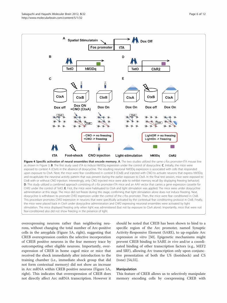
Figure 4 Specific activation of neural ensembles that encode memory. A. The two studies utilized the same c-fos promoter-tTA mouse lineas shown in Figure 3. B. The first study used tTA to induce hM3Dq expression under the control of doxycycline. C. Initially, the mice wereexposed to context A (CtxA) in the absence of doxycycline. The resulting neuronal hM3Dq expression is associated with cells that respondedupon exposure to CtxA. Next, the mice were fear conditioned in context B (CtxB) and injected with CNO to activate neurons that express hM3Dqand recapitulate the neuronal activity pattern that was present during the earlier exposure to CtxA. In the final test session, mice were exposed toCtxB with or without CNO injection. Interestingly, only CNO injected mice were able to exhibit memory recall (by displaying freezing behavior).D. This study utilized a combined approach consisting of c-fos promoter-tTA mice and an AAV vector that carries a gene expression cassette forChR2 under the control of TetO. E. First, the mice were habituated to CtxA and light stimulation was applied. The mice were under doxycyclineadministration at this stage. The mice did not freeze during this stage, confirming that light stimulation alone does not induce freezing. Next,doxycycline is withdrawn to promote ChR2 expression under the control of the c-fos promoter. Then, the mice were fear conditioned in CtxB.This procedure promotes ChR2 expression in neurons that were specifically activated by the contextual fear conditioning protocol in CtxB. Finally,the mice were placed back in CtxA under doxycycline administration and ChR2 expressing neuronal ensembles were activated by lightstimulation. The mice displayed freezing only when light was administered (but not by exposure to CtxA alone). Importantly, mice that were notfear-conditioned also did not show freezing in the presence of light.
Sakaguchi and Hayashi Molecular Brain 2012, 5:32 Page 6 of 12http://www.molecularbrain.com/content/5/1/32
overexpressing neurons rather than neighboring neu-rons, without changing the total number of Arc-positivecells in the amygdala (Figure 5A, right), suggesting thatCREB overexpression confers the selective incorporationof CREB positive neurons in the fear memory trace byoutcompeting other eligible neurons. Importantly, over-expression of CREB in home caged mice or mice thatreceived the shock immediately after introduction to thetraining chamber (i.e., immediate shock group that didnot form contextual memory) did not show an increasein Arc mRNA within CREB positive neurons (Figure 5A,right). This indicates that overexpression of CREB doesnot directly affect Arc mRNA transcription. However it
should be noted that CREB has been shown to bind to aspecific region of the Arc promoter, named SynapticActivity-Responsive Element (SARE), to up-regulate Arcexpression in vitro [50]. Epigenetic mechanisms mightprevent CREB binding to SARE in vivo and/or a coordi-nated binding of other transcription factors (e.g., MEF2and SRF), allowing Arc transcription only upon conjunc-tive presentation of both the US (footshock) and CS(tone) [54,55].
ManipulationThis feature of CREB allows us to selectively manipulatememory encoding cells by coexpressing CREB with

Figure 5 Memory allocation by CREB. A. CREB is overexpressed in the amygdala by a HSV vector before tone-fear conditioning (training). HSVrandomly infects neurons in the amygdala. During training, the memory is allocated to neurons overexpressing CREB (CREB neurons), whichresults in the preferential induction of Arc expression (neuronal activity marker) in CREB neurons. Controls that did not undergo “learning” (homecage or immediate shock groups) did not show the preferential Arc expression in CREB neurons. B. To directly show the causal link betweenCREB and memory allocation, CREB neurons were selected for deletion by expression of the diphtheria toxin receptor (DTR). The system utilizesiDTR mice in which DTR expression can be induced by CRE recombinase activity. Therefore, expressing CREB-CRE using a HSV vector makes theneurons susceptible to Diphtheria Toxin (DT). C. Injection of DT after CREB overexpression and the subsequent fear conditioning training ablatedCREB expressing neurons that resulted in a loss of the memory.
Sakaguchi and Hayashi Molecular Brain 2012, 5:32 Page 7 of 12http://www.molecularbrain.com/content/5/1/32
functional molecules. Han et al. utilized a Cre recombin-ase (Cre)-inducible diphtheria toxin receptor (iDTR)transgenic mouse line that allowed the selective elimin-ation of target cells (i.e., DTR expressing neurons) [56].They engineered a HSV vector that expresses both CREBand Cre to induce memory encoding preferentially toCREB positive neurons at the learning stage and to con-fer DT sensitivity to the same neurons (Figure 5B). After
learning, DT was administered to selectivity ablate CREBexpressing neurons. Interestingly, this process alsoresulted in the erasure of the newly acquired fear memory,suggesting that CREB positive neurons can selectively en-code fear memory, by outcompeting other neurons in theamygdala [57].Zhou et al. reported similar results utilizing an allatos-
tatin receptor (AlstR)/ligand system, originally derived

Sakaguchi and Hayashi Molecular Brain 2012, 5:32 Page 8 of 12http://www.molecularbrain.com/content/5/1/32
from insects [58]. Binding of allatostatin to heterolo-gously expressed AlstR activates endogenous mamma-lian G protein-coupled inwardly rectifying K+ (GIRK)channels, which causes membrane hyperpolarization,thereby decreasing neuronal excitability [59]. The systemallowed inducible silencing of target neurons (i.e., AlstRexpressing neurons) in a reversible manner. Silencing ofCREB overexpressing neurons by AlstR/ligand systemresulted in a reduction in freezing during a tone-fearconditioning test, providing further evidence that CREBinduces memory encoding in amygdala neurons.The authors also examined the selectivity of memory
induced by CREB. Conditioned taste aversion (CTA)memory is a type of memory known to depend on theamygdala [60]. First, mice underwent tone-fear condi-tioning, then later, CREB and AlstR were coexpressed inamygdala neurons, and CTA training was performed.Using this paradigm, CREB was active only during CTAtraining, but not during tone-fear training. The subse-quent infusion of allatostatin selectively disrupted theCTA memory but not the tone-fear memory, indicatingthat the specific memory encoding could be induced byCREB overexpression.What is the mechanism that enables neurons overex-
pressing CREB to preferentially encode memory? Elec-trophysiological recordings of hippocampal neuronsoverexpressing a constitutively active form of CREBrevealed larger N-methyl-D-aspartate type glutamate re-ceptor (NMDAR) currents and a greater magnitude ofLTP [61]. A similar experiment performed on neuronsfrom the nucleus accumbens indicates that CREBincreases overall excitability of neurons by enhancingthe Na+ current while suppressing the K+ current [62].Morphologically, neurons overexpressing a constitutivelyactive form of CREB have a higher density of dendriticspines [61]. Tone fear memory formation functionallystrengthened thalamus-to-lateral amygdala synapses inCREB neurons but not neighboring neurons [58]. Theseresults suggest that enhanced neuronal excitability isone of the mechanisms by which CREB mediates theinduction of memory encoding in amygdala dependentmemories.In the hippocampus, overexpression of CREB rescued
a spatial memory deficit in a mouse model of Alzhei-mer’s disease [63] and also enhanced fear memory inCFC [64]. It will be interesting to examine whetherCREB overexpression can also induce memory encodingin the hippocampus or other brain regions [26].
OptogeneticsAnother powerful tool that has recently emerged in thefield of memory research is the use of light-activatedproteins to control neuronal activity. Boyden et al. [65]and Ishizuka et al. [66] were first to report the usefulness
of channelrhodopsin, a blue light activated non-selectivecation channel from green algae Chlamydomonas rein-hardtii in enhancing spike generation. Further screeningof this class of micro-organisms yielded halorhodopsin, aCl- channel and Archaerhodopsin, a proton pump,which cause neuronal hyperpolarisation upon illumin-ation with yellow or green light, respectively. Such lightactivated proteins make it possible to precisely controlthe temporal and spatial activity of neurons in vivo[25,65,67-71].Johansen et al. [72] showed that optogenetic stimu-
lation of pyramidal neurons in the lateral amygdalacan replace the US in a tone fear conditioning para-digm. Choi et al. [73] succeeded in inducing neuronalensemble activity in the piriform context to controlmemory related behaviors using optogenetics. Theyshowed that the same neural ensembles could betrained to evoke both appetitive and aversive behaviorinterchangeably.Goshen et al. [25] examined whether the hippocampus
is still engaged in the remote memory using optogeneticapproaches. In contrast to previous results, where inhib-ition of hippocampal activity at remote time pointsresulted in no apparent effect in fear memory retrieval[9,12,13], inhibition of the activity of CA1 αCaMKII-positive neurons using eNpHR3.1, an improved versionof halorhodopsin, specifically during the memory re-trieval test resulted in a reduction in freezing not only atrecent but also at remote time points. Interestingly, in-hibition of activity 30 minutes before the test abolishedthe above effect (i.e., reduction in freezing), which is inagreement with other reports that utilize the othermethods (e.g., physical, pharmacological, and geneticlesions) [9,12,13]. These results showed that the hippo-campus is still engaged after memory consolidation, andhighlight the higher temporal resolution of optogeneticapproaches over other more conventional approaches.
Catching the memory engram at the level of thesynapseHebb outlined a computational model proposing thatmemories may be encoded by the associative activity ofconnected neurons at synapses, the synaptic plasticity.The proposal was substantiated by the discovery of LTP;a prolonged strengthening of the efficiency at synapses.Although its molecular mechanisms are not fully under-stood and may differ amongst different regions of thebrain, LTP is generally considered to involve two keyphenomena. One is to increase the number of AMPARs,leading to an increase in the efficiency of transmission[74,75]. For example, Rumpel et al. showed that blockingLTP by preventing synaptic trafficking of GluR1AMPARs in neurons of the lateral amygdala can led toan impairment in memory encoding of cued fear

Sakaguchi and Hayashi Molecular Brain 2012, 5:32 Page 9 of 12http://www.molecularbrain.com/content/5/1/32
conditioning in rats [76]. The other is to increase thesize of dendritic spines, where synapses reside [77,78].The distribution of potentiated synapses, synaptic en-
gram, on a neuron has been largely unknown. Govindar-ajan et al. [79] proposed two possible patterns ofdistribution, a clustered plasticity model, in which thesynaptic engrams of given learning paradigm are clus-tered in a close proximity on a dendrite and a dispersedplasticity model where the synaptic engrams are ran-domly distributed within the dendritic arborization(Figure 6).The synaptic engram can be visualized by detecting
the underlying molecular mechanisms of synaptic plasti-city. Ca2+ imaging offers a functional readout of synapticresponses in near real time. It revealed that coincidentalsynaptic input occurs on the scale of around 10 μm on asingle dendrite [80,81]. A study using a pH-sensitiveGFP-tagged AMPA receptor showed that the synapses,in which AMPA receptors were newly inserted, formedclusters through a NMDA-R dependent mechanism,adding further support to the clustered plasticity model[80]. Förster resonance energy transfer (FRET) is a
Figure 6 Two models of synaptic plasticity. Synaptic stimulation that inPlasticity Related Proteins (PRPs), but stimulation that induces only early-phactivated synapses become labeled or ‘tagged’ by certain molecules (thougproposed that once PRPs reach an activated synapse, the molecular ‘tags’ cwhen the synapse only receives E-LTP stimulation. This is called the Synaptmodels to take into account the spatial localization of participating synapsebias in the distribution of activated synapses among the dendritic brancheTherefore, most of the time PRPs may not be able to reach other potentiatthe activated synapses tend to be in close proximity to each other within tto induce L-LTP. The model also predicts a non-linear increase in the probaamong clustered synapses (not shown here). Note that for illustrative purp
sensitive method to determine if two fluorophores arewithin a small distance of each other. Sensor molecules,such as CaMKII (a major player in LTP) [82-85], andactin (a major synaptic structural protein) [78,86], wereengineered to visualize the synaptic engram using FRET.The next question to address will be whether the clus-
tering is related to the encoding of information thatcould impact animal’s behavior (e.g., CS and US), and ul-timately, whether the clustering is necessary and essentialfor associative memory. Using transcranial two-photonmicroscopy, Fu et al. showed a correlation between theextent of motor learning and the clustering of new spinesin the motor cortex. Motor learning facilitated clusteredspine formation, whereas new spines tend to avoidregions with existing spines in control conditions [87].Lai et al. [88] examined the correlation between theamount of synapse turnover and behavioral changesduring a fear conditioning paradigm. Fear learningeliminated spines in cortical neurons whereas fear ex-tinction induced the formation of new spines on thesame branch where the spine elimination took place.Importantly, re-conditioning after extinction resulted in
duces long-term LTP (L-LTP stimulation) promotes the expression ofase LTP (E-LTP) does not. A recent hypothesis has suggested thath the identity of these molecules is currently unclear). It has beenan capture the PRPs and in turn mediate the induction of L-LTP evenic Tagging and Capture theory. Now the theory has generated twos. A. Dispersed plasticity model. In the model, there is no significants of a single neuron within the PRPs expression time window.ed synapses within the time window. B. Clustered plasticity model. Ifhe time window, PRPs can translocate to other potentiated synapsesbility of neuronal firing by sharing molecules that facilitate the firingoses, L-LTP is depicted as an increase in the size of a synapse.

Sakaguchi and Hayashi Molecular Brain 2012, 5:32 Page 10 of 12http://www.molecularbrain.com/content/5/1/32
the selective elimination of synapses that were newlyformed during extinction, suggesting that the newlyformed synapses could represent the process of extinc-tion. It will be interesting to know whether such synap-tic changes are necessary and/or sufficient to changefear memory [88].If the synaptic engram is clustered, what is the under-
lying mechanism? Using the size of dendritic spines as anindex for transmission efficiency, Harvey et al. demon-strated in hippocampal slices that a spine that receivedsubthreshold stimulation which normally induces only atransient enlargement can be enlarged persistently bycombining it with suprathreshold stimulation of a nearbyspine [70]. Moreover, the amplitude of excitatory postsy-naptic potential (epsp) supralinearly sums up when stim-uli are given to adjacent synapses [89]. Importantly, theefficiency of the cross-talk between the synapses is gov-erned by the distance between two synapses and the timeinterval between stimuli [70,90].These results suggest that there is signaling cross-talk
between nearby dendritic spines. The imaging of move-ment of synaptic proteins using a photoactivatable GFPrevealed that synaptic proteins are indeed shared be-tween neighboring synapses [91,92]. FRET imagingrevealed that the activity of ras induced in a single spineby glutamate uncaging can spread to neighboring spines[93]. These observations suggest that molecules acti-vated at one synapse can spread to nearby synapses andsuch sharing may underlie the mechanisms of the cross-talk of synaptic plasticity between nearby synapses.
Future directionsResearchers hope to clarify the mechanisms of learningand memory, and ultimately to apply the techniques andknowledge to treat memory related disorders in humans[94,95]. Recent findings obtained from studies examin-ing the mechanisms of reconsolidation in rodentscould potentially be transferred to aid clinical applica-tions in humans to attenuate/prevent the return oflearnt fear [95,96]. Such studies highlight the import-ance of understanding the basic mechanisms of mem-ory to aid the establishment of viable strategies thatcan provide therapeutic relief to sufferers of memorydisorders [95-97]. To clarify the mechanisms of learningand memory, we have to identify neuronal ensembles thatencode the memory and to selectively manipulate themand observe its behavioral outcome. The main advantageof the methods discussed in this review is that they areable to selectively target memory-encoding neurons,whereas other conventional methods (such as pharmaco-logical or surgical lesions, transcranial magnetic stimula-tion) cannot. At the same time certain technologicaladvances need to be made to enable the efficient and safedelivery of genes to the human brain. The recent revival
of virus based gene delivery methods [98,99] and the es-tablishment of a method to access the deeper regions ofthe intact human brain [100] could provide a foundationfor the future development of therapeutic strategies forthe treatment of human memory disorders by directlyand selectively manipulating memory encoding neurons.
Competing interestsThe authors declare that they have no competing interests.
Authors’ contributionsMS and YH prepared the manuscript. Both authors read and approved thefinal manuscript.
AcknowledgementsWe thank Mr. Koichi Hashikawa for providing Arc-catFISH pictures and Dr LilyM.Y. Yu for critical comments on the manuscript. This work is partiallysupported by a RIKEN Special Postdoctoral Fellowship, the strategicprograms for R&D (President’s discretionary fund) of RIKEN, RIKEN Fund forSeeds (Tane) of Collaborative Research, Uehara Memorial Foundation, TakedaScience Foundation, Research Foundation for Opto-science and Technology,and Grant-in-Aid for Young Researcher (B) from the Ministry of Education,Culture, Sports, Science, and Technology in Japan (MEXT) to M.S., and byRIKEN, NIH grant R01DA17310, Grant-in-Aid for Scientific Research (A) andGrant-in-Aid for Scientific Research on Innovative Area ‘Foundation ofSynapse and Neurocircuit Pathology’ from MEXT to YH. We apologies toauthors whose work we were unable to include owing to space constraints.
Received: 18 July 2012 Accepted: 18 September 2012Published: 21 September 2012
References1. Semon R: The mneme. London: G. Allen & Unwin ltd.; 1921.2. Lashely KS: In search of the engram. Symp Soc Exp Biol 1950, 4:454.3. Scoville WB, Milner B: Loss of recent memory after bilateral hippocampal
lesions. J Neurol Neurosurg Psychiatry 1957, 20:11–21.4. Sorabji R: Aristotle on memory. Secondth edition. Chicago: University of
Chicago Press; 2006.5. Dudai Y: Memory from A to Z. Oxford: Oxford University Press; 2002.6. Pavlov IP: Conditioned reflexes. An investigation of the physiological activity of
the cerebral cortex. London: Oxford University; 1927.7. Crawley JN: What's wrong with my mouse? Behavioral Phenotyping of
Transgenic and Knockout Mice. Hoboken NJ: John Wiley & Sons; 2007.8. Phillips RG, LeDoux JE: Differential contribution of amygdala and
hippocampus to cued and contextual fear conditioning. Behav Neurosci1992, 106:274–285.
9. Kim JJ, Fanselow MS: Modality-specific retrograde amnesia of fear. Science1992, 256:675–677.
10. Frankland PW, Bontempi B, Talton LE, Kaczmarek L, Silva AJ: Theinvolvement of the anterior cingulate cortex in remote contextual fearmemory. Science 2004, 304:881–883.
11. Maviel T, Durkin TP, Menzaghi F, Bontempi B: Sites of neocorticalreorganization critical for remote spatial memory. Science 2004,305:96–99.
12. Anagnostaras SG, Maren S, Fanselow MS: Temporally graded retrogradeamnesia of contextual fear after hippocampal damage in rats: within-subjects examination. J Neurosci 1999, 19:1106–1114.
13. Shimizu E, Tang YP, Rampon C, Tsien JZ: NMDA receptor-dependentsynaptic reinforcement as a crucial process for memory consolidation.Science 2000, 290:1170–1174.
14. Riedel G, Micheau J, Lam AG, Roloff EL, Martin SJ, Bridge H, de Hoz L,Poeschel B, McCulloch J, Morris RG: Reversible neural inactivation revealshippocampal participation in several memory processes. Nat Neurosci1999, 2:898–905.
15. Winocur G, Moscovitch M, Sekeres M: Memory consolidation ortransformation: context manipulation and hippocampal representationsof memory. Nat Neurosci 2007, 10:555–557.
16. Nadel L, Moscovitch M: Memory consolidation, retrograde amnesia andthe hippocampal complex. Curr Opin Neurobiol 1997, 7:217–227.

Sakaguchi and Hayashi Molecular Brain 2012, 5:32 Page 11 of 12http://www.molecularbrain.com/content/5/1/32
17. Wang SH, Teixeira CM, Wheeler AL, Frankland PW: The precision of remotecontext memories does not require the hippocampus. Nat Neurosci 2009,12:253–255.
18. Nadel L, Samsonovich A, Ryan L, Moscovitch M: Multiple trace theory ofhuman memory: computational, neuroimaging, and neuropsychologicalresults. Hippocampus 2000, 10:352–368.
19. Moscovitch M, Nadel L, Winocur G, Gilboa A, Rosenbaum RS: The cognitiveneuroscience of remote episodic, semantic and spatial memory. CurrOpin Neurobiol 2006, 16:179–190.
20. Winocur G, Moscovitch M, Bontempi B: Memory formation and long-termretention in humans and animals: convergence towards atransformation account of hippocampal-neocortical interactions.Neuropsychologia 2010, 48:2339–2356.
21. Sutherland RJ, Lehmann H: Alternative conceptions of memoryconsolidation and the role of the hippocampus at the systems level inrodents. Curr Opin Neurobiol 2011, 21:446–451.
22. Kandel ER: The molecular biology of memory: cAMP, PKA, CRE, CREB-1,CREB-2, and CPEB. Mol Brain 2012, 5:14.
23. Morris RG: Long-term potentiation and memory. Philos Trans R Soc Lond BBiol Sci 2003, 358:643–647.
24. Nakashiba T, Young JZ, McHugh TJ, Buhl DL, Tonegawa S: Transgenicinhibition of synaptic transmission reveals role of CA3 output inhippocampal learning. Science 2008, 319:1260–1264.
25. Goshen I, Brodsky M, Prakash R, Wallace J, Gradinaru V, Ramakrishnan C,Deisseroth K: Dynamics of retrieval strategies for remote memories. Cell2011, 147:678–689.
26. Silva AJ, Zhou Y, Rogerson T, Shobe J, Balaji J: Molecular and cellularapproaches to memory allocation in neural circuits. Science 2009,326:391–395.
27. Josselyn SA: Continuing the search for the engram: examining themechanism of fear memories. J Psychiatry Neurosci 2010, 35:221–228.
28. Guzowski JF, McNaughton BL, Barnes CA, Worley PF: Environment-specificexpression of the immediate-early gene Arc in hippocampal neuronalensembles. Nat Neurosci 1999, 2:1120–1124.
29. Hayashi Y, Okamoto M, Bosch M, Futai Y: Roles of neuronal activity-induced gene products in Hebbian and homeostatic synaptic plasticity,tagging and capture. Adv Exp Med Biol 2012, 970:335–354.
30. Guzowski JF, Worley PF: Cellular compartment analysis of temporalactivity by fluorescence in situ hybridization (catFISH). Curr ProtocNeurosci 2001, Chapter 1:1–8.
31. Barot SK, Kyono Y, Clark EW, Bernstein IL: Visualizing stimulus convergencein amygdala neurons during associative learning. Proc Natl Acad Sci USA2008, 105:20959–20963.
32. Marrone DF, Schaner MJ, McNaughton BL, Worley PF, Barnes CA:Immediate-early gene expression at rest recapitulates recent experience.J Neurosci 2008, 28:1030–1033.
33. Hashikawa K, Matsuki N, Nomura H: Preferential Arc transcription at rest inthe active ensemble during associative learning. Neurobiol Learn Mem2011, 95:498–504.
34. Reijmers LG, Perkins BL, Matsuo N, Mayford M: Localization of a stableneural correlate of associative memory. Science 2007, 317:1230–1233.
35. Matsuo N, Reijmers L, Mayford M: Spine-type-specific recruitment of newlysynthesized AMPA receptors with learning. Science 2008, 319:1104–1107.
36. Wang KH, Majewska A, Schummers J, Farley B, Hu C, Sur M, Tonegawa S: Invivo two-photon imaging reveals a role of arc in enhancing orientationspecificity in visual cortex. Cell 2006, 126:389–402.
37. Clem RL, Celikel T, Barth AL: Ongoing in vivo experience triggers synapticmetaplasticity in the neocortex. Science 2008, 319:101–104.
38. Barth AL, Gerkin RC, Dean KL: Alteration of neuronal firing properties afterin vivo experience in a FosGFP transgenic mouse. J Neurosci 2004,24:6466–6475.
39. Cifani C, Koya E, Navarre BM, Calu DJ, Baumann MH, Marchant NJ, Liu QR,Khuc T, Pickel J, Lupica CR, Shaham Y, Hope BT: Medial prefrontal cortexneuronal activation and synaptic alterations after stress-inducedreinstatement of palatable food seeking: a study using c-fos-GFPtransgenic female rats. J Neurosci 2012, 32:8480–8490.
40. Eguchi M, Yamaguchi S: In vivo and in vitro visualization of geneexpression dynamics over extensive areas of the brain. NeuroImage 2009,44:1274–1283.
41. Grinevich V, Kolleker A, Eliava M, Takada N, Takuma H, Fukazawa Y,Shigemoto R, Kuhl D, Waters J, Seeburg PH, Osten P: Fluorescent Arc/
Arg3.1 indicator mice: a versatile tool to study brain activity changesin vitro and in vivo. J Neurosci Methods 2009, 184:25–36.
42. Koya E, Golden SA, Harvey BK, Guez-Barber DH, Berkow A, Simmons DE,Bossert JM, Nair SG, Uejima JL, Marin MT, Mitchell TB, Farquhar D, Ghosh SC,Mattson BJ, Hope BT: Targeted disruption of cocaine-activated nucleusaccumbens neurons prevents context-specific sensitization. Nat Neurosci2009, 12:1069–1073.
43. Farquhar D, Pan BF, Sakurai M, Ghosh A, Mullen CA, Nelson JA: Suicidegene therapy using E. coli beta-galactosidase. Cancer ChemotherPharmacol 2002, 50:65–70.
44. Santone KS, Oakes SG, Taylor SR, Powis G: Anthracycline-induced inhibitionof a calcium action potential in differentiated murine neuroblastomacells. Cancer Res 1986, 46:2659–2664.
45. Bossert JM, Stern AL, Theberge FR, Cifani C, Koya E, Hope BT, Shaham Y:Ventral medial prefrontal cortex neuronal ensembles mediate context-induced relapse to heroin. Nat Neurosci 2011, 14:420–422.
46. Garner AR, Rowland DC, Hwang SY, Baumgaertel K, Roth BL, Kentros C,Mayford M: Generation of a synthetic memory trace. Science 2012,335:1513–1516.
47. Alexander GM, Rogan SC, Abbas AI, Armbruster BN, Pei Y, Allen JA,Nonneman RJ, Hartmann J, Moy SS, Nicolelis MA, McNamara JO, Roth BL:Remote control of neuronal activity in transgenic mice expressingevolved G protein-coupled receptors. Neuron 2009, 63:27–39.
48. Liu X, Ramirez S, Pang PT, Puryear CB, Govindarajan A, Deisseroth K,Tonegawa S: Optogenetic stimulation of a hippocampal engramactivates fear memory recall. Nature 2012, 484:381–385.
49. Sassone-Corsi P, Visvader J, Ferland L, Mellon PL, Verma IM: Induction ofproto-oncogene fos transcription through the adenylate cyclasepathway: characterization of a cAMP-responsive element. Genes Dev1988, 2:1529–1538.
50. Kawashima T, Okuno H, Nonaka M, Adachi-Morishima A, Kyo N, Okamura M,Takemoto-Kimura S, Worley PF, Bito H: Synaptic activity-responsiveelement in the Arc/Arg3.1 promoter essential for synapse-to-nucleussignaling in activated neurons. Proc Natl Acad Sci USA 2009, 106:316–321.
51. Impey S, Mark M, Villacres EC, Poser S, Chavkin C, Storm DR: Induction ofCRE-mediated gene expression by stimuli that generate long-lasting LTPin area CA1 of the hippocampus. Neuron 1996, 16:973–982.
52. Impey S, Smith DM, Obrietan K, Donahue R, Wade C, Storm DR: Stimulationof cAMP response element (CRE)-mediated transcription duringcontextual learning. Nat Neurosci 1998, 1:595–601.
53. Han JH, Kushner SA, Yiu AP, Cole CJ, Matynia A, Brown RA, Neve RL,Guzowski JF, Silva AJ, Josselyn SA: Neuronal competition and selectionduring memory formation. Science 2007, 316:457–460.
54. Guan Z, Giustetto M, Lomvardas S, Kim JH, Miniaci MC, Schwartz JH, ThanosD, Kandel ER: Integration of long-term-memory-related synaptic plasticityinvolves bidirectional regulation of gene expression and chromatinstructure. Cell 2002, 111:483–493.
55. Levenson JM, Sweatt JD: Epigenetic mechanisms in memory formation.Nat Rev Neurosci 2005, 6:108–118.
56. Buch T, Heppner FL, Tertilt C, Heinen TJ, Kremer M, Wunderlich FT, Jung S,Waisman A: A Cre-inducible diphtheria toxin receptor mediates celllineage ablation after toxin administration. Nat Methods 2005, 2:419–426.
57. Han JH, Kushner SA, Yiu AP, Hsiang HL, Buch T, Waisman A, Bontempi B,Neve RL, Frankland PW, Josselyn SA: Selective erasure of a fear memory.Science 2009, 323:1492–1496.
58. Zhou Y, Won J, Karlsson MG, Zhou M, Rogerson T, Balaji J, Neve R, Poirazi P,Silva AJ: CREB regulates excitability and the allocation of memory tosubsets of neurons in the amygdala. Nat Neurosci 2009, 12:1438–1443.
59. Birgul N, Weise C, Kreienkamp HJ, Richter D: Reverse physiology indrosophila: identification of a novel allatostatin-like neuropeptide and itscognate receptor structurally related to the mammalian somatostatin/galanin/opioid receptor family. EMBO J 1999, 18:5892–5900.
60. Yamamoto T, Shimura T, Sako N, Yasoshima Y, Sakai N: Neural substratesfor conditioned taste aversion in the rat. Behav Brain Res 1994,65:123–137.
61. Marie H, Morishita W, Yu X, Calakos N, Malenka RC: Generation of silentsynapses by acute in vivo expression of CaMKIV and CREB. Neuron 2005,45:741–752.
62. Dong Y, Green T, Saal D, Marie H, Neve R, Nestler EJ, Malenka RC: CREBmodulates excitability of nucleus accumbens neurons. Nat Neurosci 2006,9:475–477.

Sakaguchi and Hayashi Molecular Brain 2012, 5:32 Page 12 of 12http://www.molecularbrain.com/content/5/1/32
63. Yiu AP, Rashid AJ, Josselyn SA: Increasing CREB function in the CA1 regionof dorsal hippocampus rescues the spatial memory deficits in a mousemodel of Alzheimer's disease. Neuropsychopharmacology 2011,36:2169–2186.
64. Restivo L, Tafi E, Ammassari-Teule M, Marie H: Viral-mediated expression ofa constitutively active form of CREB in hippocampal neurons increasesmemory. Hippocampus 2009, 19:228–234.
65. Boyden ES, Zhang F, Bamberg E, Nagel G, Deisseroth K: Millisecond-timescale, genetically targeted optical control of neural activity. NatNeurosci 2005, 8:1263–1268.
66. Ishizuka T, Kakuda M, Araki R, Yawo H: Kinetic evaluation ofphotosensitivity in genetically engineered neurons expressing greenalgae light-gated channels. Neurosci Res 2006, 54:85–94.
67. Johansen JP, Wolff SB, Luthi A, Ledoux JE: Controlling the Elements: AnOptogenetic Approach to Understanding the Neural Circuits of Fear. BiolPsychiatry 2012, 71(12):1053–1060.
68. Ciocchi S, Herry C, Grenier F, Wolff SB, Letzkus JJ, Vlachos I, Ehrlich I,Sprengel R, Deisseroth K, Stadler MB, Muller C, Luthi A: Encoding ofconditioned fear in central amygdala inhibitory circuits. Nature 2010,468:277–282.
69. Haubensak W, Kunwar PS, Cai H, Ciocchi S, Wall NR, Ponnusamy R, Biag J,Dong HW, Deisseroth K, Callaway EM, Fanselow MS, Luthi A, Anderson DJ:Genetic dissection of an amygdala microcircuit that gates conditionedfear. Nature 2010, 468:270–276.
70. Harvey CD, Svoboda K: Locally dynamic synaptic learning rules inpyramidal neuron dendrites. Nature 2007, 450:1195–1200.
71. Letzkus JJ, Wolff SB, Meyer EM, Tovote P, Courtin J, Herry C, Luthi A: Adisinhibitory microcircuit for associative fear learning in the auditorycortex. Nature 2011, 480:331–335.
72. Johansen JP, Hamanaka H, Monfils MH, Behnia R, Deisseroth K, Blair HT,LeDoux JE: Optical activation of lateral amygdala pyramidal cells instructsassociative fear learning. Proc Natl Acad Sci USA 2010, 107:12692–12697.
73. Choi GB, Stettler DD, Kallman BR, Bhaskar ST, Fleischmann A, Axel R: Drivingopposing behaviors with ensembles of piriform neurons. Cell 2011,146:1004–1015.
74. Shi SH, Hayashi Y, Petralia RS, Zaman SH, Wenthold RJ, Svoboda K, Malinow R:Rapid spine delivery and redistribution of AMPA receptors after synapticNMDA receptor activation. Science 1999, 284:1811–1816.
75. Hayashi Y, Shi SH, Esteban JA, Piccini A, Poncer JC, Malinow R: DrivingAMPA receptors into synapses by LTP and CaMKII: requirement forGluR1 and PDZ domain interaction. Science 2000, 287:2262–2267.
76. Rumpel S, LeDoux J, Zador A, Malinow R: Postsynaptic receptor traffickingunderlying a form of associative learning. Science 2005, 308:83–88.
77. Matsuzaki M, Honkura N, Ellis-Davies GC, Kasai H: Structural basis of long-term potentiation in single dendritic spines. Nature 2004, 429:761–766.
78. Okamoto K, Nagai T, Miyawaki A, Hayashi Y: Rapid and persistentmodulation of actin dynamics regulates postsynaptic reorganizationunderlying bidirectional plasticity. Nat Neurosci 2004, 7:1104–1112.
79. Govindarajan A, Kelleher RJ, Tonegawa S: A clustered plasticity model oflong-term memory engrams. Nat Rev Neurosci 2006, 7:575–583.
80. Takahashi N, Kitamura K, Matsuo N, Mayford M, Kano M, Matsuki N, Ikegaya Y:Locally synchronized synaptic inputs. Science. 2012, 335:353–356.
81. Kleindienst T, Winnubst J, Roth-Alpermann C, Bonhoeffer T, Lohmann C:Activity-dependent clustering of functional synaptic inputs ondeveloping hippocampal dendrites. Neuron 2011, 72:1012–1024.
82. Takao K, Okamoto K, Nakagawa T, Neve RL, Nagai T, Miyawaki A, Hashikawa T,Kobayashi S, Hayashi Y: Visualization of synaptic Ca2+ /calmodulin-dependent protein kinase II activity in living neurons. J Neurosci 2005,25:3107–3112.
83. Lee SJ, Escobedo-Lozoya Y, Szatmari EM, Yasuda R: Activation of CaMKII insingle dendritic spines during long-term potentiation. Nature 2009,458:299–304.
84. Kwok S, Lee C, Sanchez SA, Hazlett TL, Gratton E, Hayashi Y: Geneticallyencoded probe for fluorescence lifetime imaging of CaMKII activity.Biochem Biophys Res Commun 2008, 369:519–525.
85. Mower AF, Kwok S, Yu H, Majewska AK, Okamoto K, Hayashi Y, Sur M:Experience-dependent regulation of CaMKII activity within single visualcortex synapses in vivo. Proc Natl Acad Sci USA 2011, 108:21241–21246.
86. Okamoto K, Hayashi Y: Visualization of F-actin and G-actin equilibriumusing fluorescence resonance energy transfer (FRET) in cultured cellsand neurons in slices. Nat Protoc 2006, 1:911–919.
87. Fu M, Yu X, Lu J, Zuo Y: Repetitive motor learning induces coordinatedformation of clustered dendritic spines in vivo. Nature 2012, 483:92–95.
88. Lai CS, Franke TF, Gan WB: Opposite effects of fear conditioning andextinction on dendritic spine remodelling. Nature 2012, 483:87–91.
89. Losonczy A, Makara JK, Magee JC: Compartmentalized dendritic plasticityand input feature storage in neurons. Nature 2008, 452:436–441.
90. Govindarajan A, Israely I, Huang SY, Tonegawa S: The dendritic branch isthe preferred integrative unit for protein synthesis-dependent LTP.Neuron 2011, 69:132–146.
91. Gray NW, Weimer RM, Bureau I, Svoboda K: Rapid redistribution ofsynaptic PSD-95 in the neocortex in vivo. PLoS Biol 2006, 4:e370.
92. Tsuriel S, Geva R, Zamorano P, Dresbach T, Boeckers T, Gundelfinger ED,Garner CC, Ziv NE: Local sharing as a predominant determinant ofsynaptic matrix molecular dynamics. PLoS Biol 2006, 4:e271.
93. Harvey CD, Yasuda R, Zhong H, Svoboda K: The spread of Ras activitytriggered by activation of a single dendritic spine. Science 2008,321:136–140.
94. Debiec J, Ledoux JE: Disruption of reconsolidation but not consolidationof auditory fear conditioning by noradrenergic blockade in theamygdala. Neuroscience 2004, 129:267–272.
95. Schiller D, Monfils MH, Raio CM, Johnson DC, Ledoux JE, Phelps EA:Preventing the return of fear in humans using reconsolidation updatemechanisms. Nature 2010, 463:49–53.
96. Monfils MH, Cowansage KK, Klann E, LeDoux JE: Extinction-reconsolidationboundaries: key to persistent attenuation of fear memories. Science 2009,324:951–955.
97. Foa EB: Prolonged exposure therapy: past, present, and future. DepressAnxiety 2011, 28(12):1043–1047.
98. Kohn DB, Candotti F: Gene therapy fulfilling its promise. N Engl J Med2009, 360:518–521.
99. Gene therapy deserves a fresh chance. Nature 2009, 461:1173–1174.100. Kringelbach ML, Jenkinson N, Owen SL, Aziz TZ: Translational principles of
deep brain stimulation. Nat Rev Neurosci 2007, 8:623–635.
doi:10.1186/1756-6606-5-32Cite this article as: Sakaguchi and Hayashi: Catching the engram:strategies to examine the memory trace. Molecular Brain 2012 5:32.
Submit your next manuscript to BioMed Centraland take full advantage of:
• Convenient online submission
• Thorough peer review
• No space constraints or color figure charges
• Immediate publication on acceptance
• Inclusion in PubMed, CAS, Scopus and Google Scholar
• Research which is freely available for redistribution
Submit your manuscript at www.biomedcentral.com/submit