Review of Palaeobotany and Palynologyfaculty.etsu.edu/liuc/Xing et al. 2013. Quercus Miocene SW...
11
Research paper A new Quercus species from the upper Miocene of southwestern China and its ecological significance Yaowu Xing a, c, f , Jinjin Hu b, d , Frédéric M.B. Jacques a , Li Wang a , Tao Su a , Yongjiang Huang b, d , Yu-Sheng (Christopher) Liu e , Zhekun Zhou a, b, ⁎ a Key Laboratory of Tropical Forest Ecology, Xishuangbanna Tropical Botanical Garden, Chinese Academy of Sciences, Mengla, 666303, China b Key Laboratory of Biogeography and Biodiversity, Kunming Institute of Botany, Chinese Academy of Sciences, Kunming, 650201, China c Institute of Systematic Botany, University of Zürich, Zürich, 8008, Switzerland d Graduate University of Chinese Academy of Sciences, Beijing, 100049, China e Department of Biological Sciences, Box 70703, East Tennessee State University, Johnson City, TN 37614-1710, USA f State Key Laboratory of Loess and Quaternary Geology, Institute of Earth Environment, CAS, Xi'an, China abstract article info Article history: Received 3 May 2012 Received in revised form 5 February 2013 Accepted 8 February 2013 Available online 4 March 2013 Keywords: Quercus praedelavayi Quercus leaf morphology late Miocene Yunnan China Quercus praedelavayi Xing Y.W. et Zhou Z.K. sp. nov. is reported from the upper Miocene of the Xianfeng flora in central Yunnan, southwestern China. The fossil species is identified based on the detailed leaf morpholog- ical and cuticular examinations. The primary venation is pinnate and the major secondary venation is craspedodromous with regular spacing. Stomata are anomocytic and occur on abaxial epidermis. Trichome bases are unicellular and multicellular. The new fossil species shows the closest affinity with Quercus delavayi, an extant species distributing in southwestern China. The responses of the functional leaf traits to the climate change were studied by comparing the leaf characters of fossil species and its nearest living relative. The sto- matal density of Q. praedelavayi is higher than Q. delavayi, which suggests a lower palaeoatmospheric CO 2 concentration during the late Miocene. The trichome base density of Q. praedelavayi is higher than the extant Q. delavayi. Considering the palaeoclimatic reconstruction of Xianfeng flora, it rejected the hypothesis that in- crease in trichome density is an adaptation to the drier environment. © 2013 Elsevier B.V. All rights reserved. 1. Introduction Quercus L. is the largest genus in the family Fagaceae, with more than four hundred species (Nixon, 1993; Huang et al., 1999). It oc- curs throughout temperate and subtropical montane regions of the Northern Hemisphere, extending into the Southern Hemisphere in northern South America and Indonesia (Camus, 1936–1954; Nixon, 1993; Huang et al., 1999; Deng et al., 2008). Consistent with the present broad distribution of Quercus, the fossil record of this genus is also abundant, preserving as pollen, leaves, acorns, cupules, wood remains and inflorescences (e.g., MacGinitie, 1941; Axelrod, 1956, 1964; MacGinitie, 1969; Writing Group of Cenozoic Plants of China [WGCPC], 1978; Manchester, 1981, 1983; Kvaček and Walther, 1989; Zhou, 1993; Borgardt and Pigg, 1999; Kvaček and Walther, 2004; Liu et al., 2007). Although the geological history of this genus could be traced back to the early Paleocene, even the Creta- ceous, from the fossil records (Bell, 1957; Van Boskirk, 1998), Quercus fossils before the early Eocene are obscure due to a lack of specific morphological and anatomical characters (Zhou, 1993). The earliest re- liable Quercus fossil in North America is from the middle Eocene Oregon flora (Bones, 1979; Manchester, 1983). Fossil leaves and wood, as well as nuts and cupules showing modern forms of Quercus were found in this flora (Bones, 1979; Manchester, 1983). In Europe, the earliest leaf fossils with cuticular characters of Quercus were found in the middle Eo- cene Aschersleben flora of Germany (Kvaček and Walther, 1989). Quercus fossils were abundant in Eurasia and North America from the Oligocene to Quaternary (Zhou, 1993). The circumscription of modern Quercus has been highly contro- versial (Jones, 1986; Manos et al., 2001). The status of subgen. Cyclobalanopsis (Oersted) C. K. Schneider in Quercus has been the topic of much discussion (Jones, 1986; Deng et al., 2006). Many tax- onomists treat subgen. Cyclobalanopsis as a subgenus of Quercus (e.g., Camus, 1936–1954; Barnett, 1944; Nixon, 1993), while others treat it as a separate genus (e.g., Schottky, 1912; Hjelmqvist, 1948; Huang et al., 1999). The latter viewpoint was supported by the phylo- genetic study (Manos et al., 2001). In this study, we also consider Cyclobalanopsis as a subgenus of Quercus. The subgen. Cyclobalanopsis is mainly distributed in tropical and subtropical Asia (Huang et al., 1999). Members of subgen. Cyclobalanopsis are important evergreen trees in the subtropical to tropical forest ecosystems. They are often found as a dominant element in broad-leaved evergreen forests in Review of Palaeobotany and Palynology 193 (2013) 99–109 ⁎ Corresponding author at: Key Laboratory of Tropical Forest Ecology, Xishuangbanna Tropical Botanical Garden, Chinese Academy of Sciences, Mengla 666303, China. Tel./fax: +86 871 5219932. E-mail address: [email protected] (Z. Zhou). 0034-6667/$ – see front matter © 2013 Elsevier B.V. All rights reserved. http://dx.doi.org/10.1016/j.revpalbo.2013.02.001 Contents lists available at SciVerse ScienceDirect Review of Palaeobotany and Palynology journal homepage: www.elsevier.com/locate/revpalbo
Transcript of Review of Palaeobotany and Palynologyfaculty.etsu.edu/liuc/Xing et al. 2013. Quercus Miocene SW...

Review of Palaeobotany and Palynology 193 (2013) 99–109
Contents lists available at SciVerse ScienceDirect
Review of Palaeobotany and Palynology
j ourna l homepage: www.e lsev ie r .com/ locate / revpa lbo
Research paper
A new Quercus species from the upper Miocene of southwestern China and itsecological significance
Yaowu Xing a,c, f, Jinjin Hu b,d, Frédéric M.B. Jacques a, Li Wang a, Tao Su a, Yongjiang Huang b,d,Yu-Sheng (Christopher) Liu e, Zhekun Zhou a,b,⁎a Key Laboratory of Tropical Forest Ecology, Xishuangbanna Tropical Botanical Garden, Chinese Academy of Sciences, Mengla, 666303, Chinab Key Laboratory of Biogeography and Biodiversity, Kunming Institute of Botany, Chinese Academy of Sciences, Kunming, 650201, Chinac Institute of Systematic Botany, University of Zürich, Zürich, 8008, Switzerlandd Graduate University of Chinese Academy of Sciences, Beijing, 100049, Chinae Department of Biological Sciences, Box 70703, East Tennessee State University, Johnson City, TN 37614-1710, USAf State Key Laboratory of Loess and Quaternary Geology, Institute of Earth Environment, CAS, Xi'an, China
⁎ Corresponding author at: Key Laboratory of TropicalTropical Botanical Garden, Chinese Academy of ScieTel./fax: +86 871 5219932.
E-mail address: [email protected] (Z. Zhou).
0034-6667/$ – see front matter © 2013 Elsevier B.V. Allhttp://dx.doi.org/10.1016/j.revpalbo.2013.02.001
a b s t r a c t
a r t i c l e i n f oArticle history:Received 3 May 2012Received in revised form 5 February 2013Accepted 8 February 2013Available online 4 March 2013
Keywords:Quercus praedelavayiQuercusleaf morphologylate MioceneYunnanChina
Quercus praedelavayi Xing Y.W. et Zhou Z.K. sp. nov. is reported from the upper Miocene of the Xianfeng florain central Yunnan, southwestern China. The fossil species is identified based on the detailed leaf morpholog-ical and cuticular examinations. The primary venation is pinnate and the major secondary venation iscraspedodromous with regular spacing. Stomata are anomocytic and occur on abaxial epidermis. Trichomebases are unicellular andmulticellular. The new fossil species shows the closest affinity with Quercus delavayi,an extant species distributing in southwestern China. The responses of the functional leaf traits to the climatechange were studied by comparing the leaf characters of fossil species and its nearest living relative. The sto-matal density of Q. praedelavayi is higher than Q. delavayi, which suggests a lower palaeoatmospheric CO2
concentration during the late Miocene. The trichome base density of Q. praedelavayi is higher than the extantQ. delavayi. Considering the palaeoclimatic reconstruction of Xianfeng flora, it rejected the hypothesis that in-crease in trichome density is an adaptation to the drier environment.
© 2013 Elsevier B.V. All rights reserved.
1. Introduction
Quercus L. is the largest genus in the family Fagaceae, with morethan four hundred species (Nixon, 1993; Huang et al., 1999). It oc-curs throughout temperate and subtropical montane regions of theNorthern Hemisphere, extending into the Southern Hemisphere innorthern South America and Indonesia (Camus, 1936–1954; Nixon,1993; Huang et al., 1999; Deng et al., 2008). Consistent with thepresent broad distribution of Quercus, the fossil record of this genusis also abundant, preserving as pollen, leaves, acorns, cupules,wood remains and inflorescences (e.g., MacGinitie, 1941; Axelrod,1956, 1964; MacGinitie, 1969; Writing Group of Cenozoic Plants ofChina [WGCPC], 1978; Manchester, 1981, 1983; Kvaček andWalther, 1989; Zhou, 1993; Borgardt and Pigg, 1999; Kvaček andWalther, 2004; Liu et al., 2007). Although the geological history ofthis genus could be traced back to the early Paleocene, even the Creta-ceous, from the fossil records (Bell, 1957; Van Boskirk, 1998), Quercusfossils before the early Eocene are obscure due to a lack of specific
Forest Ecology, Xishuangbannances, Mengla 666303, China.
rights reserved.
morphological and anatomical characters (Zhou, 1993). The earliest re-liable Quercus fossil in North America is from themiddle EoceneOregonflora (Bones, 1979; Manchester, 1983). Fossil leaves and wood, as wellas nuts and cupules showing modern forms of Quercus were found inthis flora (Bones, 1979; Manchester, 1983). In Europe, the earliest leaffossilswith cuticular characters ofQuercuswere found in themiddle Eo-cene Aschersleben flora of Germany (Kvaček and Walther, 1989).Quercus fossils were abundant in Eurasia and North America from theOligocene to Quaternary (Zhou, 1993).
The circumscription of modern Quercus has been highly contro-versial (Jones, 1986; Manos et al., 2001). The status of subgen.Cyclobalanopsis (Oersted) C. K. Schneider in Quercus has been thetopic of much discussion (Jones, 1986; Deng et al., 2006). Many tax-onomists treat subgen. Cyclobalanopsis as a subgenus of Quercus(e.g., Camus, 1936–1954; Barnett, 1944; Nixon, 1993), while otherstreat it as a separate genus (e.g., Schottky, 1912; Hjelmqvist, 1948;Huang et al., 1999). The latter viewpoint was supported by the phylo-genetic study (Manos et al., 2001). In this study, we also considerCyclobalanopsis as a subgenus of Quercus. The subgen. Cyclobalanopsisis mainly distributed in tropical and subtropical Asia (Huang et al.,1999). Members of subgen. Cyclobalanopsis are important evergreentrees in the subtropical to tropical forest ecosystems. They are oftenfound as a dominant element in broad-leaved evergreen forests in

100 Y. Xing et al. / Review of Palaeobotany and Palynology 193 (2013) 99–109
East Asia (Huang et al., 1999; Luo and Zhou, 2001). The fossil history ofsubgen. Cyclobalanopsis is important to understand its biogeographyand speciation history, as well as the vegetation change in East Asia.Quercus (Cyclobalanopsis) naitoiHuzioka et Takahasi is the earliest fossilspecies of this subgenus which was found in the Eocene sediments ofJapan (Huzioka and Takahasi, 1970). China is a major center of diversityfor subgen. Cyclobalanopsis and contains 69 species in its subtropical totropical areas, of which 43 are endemic (Huang et al., 1999). The earliestreliable fossils of subgen. Cyclobalanopsis in China were reported fromthe Oligocene Jinggu flora in Yunnan Province, southwestern China(WGCPC, 1978; Zhou, 1993). During the Neogene, Cyclobalanopsis be-came common and were often found as main elements in many EastAsian fossil floras (WGCPC, 1978; Wang et al., 1982; Zhou, 1993; Geand Li, 1999; Tao, 2000). Accurate identification of fossils facilitates bet-ter understanding of the geological history and the evolution of subgen.Cyclobalanopsis.
Cupule features are used as key diagnostic feature in the taxonomyof the Fagaceae (Forman, 1966; Fey and Endress, 1983; Crepet andNixon, 1989; Chen et al., 2008). However, only a few cupules were pre-served as fossils. The leaf morphological and epidermal features alsoprovide valuable taxonomic and systematic data both in fossil and livingFagaceae (Jones, 1984, 1986; Kvaček and Walther, 1989; Zhou et al.,1995; Luo and Zhou, 2002; Liu et al., 2009). Jones (1984) examinedthoroughly the foliar characteristics of extant Fagaceae using leaf mor-phological and cuticular analysis. His study provided a basis for the in-terpretation of putatively fagaceous fossil leaves as well as usefulinformation for testing classification schemes of modern Fagaceae.Kvaček and Walther (1989) investigated various Cenozoic Fagaceaeleaves using leaf epidermal features, and their results also supportedthe efficiency of the leaf cuticular characteristics in identifyingfagaceous fossil leaves. Numerous studies on leaf morphology and epi-dermis of extant Quercus have been carried out in China which indicat-ed that foliar characters could be used successfully to distinguishChinese oak species (Zhou et al., 1995; Luo and Zhou, 2002). However,identification only based on gross morphology without cuticle charac-teristics might be problematic when some species share similar grossmorphology but differ in their cuticular features. For instance, Quercusglauca Thunberg and Quercus schottkyana Rehder et Wilson are vicari-ant species occurring between eastern and southeastern China andthe Yunnan–Guizhou Plateau respectively (Luo and Zhou, 2001). Theyare very similar in leaf shape, sizes, even the venation types but differin trichome types. Consequently, cuticular analysis appears very impor-tant for the precise identification of Cyclobalanopsis fossils.
Fig. 1. Map showing the fossil locality (black
Our present study focuses on two goals: (1) to precisely identify anew fossil Quercus species based on leaf morphological and cuticularfeatures; (2) to explore the evolutionary and ecological significance ofthis new fossil species.
2. Materials and methods
Fossils were collected from the Xiaolongtan Formation which ex-posed in the Xianfeng basin (Xing, 2010). The Xianfeng basin is locatedabout 60 km north of Kunming, Yunnan Province, southwestern China(25°25′ N, 102°51′ E, 2200 m alt.; Xing et al., 2010, Fig. 1). The geolog-ical age of the Xiaolongtan Formation has been considered to be thelate Miocene based on mammal fauna (Zhang, 1974; Dong, 2001),plant and pollen assemblages (Zhou, 1985; Wang, 1996; Zhou, 2000;Xia et al., 2009) and regional stratigraphic correlations (BGMRYP,1996). The lithological sequence of this formation has been previouslydescribed (Xing et al., 1999; Wu et al., 2006; Xing et al., 2010, 2012).It comprises four members, named as N11x–N14x. The present fossilswere collected in the layer of N13x2, which also yielded abundantplant macrofossils, shells and insect fragments (Fig. 2). A primary clas-sification showed that Fagaceae (especially the genus Quercus) andLauraceae are dominant in this flora (Xing, 2010; Xing et al., 2012).
The fossil leaves were numbered and photographed using aCanon PowerShot S5 IS digital camera. Fossil leaf fragments werephysically lifted off from the bedding surface with dissectingneedles. In order to remove the calcareous and siliceous materials,the leaf fragments were first macerated with 20% hydrochloric acid(HCl) for 6 h, then with 40% hydrofluoric acid (HF) for 12 h, ulti-mately with 20% HCl for 2 h. After being rinsed in water, the leaf frag-ments were then treated by 3.5% sodium hypochlorite (NaClO)solution for about 30 min until they became white and translucent.The bleached fragments were then immersed in water and themeso-phyll material was soon dispersed, leaving the separated upper andlower cuticles. The clean cuticles were stained in 1% aqueous solu-tion of Safranin O for 3 min. and rinsed in water to remove excessstain, after dehydrated in glycerin for 30 min, mounted in glycerinjelly for light microscopy examination. Seven slides were made intotal. Stomata density were counted based on two relatively clearslides, HLT 450A-abaxial-005 and HLT 450A-abaxial-007.
The epidermis of the extant Quercus delavayi Franchet for comparisonwere prepared with a 1:1 solution of glacial acetic acid (CH3CO2H) and30% hydrogen peroxide (H2O2) and then placed in a hot-water bath at80 °C for 3–5 h. Once the leaves turnedwhite and transparent, epidermis
triangle) of Quercus praedelavayi sp. nov.
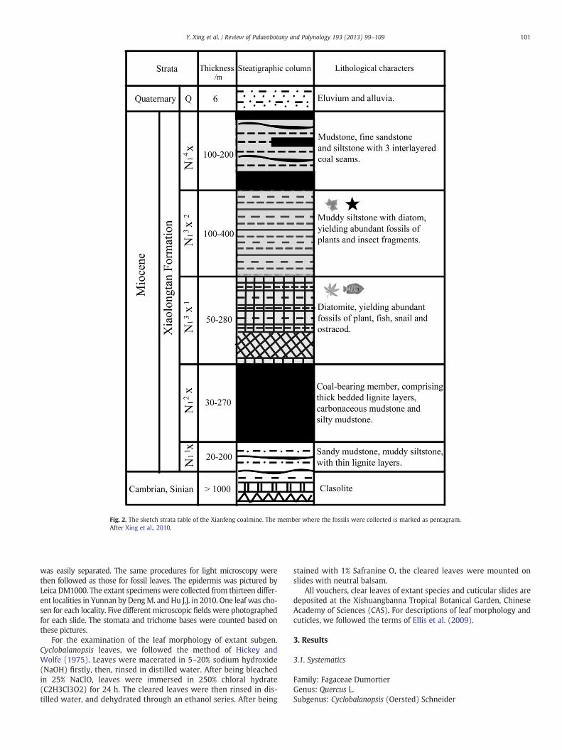
Fig. 2. The sketch strata table of the Xianfeng coalmine. The member where the fossils were collected is marked as pentagram.After Xing et al., 2010.
101Y. Xing et al. / Review of Palaeobotany and Palynology 193 (2013) 99–109
was easily separated. The same procedures for light microscopy werethen followed as those for fossil leaves. The epidermis was pictured byLeica DM1000. The extant specimens were collected from thirteen differ-ent localities in Yunnan by DengM. and Hu J.J. in 2010. One leaf was cho-sen for each locality. Five different microscopic fields were photographedfor each slide. The stomata and trichome bases were counted based onthese pictures.
For the examination of the leaf morphology of extant subgen.Cyclobalanopsis leaves, we followed the method of Hickey andWolfe (1975). Leaves were macerated in 5–20% sodium hydroxide(NaOH) firstly, then, rinsed in distilled water. After being bleachedin 25% NaClO, leaves were immersed in 250% chloral hydrate(C2H3Cl3O2) for 24 h. The cleared leaves were then rinsed in dis-tilled water, and dehydrated through an ethanol series. After being
stained with 1% Safranine O, the cleared leaves were mounted onslides with neutral balsam.
All vouchers, clear leaves of extant species and cuticular slides aredeposited at the Xishuangbanna Tropical Botanical Garden, ChineseAcademy of Sciences (CAS). For descriptions of leaf morphology andcuticles, we followed the terms of Ellis et al. (2009).
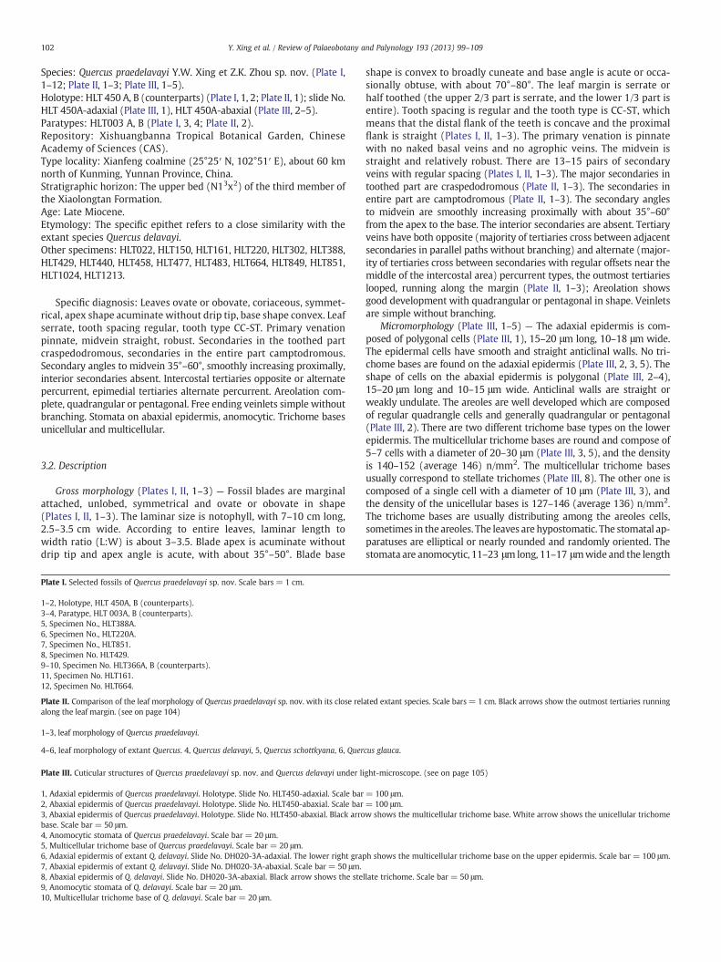
3. Results
3.1. Systematics
Family: Fagaceae DumortierGenus: Quercus L.Subgenus: Cyclobalanopsis (Oersted) Schneider
102 Y. Xing et al. / Review of Palaeobotany and Palynology 193 (2013) 99–109
Species: Quercus praedelavayi Y.W. Xing et Z.K. Zhou sp. nov. (Plate I,1–12; Plate II, 1–3; Plate III, 1–5).Holotype: HLT 450 A, B (counterparts) (Plate I, 1, 2; Plate II, 1); slide No.HLT 450A-adaxial (Plate III, 1), HLT 450A-abaxial (Plate III, 2–5).Paratypes: HLT003 A, B (Plate I, 3, 4; Plate II, 2).Repository: Xishuangbanna Tropical Botanical Garden, ChineseAcademy of Sciences (CAS).Type locality: Xianfeng coalmine (25°25′ N, 102°51′ E), about 60 kmnorth of Kunming, Yunnan Province, China.Stratigraphic horizon: The upper bed (N13x2) of the third member ofthe Xiaolongtan Formation.Age: Late Miocene.Etymology: The specific epithet refers to a close similarity with theextant species Quercus delavayi.Other specimens: HLT022, HLT150, HLT161, HLT220, HLT302, HLT388,HLT429, HLT440, HLT458, HLT477, HLT483, HLT664, HLT849, HLT851,HLT1024, HLT1213.
Specific diagnosis: Leaves ovate or obovate, coriaceous, symmet-rical, apex shape acuminate without drip tip, base shape convex. Leafserrate, tooth spacing regular, tooth type CC-ST. Primary venationpinnate, midvein straight, robust. Secondaries in the toothed partcraspedodromous, secondaries in the entire part camptodromous.Secondary angles to midvein 35°–60°, smoothly increasing proximally,interior secondaries absent. Intercostal tertiaries opposite or alternatepercurrent, epimedial tertiaries alternate percurrent. Areolation com-plete, quadrangular or pentagonal. Free ending veinlets simple withoutbranching. Stomata on abaxial epidermis, anomocytic. Trichome basesunicellular and multicellular.
3.2. Description
Gross morphology (Plates I, II, 1–3) — Fossil blades are marginalattached, unlobed, symmetrical and ovate or obovate in shape(Plates I, II, 1–3). The laminar size is notophyll, with 7–10 cm long,2.5–3.5 cm wide. According to entire leaves, laminar length towidth ratio (L:W) is about 3–3.5. Blade apex is acuminate withoutdrip tip and apex angle is acute, with about 35°–50°. Blade base
Plate I. Selected fossils of Quercus praedelavayi sp. nov. Scale bars = 1 cm.
1–2, Holotype, HLT 450A, B (counterparts).3–4, Paratype, HLT 003A, B (counterparts).5, Specimen No., HLT388A.6, Specimen No., HLT220A.7, Specimen No., HLT851.8, Specimen No. HLT429.9–10, Specimen No. HLT366A, B (counterparts).11, Specimen No. HLT161.12, Specimen No. HLT664.
Plate II. Comparison of the leaf morphology of Quercus praedelavayi sp. nov. with its close relalong the leaf margin. (see on page 104)
1–3, leaf morphology of Quercus praedelavayi.
4–6, leaf morphology of extant Quercus. 4, Quercus delavayi, 5, Quercus schottkyana, 6, Quer
Plate III. Cuticular structures of Quercus praedelavayi sp. nov. and Quercus delavayi under li
1, Adaxial epidermis of Quercus praedelavayi. Holotype. Slide No. HLT450-adaxial. Scale bar2, Abaxial epidermis of Quercus praedelavayi. Holotype. Slide No. HLT450-abaxial. Scale bar3, Abaxial epidermis of Quercus praedelavayi. Holotype. Slide No. HLT450-abaxial. Black arrobase. Scale bar = 50 μm.4, Anomocytic stomata of Quercus praedelavayi. Scale bar = 20 μm.5, Multicellular trichome base of Quercus praedelavayi. Scale bar = 20 μm.6, Adaxial epidermis of extant Q. delavayi. Slide No. DH020-3A-adaxial. The lower right grap7, Abaxial epidermis of extant Q. delavayi. Slide No. DH020-3A-abaxial. Scale bar = 50 μm.8, Abaxial epidermis of Q. delavayi. Slide No. DH020-3A-abaxial. Black arrow shows the ste9, Anomocytic stomata of Q. delavayi. Scale bar = 20 μm.10, Multicellular trichome base of Q. delavayi. Scale bar = 20 μm.
shape is convex to broadly cuneate and base angle is acute or occa-sionally obtuse, with about 70°–80°. The leaf margin is serrate orhalf toothed (the upper 2/3 part is serrate, and the lower 1/3 part isentire). Tooth spacing is regular and the tooth type is CC-ST, whichmeans that the distal flank of the teeth is concave and the proximalflank is straight (Plates I, II, 1–3). The primary venation is pinnatewith no naked basal veins and no agrophic veins. The midvein isstraight and relatively robust. There are 13–15 pairs of secondaryveins with regular spacing (Plates I, II, 1–3). The major secondaries intoothed part are craspedodromous (Plate II, 1–3). The secondaries inentire part are camptodromous (Plate II, 1–3). The secondary anglesto midvein are smoothly increasing proximally with about 35°–60°from the apex to the base. The interior secondaries are absent. Tertiaryveins have both opposite (majority of tertiaries cross between adjacentsecondaries in parallel paths without branching) and alternate (major-ity of tertiaries cross between secondaries with regular offsets near themiddle of the intercostal area) percurrent types, the outmost tertiarieslooped, running along the margin (Plate II, 1–3); Areolation showsgood development with quadrangular or pentagonal in shape. Veinletsare simple without branching.
Micromorphology (Plate III, 1–5) — The adaxial epidermis is com-posed of polygonal cells (Plate III, 1), 15–20 μm long, 10–18 μm wide.The epidermal cells have smooth and straight anticlinal walls. No tri-chome bases are found on the adaxial epidermis (Plate III, 2, 3, 5). Theshape of cells on the abaxial epidermis is polygonal (Plate III, 2–4),15–20 μm long and 10–15 μm wide. Anticlinal walls are straight orweakly undulate. The areoles are well developed which are composedof regular quadrangle cells and generally quadrangular or pentagonal(Plate III, 2). There are two different trichome base types on the lowerepidermis. The multicellular trichome bases are round and compose of5–7 cells with a diameter of 20–30 μm (Plate III, 3, 5), and the densityis 140–152 (average 146) n/mm2. The multicellular trichome basesusually correspond to stellate trichomes (Plate III, 8). The other one iscomposed of a single cell with a diameter of 10 μm (Plate III, 3), andthe density of the unicellular bases is 127–146 (average 136) n/mm2.The trichome bases are usually distributing among the areoles cells,sometimes in the areoles. The leaves are hypostomatic. The stomatal ap-paratuses are elliptical or nearly rounded and randomly oriented. Thestomata are anomocytic, 11–23 μmlong, 11–17 μmwide and the length
ated extant species. Scale bars = 1 cm. Black arrows show the outmost tertiaries running
cus glauca.
ght-microscope. (see on page 105)
= 100 μm.= 100 μm.w shows the multicellular trichome base. White arrow shows the unicellular trichome
h shows the multicellular trichome base on the upper epidermis. Scale bar = 100 μm.
llate trichome. Scale bar = 50 μm.

Plate II (caption on page 102).
104 Y. Xing et al. / Review of Palaeobotany and Palynology 193 (2013) 99–109

Plate III (caption on page 102).
105Y. Xing et al. / Review of Palaeobotany and Palynology 193 (2013) 99–109
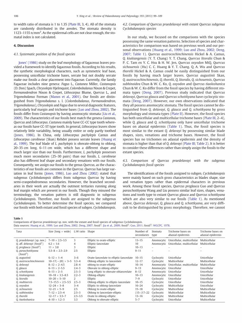
106 Y. Xing et al. / Review of Palaeobotany and Palynology 193 (2013) 99–109
to width ratio of stomata is 1 to 1.35 (Plate III, 3, 4). All of the stomataare randomly distributed in the areoles. The stomata density is1123–1155 n/mm2. As the epidermal cells are not clear enough, the sto-matal index is not calculated.
4. Discussion
4.1. Systematic position of the fossil species
Jones' (1986) study on the leaf morphology of fagaceous leaves pro-vided a framework to identify fagaceous fossils. According to his results,the synthetic morphological characters, such as symmetric leaf base,possessing unicellular trichome bases, serrate but not doubly serratemake our fossils a clear placement into Fagaceae. Currently, the familyFagaceae includes nine genera: Fagus L., Castanea Miller, Castanopsis(D. Don) Spach, ChrysolepisHjelmquist, ColombobalanusNixon & Crepet,Formanodendron Nixon & Crepet, Lithocarpus Blume, Quercus L., andTrigonobalanus Forman (Manos et al., 2001). Our fossils are distin-guished from Trigonobalanus s. l. (Colombobalanus, Formanodendron,Trigonobalanus), Chrysolepis and Fagusdue to several diagnostic features,particularly leaf margin and trichome bases (Luo and Zhou, 2002). Thefossils differ from Castanopsis by having anomocytic stomata (Liu et al.,2009). The characteristics of our fossils best match the genera Castanea,Quercus and Lithocarpus. Castaneamainly have CC-CC type toothwhere-as our fossils have CC-ST type tooth. In general, Lithocarpus leaves showrelatively little variability, being usually entire or only partly toothed(Jones, 1986). In China, only Lithocarpus pachylepis Camus andLithocarpus carolineae (Skan) Rehder possess serrate leaves (Huang etal., 1999). The leaf blade of L. pachylepis is obovate-oblong to oblong,20–35 cm long, 6–11 cm wide, which has a different shape andmuch larger size than our fossils. Furthermore, L. pachylepis possessesmuch more secondaries (25–30 pairs) than our fossils. L. carolineaealso has different leaf shape and secondary venations with our fossils.Consequently, we assign our fossils to the genus Quercus, as the charac-teristics of our fossils are common in the Quercus. Quercus has large var-iation in leaf forms (Jones, 1986). Luo and Zhou (2002) stated thatsubgenus Cyclobalanopsis differs from subgenus Quercus by havingsemi-craspedodromous secondaries. However, the branched second-aries in their work are actually the outmost tertiaries running alongleaf margin which are present in our fossils. Though they misused theterminology, the venation pattern is still diagnostic in subgenusCyclobalanopsis. Therefore, our fossils are assigned to the subgenusCyclobalanopsis. To better determine the fossil species, we comparedour fossils with both extant and fossil species of subgen. Cyclobalanopsis.
Table 1Comparisons of Quercus praedelavayi sp. nov. with the extant and fossil species of subgenuData sources: Huang et al., 1999; Luo and Zhou, 2002; Deng, 2007; fossil1: Jia et al., 2009; f
Taxa Size (long × wide) L:W ratio Shape
Q. praedelavayi (sp. nov.) 7–10 × 2.5–3.5 3 Elliptic to ovate-ellipticQ. aff. delavayi (fossil1) 6.2 × 1.6 4 Elliptic-lanceolateQ. preglauca (fossil2) 11 × 3.8 3 EllipticQ. paraschottkyana(fossil3)
5.5–8 × 2.5–2.9 2.5 Elliptic
Q. augustinii 6–12 × 1–4 3–6 Ovate-lanceolate to elliptic-laQ. austrocochinchinensis 10–17(−20) × 3–5 3.3–4 Oblong-elliptic to lanceolateQ. delavayi 8–12 × 2–4.5 2.8–4 oblong to ovate-ellipticQ. glauca 6–13 × 2–5.5 2.4–3 Obovate to oblong-ellipticQ. schottkyana 6–13 × 2–5 2.5–3 Long elliptic to obovate-oblanQ. kiukiangensis 10–18 × 3.5–8.5 2.2–3 Oblong-ellipticQ. litoralis 10–20 × 5–10 2 EllipticQ. multinervis 7.5–15.5 × 2.5–5.5 2.5–3 Oblong-elliptic to elliptic-lanQ. oxyodon 12–24 × 3–8 3–4 Elliptic to oblong-lanceolateQ. sichouensis 12–21 × 5–9 2.5 Oblong to ovate-ellipticQ. subhinoidea 7–12 × 2.5–4 2.5–3 Oblong to lanceolate-ellipticQ. thorelii 12–17 × 3.5–7 2.5–3.5 Ovate to oblong-ellipticQ. tiaoloshanica 4–10 × 1.2–3 3.3 Oblong to obovate-elliptic
4.2. Comparison of Quercus praedelavayi with extant Quercus subgenusCyclobalanopsis species
In our study, we focused on the comparisons with the speciespossessing the same venation patterns. Selection of species and char-acteristics for comparison was based on previous work and our per-sonal observations (Huang et al., 1999; Luo and Zhou, 2002; Deng,2007; Table 1). Quercus austrocochinchinensis Hickel & A. Camus,Q. kiukiangensis (Y. T. Chang) Y. T. Chang, Quercus litoralis Chun &P. C. Tam ex Y. C. Hsu & H. W. Jen, Quercus oxyodon MiQ, Quercussichouensis (Hu) C. C. Huang & Y. T. Chang. Q. A. Wu and Quercusthorelii Hickel & A. Camus could be easily distinguished from ourfossils by having much larger leaves. Quercus augustinii Skan,Q. austrocochinchinensis, Q. thorelii, Q. litoralis, Q. sichouensis, Quercussubhinoidea Chun & W. C. Ko, Q. oxyodon and Quercus tiaoloshanicaChun &W. C. Ko differ from the fossil species by having different sto-mata types (Deng, 2007). Previous study indicated that Quercusdelavayi, Quercus glauca and Quercus schottkyana have cyclocytic sto-mata (Deng, 2007). However, our own observations indicated thatthey all possess anomocytic stomata. The fossil species cannot be dis-tinguished from Q. delavayi, Q. glauca and Q. schottkyana by grossmorphology and stomata types (Plate II). However, the fossil specieshas both unicellular and multicellular trichome bases (Plate III, 2–4),while Q. glauca and Q. schottkyana only have unicellular trichomebases on abaxial epidermis (Table 1). Thus, the fossil species ismost similar to the extant Q. delavayi by possessing similar bladeshapes, sizes, venations and trichome bases. However, the fossilspecies has no trichomes on adaxial epidermis and the density ofstomata is higher than that of Q. delavayi (Plate III; Table 2). It is betterto consider these differences rather than simply assign the fossils to theextant species.
4.3. Comparison of Quercus praedelavayi with the subgenusCyclobalanopsis fossil species
The identifications of the fossils assigned to subgen. Cyclobalanopsiswere mainly based on such gross characteristics as blades shape, sizeand venation types rather than epidermal characters in previouswork. Among these fossil species, Quercus preglauca Guo and Quercusparaschottkyana Wang and Liu possess similar leaf sizes, shapes, vena-tions and tooth type to extant Quercus glauca and Quercus schottkyanawhich are also very similar to our fossils (Table 1). As mentionedabove, Quercus delavayi, Q. glauca and Q. schottkyana, are very diffi-cult to be distinguished by gross morphology. Therefore, we cannot
s Cyclobalanopsis.ossil2: Guo, 2011; fossil3: WGCPC, 1978.
Number ofsecondaries
Stomatatype
Trichome bases onabaxial epidermis
Trichome bases onadaxial epidermis
13–15 Anomocytic Unicellular, multicellular Multicellular10 Anomocytic Unicellular, multicellular Multicellular10–13 – – –
9–11 – – –
nceolate 10–15 Cyclocytic Unicellular Unicellular12–17 Cyclocytic Multicellular Multicellular10–14 Anomocytic Unicellular, multicellular Multicellular9–13 Anomocytic Unicellular Unicellular
ceolate 8–12 Anomocytic Unicellular Unicellular10–13 Anomocytic Unicellular Unicellular7–9 Cyclocytic Unicellular Unicellular
ceolate 10–15 Cyclocytic Unicellular Unicellular16–24 Cyclocytic Multicellular Unicellular15–18 Cyclocytic Multicellular Multicellular(15–)17–22 Cyclocytic Multicellular Multicellular13–16 Cyclocytic Multicellular Multicellular5–7 Cyclocytic Unicellular Multicellular
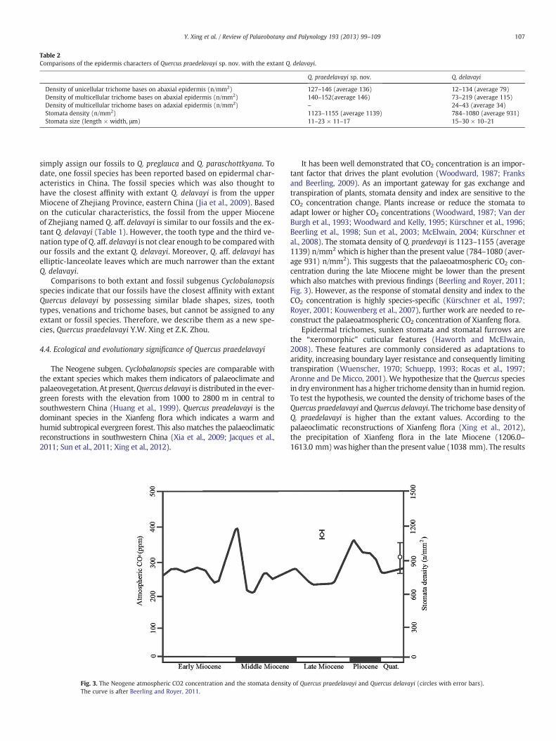
Table 2Comparisons of the epidermis characters of Quercus praedelavayi sp. nov. with the extant Q. delavayi.
Q. praedelavayi sp. nov. Q. delavayi
Density of unicellular trichome bases on abaxial epidermis (n/mm2) 127–146 (average 136) 12–134 (average 79)Density of multicellular trichome bases on abaxial epidermis (n/mm2) 140–152(average 146) 73–219 (average 115)Density of multicellular trichome bases on adaxial epidermis (n/mm2) – 24–43 (average 34)Stomata density (n/mm2) 1123–1155 (average 1139) 784–1080 (average 931)Stomata size (length × width, μm) 11–23 × 11–17 15–30 × 10–21
107Y. Xing et al. / Review of Palaeobotany and Palynology 193 (2013) 99–109
simply assign our fossils to Q. preglauca and Q. paraschottkyana. Todate, one fossil species has been reported based on epidermal char-acteristics in China. The fossil species which was also thought tohave the closest affinity with extant Q. delavayi is from the upperMiocene of Zhejiang Province, eastern China (Jia et al., 2009). Basedon the cuticular characteristics, the fossil from the upper Mioceneof Zhejiang named Q. aff. delavayi is similar to our fossils and the ex-tant Q. delavayi (Table 1). However, the tooth type and the third ve-nation type of Q. aff. delavayi is not clear enough to be comparedwithour fossils and the extant Q. delavayi. Moreover, Q. aff. delavayi haselliptic-lanceolate leaves which are much narrower than the extantQ. delavayi.
Comparisons to both extant and fossil subgenus Cyclobalanopsisspecies indicate that our fossils have the closest affinity with extantQuercus delavayi by possessing similar blade shapes, sizes, toothtypes, venations and trichome bases, but cannot be assigned to anyextant or fossil species. Therefore, we describe them as a new spe-cies, Quercus praedelavayi Y.W. Xing et Z.K. Zhou.
4.4. Ecological and evolutionary significance of Quercus praedelavayi
The Neogene subgen. Cyclobalanopsis species are comparable withthe extant species which makes them indicators of palaeoclimate andpalaeovegetation. At present,Quercus delavayi is distributed in the ever-green forests with the elevation from 1000 to 2800 m in central tosouthwestern China (Huang et al., 1999). Quercus preadelavayi is thedominant species in the Xianfeng flora which indicates a warm andhumid subtropical evergreen forest. This also matches the palaeoclimaticreconstructions in southwestern China (Xia et al., 2009; Jacques et al.,2011; Sun et al., 2011; Xing et al., 2012).
Fig. 3. The Neogene atmospheric CO2 concentration and the stomata densityThe curve is after Beerling and Royer, 2011.
It has been well demonstrated that CO2 concentration is an impor-tant factor that drives the plant evolution (Woodward, 1987; Franksand Beerling, 2009). As an important gateway for gas exchange andtranspiration of plants, stomata density and index are sensitive to theCO2 concentration change. Plants increase or reduce the stomata toadapt lower or higher CO2 concentrations (Woodward, 1987; Van derBurgh et al., 1993; Woodward and Kelly, 1995; Kürschner et al., 1996;Beerling et al., 1998; Sun et al., 2003; McElwain, 2004; Kürschner etal., 2008). The stomata density of Q. praedevayi is 1123–1155 (average1139) n/mm2 which is higher than the present value (784–1080 (aver-age 931) n/mm2). This suggests that the palaeoatmospheric CO2 con-centration during the late Miocene might be lower than the presentwhich also matches with previous findings (Beerling and Royer, 2011;Fig. 3). However, as the response of stomatal density and index to theCO2 concentration is highly species-specific (Kürschner et al., 1997;Royer, 2001; Kouwenberg et al., 2007), further work are needed to re-construct the palaeoatmospheric CO2 concentration of Xianfeng flora.
Epidermal trichomes, sunken stomata and stomatal furrows arethe “xeromorphic” cuticular features (Haworth and McElwain,2008). These features are commonly considered as adaptations toaridity, increasing boundary layer resistance and consequently limitingtranspiration (Wuenscher, 1970; Schuepp, 1993; Rocas et al., 1997;Aronne and De Micco, 2001). We hypothesize that the Quercus speciesin dry environment has a higher trichomedensity than in humid region.To test the hypothesis, we counted the density of trichome bases of theQuercus praedelavayi andQuercus delavayi. The trichome base density ofQ. praedelavayi is higher than the extant values. According to thepalaeoclimatic reconstructions of Xianfeng flora (Xing et al., 2012),the precipitation of Xianfeng flora in the late Miocene (1206.0–1613.0 mm)was higher than the present value (1038 mm). The results
of Quercus praedelavayi and Quercus delavayi (circles with error bars).

108 Y. Xing et al. / Review of Palaeobotany and Palynology 193 (2013) 99–109
indicate that the evolution of trichome density of Q. delavayi is not drivenby the decrease in precipitation.
Acknowledgments
The authors thank the editor and two reviewers, Dr. V. Teodoridisand another anonymous reviewer for their constructive suggestionsto the manuscript. We also thank Dr. Richard Carter from the Insti-tute of Systematic Botany, University of Zurich and Mr. WarrenThomas Kellie for the English phrasing. This work is supported by theNational Basic Research Program of China, 973 Program (No.2012CB821901), the National Natural Science Foundation of China,NNSFC (no. 41030212) to Zhekun Zhou, and the Open Foundation ofState Key Laboratory of Loess and Quaternary Geology, Institute ofEarth Environment, CAS (no. SKLLQG 1014) to Yaowu Xing.
References
Aronne, G., De Micco, V., 2001. Seasonal dimorphism in the Mediterranean Cistusincanus L. subsp. incanus. Annals of Botany 87, 789–794.
Axelrod, D.I., 1956. Mio-Pliocene Floras from West Central Nevada. University of California.Publications in Geological Science 33, 1–322.
Axelrod, D.I., 1964. The Miocene Trapper Creek Flora of Southern Idaho. University ofCalifornia. Publications in Geological Science 51, 1–181.
Barnett, E.C., 1944. Keys to the species groups of Quercus, Lithocarpus and Castanopsis ofEastern Asia, with notes on their distribution. Transactions of the Botanical Societyof Edinburgh 34, 159–204.
Beerling, D.J., Royer, D.L., 2011. Earth's atmospheric CO2 history by proxy. Nature Geo-science 4, 1–2.
Beerling, D.J., McElwain, J.C., Osborne, C.P., 1998. Stomatal responses of the ‘living fos-sil’ Ginkgo biloba to changes in atmospheric CO2 concentrations. Journal of Experi-mental Botany 49, 1603–1607.
Bell, W.A., 1957. Flora of the Upper Cretaceous Nanaimo Group of Vancouver Island.British Columbia Geological Survey of Canada Memoir 293, 1–84.
Bones, T.J., 1979. Atlas of fossil fruits and seeds from north central Oregon. Oregon Mu-seum of Science and Industry Occasional Papers in Natural Science 1, 1–23.
Borgardt, S.J., Pigg, P.B., 1999. Anatomical and developmental study of petrified Quercus(Fagaceae) fruit from the Middle Miocene, Yakima Canyon, Washington, USA.American Journal of Botany 86, 307–325.
Bureau of Geology and Mineral Resources of Yunnan Province (BGMRYP), 1996. Stra-tigraphy (Lithostratic) of Yunnan Province. China University of GeosciencesPress, Beijing 220–222 (in Chinese).
Camus, A., 1936–1954. Les Chenes. Monographie du genre Quercus and monographie dugenre Lithocarpus. : Encyclopedie Econnomique de SylvicultureLechevalier, Paris 6–8.
Chen, Y.Q., Deng, M., Zhou, Z.K., 2008. A hypothesis on cupule evolution and the evi-dence from molecular phylogenies and fossils. Journal of Systematics and Evolu-tion 46 (1), 41–52.
Crepet, W.L., Nixon, K.C., 1989. Earliest megafossil evidence of Fagaceae: phylogeneticand biogeographic implications. American Journal of Botany 76, 842–855.
Deng, M., 2007. Anatomy, Taxonomy, Distribution, and Phylogeny of Quercus SubgenusCyclobalanopsis (Oersted) Schneid. (Fagaceae). Ph.D. Thesis, Kunming Institute ofBotany, Chinese Academy of Sciences, Kunming, China (in Chinese, with EnglishAbstr.).
Deng, M., Zhou, Z.K., Coombes, A., 2006. Taxonomic notes on the Genus Cyclobalanopsis(Fagaceae). Annales Botanici Fennici 43, 57–61.
Deng, M., Zhou, Z.K., Chen, Y.Q., Sun, W.B., 2008. Systematic significance of the devel-opment and anatomy of flowers and fruit of Quercus schottkyana (subgenusCyclobalanopsis: Fagaceae). International Journal of Plant Sciences 169 (9),1261–1277.
Dong, W., 2001. Upper Cenozoic stratigraphy and paleoenvironment of XiaolongtanBasin, Kaiyuan, Yunnan Province. Proc. Eighth Ann. Meeting, Chinese: Soc. Vert.Paleo. China, vol. 8 (in Chinese).
Ellis, B., Daly, D.C., Hickey, L.J., Johnson, K.R., Mitchell, J.D., Wilf, P., Wing, S.L., 2009.Manual of Leaf Architecture. Cornell University Press, New York.
Fey, B.S., Endress, P.K., 1983. Development and morphological interpretation of the cu-pule in Fagaceae. Flora 173 (5–6), 451–468.
Forman, L.L., 1966. On the evolution of cupules in the Fagaceae. Kew Bulletin 18, 385–419.Franks, P.J., Beerling, D.J., 2009. Maximum leaf conductance driven by atmospheric CO2
effects on stomatal size and density over geologic time. Proceedings of the NationalAcademy of Sciences of the United States of America 106, 10343–10347.
Ge, H.R., Li, D.Y., 1999. Cenozoic Coal-bearing Basins and Coal, Forming Regularity inWest Yunnan. Yunnan Science and Technology Press, Kunming (in Chinese, withEnglish Abstr.).
Guo, S.X., 2011. The late Miocene Bangmai flora from Lincang County of Yunnan, south-western China. Acta Palaeontologica Sinica 50 (3), 353–408.
Haworth, M., McElwain, J.C., 2008. Hot, dry, wet, cold or toxic? Revisiting the ecological sig-nificance of leaf and cuticular micromorphology. Palaeogeography, Palaeoclimatology,Palaeoecology 262 (1–2), 79–90.
Hickey, L.J., Wolfe, J.A., 1975. The bases of angiosperm phylogeny: vegetative morphol-ogy. Annals of the Missouri Botanical Garden 62, 538–590.
Hjelmqvist, H., 1948. Studies on the floral morphology and phylogeny of theAmentiferae. Botaniska Notiser (Supplement 2), 1–171.
Huang, C.C., Chang, Y.T., Bartholomew, B., 1999. Fagaceae. In: Wu, C.Y., Raven, P.H.(Eds.), Flora of China. Science Press, Beijing.
Huzioka, K., Takahasi, E., 1970. The Eocene flora of the Ube coal-field, southwest Hon-shu, Japan. J. Mining College, Akita Univ. Journal of the Mining College, Akita Uni-versity, Series A: Mining Geology 4 (5), 1–88.
Jacques, F.M.B., Guo, S.X., Su, T., Xing, Y.W., Huang, Y.J., Liu, Y.S., Ferguson, D.K., Zhou,Z.K., 2011. Quantitative reconstruction of the Late Miocene monsoon climates ofsouthwest China: a case study of the Lincang flora from Yunnan Province.Palaeogeography, Palaeoclimatology, Palaeoecology 304 (3–4), 318–327.
Jia, H., Sun, B.N., Li, X.C., Xiao, L., Wu, J.Y., 2009. Microstructures of one species ofQuercus from the Neogene in Eastern Zhejiang and its palaeoenvironmental indi-cation. Frontiers of Earth Science 16 (5), 79–90 (in Chinese, with English Abstr.).
Jones, J. H., 1984. Leaf Architectural and Cuticular Analyses of Extant Fagaceae and‘Fagaceous’ Leaves From the Paleogene of Southeastern North America. Ph.D. The-sis, Indiana Univ., Bloomington.
Jones, J.H., 1986. Evolution of the Fagaceae: the implications of foliar features. Annals ofthe Missouri Botanical Garden 73 (2), 228–275.
Kouwenberg, L.R., Kürschner, W.M., McElwain, J.C., 2007. Stomatal frequency changeover altitudinal gradients: prospects for paleoaltimetry. Reviews in Mineralogyand Geochemistry 66, 215–241.
Kürschner, W.M., van der Burgh, J., Visscher, H., Dilcher, D.L., 1996. Oak leaves as bio-sensors of late Neogene and early Pleistocene paleoatmospheric CO2 concentra-tions. Marine Micropaleontology 27, 299–312.
Kürschner, W.M., Wagner, F., Visscher, E.H., Visscher, H., 1997. Predicting the responseof leaf stomatal frequency to a future CO2 enriched atmosphere: constraints fromhistorical observations. Geologische Rundschau 86, 512–517.
Kürschner, W.M., Kvaček, Z., Dilcher, D.L., 2008. The impact of Miocene atmosphericcarbon dioxide fluctuations on climate and the evolution of terrestrial ecosystems.Proceedings of the National Academy of Sciences of the United States of America105 (2), 449–453.
Kvaček, Z., Walther, H., 1989. Paleobotanical studies in Fagaceae of the European Tertiary.Plant Systematics and Evolution 162 (1–4), 2130–2229.
Kvaček, Z., Walther, H., 2004. Oligocene flora of Bechlejovice at Decin from theneovolcanic area of the Ceske Stredohori Mountains, Czech Republic. Acta MuseiNationalis Pragae, Series B, Historia Naturalis 60, 9–60.
Liu, Y.S., Zetter, R., Ferguson, D.K., Mohr, B.A.R., 2007. Discriminating fossil evergreenand deciduous Quercus pollen: a case study from the Miocene of eastern China. Re-view of Palaeobotany and Palynology 145, 289–303.
Liu, M.Q., Deng, M., Zhou, Z.K., 2009. Taxonomic and ecological implications of leaf cu-ticular morphology in Castanopsis, Castanea, and Chrysolepis. Plant Systematics andEvolution 283 (1–2), 111–123.
Luo, Y., Zhou, Z.K., 2001. Phytogeography of Quercus subg. Cyclobalanopsis. Acta Botan-ica Yunnanica 23 (1), 1–16 (in Chinese, with English Abstr.).
Luo, Y., Zhou, Z.K., 2002. Leaf architecture in Quercus subgenus Cyclobalanopsis(Fagaceae) from China. Botanical Journal of the Linnean Society 140 (3), 283–295.
MacGinitie, H.D., 1941. A Middle Eocene Flora from the Central Sierra Nevada. CarnegieInstitution of Washington Publication, USA.
MacGinitie, H.D., 1969. The Eocene Green River Flora of North-western Colorado andNortheastern Utah. University of California. Publications in Geological Science 83,1–203.
Manchester, S.R., 1981. Fossil plants of the Eocene Clarno nut beds. Oregon Geology 43,75–81.
Manchester, S.R., 1983. Eocene fruits, wood and leaves of the Fagaceae from the ClarnoFormation of Oregon. American Journal of Botany 70 (5, 2), 74 (Abstract).
Manos, P.S., Zhou, Z.K., Cannon, C.H., 2001. Systematics of Fagaceae: phylogenetic tests ofreproductive trait evolution. International Journal of Plant Sciences 162 (6),1361–1379.
McElwain, J.C., 2004. Climate-independent paleoaltimetry using stomatal density infossil leaves as a proxy for CO2 partial pressure. Geology 32 (12), 1017–1021.
Nixon, K.C., 1993. Infrageneric classification of Quercus (Fagaceae) and typification ofsectional names. Annales des Sciences Forestières 50 (Suppl. 1), S25–S34.
Rocas, G., Barros, C.F., Scarano, F.R., 1997. Leaf anatomy plasticity of Alchorneatriplinervia (Euphorbiaceae) under distinct light regimes in a Brazilian montane At-lantic rain forest. Trees—Structure and Function 11 (8), 469–473.
Royer, D.L., 2001. Stomatal density and stomatal index as indicators of paleoatmosphericCO2 concentration. Review of Palaeobotany and Palynology 114 (1–2), 1–28.
Schottky, E.M., 1912. Die Eichen des extratropischen Ostasiens und ihrepflanzengeographische Bedeutung. Botanische Jahrbücher für Systematik 47,617–708.
Schuepp, P.H., 1993. Leaf boundary layers. New Phytologist 125, 477–507.Sun, B.N., Dilcher, D.L., Beerling, D.J., Zhang, C.J., Yan, D.F., Kowalski, E., 2003. Variation in
Ginkgo biloba L. leaf characters across a climatic gradient in China. Proceedings of theNational Academy of Sciences of the United States of America 100 (12), 7141–7146.
Sun, B.N., Wu, J.Y., Liu, Y.S., Ding, S.T., Li, X.C., Xie, S.P., Yan, D.F., Lin, Z.C., 2011.Reconstructing Neogene vegetation and climates to infer tectonic uplift in westernYunnan, China. Palaeogeography, Palaeoclimatology, Palaeoecology 304 (3–4),328–336.
Tao, J.R., 2000. The Evolution of the Late Cretaceous–Cenozoic Floras in China. SciencePress, Beijing (in Chinese).
Van Boskirk, M.C., 1998. The Flora of the Eagle Formation and Its Significance for LateCretaceous Floristic Evolution. Ph.D. Thesis, Yale University, New Haven, Connecticut,USA.
Van der Burgh, J., Visscher, H., Dilcher, D.L., Kürschner, W.M., 1993. Paleoatmosphericsignatures in neogene fossil leaves. Science 260 (5115), 1788–1790.
109Y. Xing et al. / Review of Palaeobotany and Palynology 193 (2013) 99–109
Wang, W.M., 1996. A palynological survey of Neogene strata in Xiaolongtan basin,Yunnan Province of South China. Acta Botanica Sinica 38, 743–748 (in Chinese,with English Abstr.).
Wang, G.P., Chen, Q.S., Li, Y.T., Li, H.M., Guo, S.X., Lan, S.X., Ju, K.X., 1982. Fossil plants.In: Nanjing Institute of Geology and Mineral Resources (Ed.), PalaeontologicalAtlas of East China. Part 3. : Volume of Mesozoic and Cenozoic. Geological Publish-ing House, Beijing, pp. 236–316.
Woodward, F.I., 1987. Stomatal numbers are sensitive to increases in CO2 from pre-industrial levels. Nature 327, 617–618.
Woodward, F.I., Kelly, C.K., 1995. The influence of CO2 concentration on stomatal den-sity. New Phytologist 131 (3), 311–327.
Writing Group of Cenozoic Plants of China (WGCPC), 1978. Cenozoic Plants from China,Fossil Plants of China, vol. 3. Science Press, Beijing (in Chinese).
Wu, C.L., Li, S.H., Wang, G.F., Liu, G., Kong, C.F., 2006. The allochthonous genesis modelabout the extra-thick and high-quality coal bed in Xianfeng basin, Yunnan Prov-ince, China. Frontiers of Earth Science in China 1 (1), 97–105.
Wuenscher, J.E., 1970. The effect of leaf hairs of Verbascum thapsus on leaf energy ex-change. New Phytologist 69, 65–73.
Xia, K., Su, T., Liu, Y.S., Xing, Y.W., Jacques, F.M.B., Zhou, Z.K., 2009. Quantitative climatereconstructions of the late Miocene Xiaolongtan megaflora from Yunnan, south-west China. Palaeogeography, Palaeoclimatology, Palaeoecology 276 (1–4), 80–86.
Xing, Y.W., 2010. The Late Miocene Xianfeng Flora, Yunnan, Southwest China and ItsQuantitative Palaeoclimatic Reconstructions. Ph.D. Thesis, Kunming Institute ofBotany, Chinese Academy of Sciences, Kunming, China (in Chinese, with EnglishAbstr.).
Xing, J., Liu, G.X., Xu, G.Q., 1999. The characteristics of coal facies of mega coal seam inXianfeng basin (Xiaolongtan Formation), Yunnan, China. Coal Geology Exploration27, 1–4 (in Chinese, with English Abstr.).
Xing, Y.W., Liu, Y.S., Su, T., Jacques, F.M.B., Zhou, Z.K., 2010. Pinus prekesiya sp. nov. fromthe upper Miocene of Yunnan, southwestern China and its biogeographical impli-cations. Review of Palaeobotany and Palynology 160, 1–9.
Xing, Y.W., Utescher, T., Jacques, F.M.B., Su, T., Liu, Y.S., Huang, Y.J., Zhou, Z.K., 2012. Pa-leoclimatic estimation reveals a weak winter monsoon in southwestern China dur-ing the late Miocene: evidence from plant macrofossils. Palaeogeography,Palaeoclimatology, Palaeoecology 358–360, 19–26.
Zhang, Y.P., 1974. Miocene suids from Kaiyuan, Yunnan and Linchu, Shantung.Vertebrata Palasiatica 2, 117–125 (in Chinese, with English Abstr.).
Zhou, Z.K., 1985. The Miocene Xiaolongtan Fossil Flora in Kaiyuan, Yunnan, China. M.Sc.Thesis, Nanjing Institute of Geology and Palaeontology, Chinese Academy of Sci-ences, Nanjing, China (in Chinese).
Zhou, Z.K., 1993. The fossil history of Quercus. Acta Botanica Yunnanica 15 (1), 21–33(in Chinese, with English Abstr.).
Zhou, Z.K., 2000. On the Miocene Xiaolongtan flora from Kaiyuan, Yunnan Province. In:Tao, J.R. (Ed.), The Evolution of the Late Cretaceous–Cenozoic Floras in China. Sci-ence Press, Beijing, pp. 64–72 (in Chinese).
Zhou, Z.K., Wilkinson, H., Wu, Z.Y., 1995. Taxonomical and evolutionary implications ofthe leaf anatomy and architecture of Quercus L. Subgenus Quercus from China.Cathaya 7, 1–34.