Review of Osteosarcoma and Current Management · REVIEW Review of Osteosarcoma and Current...
23
REVIEW Review of Osteosarcoma and Current Management Ryan A. Durfee . Maryam Mohammed . Hue H. Luu Received: August 5, 2016 / Published online: October 19, 2016 Ó The Author(s) 2016. This article is published with open access at Springerlink.com ABSTRACT Osteosarcoma is the most common primary malignancy of bone in children and young adults. This tumor has a very heterogeneous genetic profile and lacks any consistent unifying event that leads to the pathogenesis of osteosarcoma. In this review, some of the important genetic events involved in osteosarcoma will be highlighted. Additionally, the clinical diagnosis of osteosarcoma will be discussed, as well as contemporary chemotherapeutic and surgical management of this tumor. Finally, the review will discuss some of the novel approaches to treating this disease. Keywords: Limb preservation surgery; Osteosarcoma; Review; Targeted therapy OSTEOSARCOMA Osteosarcoma (OS) is a high-grade primary skeletal malignancy characterized by spindle cells of mesenchymal origin depositing immature osteoid matrix [1, 2]. With an annual incidence rate of 3.1 cases per million in the US, OS accounts for less than 1% of all newly diagnosed cancers in adults and 3–5% of those in children, but it is the most common primary malignancy in adolescents outside of leukemia and lymphoma [3]. Although rare overall, OS is the most common primary malignancy of bone in children [3–5]. OS incidence is distributed bimodally across age. An initial peak is observed between the ages of 10 and 14 years during the pubertal growth spurt, and is followed by a smaller second peak after the age of 60 years [6]. OS develops in adolescents most often at the metaphysis of lower extremity long bones (*75% of cases), and these findings suggest a relationship between the hormonal changes of puberty and/or physiologic bone growth and the pathogenesis of OS [4, 6–8]. While long bones of the extremities continue to be the most common site for OS after the age of 60 years, Enhanced content To view enhanced content for this article go to http://www.medengine.com/Redeem/D1E6 F06013338352. R. A. Durfee Á M. Mohammed Á H. H. Luu (&) Department of Orthopaedic Surgery and Rehabilitation, University of Chicago, Chicago, IL, USA e-mail: [email protected] Rheumatol Ther (2016) 3:221–243 DOI 10.1007/s40744-016-0046-y
Transcript of Review of Osteosarcoma and Current Management · REVIEW Review of Osteosarcoma and Current...

REVIEW
Review of Osteosarcoma and Current Management
Ryan A. Durfee . Maryam Mohammed . Hue H. Luu
Received: August 5, 2016 / Published online: October 19, 2016� The Author(s) 2016. This article is published with open access at Springerlink.com
ABSTRACT
Osteosarcoma is the most common primary
malignancy of bone in children and young
adults. This tumor has a very heterogeneous
genetic profile and lacks any consistent
unifying event that leads to the pathogenesis
of osteosarcoma. In this review, some of the
important genetic events involved in
osteosarcoma will be highlighted.
Additionally, the clinical diagnosis of
osteosarcoma will be discussed, as well as
contemporary chemotherapeutic and surgical
management of this tumor. Finally, the review
will discuss some of the novel approaches to
treating this disease.
Keywords: Limb preservation surgery;
Osteosarcoma; Review; Targeted therapy
OSTEOSARCOMA
Osteosarcoma (OS) is a high-grade primary
skeletal malignancy characterized by spindle
cells of mesenchymal origin depositing
immature osteoid matrix [1, 2]. With an
annual incidence rate of 3.1 cases per million
in the US, OS accounts for less than 1% of all
newly diagnosed cancers in adults and 3–5% of
those in children, but it is the most common
primary malignancy in adolescents outside of
leukemia and lymphoma [3]. Although rare
overall, OS is the most common primary
malignancy of bone in children [3–5]. OS
incidence is distributed bimodally across age.
An initial peak is observed between the ages of
10 and 14 years during the pubertal growth
spurt, and is followed by a smaller second peak
after the age of 60 years [6]. OS develops in
adolescents most often at the metaphysis of
lower extremity long bones (*75% of cases),
and these findings suggest a relationship
between the hormonal changes of puberty
and/or physiologic bone growth and the
pathogenesis of OS [4, 6–8]. While long bones
of the extremities continue to be the most
common site for OS after the age of 60 years,
Enhanced content To view enhanced content for thisarticle go to http://www.medengine.com/Redeem/D1E6F06013338352.
R. A. Durfee � M. Mohammed � H. H. Luu (&)Department of Orthopaedic Surgery andRehabilitation, University of Chicago, Chicago, IL,USAe-mail: [email protected]
Rheumatol Ther (2016) 3:221–243
DOI 10.1007/s40744-016-0046-y

they no longer account for the majority of cases
due to an increase in the diversity of primary
tumor sites. Craniofacial and axial tumors
increase in frequency with age, accounting for
40% of all OS cases after 60 years of age,
compared to less than 12% before the age of
24 years [6]. Juxtacortical osteosarcomas that
occur along the surface of bones are usually
lower grade, although there are some
exceptions [9].
Compliance with Ethics Guidelines
This article is based on previously conducted
studies and does not involve any new studies of
human or animal subjects performed by any of
the authors.
PREDISPOSING CONDITIONSAND RISK FACTORS FOR OS
Genetics of OS
Phenotypic risk factors for OS are related to
physiologic growth and include both a tall
height and a high birth weight [10]. The vast
majority of cases are the result of sporadic
mutations, but loss of tumor suppressor
function is commonly identified in OS and
represents a critical step in its pathogenesis
[11–13]. Overall, there is no unifying genetic
event that leads to the development of OS. In
addition to somatic mutations, there are a few
well-identified syndromes that predispose to
OS, and these are usually discussed to
highlight some of the sentinel genetic events
that are involved in pathogenesis.
Li–Fraumeni syndrome (LFS) is the most
common syndrome predisposing to pediatric
sarcomas and involves a germline mutation of
the TP53 gene. TP53 encodes for p53, a master
transcription factor regulating expression of
DNA repair genes and initiating apoptosis
when damage is irreparable [14]. Loss of this
tumor suppressor function predisposes to a
multitude of malignancies, and an estimated
30% of patients with LFS develop OS during
their lives [15, 16]. Although LFS is rare, damage
to the p53 pathway is not. Mutations at TP53
represent the most frequently identified genetic
alterations in human cancers. Somatic loss of
p53 has also been identified in 18–26.5% of
sporadic cases of OS [8, 17].
Retinoblastoma is another condition
commonly identified to predispose to OS. The
retinoblastoma protein pRb (encoded by RB1)
binds the E2F family of transcription factors and
halts progression through the G1 phase of the
cell cycle [18]. Loss of pRb induces unregulated
cell cycle progression. Germline loss of RB1 in
13q14 microdeletion syndrome (hereditary
retinoblastoma) is associated with an increased
risk for retinoblastoma and, to a lesser degree,
soft-tissue sarcomas, melanoma, and OS
[19–21]. Sarcomas are the most common
secondary tumors in retinoblastoma patients,
representing 60% of cases, and may be due in
part to the use of radiation in the treatment of
retinoblastoma [21, 22]. Loss of pRb is common
in sporadic cases of OS as well ([60% of cases in
one series), and is predictive of unfavorable
outcomes [23, 24]. Loss of other genes in this
pathway are functionally equivalent to loss of
RB1 and have been identified in OS tumors
lacking RB1 alterations [25, 26].
RecQ helicases are members of a conserved
family of proteins that unwind
double-stranded DNA prior to replication.
Loss of RecQ helicases is an inheritable risk
factor for OS [27, 28]. Germline mutations in
genes in the RecQ family give rise to the rare
autosomal recessive cancer predisposition
disorders (e.g., Bloom’s syndrome, Werner’s
syndrome, and Rothmund–Thomson
222 Rheumatol Ther (2016) 3:221–243

syndrome), which are all associated with
increased incidence of OS [29].
In addition to genetic alterations due to
chromosomal instability and loss of tumor
suppressor genes, OS can also have disruptions
in major signaling pathways, creating a bone
microenvironment that promotes proliferation
and metastasis. The TGF-b proteins are part of a
superfamily of five isoforms (TGF-b1–5) and the
bone morphogenic proteins (BMP1–15) [30].
Skeletal tissue harbors the largest reserve of
TGF-b [31]. TGF-b has a broad range of activities,
including stimulating mesenchymal cell growth,
immunosuppression, and enhancing
extracellular matrix production; TGF-b1 has a
mitogenic effect on OS cell lines [32].
Alterations in the insulin-like growth factor-I
(IGF-RI) receptor pathway have been identified
in the development of OS [33–40]. Upon
binding the IGF-RI, IGF-I/II activate
downstream PI3K/Akt/mTOR and MAPK/ERK
cascades promoting proliferation, migration,
and survival. Burrow et al. found
overexpression of IGF-IR, IGF-I, and IGF-II in a
significant proportion of 48 OS primary tumors,
with no difference in expression between
primary and metastatic samples [38]. In
preclinical studies, OS proliferation was
enhanced by IGF-I/II and inhibited when
IGF-IR was silenced by monoclonal antibodies,
RNA interference, or microRNA [39, 40].
Increased IGF-1 expression leads to more
aggressive phenotypes in vitro and is a
negative prognosticator when found in
primary tumors [36, 37]. mTOR, a downstream
target in the IGF-I/II pathway, is an attractive
target in many cancers, and recent efforts have
attempted to target this area in OS [41].
Metastatic OS has its own set of identifiable
genetic alterations that allow tumor cells to
migrate into the bloodstream, avoid apoptosis
and immune destruction, and adhere and
proliferate in distant tissues. Promotion of the
Wnt/B-catenin and src pathways have been
implicated in the migration of tumor cells into
the circulation, and upregulation of the Notch1
and Notch2 receptors has been identified in
highly metastatic OS specimens. The Fas/Fas
ligand pathway is a death receptor pathway that
is often downregulated in OS [42, 43]. Besides
triggering apoptosis, Fas receptors also function to
target the cell for elimination by natural killer
(NK) cells. Elimination of this pathway allows OS
cells to both avoid apoptosis and evade the
immune system, and it is not surprising that
samples from pulmonary OS metastases have
been shown to be Fas negative [44, 45]. Once in
target cells, tumor growth and progression is
assisted by growth factors and angiogenic
enzymes such as PDGF-R, VEGF, EGFR, and IL-8.
The src pathway is again active at this step and is
responsible for hyperproliferation of tumor cells
and induction of neovascularity [46, 47].
As the science of molecular genetics
advances, so does our understanding of
osteosarcoma. A recent genome-wide
association study of 941 cases of OS and 3291
controls was able to identify 2 loci with
genome-wide significance, one at 6p21.3 in
the glutamate receptor metabotropic 4 (GRM4)
gene, and another in a gene desert on 2p25.2
[48]. The GRM4 gene is involved in cyclic AMP
signaling and may have important interactions
in bone metabolism. Another multi-institution
genome-wide scan in 935 patients with
metastatic OS found significance in a mutation
of the NF1B gene. The mutation decreased NF1B
activity, leading to increased OS cell migration,
proliferation, and colony formation [49].
Risk Factors for Secondary OS
Secondary OS can develop following malignant
degeneration of benign bone lesions or
Rheumatol Ther (2016) 3:221–243 223

exposure to ionizing radiation [50–52]. It
accounts for one-third of cases in patients
[65 years old [6]. Geographic variation in the
incidence of risk factors for secondary OS,
namely radiation exposure and Paget’s disease,
has been used to explain OS incidence rates in
the elderly [10]. The etiology of secondary OS in
the setting of radiation is likely due to DNA
damage from the ionizing radiation. In Paget’s
disease, the pathogenesis to OS development is
not well understood.
CLINICAL PRESENTATIONAND MANAGEMENTOF OSTEOSARCOMA
Conventional OS is the most common
histologic type and accounts for
approximately 75% of all cases [53]. These
tumors represent the classic form of OS: a
high-grade mass of malignant mesenchymal
cells with osteoid production and local tissue
invasion. Conventional osteosarcomas are
further classified into osteoblastic,
chondroblastic, or fibroblastic types,
depending on which matrix-producing cells
dominate, but generally behave similarly in
regards to appearance and prognosis [54].
Other high-grade central osteosarcomas
include telangiectatic, giant cell-rich, small
cell, and epithelioid variants, each with
characteristic histology and small differences
in survival [55]. A low-grade intramedullary
type termed low-grade central osteosarcoma
(LCOS) has a much lower rate of metastasis
and greater overall survival [54, 55].
OS can originate along the cortex or
periosteum as well. The most common of
these juxtacortical lesions is parosteal sarcoma,
which constitutes about 1–6% of all OS cases
[54]. These lesions are found on the
metaphyseal regions of long bones, typically
the distal femur, and have a ‘‘stuck on’’
appearance. They are slow-growing and
low-grade in comparison to conventional OS,
and histologic examination shows
well-differentiated fibrous stroma with osseous
components. They may have a cartilage cap and
can be confused with osteochondromas, but
will not have the characteristic
cortical–medullary continuity characteristic of
those benign lesions [56]. Periosteal sarcomas
are another type of juxtacortical OS with a
similar ‘‘stuck on’’ appearance, but they exhibit
more aggressive characteristics on radiographs
and histology. These tumors represent
mid-grade lesions, but rarely metastasize when
treated appropriately [57]. Finally, high-grade
surface OS is the most aggressive type and has a
course similar to conventional high-grade OS
[9].
Presenting Signs and Evaluation
Patients with OS often present with nonspecific
complaints, including pain in the affected area.
Pain during sleep, enlarging mass, and
worsening pain without clear signs of
infection or injury are particularly worrisome
signs. Physical exam findings may reveal a
palpable mass, restricted joint motion, pain
with weight bearing, or localized warmth/
erythema. An estimated 5–10% of patients will
present with a pathological fracture as their first
sign of illness [58]. The traditional signs of
cancer—weight loss, malaise and fever—are
usually only present in advanced disease and
are not sensitive signs in children [59].
Workup should begin with orthogonal X-ray
imaging of the affected extremity. Radiographs
will typically demonstrate a poorly marginated
or moth-eaten appearance of the bone with
mixed amounts of cloudy mineralized matrix
and areas of bone resorption. Alternatively, a
224 Rheumatol Ther (2016) 3:221–243

cartilage or fibrous matrix may be present, or
there may be tremendous bone resorption,
depending on the subtype [56]. If the lesion
has an associated soft tissue mass, a
discontinuous or broken periosteal reaction is
usually present (Fig. 1). Lab work is
nondiagnostic, but high levels of alkaline
phosphatase (ALK-P) and lactate
dehydrogenase (LDH) have been shown to
predict a poorer prognosis [60–63]. Advanced
imaging is best accomplished with magnetic
resonance imaging (MRI) and should be
performed for the entire bone. MRI will clearly
demonstrate the extent of the bone marrow
invasion, the presence and size of any soft-tissue
mass, and the relationship to surrounding vital
structures (Fig. 2). Tumors are hypointense on
T1, hyperintense on T2 and STIR imaging,
usually exhibit mixed heterogeneity and
surrounding peritumoral edema, and show
abundant enhancement with contrast
administration. It is important to image the
entire bone involved to detect potential skip
metastases and accurately plan resection and
reconstruction efforts. Generally speaking,
computed tomography (CT) is inferior to MRI,
unless further information is needed regarding
cortical integrity or the presence of fracture
[54].
When a diagnosis of malignancy is
suspected, a biopsy is required for tissue
confirmation. This can usually be
accomplished with a core needle biopsy using
either ultrasound or CT guidance. The specialist
performing the biopsy should communicate
with the treating physician to plan the
incision such that the biopsy tract can be
easily removed with the tumor. Multiple cores
can be obtained from the same incision, which
increases the accuracy of diagnosis [56]. If
needle biopsy is insufficient, an open biopsy
can be performed, but it should be done
Fig. 1 AP (a) and lateral (b) X-rays of an 11-year-old patient with an osteosarcoma of the distal femur. Note the wide zoneof transition, discontinuous periosteal reaction, and areas of increased mineralization
Rheumatol Ther (2016) 3:221–243 225

226 Rheumatol Ther (2016) 3:221–243

through a small incision, with meticulous
hemostasis. It is best performed by the surgeon
who will carry out the final resection [64]. There
is some evidence that not all needle biopsy
tracts need to be resected [65], but an open
biopsy tract should always be removed along
with the tumor. At our institution, needle
biopsy tracts are removed along with the final
resection to prevent any chance of recurrence
from residual tumor cells.
Histologic Findings
Histologic examination of conventional OS
demonstrates malignant spindle or polyhedral
mesenchymal cells with pleomorphic nuclei,
scattered mitotic figures, and varying levels of
anaplasia (Fig. 3). Immature and disorganized
osteoid production is a characteristic hallmark
and must be present for diagnosis.
Conventional osteosarcomas may have a
matrix dominated by osseous, cartilaginous,
or fibrous elements, and are further subtyped
depending on which of these matrix cells
dominate. Other types of OS will show similar
high-grade morphology along with areas of
abundant giant cells, small cells, or
epithelioid morphology, but must also
contain osteoid somewhere in the sample.
Lower-grade central and surface OS will
demonstrate woven microtrabeculae of bone
within a bland to moderately cellular fibrous
stroma [54, 55].
Staging
Staging is important for detecting metastasis,
establishing prognosis, and determining
appropriate medical therapy and surgery
[53, 63, 66]. Since over 75% of metastases
involve the lungs, all patients with bone
sarcoma should receive a CT scan of the chest
[67, 68]. At presentation, 20% of patients have
metastatic disease detectable with current CT
imaging. However, the majority of metastatic
disease is microscopic, and it is estimated that
another 60% of patients have micrometastatic
disease [69–71]. A bone scan or positron
emission tomography (PET) scan is
recommended to detect metastatic bone and
Fig. 3 Medium-power (a) and high-power(b) microscopic images of an osteosarcoma specimen,showing high cellularity, nuclear polymorphism, atypia,and disorganized osteoid production
Fig. 2 MRI images from the patient in Fig. 1. a Coronalimages showing the extent of the marrow abnormality andthe soft-tissue mass which appears hypointense on T1imaging. b STIR imaging illustrating the reaction zone ofperitumoral inflammation. c, d Axial images through thetumor reveal a large soft tissue mass with surroundingedema. Pre- (e) and post-contrast (f) axial images showareas of enhancement in the bone and soft tissue,corresponding to sites of increased metabolic activity
b
Rheumatol Ther (2016) 3:221–243 227
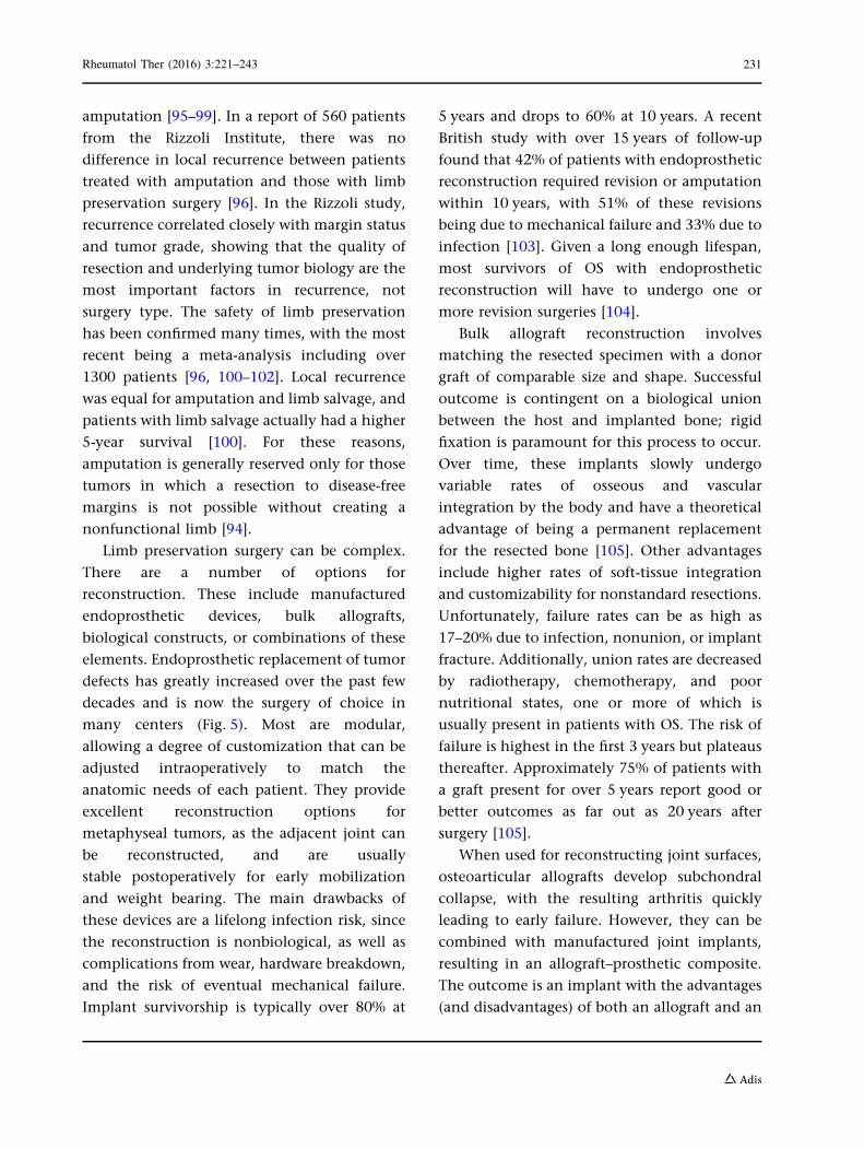
soft-tissue disease (Fig. 4). Bone scan is more
cost-effective and superior to PET for bony
disease, but PET allows for better detection in
soft tissue, and includes the chest and
abdomen. Both are effective scanning
techniques; the choice made usually varies by
institution. An additional advantage of PET is
that it may be able to identify tumors with
higher metabolic activity and, therefore,
higher-grade malignancies [72, 73]. Finally, if
not already available, an MRI of the entire bone
involved is important to rule out any skip
metastases, which must be addressed with
primary resection and predict a poor survival
[74].
The two staging systems currently employed
for staging OS are the Enneking system and the
AJCC (Tables 1, 2). Enneking was the first to
organize bone sarcoma into a comprehensive
staging system, and the AJCC later used these
principles to develop its own staging system
with nomenclature similar to that used for
other cancers. Both use histological grade and
the presence/absence of metastases and differ in
their evaluation of the size of the primary
tumor. The Enneking system makes a
distinction between whether the mass is
intracompartmental or has become
extracompartmental, while the AJCC system
uses tumor size (\8 or[8 cm) to determine a T1
from a T2 tumor. Despite these differences,
most tumors will be a similar stage in both
systems, as the major driver of prognosis is the
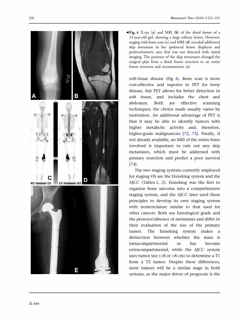
bFig. 4 X-ray (a) and MRI (b) of the distal femur of a13-year-old girl, showing a large solitary lesion. However,staging with bone scan (c) and MRI (d) revealed additionalskip metastasis in her ipsilateral femur diaphysis andperitrochanteric area that was not detected with initialimaging. The presence of the skip metastases changed thesurgical plan from a distal femur resection to an entirefemur resection and reconstruction (e)
228 Rheumatol Ther (2016) 3:221–243

presence of metastases, which both systems
define similarly [75].
Treatment
Prior to the advent of chemotherapy, OS was
almost a universally fatal disease. Patients with
metastasis at diagnosis would typically survive
only months, and those with localized disease
would soon develop metastatic spread, despite
radical and disabling surgical procedures. In the
1970s, Jaffe published the first significant
success of chemotherapy, showing that
methotrexate was a useful agent to manage
metastases in advanced disease [76]. As new
cytotoxic agents were discovered, the use of
chemotherapy blossomed, but the practice
remained controversial until a landmark study
in 1985 which showed an increase in 6-year
survival from 11 to 61% with the addition of
multi-agent chemotherapy [69]. A study
performed during the same time period at
Memorial Sloan Kettering found similar
increases in survival with chemotherapy that
was given before surgery (neoadjuvant),
showing that it was safe to delay surgery for
treatment [77]. The authors preferred
neoadjuvant chemotherapy because it allowed
more time to fabricate endoprosthetic devices,
decreased tumor size, and permitted an analysis
of the surgical specimen for its response to
chemotherapy [66].
Chemotherapy
Today, most OS patients receive neoadjuvant
chemotherapy, followed by surgical resection of
all detectable disease and a regimen of adjuvant
chemotherapy postoperatively [78]. The current
regimen of methotrexate, adriamycin, and
Table 1 Enneking MSTS staging system
Stage Grade Size Metastasis
IA Low T1—intracompartmental M0—none
IB Low T2—extracompartmental M0—none
IIA High T1—intracompartmental M0—none
IIB High T2—extracompartmental M0—none
III Any Any M1—regional or distant
Table 2 AJCC staging system for bone sarcoma
Stage Grade (G) Size (T) Lymph node (N) Metastasis (M)
IA G1—low T1\8 cm N0—none M0—none
IB G1—low T2[8 cm N0—none M0—none
IIA G2—high T1\8 cm N0—none M0—none
IIB G2—high T2[8 cm N0—none M0—none
III Any G Any T Skip metastasis Skip metastasis
IVA Any G Any T N0—none M1—lung metastasis
IVB Any G Any T N1—lymph node metastasis or N0 M1—non-lung metastasis
Rheumatol Ther (2016) 3:221–243 229

cisplatin (MAP) has become standard in North
America and Europe [66, 79]. This is typically
started after pathological diagnosis and staging
studies have been completed, and continues for
a period of 6–8 weeks depending on the
institution [80]. Some centers will also add
ifosfamide with or without etoposide, but this
increases the toxicity of therapy, and recent
randomized clinical trials have failed to show a
survival advantage [81, 82]. Unfortunately,
many agents are limited by their toxicity
profile, and some side effects, such as
adriamycin-induced cardiomyopathy, can be
permanent [83]. There is a dose effect on
treatment response, but recent research has
shown that high-dose chemotherapy does not
increase survival when compared to less toxic
moderate doses [84]. Much research has been
focused on changing chemotherapy to improve
survival in patients with a poor histologic
treatment response, but this has so far been
unsuccessful [85, 86]. Resistance to
chemotherapy is often multifactorial and an
area of much recent research [87].
Chemotherapy has little effect on lower-grade
types of OS, such as parosteal and periosteal
sarcoma, and these tumors have good rates of
survival without systemic therapy [9, 57, 88].
Radiation treatment is rarely included as an
adjuvant, but has been used in
unresectable cases [89–91].
Surgery
Regardless of the chemotherapy regimen,
surgical removal of all evidence of disease
remains critical to obtaining a remission and
improving patient survival. Patients will
undergo resection of the OS tumor by
amputation or limb salvage surgery techniques
as well as resection of any metastases if possible.
All tumors should be removed with a wide
margin to prevent residual disease; the
adequacy of this margin is critical in
preventing recurrences [92]. Recurrent disease
is closely linked to a poor prognosis and the risk
of metastatic disease. Following resection,
pathological specimens are examined to see
the effect that chemotherapy has had on the
tumor. A necrosis rate of C90% is considered a
‘‘good response,’’ and these patients will have a
better prognosis than those with less than 90%
necrosis. Besides its prognostic value,
histological response has been used to guide
therapy. Patients with a good response are
restarted on their preop chemo regimen, while
patients with a poor response may be switched
to a different combination of drugs. Despite
some favorable results in smaller studies, no
regimen has been shown to be superior in a
poor responder, but this is an area of continued
research [86]. Chemotherapy can usually be
resumed 2–3 weeks after surgery, once the
wound is healed [80, 93].
Historically, amputation was believed to be
necessary to control local disease, but that has
changed in recent decades, as advances in
chemotherapy, imaging, and reconstruction
techniques have made limb salvage surgeries
more feasible [94, 95]. Today, about 85% of
high-grade appendicular OS cases can be
successfully resected and reconstructed with
preservation of the affected limb and its
function [94, 95]. In limb preservation surgery,
the tumor is removed while maintaining a wide
cuff of tissue around the tumor when possible,
but allowing more narrow margins around vital
neurovascular structures. Careful dissection
preserves limb function but still removes all
disease safely.
Theoretically, limb preservation increases
the rate of local recurrence, but in experienced
hands it can be performed with little or no
increase in local recurrence compared to
230 Rheumatol Ther (2016) 3:221–243
amputation [95–99]. In a report of 560 patients
from the Rizzoli Institute, there was no
difference in local recurrence between patients
treated with amputation and those with limb
preservation surgery [96]. In the Rizzoli study,
recurrence correlated closely with margin status
and tumor grade, showing that the quality of
resection and underlying tumor biology are the
most important factors in recurrence, not
surgery type. The safety of limb preservation
has been confirmed many times, with the most
recent being a meta-analysis including over
1300 patients [96, 100–102]. Local recurrence
was equal for amputation and limb salvage, and
patients with limb salvage actually had a higher
5-year survival [100]. For these reasons,
amputation is generally reserved only for those
tumors in which a resection to disease-free
margins is not possible without creating a
nonfunctional limb [94].
Limb preservation surgery can be complex.
There are a number of options for
reconstruction. These include manufactured
endoprosthetic devices, bulk allografts,
biological constructs, or combinations of these
elements. Endoprosthetic replacement of tumor
defects has greatly increased over the past few
decades and is now the surgery of choice in
many centers (Fig. 5). Most are modular,
allowing a degree of customization that can be
adjusted intraoperatively to match the
anatomic needs of each patient. They provide
excellent reconstruction options for
metaphyseal tumors, as the adjacent joint can
be reconstructed, and are usually
stable postoperatively for early mobilization
and weight bearing. The main drawbacks of
these devices are a lifelong infection risk, since
the reconstruction is nonbiological, as well as
complications from wear, hardware breakdown,
and the risk of eventual mechanical failure.
Implant survivorship is typically over 80% at
5 years and drops to 60% at 10 years. A recent
British study with over 15 years of follow-up
found that 42% of patients with endoprosthetic
reconstruction required revision or amputation
within 10 years, with 51% of these revisions
being due to mechanical failure and 33% due to
infection [103]. Given a long enough lifespan,
most survivors of OS with endoprosthetic
reconstruction will have to undergo one or
more revision surgeries [104].
Bulk allograft reconstruction involves
matching the resected specimen with a donor
graft of comparable size and shape. Successful
outcome is contingent on a biological union
between the host and implanted bone; rigid
fixation is paramount for this process to occur.
Over time, these implants slowly undergo
variable rates of osseous and vascular
integration by the body and have a theoretical
advantage of being a permanent replacement
for the resected bone [105]. Other advantages
include higher rates of soft-tissue integration
and customizability for nonstandard resections.
Unfortunately, failure rates can be as high as
17–20% due to infection, nonunion, or implant
fracture. Additionally, union rates are decreased
by radiotherapy, chemotherapy, and poor
nutritional states, one or more of which is
usually present in patients with OS. The risk of
failure is highest in the first 3 years but plateaus
thereafter. Approximately 75% of patients with
a graft present for over 5 years report good or
better outcomes as far out as 20 years after
surgery [105].
When used for reconstructing joint surfaces,
osteoarticular allografts develop subchondral
collapse, with the resulting arthritis quickly
leading to early failure. However, they can be
combined with manufactured joint implants,
resulting in an allograft–prosthetic composite.
The outcome is an implant with the advantages
(and disadvantages) of both an allograft and an
Rheumatol Ther (2016) 3:221–243 231
232 Rheumatol Ther (2016) 3:221–243

endoprosthesis. The allograft side allows for a
stronger and more complete reconstruction of
the periarticular soft-tissue envelope and
incorporates into the patient’s host bone,
while the prosthetic side creates a stable and
predictable joint articulation. These implants
represent a high degree of complexity for the
surgeon, but have been shown by several
centers to provide a stable reconstruction with
similar failure rates to other methods [106, 107].
Most OS cases occur in younger patients,
many of which have active growth plates at the
time of diagnosis. Since most of these tumors
originate in the metaphysis and expand
circumferentially, the physis is often at risk.
When the physis must be sacrificed along with
the tumor mass, reconstruction must plan to
resolve or prevent significant limb length
discrepancy resulting from growth. In lower
extremities, this is usually agreed to be a limb
length discrepancy of greater than 2 cm at
maturity, but is less defined in the upper
extremity. A variety of options exist, and the
appropriate choice is often a subject of
controversy. Reconstruction strategies include
leaving the operative extremity longer than the
contralateral side to allow growth, slowing or
halting the growth of the nonoperative limb,
replacing the defect with an implant that can be
expanded as the child grows, or choosing a
functional amputation, such as Van Ness
rotationplasty (Fig. 6).
In our institution, we have found success with
a physeal sparing resection with allograft
reconstruction when possible (Fig. 7). This
technique has been recently reported in a series
of 35 Argentinean patients with 95% survival of
the limb at 5 and 10 years [108].When the physis
cannot be preserved, we prefermodulated growth
for defects of less than 3 cm and an expandable
prosthesis for larger defects. Several expandable
implants exist on the market. Some require
minimal surgery for mechanical expansion,
while others rely on an electromagnet for
noninvasive lengthening. The noninvasive
implants have the advantage of avoiding further
surgery, but the first generation has had an
intolerable failure rate [109, 110].
Second-generation noninvasive implants have
only been available for a short time, but may be
more stable. Most expandable prostheses will
eventually have to undergo revision, given the
age and activity level of their patients.
Rotationplasty remains a useful and lasting
option in patients with distal femur OS,
especially for patients who desire high-demand
activity, but few patients and their parents are
comfortable with this type of amputation [111].
OS in axial locations is rare, but often
presents unique challenges. Pelvis OS accounts
for only about 8% of all cases, but these tumors
tend to be larger, more biologically active, and
have metastatic disease more often compared to
extremity OS [112, 113]. Resection is still
necessary for cure, but is more complicated
given the three-dimensional anatomy of the
bony pelvis and surrounding vital structures.
Reconstruction is also more difficult and should
only be attempted in certain cases, as many
reconstructions only increase the complication
rate without improving postoperative function
[114, 115]. In addition to surgery, radiotherapy
can be used for added local control and may
improve overall survival [116]. OS of the spine is
rare as well but, when present, usually occurs in
the vertebral body and requires en-bloc
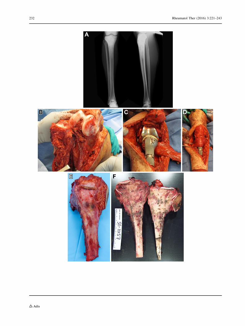
bFig. 5 A 22-year-old male with an osteosarcoma of theproximal tibia. XR images (a) show an aggressive lesion ofthe proximal tibia with abundant osseous matrix. Resectionof the tumor mass was performed with care taken to sparethe neurovascular bundle (b), and then the defect wasreconstructed with a proximal tibia endoprosthesis (c) anda medial gastrocnemius muscle flap for soft-tissue coverage(d). Resected tumor specimen (e, f) and gross pathologyexamination
Rheumatol Ther (2016) 3:221–243 233

resection for cure [117]. OS metastases, when
present, should be removed in close chronology
to the main tumor. Although lung metastasis
predicts a poorer prognosis, resection of
metastatic disease can still lead to remission in
some cases and has been shown to improve
survival [118, 119].
Following completion of treatment, patients
with OS must be observed closely for signs of
recurrence. The National Comprehensive
Cancer Network (NCCN) recommends imaging
of both the chest and the surgical site as often as
every 3 months during the first 2 years, and
continued surveillance at increasing intervals
thereafter [67]. Recurrence, either local or
distant, was found to occur in 20–30% of
patients who presented with localized disease
and 80% of patients who presented with
metastasis within the first 3 years [120].
Recurrent OS is treated with second-line
chemotherapy and surgery. If the recurrence
can be completely surgically removed, the
patient has a greatly increased chance of
survival [121].
Prognosis
Survival rates for patients with OS increased
dramatically with the introduction of
chemotherapy, but have since plateaued.
Today, 5-year survival for all patient groups
with high-grade OS is 60–66% but remains
highly dependent on stage at diagnosis.
Patients with localized disease can expect
5-year survival rates as high as 60–78%, but
survival drops to 20–30% for those with
metastatic disease [66, 78, 92, 101, 122]. Other
poor predictors of survival are increased tumor
size, increased serum alkaline phosphatase,
axial location, and secondary OS [102].
Fig. 6 An11-year-oldmale patientwith a large osteosarcomaof the distal femur that was reconstructed with anexpandable endoprosthesis. Preop (a), immediate postop
(b), and 2-year postoperative (c) X-ray images are shown.A lengthening of almost 5 cm was achieved with theexpandable construct by age 13
234 Rheumatol Ther (2016) 3:221–243

Advanced adult age is associated with increases
in both higher-grade tumors and axial tumors,
along with decreased response to and toleration
of chemotherapy. Subsequently, older adults
have a poorer prognosis [123]. Recurrent
disease, either local or distant, decreases
average 5-year survival to 20%, but can be as
high as 45% for relapses greater than 2 years out
that can be surgically resected. Lower-grade
osteosarcomas, including parosteal and
Fig. 7 17-year-old male with periosteal osteosarcoma.MRI images (a, b) show the cortically based tumor islocated in the metaphysis with some invasion of themarrow and a soft-tissue mass. A polyhedral,metaphysis-sparing bone cut was planned with navigation
software (c), and intraoperative guidance was used to assistwith the bone cuts according to the preoperative plan (d).A matching allograft was used to fill the defect and fixed tothe patient’s remaining bone (e). Two-year follow-upshows robust union at the junction sites (f)
Rheumatol Ther (2016) 3:221–243 235

periosteal sarcomas, have a much better
prognosis than the high-grade conventional
type. The 5-year survival of periosteal sarcoma
is around 83%, and parosteal sarcoma has a
reported 5-year survival of 91%. This is
primarily due to the low rate of metastasis.
New Therapies
A number of preclinical and clinical agents are
currently being investigated for OS. One area of
significant research involves using specific agents
to target known processes important in OS
pathogenesis. One attractive target is the mTOR
pathway, a downstream pathway of IGF-1 that
stimulates proliferation, survival, and
angiogenesis. Sirolimus, an mTOR inhibitor, was
found to inhibit metastasis and OS xenograft
growth in mice [124, 125]. Everolimus, an oral
mTOR inhibitor, also showed activity against
human and mice OS cells, an effect that was
enhanced by combination with zoledronic acid
[126]. A recent phase I trial has shown that oral
everolimus is safe in pediatric populations [127],
and a phase II study is currently ongoing in
refractory OS [128]. Disruption of angiogenesis is
another strategy, and several targeted agents have
been tested in OS. Pazopanib is an inhibitor of
VEGFR, PDGFR, and c-kit that has shown some
efficacy in metastatic OS [129]. The drug is well
tolerated in children and currently under
investigation in a phase II clinical trial among
OS patients with lung metastases. Sorafenib is an
oral anti-angiogenic agent with activity against
VEGFR-2 and PDFGR-B that has shown good
activity as a second- or third-line agent in
refractory OS [130]. Surprisingly, a recent clinical
study failed to show efficacy when sorafenib was
combined with everolimus in inoperable
high-grade progressive OS patients [131].
Immune modulation is another area of
increased OS research. As discussed earlier,
inhibition of the Fas pathway helps OS cells
avoid apoptosis and immune-mediated
destruction [43]. Targeting this pathway has
successfully been accomplished in preclinical
models with the use of interleukin 12 (IL-12).
IL-12 functions to activate Fas on cell surfaces,
leading to increased cell death and immune
clearance [42, 44]. Unfortunately, IL-12 is toxic
when administered systemically. Liposomal
muramyl tripeptide phosphatidyl
ethanolamine (liposomal MTP-PE) is a
promising agent that functions to induce
endogenous IL-12 and thus provides the effect
of IL-12 without the toxicity. In a recent
Children’s Oncology Group (COG) phase III
trial, liposomal MTP-PE improved overall
survival regardless of treatment regimen
[45, 132].
Another class of drugs currently garnering
attention in OS are bisphosphonates. Besides
their effects on osteoclast activity,
bisphosphonates also act to inhibit cell growth
and proliferation, can induce apoptosis, and
downregulate angiogenic growth factors [133].
In a preclinical study, zoledronate successfully
suppressed tumor growth and lung metastasis
in a mouse model and is now the subject of an
ongoing trial with combination chemotherapy
[126, 133]. In recent years, an international
collaboration, the EURAMOS group, has been
successful at overcoming the small numbers of
patients at each institution by designing
cooperative randomized trials across
institutions, and even nations. The first phase,
EURAMOS-1, tested the addition of pegylated
interferon to chemotherapy as maintenance
therapy in good responders, and, while the
treatment was unsuccessful, the trial showed
that international collaboration is possible in
this rare tumor [86, 134].
The field of pharmacogenetics seeks to
predict response to therapy and prognosis, and
236 Rheumatol Ther (2016) 3:221–243

is being used to personalize treatment across
healthcare. Recently, Caronia et al. used single
nucleotide polymorphisms (SNPs) to identify
four variations in two genes responsible for
chemotherapy resistance. Their work identified
polymorphisms in the ABCC3 gene, a member
of the multi-drug resistance protein family, and
ABCB1, which encodes for an ATP-mediated
efflux pump. Patients with these
polymorphisms had inferior estimated 5-year
survival [135]. Other authors have used similar
techniques to describe variations in different
genes leading to changes in prognosis or
resistance to chemotherapy [136, 137]. As this
field evolves, new information about a patient’s
genetic profile can be used to select the most
efficacious therapy, minimize side effects, and
better inform prognosis.
CONCLUSION
Advances in chemotherapy and surgery have
taken OS from an almost universally fatal
disease to one in which the majority of
patients will survive with a meaningful quality
of life. Despite this, a fair number of those
affected will still develop fatal metastatic disease
or serious complications of treatment,
emphasizing the need for further clinical
advancements. Accurate and efficient
diagnosis, preoperative chemotherapy, surgical
resection, postoperative chemotherapy, and
lifelong surveillance are all vital in managing
this complicated and potentially deadly disease.
ACKNOWLEDGMENTS
No funding or sponsorship was received for this
study or publication of this article.
All named authors meet the International
Committee of Medical Journal Editors
(ICMJE) criteria for authorship for this
manuscript, take responsibility for the
integrity of the work as a whole, and have
given final approval for the version to be
published.
All tables/figures are original and have been
produced by the authors for this particular
publication.
Disclosures. R. A. Durfee, M. Mohammed
and H. H. Luu have nothing to disclose.
Compliance with Ethics Guidelines. This
article is based on previously conducted
studies and does not involve any new studies
of human or animal subjects performed by any
of the authors.
Open Access. This article is distributed
under the terms of the Creative Commons
Attribution-NonCommercial 4.0 International
License (http://creativecommons.org/licenses/
by-nc/4.0/), which permits any noncommercial
use, distribution, and reproduction in any
medium, provided you give appropriate credit
to the original author(s) and the source, provide
a link to the Creative Commons license, and
indicate if changes were made.
REFERENCES
1. Campanacci M. Bone and soft tissue tumors:clinical features, imaging, pathology andtreatment. Berlin: Springer; 2013.
2. Sissons HA. The WHO classification of bone tumors.Recent Res Cancer Res. 1976;54:104–8.
3. Damron TA, Ward WG, Stewart A. Osteosarcoma,chondrosarcoma, and Ewing’s sarcoma: NationalCancer Data Base Report. Clin Orthop Relat Res.2007;459:40–7.
4. Dorfman HD, Czerniak B. Bone cancers. Cancer.1995;75(1 Suppl):203–10.
Rheumatol Ther (2016) 3:221–243 237
5. dos Santos Silva I, Swerdlow AJ. Sex differences inthe risks of hormone-dependent cancers. Am JEpidemiol. 1993;138(1):10–28.
6. Mirabello L, Troisi RJ, Savage SA. Osteosarcomaincidence and survival rates from 1973 to 2004: datafrom the Surveillance, Epidemiology, and EndResults Program. Cancer. 2009;115(7):1531–43.
7. Fraumeni JF Jr. Stature and malignant tumors ofbone in childhood and adolescence. Cancer.1967;20(6):967–73.
8. Miller RW. Contrasting epidemiology of childhoodosteosarcoma, Ewing’s tumor, andrhabdomyosarcoma. Natl Cancer Inst Monogr.1981;56:9–15.
9. Temple HT, Scully SP, O’Keefe RJ, Katapurum S,Mankin HJ. Clinical outcome of 38 patients withjuxtacortical osteosarcoma. Clin Orthop Relat Res.2000;373:208–17.
10. Mirabello L, Troisi RJ, Savage SA. Internationalosteosarcoma incidence patterns in children andadolescents, middle ages and elderly persons. Int JCancer. 2009;125(1):229–34.
11. Kruzelock RP, Murphy EC, Strong LC, Naylor SL,Hansen MF. Localization of a novel tumorsuppressor locus on human chromosome 3qimportant in osteosarcoma tumorigenesis. CancerRes. 1997;57(1):106–9.
12. Nellissery MJ, Padalecki SS, Brkanac Z, et al.Evidence for a novel osteosarcomatumor-suppressor gene in the chromosome 18region genetically linked with Paget disease ofbone. Am J Hum Genet. 1998;63(3):817–24.
13. Toguchida J, Ishizaki K, Sasaki MS, et al. Preferentialmutation of paternally derived RB gene as the initialevent in sporadic osteosarcoma. Nature.1989;338(6211):156–8.
14. Beckerman R, Prives C. Transcriptional regulationby p53. Cold Spring Harb Perspect Biol.2010;2(8):a000935.
15. Zhang J, Walsh MF, Wu G, et al. Germlinemutations in predisposition genes in pediatriccancer. N Engl J Med. 2015;373(24):2336–46.
16. Bougeard G, Renaux-Petel M, Flaman JM, et al.Revisiting Li–Fraumeni syndrome from TP53mutation carriers. J Clin Oncol.2015;33(21):2345–52.
17. Lonardo F, Ueda T, Huvos AG, Healey J, Ladanyi M.p53 and MDM2 alterations in osteosarcomas:correlation with clinicopathologic features andproliferative rate. Cancer. 1997;79(8):1541–7.
18. Goodrich DW, Wang NP, Qian YW, Lee EY, LeeWH. The retinoblastoma gene product regulatesprogression through the G1 phase of the cell cycle.Cell. 1991;67(2):293–302.
19. DerKinderen DJ, Koten JW, Nagelkerke NJ, Tan KE,Beemer FA, Den Otter W. Non-ocular cancer inpatients with hereditary retinoblastoma and theirrelatives. Int J Cancer. 1988;41(4):499–504.
20. Kleinerman RA, Schonfeld SJ, Tucker MA. Sarcomasin hereditary retinoblastoma. Clin Sarcoma Res.2012;2(1):15.
21. Wong FL, Boice JD Jr, Abramson DH, et al. Cancerincidence after retinoblastoma. Radiation dose andsarcoma risk. JAMA. 1997;278(15):1262–7.
22. Hawkins MM, Draper GJ, Kingston JE. Incidence ofsecond primary tumours among childhood cancersurvivors. Br J Cancer. 1987;56(3):339–47.
23. Feugeas O, Guriec N, Babin-Boilletot A, et al. Loss ofheterozygosity of the RB gene is a poor prognosticfactor in patients with osteosarcoma. J Clin Oncol.1996;14(2):467–72.
24. Wadayama B, Toguchida J, Shimizu T, et al.Mutation spectrum of the retinoblastoma gene inosteosarcomas. Cancer Res. 1994;54(11):3042–8.
25. Nevins JR. The Rb/E2F pathway and cancer. HumMol Genet. 2001;10(7):699–703.
26. Nielsen GP, Burns KL, Rosenberg AE, Louis DN.CDKN2A gene deletions and loss of p16 expressionoccur in osteosarcomas that lack RB alterations. AmJ Pathol. 1998;153(1):159–63.
27. Chakraverty RK, Hickson ID. Defending genomeintegrity during DNA replication: a proposed rolefor RecQ family helicases. Bioessays.1999;21(4):286–94.
28. Karow JK, Wu L, Hickson ID. RecQ family helicases:roles in cancer and aging. Curr Opin Genet Dev.2000;10(1):32–8.
29. Mohaghegh P, Hickson ID. DNA helicasedeficiencies associated with cancer predispositionand premature ageing disorders. Hum Mol Genet.2001;10(7):741–6.
30. Kloen P, Gebhardt MC, Perez-Atayde A, et al.Expression of transforming growth factor-beta(TGF-beta) isoforms in osteosarcomas: TGF-beta3 isrelated to disease progression. Cancer.1997;80(12):2230–9.
31. Assoian RK, Komoriya A, Meyers CA, Miller DM,Sporn MB. Transforming growth factor-beta inhuman platelets. Identification of a major storage
238 Rheumatol Ther (2016) 3:221–243

site, purification, and characterization. J Biol Chem.1983;258(11):7155–60.
32. Kloen P, Jennings CL, Gebhardt MC, Springfield DS,Mankin HJ. Expression of transforming growthfactor-beta (TGF-beta) receptors, TGF-beta 1 andTGF-beta 2 production and autocrine growthcontrol in osteosarcoma cells. Int J Cancer.1994;58(3):440–5.
33. Lamplot JD, Denduluri S, Qin J, et al. The currentand future therapies for human osteosarcoma. CurrCancer Ther Rev. 2013;9(1):55–77.
34. Luther GA, Lamplot J, Chen X, et al. IGFBP5domains exert distinct inhibitory effects on thetumorigenicity and metastasis of humanosteosarcoma. Cancer Lett. 2013;336(1):222–30.
35. Su Y, Wagner ER, Luo Q, et al. Insulin-like growthfactor binding protein 5 suppresses tumor growthand metastasis of human osteosarcoma. Oncogene.2011;30(37):3907–17.
36. Jentzsch T, Robl B, HusmannM, Bode-Lesniewska B,Fuchs B. Worse prognosis of osteosarcoma patientsexpressing IGF-1 on a tissue microarray. AnticancerRes. 2014;34(8):3881–9.
37. MacEwen EG, Pastor J, Kutzke J, et al. IGF-1 receptorcontributes to the malignant phenotype in humanand canine osteosarcoma. J Cell Biochem.2004;92(1):77–91.
38. Burrow S, Andrulis IL, Pollak M, Bell RS. Expressionof insulin-like growth factor receptor, IGF-1, andIGF-2 in primary and metastatic osteosarcoma.J Surg Oncol. 1998;69(1):21–7.
39. Chen G, Fang T, Huang Z, et al. MicroRNA-133ainhibits osteosarcoma cells proliferation andinvasion via targeting IGF-1R. Cell PhysiolBiochem. 2016;38(2):598–608.
40. Kappel CC, Velez-Yanguas MC, Hirschfeld S,Helman LJ. Human osteosarcoma cell lines aredependent on insulin-like growth factor I forin vitro growth. Cancer Res. 1994;54(10):2803–7.
41. Heymann D, Redini F. Targeted therapies for bonesarcomas. Bonekey Rep. 2013;2:378.
42. Lafleur EA, Koshkina NV, Stewart J, et al. IncreasedFas expression reduces the metastatic potential ofhuman osteosarcoma cells. Clin Cancer Res.2004;10(23):8114–9.
43. Worth LL, Lafleur EA, Jia SF, Kleinerman ES. Fasexpression inversely correlates with metastaticpotential in osteosarcoma cells. Oncol Rep.2002;9(4):823–7.
44. Lafleur EA, Jia SF, Worth LL, Zhou Z, Owen-SchaubLB, Kleinerman ES. Interleukin (IL)-12 and IL-12gene transfer up-regulate Fas expression in humanosteosarcoma and breast cancer cells. Cancer Res.2001;61(10):4066–71.
45. PosthumaDeBoer J, Witlox MA, Kaspers GJ, vanRoyen BJ. Molecular alterations as target for therapyin metastatic osteosarcoma: a review of literature.Clin Exp Metastasis. 2011;28(5):493–503.
46. Hingorani P, Zhang W, Gorlick R, Kolb EA.Inhibition of Src phosphorylation alters metastaticpotential of osteosarcoma in vitro but not in vivo.Clin Cancer Res. 2009;15(10):3416–22.
47. Kim LC, Song L, Haura EB. Src kinases as therapeutictargets for cancer. Nat Rev Clin Oncol.2009;6(10):587–95.
48. Savage SA, Mirabello L, Wang Z, et al. Genome-wideassociation study identifies two susceptibility locifor osteosarcoma. Nat Genet. 2013;45(7):799–803.
49. Mirabello L, Koster R, Moriarity BS, et al. Agenome-wide scan identifies variants in NFIBassociated with metastasis in patients withosteosarcoma. Cancer Discov. 2015;5(9):920–31.
50. Desai P, Perino G, Present D, Steiner GC. Sarcoma inassociation with bone infarcts. Report of five cases.Arch Pathol Lab Med. 1996;120(5):482–9.
51. Hansen MF, Seton M, Merchant A. Osteosarcoma inPaget’s disease of bone. J Bone Miner Res.2006;21(Suppl 2):P58–63.
52. Shaheen M, Deheshi BM, Riad S, et al. Prognosis ofradiation-induced bone sarcoma is similar toprimary osteosarcoma. Clin Orthop Relat Res.2006;450:76–81.
53. Spina V, Montanari N, Romagnoli R. Malignanttumors of the osteogenic matrix. Eur J Radiol.1998;27(Suppl 1):S98–109.
54. Fox MG, Trotta BM. Osteosarcoma: review of thevarious types with emphasis on recentadvancements in imaging. Semin MusculoskeletRadiol. 2013;17(2):123–36.
55. Klein MJ, Siegal GP. Osteosarcoma: anatomic andhistologic variants. Am J Clin Pathol.2006;125(4):555–81.
56. Clayer M. Many faces of osteosarcoma on plainradiographs. ANZ J Surg. 2015;85(1–2):22–6.
57. Grimer RJ, Bielack S, Flege S, et al. Periostealosteosarcoma—a European review of outcome. EurJ Cancer. 2005;41(18):2806–11.
Rheumatol Ther (2016) 3:221–243 239

58. Scully SP, Ghert MA, Zurakowski D, Thompson RC,Gebhardt MC. Pathologic fracture in osteosarcoma:prognostic importance and treatment implications.J Bone Jt Surg Am. 2002;84-A(1):49–57.
59. Dadia S, Grimer R. Characteristics, diagnosis andtreatment of bone and soft tissue sarcomas. Br JHosp Med (Lond). 2007;68(11):589–93.
60. Chen J, Sun MX, Hua YQ, Cai ZD. Prognosticsignificance of serum lactate dehydrogenase level inosteosarcoma: a meta-analysis. J Cancer Res ClinOncol. 2014;140(7):1205–10.
61. Marais LC, Bertie J, Rodseth R, Sartorius B, FerreiraN. Pre-treatment serum lactate dehydrogenase andalkaline phosphatase as predictors of metastases inextremity osteosarcoma. J Bone Oncol. 2015;4(3):80–4.
62. Ren HY, Sun LL, Li HY, Ye ZM. Prognosticsignificance of serum alkaline phosphatase level inosteosarcoma: a meta-analysis of published data.Biomed Res Int. 2015;2015:160835.
63. Vasquez L, Tarrillo F, Oscanoa M, et al. Analysis ofprognostic factors in high-grade osteosarcoma ofthe extremities in children: a 15-yearsingle-institution experience. Front Oncol.2016;6:22.
64. Mankin HJ, Mankin CJ, Simon MA. The hazards ofthe biopsy, revisited. Members of theMusculoskeletal Tumor Society. J Bone Jt Surg Am.1996;78(5):656–63.
65. Binitie O, Tejiram S, Conway S, Cheong D, TempleHT, Letson GD. Adult soft tissue sarcoma localrecurrence after adjuvant treatment withoutresection of core needle biopsy tract. Clin OrthopRelat Res. 2013;471(3):891–8.
66. Isakoff MS, Bielack SS, Meltzer P, Gorlick R.Osteosarcoma: current treatment and acollaborative pathway to success. J Clin Oncol.2015;33(27):3029–35.
67. Biermann JS, Adkins DR, Agulnik M, Benjamin RS,Brigman B, et al. NCCN guidelines version 1.2013.Bone cancer. Fort Washington: NCCN; 2012. p. 58.
68. Kager L, Zoubek A, Potschger U, et al. Primarymetastatic osteosarcoma: presentation andoutcome of patients treated on neoadjuvantCooperative Osteosarcoma Study Group protocols.J Clin Oncol. 2003;21(10):2011–8.
69. Link MP, Goorin AM, Miser AW, et al. The effect ofadjuvant chemotherapy on relapse-free survival inpatients with osteosarcoma of the extremity. N EnglJ Med. 1986;314(25):1600–6.
70. Yonemoto T, Tatezaki S, Ishii T, Satoh T, Kimura H,Iwai N. Prognosis of osteosarcoma with pulmonarymetastases at initial presentation is not dismal. ClinOrthop Relat Res. 1998;349:194–9.
71. Kaste SC, Pratt CB, Cain AM, Jones-Wallace DJ, RaoBN. Metastases detected at the time of diagnosis ofprimary pediatric extremity osteosarcoma atdiagnosis: imaging features. Cancer. 1999;86(8):1602–8.
72. Eary JF, Mankoff DA. Tumor metabolic rates insarcoma using FDG PET. J Nucl Med.1998;39(2):250–4.
73. Eary JF, O’Sullivan F, Powitan Y, et al. Sarcomatumor FDG uptake measured by PET and patientoutcome: a retrospective analysis. Eur J Nucl MedMol Imaging. 2002;29(9):1149–54.
74. Kager L, Zoubek A, Kastner U, et al. Skip metastasesin osteosarcoma: experience of the CooperativeOsteosarcoma Study Group. J Clin Oncol.2006;24(10):1535–41.
75. Moore DD, Luu HH. Osteosarcoma. Cancer TreatRes. 2014;162:65–92.
76. Jaffe N. Recent advances in the chemotherapy ofmetastatic osteogenic sarcoma. Cancer.1972;30(6):1627–31.
77. Rosen G, Caparros B, Huvos AG, et al. Preoperativechemotherapy for osteogenic sarcoma: selection ofpostoperative adjuvant chemotherapy based on theresponse of the primary tumor to preoperativechemotherapy. Cancer. 1982;49(6):1221–30.
78. Friebele JC, Peck J, Pan X, Abdel-Rasoul M,Mayerson JL. Osteosarcoma: a meta-analysis andreview of the literature. Am J Orthop (Belle MeadNJ). 2015;44(12):547–53.
79. Ferrari S, Serra M. An update on chemotherapy forosteosarcoma. Expert Opin Pharmacother.2015;16(18):2727–36.
80. Bielack S, Jurgens H, Jundt G, et al. Osteosarcoma:the COSS experience. Cancer Treat Res.2009;152:289–308.
81. Carrle D, Bielack SS. Current strategies ofchemotherapy in osteosarcoma. Int Orthop.2006;30(6):445–51.
82. Ferrari S, Ruggieri P, Cefalo G, et al. Neoadjuvantchemotherapy with methotrexate, cisplatin, anddoxorubicin with or without ifosfamide innonmetastatic osteosarcoma of the extremity: anItalian Sarcoma Group trial ISG/OS-1. J Clin Oncol.2012;30(17):2112–8.
240 Rheumatol Ther (2016) 3:221–243
83. Schwartz CL, Wexler LH, Krailo MD, et al.Intensified chemotherapy with dexrazoxanecardioprotection in newly diagnosednonmetastatic osteosarcoma: a report from theChildren’s Oncology Group. Pediatr Blood Cancer.2016;63(1):54–61.
84. Wang WG, Wan C, Liao GJ. The efficacy ofhigh-dose versus moderate-dose chemotherapy intreating osteosarcoma: a systematic review andmeta-analysis. Int J Clin Exp Med.2015;8(9):15967–74.
85. Anninga JK, Gelderblom H, Fiocco M, et al.Chemotherapeutic adjuvant treatment forosteosarcoma: where do we stand? Eur J Cancer.2011;47(16):2431–45.
86. Bielack SS, Smeland S, Whelan JS, et al.Methotrexate, doxorubicin, and cisplatin (MAP)plus maintenance pegylated interferon Alfa-2bversus MAP alone in patients withresectable high-grade osteosarcoma and goodhistologic response to preoperative MAP: firstresults of the EURAMOS-1 good responserandomized controlled trial. J Clin Oncol.2015;33(20):2279–87.
87. He H, Ni J, Huang J. Molecular mechanisms ofchemoresistance in osteosarcoma (review). OncolLett. 2014;7(5):1352–62.
88. Hang JF, Chen PC. Parosteal osteosarcoma. ArchPathol Lab Med. 2014;138(5):694–9.
89. Mavrogenis AF, Rossi G, Palmerini E, et al. Palliativetreatments for advanced osteosarcoma. J BUON.2012;17(3):436–45.
90. Oertel S, Blattmann C, Rieken S, et al. Radiotherapyin the treatment of primary osteosarcoma—a singlecenter experience. Tumori. 2010;96(4):582–8.
91. DeLaney TF, Park L, Goldberg SI, et al. Radiotherapyfor local control of osteosarcoma. Int J Radiat OncolBiol Phys. 2005;61(2):492–8.
92. Bacci G, Longhi A, Versari M, Mercuri M, BriccoliA, Picci P. Prognostic factors for osteosarcoma ofthe extremity treated with neoadjuvantchemotherapy: 15-year experience in 789 patientstreated at a single institution. Cancer.2006;106(5):1154–61.
93. Bacci G, Longhi A, Bertoni F, et al. Bone metastasesin osteosarcoma patients treated with neoadjuvantor adjuvant chemotherapy: the Rizzoli experiencein 52 patients. Acta Orthop. 2006;77(6):938–43.
94. Grimer RJ. Surgical options for children withosteosarcoma. Lancet Oncol. 2005;6(2):85–92.
95. Marulanda GA, Henderson ER, Johnson DA, LetsonGD, Cheong D. Orthopedic surgery options for thetreatment of primary osteosarcoma. CancerControl. 2008;15(1):13–20.
96. Bacci G, Ferrari S, Lari S, et al. Osteosarcoma of thelimb. Amputation or limb salvage in patientstreated by neoadjuvant chemotherapy. J Bone JtSurg Brit. 2002;84(1):88–92.
97. Rougraff BT, Simon MA, Kneisl JS, Greenberg DB,Mankin HJ. Limb salvage compared withamputation for osteosarcoma of the distal end ofthe femur. A long-term oncological, functional, andquality-of-life study. J Bone Jt Surg Am.1994;76(5):649–56.
98. Simon MA. Limb salvage for osteosarcoma in the1980s. Clin Orthop Relat Res. 1991;270:264–70.
99. Simon MA, Aschliman MA, Thomas N, Mankin HJ.Limb-salvage treatment versus amputation forosteosarcoma of the distal end of the femur.J Bone Jt Surg Am Vol. 1986;68(9):1331–7.
100. Li X, Zhang Y, Wan S, et al. A comparative studybetween limb-salvage and amputation for treatingosteosarcoma. J Bone Oncol. 2016;5(1):15–21.
101. Haddox CL, Han G, Anijar L, et al. Osteosarcoma inpediatric patients and young adults: a singleinstitution retrospective review of presentation,therapy, and outcome. Sarcoma.2014;2014:402509.
102. Bielack SS, Kempf-Bielack B, Delling G, et al.Prognostic factors in high-grade osteosarcoma ofthe extremities or trunk: an analysis of 1702patients treated on neoadjuvant cooperativeosteosarcoma study group protocols. J Clin Oncol.2002;20(3):776–90.
103. Jeys LM, Kulkarni A, Grimer RJ, Carter SR, TillmanRM, Abudu A. Endoprosthetic reconstruction forthe treatment of musculoskeletal tumors of theappendicular skeleton and pelvis. J Bone Jt Surg AmVol. 2008;90(6):1265–71.
104. Grimer RJ, Aydin BK, Wafa H, et al. Very long-termoutcomes after endoprosthetic replacement formalignant tumours of bone. Bone Jt J.2016;98-B(6):857–64.
105. Mankin HJ, Gebhardt MC, Jennings LC, SpringfieldDS, Tomford WW. Long-term results of allograftreplacement in the management of bone tumors.Clin Orthop Relat Res. 1996;324:86–97.
106. Campanacci L, Manfrini M, Colangeli M, Ali N,Mercuri M. Long-term results in children withmassive bone osteoarticular allografts of the knee
Rheumatol Ther (2016) 3:221–243 241

for high-grade osteosarcoma. J Pediatr Orthop.2010;30(8):919–27.
107. Campanacci L, Ali N, Casanova JM, Kreshak J,Manfrini M. Resurfaced allograft-prostheticcomposite for proximal tibial reconstruction inchildren: intermediate-term results of an originaltechnique. J Bone Jt Surg Am Vol.2015;97(3):241–50.
108. Aponte-Tinao L, Ayerza MA, Muscolo DL, FarfalliGL. Survival, recurrence, and function afterepiphyseal preservation and allograftreconstruction in osteosarcoma of the knee. ClinOrthop Relat Res. 2015;473(5):1789–96.
109. Benevenia J, Patterson F, Beebe K, et al. Results of 20consecutive patients treated with the Repiphysisexpandable prosthesis for primary malignant bone.SpringerPlus. 2015;4:793.
110. Staals EL, Colangeli M, Ali N, Casanova JM, DonatiDM, Manfrini M. Are complications associated withthe repiphysis (R) expandable distal femoralprosthesis acceptable for its continued use? ClinOrthop Relat Res. 2015;473(9):3003–13.
111. Gradl G, Postl LK, Lenze U, et al. Long-termfunctional outcome and quality of life followingrotationplasty for treatment of malignant tumors.BMC Musculoskelet Disord. 2015;16:262.
112. Parry MC, Laitinen M, Albergo J, et al.Osteosarcoma of the pelvis. Bone Jt J.2016;98-B(4):555–63.
113. Isakoff MS, Barkauskas DA, Ebb D, Morris C, LetsonGD. Poor survival for osteosarcoma of the pelvis: areport from the Children’s Oncology Group. ClinOrthop Relat Res. 2012;470(7):2007–13.
114. Farfalli GL, Albergo JI, Ritacco LE, Ayerza MA,Muscolo DL, Aponte-Tinao LA. Oncologic andclinical outcomes in pelvic primary bone sarcomastreated with limb salvage surgery. MusculoskeletSurg. 2015;99(3):237–42.
115. Mayerson JL, Wooldridge AN, Scharschmidt TJ.Pelvic resection: current concepts. J Am AcadOrthop Surg. 2014;22(4):214–22.
116. Ozaki T, Flege S, Kevric M, et al. Osteosarcoma ofthe pelvis: experience of the CooperativeOsteosarcoma Study Group. J Clin Oncol.2003;21(2):334–41.
117. Dekutoski MB, Clarke MJ, Rose P, et al.Osteosarcoma of the spine: prognostic variablesfor local recurrence and overall survival, amulticenter ambispective study. J NeurosurgSpine. 2016;25(1):59–68.
118. Gok Durnali A, Paksoy Turkoz F, Ardic Yukruk F,et al. Outcomes of adolescent and adult patientswith lung metastatic osteosarcoma and comparisonof synchronous and metachronous lung metastaticgroups. PLoS One. 2016;11(5):e0152621.
119. Bacci G, Rocca M, Salone M, et al. High gradeosteosarcoma of the extremities with lungmetastases at presentation: treatment withneoadjuvant chemotherapy and simultaneousresection of primary and metastatic lesions. J SurgOncol. 2008;98(6):415–20.
120. Longhi A, Errani C, De Paolis M, Mercuri M, BacciG. Primary bone osteosarcoma in the pediatric age:state of the art. Cancer Treat Rev.2006;32(6):423–36.
121. Kempf-Bielack B, Bielack SS, Jurgens H, et al.Osteosarcoma relapse after combined modalitytherapy: an analysis of unselected patients in theCooperative Osteosarcoma Study Group (COSS).J Clin Oncol. 2005;23(3):559–68.
122. Smith MA, Seibel NL, Altekruse SF, et al. Outcomesfor children and adolescents with cancer:challenges for the twenty-first century. J ClinOncol. 2010;28(15):2625–34.
123. Grimer RJ, Cannon SR, Taminiau AM, et al.Osteosarcoma over the age of forty. Eur J Cancer.2003;39(2):157–63.
124. Houghton PJ, Morton CL, Kolb EA, et al. Initialtesting (stage 1) of the mTOR inhibitor rapamycinby the pediatric preclinical testing program. PediatrBlood Cancer. 2008;50(4):799–805.
125. Wan X, Mendoza A, Khanna C, Helman LJ.Rapamycin inhibits ezrin-mediated metastaticbehavior in a murine model of osteosarcoma.Cancer Res. 2005;65(6):2406–11.
126. Moriceau G, Ory B, Mitrofan L, et al. Zoledronicacid potentiates mTOR inhibition and abolishes theresistance of osteosarcoma cells to RAD001(everolimus): pivotal role of the prenylationprocess. Cancer Res. 2010;70(24):10329–39.
127. Fouladi M, Laningham F, Wu J, et al. Phase I studyof everolimus in pediatric patients with refractorysolid tumors. J Clin Oncol. 2007;25(30):4806–12.
128. Hattinger CM, Fanelli M, Tavanti E, et al. Advancesin emerging drugs for osteosarcoma. Expert OpinEmerg Drugs. 2015;20(3):495–514.
129. Safwat A, Boysen A, Lucke A, Rossen P. Pazopanib inmetastatic osteosarcoma: significant clinicalresponse in three consecutive patients. ActaOncol. 2014;53(10):1451–4.
242 Rheumatol Ther (2016) 3:221–243
130. Grignani G, Palmerini E, Dileo P, et al. A phase IItrial of sorafenib in relapsed andunresectable high-grade osteosarcoma after failureof standard multimodal therapy: an Italian SarcomaGroup study. Ann Oncol. 2012;23(2):508–16.
131. Grignani G, Palmerini E, Ferraresi V, et al. Sorafeniband everolimus for patients withunresectable high-grade osteosarcoma progressingafter standard treatment: a non-randomised phase 2clinical trial. Lancet Oncol. 2015;16(1):98–107.
132. Chou AJ, Kleinerman ES, Krailo MD, et al. Additionof muramyl tripeptide to chemotherapy for patientswith newly diagnosed metastatic osteosarcoma: areport from the Children’s Oncology Group.Cancer. 2009;115(22):5339–48.
133. Kansara M, Teng MW, Smyth MJ, Thomas DM.Translational biology of osteosarcoma. Nat RevCancer. 2014;14(11):722–35.
134. Whelan JS, Bielack SS, Marina N, et al. EURAMOS-1,an international randomised study forosteosarcoma: results from pre-randomisationtreatment. Ann Oncol. 2015;26(2):407–14.
135. Caronia D, Patino-Garcia A, Perez-Martinez A, et al.Effect of ABCB1 and ABCC3 polymorphisms onosteosarcoma survival after chemotherapy: apharmacogenetic study. PLoS One.2011;6(10):e26091.
136. Hagleitner MM, Coenen MJ, Gelderblom H, et al. Afirst step toward personalized medicine inosteosarcoma: pharmacogenetics as predictivemarker of outcome after chemotherapy-basedtreatment. Clin Cancer Res. 2015;21(15):3436–41.
137. Hattinger CM, Serra M. Role of pharmacogenetics ofdrug-metabolizing enzymes in treatingosteosarcoma. Expert Opin Drug Metab Toxicol.2015;11(9):1449–63.
Rheumatol Ther (2016) 3:221–243 243