
REVIEW OF LITERATURE 1.1 Historical overview of...
719
DEPARTAMENTO DE DERECHO ADMINISTRATIVO Y PROCESAL LA CORRELACIÓN DE LA SENTENCIA CON LA ACUSACIÓN Y LA DEFENSA. ESTUDIO COMPARADO DEL DERECHO ESPAÑOL CON EL CHILENO CARLOS DEL RÍO FERRETTI UNIVERSITAT DE VALENCIA Servei de Publicacions 2007
Transcript of REVIEW OF LITERATURE 1.1 Historical overview of...
3
REVIEW OF LITERATURE
1.1 Historical overview of typhoid
Typhoid fever was first described as long back as 1659
by Thomas Willis. However, it was only in 1829 that
Dr.P.Ch.A.Louis gave a clear definition of the disease,
separating it from other fevers and associated the
clinical expression of the infection with the essential
pathologic lesions in the intestines, mesentric lymph
nodes and spleen. He also described the rose spots,
intestinal perforation and hemorrhage. William Budd in
1873 provided evidence that bowel aischarges were the
main source of infection; that the disease was water
borne; that milk, food, contaminated linen and other
fomites were sources of dissemination and insisted
without direct proof that a specific germ caused
typhoid. It was in 1884 that Gaffkey, a German, first
cultivated and isolat~d Salmonella typhi as a pure
culture from the spleens of infected patients. This was
followed shortly by the cultivation of the organism
from stool, urine, rose spots and gall bladder of the
patients by other German investigators.
Pfeiffer and Kolle in 1896 made the first vaccine for
human use against typhoid with heat-killed organisms and
demonstrated the development of antibodies that
passively protected guinea pigs against experimental
4
infection. It was in that year that Gruber, Durham and
Widal each independently reported that convalescent
phase serum when mixed with S.typhi caused agglutination
of the organisms. Thus came in existence the classic
serologic test for infection by S.typhi, the Widal's
agglutintion test. In 1903 Robert Koch outlined three
logical methods for typhoid control: disinfect the
excreta at its source, improve sewage handling and
isolate convalescent patients until they become bacillus
free. However, case fatality continued to range upto 30%
until Theodore Woodward and his colleagues reported in
1948 that chloromycetin could sterilize blood cultures
from typhoid patients. There started the era of
antibiotics for treatment of ~yphoid fever.
1.2 Epidemiology
The first and the only attempt to quantitate the extent
of typhoid fever throughout the contemporary world was
reported in 1984 by Dr. Dhimon Barua in an international
workshop on typhoid fever held at the Pan American
Health Organisation in Washington,D.C. Dr.Barua gave an
annual estimate of approximately 12.5 million cases in
the world (excluding China). This estimate included 6.98
million cases per year in South and East Asia
(population 1369 million), 749 thousand in West Asia
(population 98 million), 4.36 million in Africa

5
(popu1ation 427 million), 15 thousand in Egypt
(population 43 million), 406 thousand in Latin America
and South Pacific Islands (population 369 ~illion) and
23,000 in the developed world (population 1131 million).
The countries that report increased typhoid severity
share several characteristics 1) rapidly increasing
population, 2) rapidly increasing urbanization,
3) inadequate facilities for processing human wastes, ·4)
decreasing water supply per capita, 5) intimate contact
between humans, food and heavily contaminated water
supply and 6) overburdened health care delivery systems
(Gangarosa, 1982). All these factors together have led
to increased numbers of people coming into contact with
larger inocula of S.typhi. This has probably led to an
absolute increase in number of cases of typhoid fever
with an increase in the incidence and an increase in the
prevalence and per9entage of severe typhoid cases. The
situation has been furtDer complicatd by the emergence
of strains of S.typhi resistant to commonly used and
inexpensive antibiotics (Taylor et al., 1983). In India,
S. typhi is the major cause of enteric fevers. Of the
373,522 cases reported in 1979 by W.H.O., there were no
fewer than 266,204 from India (Huckstep, 1985).
1.3 Bacteriology
Salmonella typhi is a gram negative, non-sporing
6
bacterium, about 2-4 J.l by 0. 5 )1, actively motile with
numerous long peritrichous flageala. Almost all the
clinical isolates are encapsulated on primary isolation.
It is an aerobe and facultative anaerobe and grows well
on ordinary culture media. MacConkey's agar and
Deoxycholate citrate agar (DCA) are the selective media.
1.4 Pathogenecity
S.typhi is primarily an intestinal parasite. It exhibits
strong host specific! ty for man. A disease similar to
typhoid fever in man Gan be produced in Chimpanzees if
massive doses of living bacilli are administered orally.
However, the incubation is shorter and ulceration of
lymphoid tissue of the small intestine does not occur
(Metchnikoff & Ber~edka, 1911; Edsall et al., 1960;
Gaines et al., 1968b). Oral administration of typhoid
bacilli to small labortary animals (rabbit, guinea pig,
rat or mouse) does not give rise to any infection of
this type or usually to any harmful effect. How~~er,
massive doses given intraperitoneally or intravenously
would produce fatal infection.
1.5 Antigenic.structure
The most extensively studied antigens of S.typhi which
have been suggested to play a role in the pathogenesis
or in the development of immune response a~e 1) the
7
somatic or the a-antigen 2) the flagellar or the H
antigen and 3) the capsular or the Vi antigen.
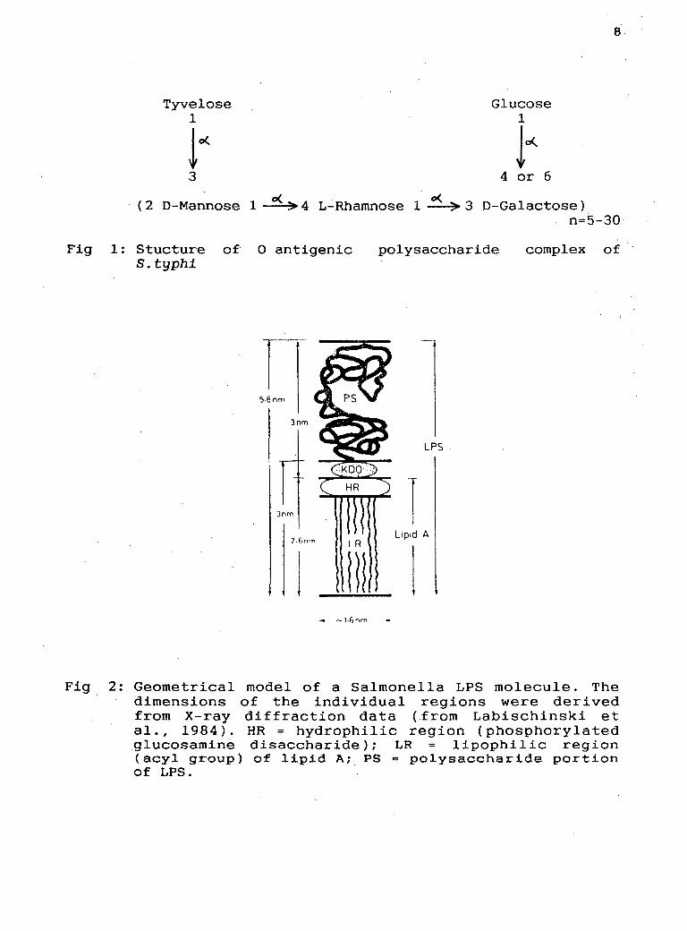
1.5.1 Somatic antigen: The somatic or the a-antigen
forms the side chain component of the lipopolysaccharide
present in bacterial cell envelope. It is composed of a
polymer of oligosaccharide molecules in repeating units.
Each serogroup within a genus of gram negative bacteria
is characterized by a unique a specific chain structure.
This is exploited in the Kauffman-White scheme for
serogrouping of Salmonellae in which specific antisera
are used to classify different Salmonella species
(Kauffman, 1975). According to this scheme,S.typhi falls
in the serogroup D and contains a antigens 9 and 12. The
structure of a-antigenic polysaccharide complex of
S.typhi LPS is shown in Figure 1. a9 consists of
Tyvelose ( dideoxyhexose) coupled to D mannose and 012
is constituted by a sequence in the chain 2 D-mannose
1~ 4 L-rhamnose 1~ 3 D-galactose. The a-antigen is
linked through 2-keto-3-deoxy-D-:-mannooctonate ( KDa) of
the core oligosaccharide to lipid A as shown
diagramatically in Fig 2.
1.5.2 Flagellar antigen: The flagellar antigen consists
of a single protein o~ around 60,000 daltons molecular
weight. It has both common antigenic determinants shared
by different Salmonella species and serotype specific
Tyvelose 1
!o( 3
Glucose 1
lo( 4 or 6
8
( 2 D-Mannose 1 ~ 4 L-Rhamnose 1 ~ 3 D-Ga1actose) n=5-30
Fig 1: Stucture of 0 antigenic S.typhi
n s.6nm
3nm
3nm
7-6nrn l R
Ill
polysaccharide complex of
LPS
Lipid A
Fig 2: Geometrical model of a Salmonella LPS molecule. The dimensions of the individual regions were derived from X-ray diffraction data (from Labischinski et al., 1984). HR = hydrophilic region (phosphorylated glucosamine disaccharide); LR lipophilic region (ecyl group) of lipid A; PS ~ polysaccharide portion of LPS.

9
determinants; the latter form the basis of Kauffman-
White scheme for serotyping. In most Salmonellae, the
flagellar antigens exist in two alternate phases, in
phase 1 there are one or more antigens and in phase 2
two or more. The phase 1 antigen of S.tyl(hi is
designated as d; it has no phase 2 antigens.
1.5.3 Capsular antigen : The Vi capsular polysaccharide
of S.typhi is a linear homopolymer of 1~ 4, 2 deoxy t.
2N-acetylgalacturonic acid, with variable a-acetylation
to about 90% at the C3 position.It has a molecular
weight of more than 5x10⁶ daltons but less than 20x106
dal tons (Robbins & Robbins, unpublished data) . It has
been shown to have two antigenic determinants, one
constituted by the 0-acetyl galacturonic acid moiety and
the other by N-acetyl and carboxyl groups together
(Szewczyk & Taylor, 1980).
1.6 Pathogenesis
S.typhi enters the body by the oral route. The bacilli
after passing through the stomach penetrate the
intestinal mucosa to be carried to the blood stream.
Primary colonization of the bacteria in the small
intestine, as was thought earlier, has been ruled out
after it was demonstrated that multiplication of
bacteria in the blood stream precedes their

10
multiplication in the intestine (Christie, 1980).The
bacilli have been isolated from the feces during the
incubation period only on very rare occasions (Wilson,
1881; Ledingham & Arkwright, 1912). Their presence in
large numbers in feces during the 2nd and 3rd weeks of
an attack of typhoid fever is due in large part to
bacteria re-entering the lumen from the biliary tract or
from the intestinal lesions that develop later in the
disease. The bacteria find their way into the blood
stream after entering the intestinal lymphatics perhaps
via Peyer's patches. A transitory bacteraemia which
apparently takes place within 24 to 72 hours after
ingestion of bacteria is rapidly brought to an end by
the removal of bacilli by macrophages and monocytes of
the reticuloendothelial system. Nevertheless, viable
bacilli are disseminated throughout the body and
apparently persist within the reticuloendothelial cells.
The patients are usually asymptomatic during this
period. After this, a phase of secondary bacteraemia
starts ~hich is associated with the dissemination of the
organisms throughout the tissues and a secondary
invasion of the intestine. The organisms re-enter the
blood stream producing continuous bacteraemia for days
or weeks and the symptomatic phase of infection starts.
There is an' initial hyperplasia of the endothelial cells
which leads to lesions in Peyer's patches followed by

11
necrosis and slough~ng. The gall bladder is one of the
most frequent sites of infection and bacilli may remain
lat~nt for long periods of time. The organisms multiply
in the bile to a high titre, usually without
manifestations of cholycysti tis and are excreted with
bile into the intestinal tract. Stool cultures, which
are usually negative for S.typhi during the incubation
period and early phase of the disease, become positive
in a large proportion of cases during the third or
fourth week of illness.·
The number of organisms ingested seems to be playing an
important role in determining the chances of contracting
the infection, with no significant effect on the
clinical course of the disease (Hornick et al., 1970).
Studies in volunteers have shown that with the Quailes
strain (Vi +), the dose necessary to caus~ typhoid fever
in 50 percent of the subjects was 107 organisms, though
a few volunteers remained well after ingesting 100 times
this dose. Vi negative strains, though ~ess infective,
caused clinically typical disease in some of the
volunteers and symptomless infection in others. Thus it
is evident that different strains of typhoid bacilli
vary considerably in their capacity to produce disease
in humans.
An important protective mechanism against invasion by
S.typhi resides in the upper intestinal tract. Studies.

12
in volunteers have demonstrated that antimicrobial
therapy a day or so before oral challenge with S.typhi
markedly decreases the number of bacilli required to
produce disease. It is possible that certain factors
known to be associated with typhoid outbreaks, such as
malnutrition, enhance susceptibility to typhoid
infection by alterations in the intestinal flora or
other host defenses.
The factors responsible for fever, leukopenia and other
manifestations of typhoid have been inadequately
defined. Biologically active lipopolysaccharides or
endotoxins pres~nt in typhoid bacilli have been shown to
produce fever, leukopenia, thrombocytopenia and
hyperplasia of reticuloendothelial cells when injected
into animals or · humans (Morgan, 1943; Hornick et
al.,1970; Greisman et al., 1963; Woodward, 1963; Morgan,
1941; Favorite & Morgan 1942). However, the evidence
regarding the role of endotoxin in causing the
manifestations of typhoid is inconclusive. The
development of tolerance to pyrogenic effects of
endotoxins during con·.ralescence phase of illness in
patients and volunteers infected with S.typhi suggested
the release of endotoxins during infection (Greisman et
al., 1963; Woodward, 1963; Neva, 1950). However,
studies aimed at detection of circulating endotoxin in
patients with typhoid fever have yielded conflicting
13
results (Butler et al., 1978; Maglilulo et al., 1976).
The endotoxin tolerance was proposed as an important
mechanism in recovery from typhoid fever ( Greisman et
al., 1963,64). But in subsequent studies it was shown
that typhoid fever follows a normal course in volunteers
rendered tolerant to endotoxins prior to challenge
(Hornick et al., 1970; Greisman et al., 1964; Greisman
et al., 1969; Woodward, 1963). Thus it has been
suggested that endogenous pyrogens released by local
inflammatory effects of S. typhi may sustain pyrexia in
typhoid fever. The role of flagella in pathogenesis has
been demonstrated in murine typhoid (Carsiotis et al.,
1984; Weinstein et al. , 1984). The studies suggested
that flagella either protected S. typhimurium from
intracellular killing mechanism of murine macrophages or
that flagella enhanced the ability of S.typhimurium to
multiply within the macrophages.
1.7 Patho1ogy
The most prominent microscopic lesion in typhoid fever
is proliferation of large mononuclear cells in many
different tissues. Mononuclear hyperplasia leads to
lymphadenopathy, splenomegaly and enlargement of
lymphoid tissues in the intestines, especially in
Peyer's patches. Proliferation of mononuclear cells may
also be observed in bone marrow, 1 i ver and 1 ung.

14
Necrosis in Peyer's patches may be associated with
erosion of blood vessels in the lesions in the
intestinal tract which leads to oozing of blood or
massive hemorrhage. Lesions may extend deep into the
intestinal wall and cc.use perforation, usually in the
distal ileum. This takes place most often in the third
febrile week.
The gall bladder and bile ducts are routinely infected
during the disease. The biliary infection is mostly
asymptomatic although acute cholycystitis may occur
occasionally. In addition to intestinal perforation and
hemorrhage other complications which could arise due to
localized infection include meningitis, chondritis,
periostitis, osteomyelitis, arthritis and
pyelonephritis. However, these complications are very
rare. Jaundice secondary to extensive infiltration of
mononuclear cells in the liver and hepatic cell necrosis
is again a rare complication. Acute renal failure
leading to so called typhoid nephritis is also observed
rarely.
1.8 Clinical manifesta~~ons
The clinical manifestations show marked variation from
one patient to another. The incubation period averages
about two weeks but may go upto eight weeks depending
upon the infecting dose. Mild form of the disease,

15
characterised primarily by fever, may last only a week.
The onset of the disease is insidious with headache,
malaise, anorexia and fever. The fever is remittant,
frequently increasing in a step ladder like manner from
day to day as the disease progresses. A relative
bradycardia occurs in 30 to 40 percent of the patients.
Mental dullness and· delirium are associated with
prolonged persistent fever. Abdominal pain and marked
distention are usual. Constipation during the early
phase of illness may give rise to diarrhea later in the
course of the disease.
The characteristic rash (rose spots) are often observed
during the second week of the disease. The lesions are
small, 2 to 4mm, erythromatous macules which occur in
small numbers on the upper abdomen and anterior thorax.
The lesions blanch on pressure and last oqly 2 to 3
days. The spleen and liver are frequently enlarged and
palpable after the first week of illness. The spleen is
palpable in about 75% of the patients. The liver may be
tender and occasionally a friction rubber is audible
over the spleen.
The symptoms slowly abate after the third week and
temperature returns to normal over a period of days. The
incidence of relapse is 5 to 10 percent. It is usually
milder and of shorter duration than the original
illness.

16
1.9 Chronic carriers
About 3% of typhoid patients become chronic carriers and
continue to excrete organisms in feces for years. The •
chronic carrier state is rare in children and occurs
more commonly in women than men. The chronic biliary
carrier is usually asymptomatic. Despite millions of
organisms entering the intestine in each milliliter of
bile, patients show no systemic manifestations.
1.10 Immune response during Typhoid fever
The mechanism of host defense in typhoid fever is not
very well understood. Both humoral and cell mediated
immune responses develop during the disease. A majority,
if not all, of typhoid patients develop antibodies to
somatic(O), flagellar(H) and capsular(Vi) antigens of
S. typhi ( Dham & Thompson, 1982; Oli t.zki, 19 82). The
antibodies start appearing about one week after the
illness and gradually increase during the following
days. Anti-0 antibodies appear earlier than anti-H
antibodies and fall more quickly. Tsang et al., (1981)
reported anti-LPS antibodies of all three classes
(IgM,IgG,IgA) in patients suffering from typh6id fever.
Antibodies to the flagellar antigen are predominantly of
IgG type and persist for a long time even after the
disease is over. These may also be stimulated by
subsequent non-specific febrile illness
17
and
are,therefore, of less diagnostic value than anti-0
antibodies. Antibodies to Vi capsular polysaccharide do
not appear with any regularity during typhoid fever.
However,these have been found to be very useful in
detecting the carrier state ( Lanata et al. , 19 83;
Losonsky et al., 1987; Lin et al., 1988). In addition
antibodies of IgM, IgG and IgA classes against protein
antigens have also been reported but the nature of these
antigens has not been defined (Tsang at al., 1981). Chau
et al.(1984) have reported antibodies to non-O,non-H and
non-Vi antigen in the sera of typhoid patients and
carriers. Calderon et al.(1986) reported high antibody
levels against outer membrane protein antigens (OMP) of
S. typhi in sera from typhoid patients in acute phase
and convalescent phase.
The antibodies to S. typhi do not seem to afford any
protection in patient~ with naturally acquired typhoid
infection ( Dham & Thompson, ,1982; Sarma et al., 1977).
The levels of antibodies do not have any relationship
with the severity of illness or relapses. However, it
has been reported that patients with low antibody titres
seem to suffer from intestinal perforation more often
(McKendrick,~978). This could_be due to diminished local
immune response (IgA synthesis) in these patients. High
levels of IgA antibodies against LPS and protein
18
antigens of . s. typhi have been reported in typhoid
carriers (Chau et al., 1981).
The development of cell mediated immunity (CMI) and its
possible role in protection in typhoid fever has been
demonstrated by many workers. Dham & Thompson (19a2)
studied CMI and antibody response in typhoid patients
and TAB (vaccine comprising of S.typhi, S.paratyphi A
and S.paratyphi B) vaccinated subjects. CMI as assessed
by l.ymphocyte migration inhibition ( LMI ) developed in
all cases of typhoid fever in the first week of illness
and was maintained during the progression of the
disease. In some patients LMI could be seen even after a
year. Five out of nine TAB vaccinated subjects also
developed this response at the end of three weeks.
Similar studies have been carried out by Kumar et al.
(1974). Rajagopalan et al. (1982) studied LMI, blast
transformation and E-rosetting in patients suffering
from typhoid fever. Specific lymphocyte sensitivity
could be demonstrated in the uncomplicated cases of
typhoid fever during second week of illness. In
complicated cases these tests were usually negative.
However, the antibody levels were comparable in the two
categories of patients. A relationship between
complications and lack of cell mediated immune response
was thus a reasonable assumption. The presence of high
levels of circulating immune complexes (CICs) and high
19
levels of Fe~ bearing T cells has been suggested to be
responsible for suppressed CMI in these patients (CICs
are known to stimulate suppressor T cells and other
suppressor factors- Perry et al., 1978).
Studies on efficacy of typhoid vaccine by Nath et al.
(1977) have shown that TAB vaccine could induce good
anti-C and anti-H response three weeks after vaccination
but no cell mediated immune response. Moreover,
vaccination produced transient unresponsiveness in LMI
positive subjects; the subjects became LMI negative and
reverted back to normalcy within eight weeks. This would
imply that typhoid vaccine should be avoided during
epidemics.
In an experimental model of typhoid, mice depleted of T
cells were able to survive the infection with Salmonella
enteritidis but could not get rid of bacilli suggesting
that the defect in CMI may have something to do with the
carrier state, at least in mice. These mice were not
protected with live Salmonella vaccine against
subsequent challenge with virulent strains of
Salmonellae (Davies & Kotlarski,l976). Eisenstein et al.
(1984) and Killar & Eisenstein (1984) have shown in
mouse model of typhoid that antibody is sufficient to
protect inherently resistant mice ( C3H/HeN. Cr. lBR)
against the infection but is ineffective or poorly
effective in protecting inherently susceptible mice
20
(C3H/HeJ). However, since mouse is not a natural host of
S.typhi, it is difficult to argue about any correlation
.between these studies and typhoid fever in man.
Udhayakumar and Muthukkaruppan ( 1987) have shown that
outer membrane proteins of S. typl~imurium could elicit a
long lasting protective response to infective doses
equivalent to 50 times the half-lethal dose. Recently
Armando Isibase et al. (1988) have demonstrated that
mice immunized with outer membrane proteins isolated
from S.typhi 9,12 d Vi were protected against Salmonella
typhi infection.
Natural antibacterial activity against S.typhi by human
T4 lymphocytes armed with IgA antibodies has been
demonstrated by Tagliabue et al. (1985).
Antimicrobial treatment does not affect lymphocyte
migration inhibition (Sarma et al., 1977). There are
conflicting reports about the.effect of chloramphenicol
therapy on the development of anti-0 and anti-H
antibodies (Kumar et al., 1974; Gulati et al., 1968).
1.11 piagnosis
1.11.1 Bacteriological culture: The most reliable way of
establishing a definite diagnosis of typhoid fever is by
blood culture. About 70-90 percent of the patients show
positive blood culture in the first week of illness and
about 30-40% during the third week (Guerrant, 1987).
21
Blood cultures are frequently positive during relapses.
The number of organisms in the blood during typhoid
fever is generally very small and may be as low as one
to ten organisms per ml of blood. Therefore, sometimes
the blood sample has to be incubated for a longer time.
Bone marrow culture has been shown to increase the
~~~s~ensitivity of diagnosis especially in cases where the
ients are admitted while on antibiotic treatment
ng et al., 1948). In a study carried out by Gilman et
found that diagnosis would have been
missed in 24 of 62 cases if cultures of bone marrow and
of skin biopsies from rose spots had not been undertaken
alongwith blood cultures.
Only about 10-15% of typhoid patients have positive
stool cultures during the first week of illness.
However, the frequency of positive stool culture
increases as the disease progresses, with about 75% of
the cases showing positive stool culture during thi-rd or
fourth week of illness. After that it starts declining
so that by eigtth week 10% of the patients are positive
except for the chronic carriers who secrete the
organisms. in the feces even after one year. The
incidence of positive urine cultures varies markedly
during the course of typhoid fever and parallels the
frequency of positive stool cultures.
22
1.11.2 Serodiagnosis: The most commonly used serological
test for the diagnosis of typhoid fever is Widal's
agglutination test (Widal, 1896). The test is based upon
detection of serum antibodies against 0 and .H antigens
of S.typhi, capable of agglutinating fixed, killed
bacteria. A fourfold increase in anti-0 antibody titre
is very much suggestive of S.typhi infection (H titre is
regarded of little diagnostic value). Since a rise in
titre must be demonstrated, at least two samples taken a
week or more apart are required. The test becomes
positive only a week after the onset of fever and lacks
specificity. Due to high endemicity of the disease
agglutinins are frequently found in normal and febrile
pan-typhoidal sera. The antibodies ~re also produced in
response to vaccinatiou. Hence the value of a single
sample obtained from a patient in a typhoid endemic area
or a putative typhoid patient who has been vaccinated is
limited. The value of this test has been questioned time
and agairi (Abraham et al., 1981; Levine et al., 1978)
although it still remains the most widely used
diagnostic test.
Many workers have reported more sensitive antibody-based
tests for diagnosis of typhoid fever. Carlsson et al.
( 1975) used enzyme linked immunosorbent assay for the
detection of anti-0 antibodies. The assay although more
sensitive and more reproducible did not of fer any
23
advantage over Widal' s agglutination test in terms of
specificity. Antibodies bound to LPS from serogroup D as
well as serogroup B Salmonellae. Several other workers
have used enzyme immunoassays (Beasley et al., 1981;
Nardiello et al., 1984) for serodiagnosis of enteric
fevers. Tsang et al.(1981) demonstrated high levels of
antibodies against lipopolysaccharide and protein
antigens of S.typhi in the sera of typhoid patients, by
radioimmunoassay. Chau et al. (1981) showed the presence
of IgM and IgA class of antibodies in the intestinal
lavage of typhoid patients and IgA type of antibodies in
typhoid carriers.
Lim and Ho (1983) used a competitive enzyme immunoassay
based upon anti-09 monoclonal antibody for the diagnosis
of typhoid fever. However, even this assay could not
differentiate patients, carriers and TAB vaccinated
subjects nor could it discriminate typhoid fever from
paratyphoid.
When combined with the determination of class of
immunoglobulins, the antibody-based assay may provide a
more sensitive means of diagnosing typhoid fever.
However, since the antibodies appear 5 to 6 days after
the onset of fever and are also produced in response to
vaccination, antibody-based test may not be ideal for an
early and definite diagnosis of the disease.
A number of studies have shown the presence of
24
circulating antigen in typhoid fever, usin~
counterimmunoelectrophoresis ( CIE) and coagglutination
assays based upon polyclonal antibodies ( Harish &
Sambasiva Rao, 1983; John et al., 1984; Gupta & Rao,
1979). CIE, however, is less sensitive and cannot be
performed routinely in primary health centres.
Coagglutination test, though less time consuming needs
prior absorption of the serum with Staphylococcus
aureus. Rockhill et al.(l980) demonstrated, by slide
agglutination, the pre3ence of D, d and Vi antigens in
urine from typhoid patients~ Barrett et al.(l982) used
ELISA for the detection of Vi in the urine. Taylor et
al. (1983) compared these two assays for Vi antigen
detection in urine samples obtained from patients with
acute typhoid fever, paratyphoid fever, other febrile
illnesses and afebrile control subjects. Both the tests
were found to have low sensitivity and specificity and
were,therefore, of little value for the diagnosis of
typhoid fever. In both the assays false positivity with
paratyphoid fevers and febrile non-typhoidal subjects
was very high. The relative lack of specificity of the
antibodies was suggested to be responsible for low
sensitivity and specificity. In fact in all the
· polyclonal antibody-based assays for antigen detection
it has to be made sure that the antiserum is extensively
absorbed with related bacteria otherwise one would end
25
-up with high false positivity owing to extensive cross-
reactivity amongst the gram negative bacteria.
Recently attempts have been made to produce monoclonal
antibodies against S.typhi and use them for diagnostic
purposes. Lim and Fok ( 1987) used anti-09 monoclonal
antibody for the detection of group D Salmonellae in
blood culture broths. Tsang and Chau (1987) raised
monoclonal antibodies against Vi and propose to use them
for diagnosis of typhoid fever. Monoclonal antibodies
have· also been generated against a 34Kd protein antigen
of S.typhi and may find application in the serodiagnosis
of typhoid (Sarasombath et al., 1988; Appasakij, 1987).
An 8.6 kilobase DNA probe encoding a genetic locus via B
involved in the synthesis of Vi antigen has been
developed by Frank Rubin (1985) and his colleagues.
Initial studies with bacterial isolates from blood,
bile, bone marrow and stool specimen from patients in
Peru and Indonesia, using in situ hybridization, have
shown the probe to be highly sensitive and specific.
Attempts to detect S.typhi directly in specimens of
blood and stool are in progress (Rubin et al., 1988).
1.12 Treatment
Chloramphenicol remains the drug of choice for the
treatment of typhoid fever. It has been consistently
shown to be more effective .than other antimicrobial
26
agents in terminating the febrile toxic course of the
disease in the greatest proportion of patients in the
shortest period of time. This antibiotic is
bacteriostatic. Bacteraemia usually clears within hours
after therapy is instituted but occasionally organisms
can be recovered from blood 24 to 48 hours after the
treatment (Guerrant, 1987). Bone marrow damage and
hemolytic crisis in persons with Glucose-6-phosphate
dehydrogenase deficiency are the two major drawbacks of
this drug (Brauer & Damashe, 1967; Wallerstein et al.,
1969; Galpine, 1949). Chloramphenicol resistant strains
have been reported since 1972 from many parts of the
world especially Mexico, South-east Asia and India
(Anderson, 1975; Panikar & Vimala, 1972; Sharma et al.,
1979; Vongsthongsri & Tharavanij, 1980; Taylor, Pollard
· & Blake, 1983). Resistance is due to a transferable R
factor which also codes for resistance to sulfonamides,
tetracycline and streptomycin. Chloramphenicol does not
work with typhoid carriers. Ampicillin, amoxicillin and
trimethoprin-sulfamethoxazole are other useful drugs
which have been found to be effective in the treatment
of typhoid fever (Guerrant, 1987). Ampicillin is the
drug of choice for the treatment of typhoid carriers
(Christie, 1964; Scioli et al., 1972). In severe cases
of typhoid fever administration of dexamethasone
alongwith chloramphenicol has been found to reduce the

27
mortality (Hoffman et al., 1984).
1.13 Vaccines
The first vaccine for human use against typhoid was made
by Pfeiffer and Kolle (1896), with heat killed
organisms. However, it was not before 1960s that well
controlled field trials were carried out to study the
efficacy of the vaccines against typhoid fever. The
first trial . in Yugoslavia ( W. H. 0. report, 19 57, 62)
showed that two doses, at three week interval, of h~at
killed phenolized vaccine were followed during the next
year by a fall in the annual average attack rate per ten
thousand from 19.2 to 6. 5 but that two doses of
alcoholized vaccine made from the same strain resulted
in an attack rate of 14.1. The next trial ~n Guiana
(W.H.O. report, 1964) compared a heat killed phenolized
vaccine designated as L and acetone killed vaccine
designated as K (acetone was used with the objective of
preserving Vi antigen). Both the vaccines were found to
be effective, with K vaccine slightly better. This was
followed by efficacy studies in Yugoslavia, Guiana,
Poland and U.S.S.R.(Cvjetanovic & Uemura, 1965).The
efficacy of these vaccines was confirmed by the
protection studies with typhoid bacilli. With an
inoculum of 105 organisms per os of a strain that gave
. 40 percent attack rate in unvaccinated subjects, the .

28
. percentage effectiveness of L vaccine was 67 percent and
that of K vaccine 75 percent (Hornick & Woodward, 1967;
Woodward, 1980). However, it was found that vaccination
was not effective when the volunteers were infected with
107 organisms: Furthermore these vaccines provoked
unpleasant side reactions and there was no relationship
between antibody levels against O,H and Vi antigens and
resistanc~ to the dise0se, relapses or reinfection. The
vaccine had no effect on the severity and course of
illness compared to the disease in the unvaccinated
persons.
The killed oral vaccines have been found to confer only
minimal protection against experimental infection in man
(Woodward, 1980; Chutani et al., 1971). Infact, their
effect~veness has been questioned even more than the
killed parenteral vaccine.
The present human typhoid vaccine consisting of heat
killed bacteria (S.typhi, S.paratyphi A and S.paratyphi
B) confers only short term immunity and does not prevent
relapses (Cvjetanovic, 1978). Little is known about the
eff;icacy of its paratyphoid A component and since
paratyphoid B is seldom of much importance in the
geographical areas in which typhoid infection is
prevalent (Santiago is one exception - Edelman &
Levine, 1986), opinion now favours the use of a vaccine
composed only of typhoid bacilli.

29
Work has been going on to study the efficacy of live
oral vaccines for prophylaxis against typhoid fever for
the last ten years or so. The results seem to be quite
promising. The first to be studied was a streptomycin-
dependent mutant of S.typhi. When freshly harvested and
given orally it was effective in 66 to 78 percent of the
subjects challenged with a dose of 105 organisms (Levine
et al., 1976). However, this vaccine lost its efficacy
on freeze drying. In 1975 Germanier and Furer described
a stable double mutant of the virulent but Vi negative
strain Ty2 that lacks the enzyme UDP-galactose-4-
epimerase. This gal E mutant designated Ty2la, proved to
be safe to administer and stable in human body and when
given orally was 87 percent effective in preventing
experimental typhoid fever in man (Gilman et al., 1977).
A large scale field trial of the vaccine was conducted
in school children in' Egypt ( Wahdan et al., 1980,82).
Three doses of the vaccine (freeze dried material
reconstituted in sucrose phosphate buffer) were given at
two.day intervals, 10 minutes after a tablet of sodium
citrate had been chewed. The vaccine given to 19,000
children had 96 percent efficacy. Another large scale '
field trial was conducted in 1982-83 in school children
in Santiago, Chile. The level of protection against
typhoid fever was found to be substantially lower (60 to
90 percent) than the efficacy of 95 percent in Egyptian

30
field trial. Several reasons were considered for this
reduced efficacy. First, the vaccine dose schedule was
changed from three doses within one week to two doses
one week apart or one dose only. Second, the vaccine
organisms were administered in lyophilized form
contained within enteric-coated capsules, instead of
being ingested as a liquid, after neutralization of
gastric acid with sodium bicarbonate. Third, the
incidence of bacteriologically confirmed typhoid fever
in Santiago .children was five times that of Egyptian
children. Thus a second field trial was conducted in
1983-84 in Santiago with some modifications, in about
30,000 children. Three doses of enteric-coated vaccine
given every other day had an efficacy ot 75 percent
while the one given every 21 · day~r-'was. 71 percent J
efficient. Children who were > 15 years of age were
better protected than were children 5 to 9 years old. A
third large scale field trial is underway to compare the
efficacy of two, three or four doses of enteric coated
vaccine given within one week.
The vaccine S.typhi Ty21a described above was made by
extensive non-specific mutagenesis which induced
mutations other than gal E. These include i) a via
mutation blocking Vi antigen synthesis (Germanier &
Furer, 1983) ii) one or more mutations giving Ty21a a
growth rate half that of its parent Ty2 ( Germanier &

31
Furer, 1975) iii) a mutation( s) causing a requirement
for valine and isoleucine (unpublished observation) and
iv) an inability to produce H2s (Germanier & Furer,
1983). Some of these mutations and perhaps others yet to
be detected, may contribute to the attenuation of Ty2la.
More recently one or more mutations of molecular
character have been introduced to convert a virulent . .
into a non-virulent strain. These strains are obtained
by procuring non-reverting mutations causing req~irement
for certain metabolites. Two such strains S. typhi 541
Ty, strain bearing deletions in pur A and aro A and its
Vi negative derivative S.typhi 543 Ty, have been tested
for safety and immunogenecity in humans (Levine et al.,
1987). The mutation in aro A made .these strains
dependent on external supply of essential aromatic
metabolites and the one in pur A resulted in the loss of
enzyme activity for three steps in purine biosynthesis
pathway before inosine monophosphate thus causing a
purine requirement. The two together led to a virtually
complete loss of virulence. Both the S.typhi strains
induced -good CMI response but virtually no antibody
response. Since the vaccinees were not challenged with
virulent S.typhi, the protective efficacy of these
vaccines remains unknown. Similar derivatives of
.S. typhimurium have been found to be efficient vaccines
in mice and calves (Stocker et al., 1983; Smith et al.,
32
1984; Robertson et al., 1983). Work is in progress to
construct better live attenuated oral vaccines against
typhoid fever (Stocker, 1988).
Another candidate vaccine which has been shown to confer
protection against typhoid fever is the Vi capsular
polysaccharide (Hornick, 1985; Robbins & Robbins, 1984;
Landy, 1954). The Vi vaccine elicited a four fold or
greater rise in serum antibodies in 75% of the children
and adults in Nepal and in Eastern Transvaal, Republic
of South Africa (Acharya et al., 1987; Klugman et al.,
1987). The protective efficacy of the Vi antigen in
these two trials was approximately 70%. In contrast,
same Vi elicited >4 fold antibody rise in 97% of young
adults in France and United States (Tacket et al.,
1986). Studies have been undertaken to increase the
immunogenecity of Vi by coupling it to a carrier protein
like tetanus toxoid or cholera toxin (Szu et al., 1987)
which might be more protective in high risk populations.
It might be worth mentioning here that effective
protection against S.typhi can occur in the absence of
Vi antibody because the protective Ty21a oral vaccine
lacks Vi antigen.