Review of Basic Anatomy: - Dental Learning Network
20
Academy of Dental Learning ©2013 The Academy of Dental Learning & OSHA Training 1101 Sibley Memorial Hwy Ste. 211 St. Paul, MN 55118 (800) 522-1207 [email protected] Academy - Dental Learning & OSHA Training is an ADA CERP Recognized provider. ADA CERP is a service of the American Dental Association to assist dental professionals in identifying quality providers of continuing dental education. ADA CERP does not approve or endorse individual courses or instructors, nor does it imply acceptance of credit hours by boards of dentistry. Concerns or complaints about a CE provider may be directed to the provider or to ADA CERP at www.ada.org/cerp. Provider Disclosure: Dental Learning or its authors have no commercial interest in the content of this educational activity. Cancellation/Refund Policy: Any participant not 100% satisfied with this course may request a full refund by contacting: [email protected] Review of Basic Anatomy: Treatment of TMJ April 2013 2 credit hours (2 CEs) Authored by ADL Faculty: William D. Bellavia, DDS Publication Date: April 2013 Expiration Date: March 2016
Transcript of Review of Basic Anatomy: - Dental Learning Network

Academy of Dental Learning ©2013 All rights reserved.
The Academy of Dental Learning & OSHA Training 1101 Sibley Memorial Hwy Ste. 211
St. Paul, MN 55118 (800) 522-1207
Academy - Dental Learning & OSHA Training is an ADA CERP Recognized provider. ADA CERP is a service of the American Dental Association to assist dental professionals in identifying quality providers of continuing dental education. ADA CERP does not approve or endorse individual courses or instructors, nor does it imply acceptance of credit hours by boards of dentistry. Concerns or complaints about a CE provider may be directed to the provider or to ADA CERP at www.ada.org/cerp.
Provider Disclosure: Dental Learning or its authors have no commercial interest in the content of this educational activity.
Cancellation/Refund Policy: Any participant not 100% satisfied with this course may request a full refund by contacting: [email protected]
Review of Basic Anatomy: Treatment of TMJ
April 2013
2 credit hours (2 CEs)
Authored by ADL Faculty:
William D. Bellavia, DDS
Publication Date: April 2013
Expiration Date: March 2016

Academy of Dental Learning ©2013 All rights reserved.
Academy of Dental Learning & OSHA Training
COURSE AND EXAMINATION INSTRUCTIONS
1. Review the Objectives
Objectives provide an overview of the entire course and each chapter. Read the Course Description and focus on the Learning Objectives listed.
2. Study the Chapters in Order
Each chapter contains information essential to understanding subsequent sections. Keep your learning ‘programmed’ by reviewing the materials in order.
3. Complete the Post-Examination Online or by Fax
After studying the course take the test. You can access the exam by clicking on the red exam box which is located in the upper right corner of this page and at the end of the last chapter.
Answer each question by clicking on the button corresponding to the correct answer. All questions must be answered before the test can be graded. There is no time limit on the test. You may refer back to the course at any time with the back arrow on your browser.
You may also choose to print the exam and complete it manually. If you choose this option, please FAX your answer sheet to (703) 935-2190.
4. Grade the Test
If you completed the test online, click on ‘Grade Test’. You will then have the option to Register your name and license number or Login if you have previously registered. Finally, you will be required to provide a credit card number for secure transmission to pay the exam processing fee.
If you completed the test manually and faxed it to us, someone from our office will grade it and contact you with the results and your certificate.
A score of 70% or more is required to pass the test. If your score is less than 70%, you may try again.
5. Fill out the Evaluation Form
Your opinion matters! After you pass the online test our evaluation form will be displayed on-screen. Please answer the questions, enter the amount of time spent completing the entire course and post-examination, and submit the form.
6. CE Certificate
Your CE Certificate will be displayed for you to print for your records.
THANK YOU FOR CHOOSING THE DENTAL LEARNING NETWORK!
If you have any questions, please email us at [email protected] or call our friendly customer service department at 1-800-522-1207
Click here to take the online test.

Academy of Dental Learning & OSHA Training
TABLE OF CONTENTS
Course Instructions and Link to examination precedes the course material
Introduction 2 Learning Objectives 2 Osteology 3 Temporomandibular Joints 5 Teeth 5 Cartilage 6 Opening Muscles of the Mandible 6 Closing Muscles of the Mandible 7 Ligaments 10 Neurology 10 Conclusion 12 Examination 13 FAX Answer Sheet (if necessary) 17

Page 2
Academy of Dental Learning & OSHA Training
Head and Neck Anatomy Review in Preparation for the Diagnosis and Treatment of TMD
Introduction
The purpose of this course is not to dazzle you with knowledge of anatomy or to test you
according to origins and insertions of muscles; rather it is to impart to you the basic information
necessary for a clinician to diagnose and treat the malady of Temporomandibular Dysfunction
(Myofascial Pain Dysfunction; TMJ Pathology; Occlusal disharmony; Fibromyalgia, etc.) This course
is the introductory module for a discussion of TMJ pathology, and is also a stand alone course for a
basic review of anatomic structures of the head and neck.
Learning Objectives
Upon completion of the course, the student will:
Understand the basic anatomy of the head and neck
Identify the major muscles of the head and neck
Know the role of ligaments in head and neck anatomy.
Identify major nerves and their pathways
Know the implications of the anatomical structures for TMJ disease discussions in additional
modules

Page 3
Academy of Dental Learning & OSHA Training
Osteology
Mandible
The Mandible is a horseshoe like bone with a bilateral paired joint called the Temporomandibular
Joints; its angles (body of the mandible) house the insertion of both the masseter muscles and
Internal Pterygoid Muscles (see individual muscles below):
The mandible houses both the mandibular teeth as well as the Third Division of the Trigeminal
Nerve as it enters the mandibular canal via the foramen called the lingulae.
This unique bone has a forward process dubbed the coronoid process, which tucks under the
zygomatic process (temporal bone/cheek bone). The process is home to the insertion of the
temporalis muscle.
Maxillae
The Maxillae houses the maxillary teeth and the major sinus (maxillary sinus) of the head as well
as the floor of the orbit.

Page 4
Academy of Dental Learning & OSHA Training
Temporal Bone
The Temporal Bone is a major bone of the skull. It resides on the side of the skull and has as its
most inferior process the Styloid process. Just above and in front of this process (anterior to the
ear (auditory) canal) is the glenoid fossa which in turn houses the mandibular condyle and makes
up one half of the Temporomandibular Joint (TMJ).
Just in front and above this joint is a process known as the zygomatic arch (cheek bone).
Hyoid Bone
The hyoid bone is located in the mid-throat. It is unique in that it is the only bone in the body
which does not articulate with another bone. It literally floats in the throat and acts as bracing
anchor enabling the weaker opening muscles of the mandible to perform their duty (as well as aid
in the swallowing process).

Page 5
Academy of Dental Learning & OSHA Training
Temporomandibular Joints
The joints are paired and oblong. They are suspended to the skull via ligaments, tendons, and
muscles.
For ease of discussion we will speak of the joint as individual. It articulates with the temporal bone
via a capsular ligament and is cushioned (separated) by a fibro cartilaginous disc as it articulates
with the glenoid fossae of the temporal bone.
Teeth
Teeth articulate (and thus are categorized as a joint) when the mandible closes. Their major
function is to chew food, breaking it into small enough pieces to swallow and aid in digestion. The
way in which the teeth articulate may enhance or be causative in Temporomandibular Joint
Disease.

Page 6
Academy of Dental Learning & OSHA Training
Cartilage
The joint itself (glenoid fossae and head of the mandibular condyles as well as the disc) is
composed mainly of tough fibrous tissue rather than elastic and smooth cartilaginous tissue found
in most joints, hence the name, fibro cartilaginous joint rather than the more elastic cartilaginous
(hyaline cartilage) joint.
Page 7
Academy of Dental Learning & OSHA Training
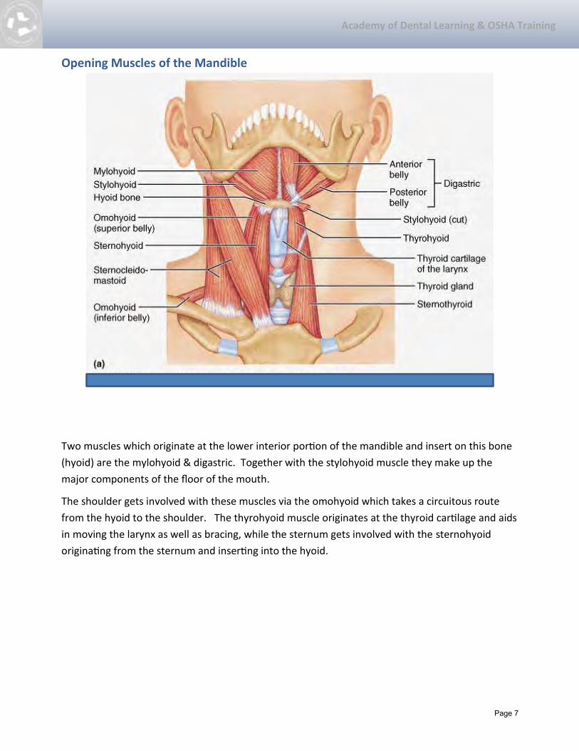
Opening Muscles of the Mandible
Two muscles which originate at the lower interior portion of the mandible and insert on this bone
(hyoid) are the mylohyoid & digastric. Together with the stylohyoid muscle they make up the
major components of the floor of the mouth.
The shoulder gets involved with these muscles via the omohyoid which takes a circuitous route
from the hyoid to the shoulder. The thyrohyoid muscle originates at the thyroid cartilage and aids
in moving the larynx as well as bracing, while the sternum gets involved with the sternohyoid
originating from the sternum and inserting into the hyoid.

Page 8
Academy of Dental Learning & OSHA Training
These muscles are much weaker than the closing musculature of the mandible.
Closing Muscles of the Mandible Masseter
One of the ‘sling’ muscle, it is the strongest of the group and functions in a straight closing action.
It originates at angle of the mandible and inserts on the zygomatic process.
Internal Pterygoid resides opposite the masseter and completes the sling. It too functions in a
straight closing action. The Internal Pterygoid originates at the internal aspect of the angle of the
mandible and inserts on the Pterygoid process of the sphenoid bone.

Page 9
Academy of Dental Learning & OSHA Training
Masseter
External Pterygoid
Assists the disc and mandible to pull forward when mandible closes and moves laterally during
chewing (deglutition). It originates on the pterygoid process of the sphenoid bone and inserts
onto the disc (may be via a ligament) and head of the condyle. It has two bellies (superior and
inferior (see above).
Temporalis
Assists in closing mandible; it is a powerful muscle that virtually covers the temporalis bone of the
skull. As it descends it ducks under the zygomatic process and inserts onto the coronoid process of
the mandible.

Page 10
Academy of Dental Learning & OSHA Training
The long tendon insertion onto the coronoid process of the mandible is often associated with
pathology.
Sternocleidomastoid
Acts as a bracing muscle for the opening and closing of the mandible. It is a unique muscle in that
it is a muscle of mastication (bracing, opening mandible), side bending (head), rotating (neck), and
flexing (neck) muscle and as such is associated with more individual aspects of movement than any
other muscle. It also covers and is in close proximity to the jugular vein, carotid artery, and
stellate ganglion… all vital to life.
Sternocleidomastoid

Page 11
Academy of Dental Learning & OSHA Training
Ligaments
Ligaments attach bone to bone and act as breaks for excessive movements. There are three
ligaments which attach the mandible to the skull. They are: stylomandibular ligament,
spenomandibular ligament; and the articular or capsular ligament.
The other major ligament residing in the area is the stylohyoid ligament.
Neurology
The Trigeminal Nerve is the fifth cranial nerve (nerve which originates in the brain instead of first
synapsing in the spinal cord). It is a sensory nerve (carries message from stimulus to the brain).
After it exits the base of the brain it separates and leaves the skull via the foramen (see below).
The three major branches of the Trigeminal Nerve are: the Ophthalmic V1 (upper face; superior
orbital fissure), the Maxillary V2 (mid face, foramen rotundum), and the Mandibular V3 (lower
face, foramen ovale).
The first major nerve to branch from the V3 is the Auriculo-temporal nerve. It innervates the TMJ
as well as the other structures in the area (pterygoid region). As the mandibular branch readies
itself to enter the mandible via the lingulae (spot where dentists access to give mandibular block)
it sends a separate branche to innervate the tongue (lingual nerve).

Page 12
Academy of Dental Learning & OSHA Training
.

Page 13
Academy of Dental Learning & OSHA Training
Facial Nerve (VII)
The Seventh Cranial Nerve is a mainly a motor nerve (carries the impulse from the brain to the
muscle) and is responsible for all facial movement and expression. It exits the brain behind the ear
(internal acoustic meatus) and after two tight turns within the skull (facial canal) it exits via the
stylomastoid foramen. After it passes through the parotid salivary gland (but not innervating it) it
divides into five branches: Temporal; Zygomatic; Buccal; Masseter; Cervical.
Conclusion
There you have it… dental anatomy in its most basic form, broken up as to its structures: bone,
cartilage, joints, etc. This is no way is this meant to be a comprehensive course, but one
meaningful and practical to the clinician as a review of the major anatomical features of the head
and neck—to prepare for a discussion of TMJ / Disease in other study modules.

Page 14
Academy of Dental Learning & OSHA Training
Examination – Basic Anatomy Review for TMJ Treatment
1. The major bones involved with the TMJ are:
a. temporal
b. mandible
c. hyoid
d. Maxillae
e. A, B, C
2. The Mandible articulates with:
a. the coronoid process
b. the maxillae
c. the temporal bone
3. The Temporal Muscle inserts on the Coronoid Process of the Mandible via a:
a. tendon
b. ligament
4. The mandible is articulated with the skull via:
a. ligaments
b. tendons
c. A & B
5. The Hyoid Bone is unique in that it:
a. does not articulate with any other bone
b. is made mostly of fibrous tissue
c. stabilizes the sternocleidomastoid muscle
d. is mainly involved with closing the mandible

Page 15
Academy of Dental Learning & OSHA Training
6. The TMJ is separated from the skull via cartilagino-fibrous disc
a. True
b. False
7. The only other joint with the make-up of the TMJ Disc is the
a. Knee
b. Glenoid Fossae
c. Shoulder
d. Sternoclavicular joint
8. The muscle which boasts the most individual movements is:
a. Masseter
b. Temporalis
c. External Pterygoid
d. Sternocleidomastoid
9. The closing muscles of the mandible are:
a. Internal Pterygoid and Temporalis
b. Masseter and External Pterygoid
c. All of the Above
10. Teeth cannot affect the wellbeing of the TMJ.
a. True
b. False
11. The main purpose of teeth is to aid in the closing of the mandible.
a. True
b. False
12. The main function of the External Pterygoid Muscle is to:
a. Close the mandible
b. Guide the disc over the Hyoid Bone
c. Guide the disc over the glenoid fossae

Page 16
Academy of Dental Learning & OSHA Training
13. Ligaments attach bone to muscle.
a. True
b. False
14. There are three major ligaments attaching the mandible to the skull.
a. True
b. False
15. The Capsular ligament attaches the mandible to:
a. Hyoid Bone
b. Temporal Bone
c. Maxillae
16. A sensory nerve carries stimuli away from the CNS.
a. True
b. False
17. The Facial Nerve is a Sensory Nerve.
a. True
b. False
18. The Trigeminal Nerve has five major branches.
a. True
b. False
19. The mandibular branch of the Trigeminal Nerve breaks into three branches before it enters
the mandible.
a. True
b. False

Page 17
Academy of Dental Learning & OSHA Training
20. The _____________ nerve innervates the TMJ.
a. Temporal Nerve
b. Lingual Nerve
c. Maxillary Nerve
d. Auriculo-temporal Nerve

Page 18
Academy of Dental Learning & OSHA Training
FAX ANSWER SHEET (703) 935-2190
Basic Anatomy Review: Treatment of TMJ Please complete and fax this form only to (703) 935-2190. Be sure to include the name of the course you are submitting answers for. Please print your answers clearly.
Name: Profession:
License State: License #: Exp. Date:
mm yy
Address 1:
Address 2:
City: State: Zip Code:
Phone: Fax: Email:
Please print the corresponding letter for each answer below:
1. 6. 11. 16.
2. 7. 12. 17.
3. 8. 13. 18.
4. 9. 14. 19.
5. 10. 15. 20.
Please help us improve by circling the appropriate answer.
Ordering experience was convenient. Yes No I received my workbook or file in a timely manner. Yes No Course text and test is clear and understandable. Yes No I will use the course information in my daily practice. Yes No Overall, I would give this course a grade of _________. Comments:
Fax completed exam to: 703-935-2190