Review Hyperthermia v11b
21
- 1 - Carbon nanotube based biomedical agents for heating, temperatur e sensoring and drug delivery Rüdiger Klingeler ([email protected]) Silke Hampel Bernd Büchner Leibniz Institute for Solid State and Materials Research (IFW) Dresden, Helmholtzstr. 20, D-01069 Dresden, Germany Keywords (1-5): Carbon Nanotubes, Hyperthermia, Magnetic Nanoparticles, Temperature Control, NMR Abstract Due to their extraordinary physical and chemical properties carbon nanotubes reveal a promising potential as biomedical agents for heating, temperature sensoring and drug delivery on the cellular level. Filling carbon nanotubes with tailored materials realises nanoscaled containers in which the active content is encapsulated by a protecting carbon shell. We describe different synthesis routes and show the structural and magnetic properties of carbon nanotubes. In particular, the filling with magnetic materials offers the potential for hyperthermia applications while the insertion of NMR active substances allows the usage as markers and sensors. The potential of carbon nanotubes for biomedical applications is highlighted by hyperthermia studies which prove their applicability for local in-situ heating. In addition we have shown that a non-invasive temperature control by virtue of a carbon-wrapped nanoscaled thermometer and filling with anti-cancer drugs is possible.
-
Upload
spokenfear -
Category
Documents
-
view
231 -
download
0
Transcript of Review Hyperthermia v11b
8/3/2019 Review Hyperthermia v11b
http://slidepdf.com/reader/full/review-hyperthermia-v11b 1/21
- 1 -
Carbon nanotube based biomedical agents for heating, temperature sensoring
and drug delivery
Rüdiger Klingeler ([email protected])
Silke Hampel
Bernd Büchner
Leibniz Institute for Solid State and Materials Research (IFW) Dresden, Helmholtzstr.
20, D-01069 Dresden, Germany
Keywords (1-5):
Carbon Nanotubes, Hyperthermia, Magnetic Nanoparticles, Temperature Control,
NMR
Abstract
Due to their extraordinary physical and chemical properties carbon nanotubes reveal
a promising potential as biomedical agents for heating, temperature sensoring and
drug delivery on the cellular level. Filling carbon nanotubes with tailored materials
realises nanoscaled containers in which the active content is encapsulated by a
protecting carbon shell. We describe different synthesis routes and show the
structural and magnetic properties of carbon nanotubes. In particular, the filling with
magnetic materials offers the potential for hyperthermia applications while the
insertion of NMR active substances allows the usage as markers and sensors. The
potential of carbon nanotubes for biomedical applications is highlighted by
hyperthermia studies which prove their applicability for local in-situ heating. In
addition we have shown that a non-invasive temperature control by virtue of a
carbon-wrapped nanoscaled thermometer and filling with anti-cancer drugs ispossible.
8/3/2019 Review Hyperthermia v11b
http://slidepdf.com/reader/full/review-hyperthermia-v11b 2/21
- 2 -
Strong adverse effects on the healthy tissue in the vicinity of a tumour are a major
drawback in current cancer therapies. One innovative technological approach to
solve this problem focuses on therapies on the cellular level by applying intracellular
probes, i.e. the transfer of nano-sized biocompatible devices into the cells. Most
promising materials in this regard are magnetic nanoparticles which can be
addressed by external magnetic fields. A great advantage of magnetic fields is their
biocompatibility, i.e. they penetrate tissue non-invasively and without known adverse
effects, and their weak interaction with organic matter so that deep layers of (human)
tissue can be reached. In particular, magnetic nanoparticles can be localized in deep
tissue, external static magnetic fields can fix them at a precise position, gradient
fields can move them and alternating (AC) fields lead to local heating. The latter can
be utilized for hyperthermia applications which is shown e.g. by the use of
superparamagnetic nanoparticles for magnetic fluid hyperthermia [1] Much of the
current research is focused on iron oxide nanoparticles which have proven their
feasibility in animal experiments [2,3] and are now under clinical trials [4]. Other
magnets usually present limitations due to biocompatibility issues, since most of
them contain potentially toxic elements.
Due to its particular magnetic properties, i.e. large anisotropy and saturation
magnetisation, metallic iron could generate heat more efficiently in comparison to
iron oxides since various mechanisms yield dissipative effects such as domain wall
motion etc. [5]. In practice, higher heating efficiency means that less nanoscaled
material would have to be introduced into the biological system in order to achieve
the targeted hyperthermia effect. The use of nanoparticles made of iron, however, is
hindered by the fact that oxidation in ambient or biological conditions has to be
avoided. A promising way to overcome this problem appears to be the coating of theiron with a carbon shell by insertion of the material inside of carbon nanotubes (CNT)
and thereby protecting the biological environment and the filling material against
each other. Degradation of filling materials is avoided and their potential toxicity and
adverse effects are suppressed so that CNT provide a smart carrier system on the
nanometer scale.
A targeted hyperthermia therapy using iron filled CNT and, in general, utilisation of tailored carbon nanotube based biomedical agents for therapeutic and diagnostic
8/3/2019 Review Hyperthermia v11b
http://slidepdf.com/reader/full/review-hyperthermia-v11b 3/21
- 3 -
purposes can hence be envisioned which will be detailed in the following sections.
After introducing some basic properties of CNT, a short review of
biofunctionalisation, biocompatibility and toxicity aspects is provided. The synthesis
of iron filled CNT as well as magnetic and hyperthermia studies are discussed.
Adding additional material such as a thermometer in Fe-filled CNT increases the
potential of CNT as hyperthermia inducing agents since simultaneous local heating
and temperature control might be feasible. Moreover, the container feature of CNT
offers the step beyond the controlled heat treatment but also suggests the additional
local release of therapeutics.
2. Biocompatibility and Toxicity
CNT are hollow carbon structures with one or more walls, a small diameter on the
nanometre scale and a large length in comparison. Their mechanically and
chemically stable carbon shells can be opened, filled and closed again without losing
their stability. Experiences in filling CNT range back to their discovery in 1991 [6].
Since then, extensive work has been performed to synthesise CNT and to
functionalise them both exohedrally, i.e. by attaching functional elements to the outer
shell, and endohedrally by filling with various materials. CNT can be filled with
metals, semiconductors, salts, organic materials, fullerenes, etc., either during the
synthesis process or through subsequent opening, filling, and closing of the CNT. In
particular, ferromagnetic materials such as Fe, Ni, and Co can be encapsulated
which is relevant to hyperthermia applications. Introducing fluorescent markers offers
a route for visualising CNT [7], which is also possible by exohedral functionalisation
[8,9]. The container feature of CNT allows, in principle, simultaneous filling of CNT
with different materials thereby combining multiple functionalities in one kind of
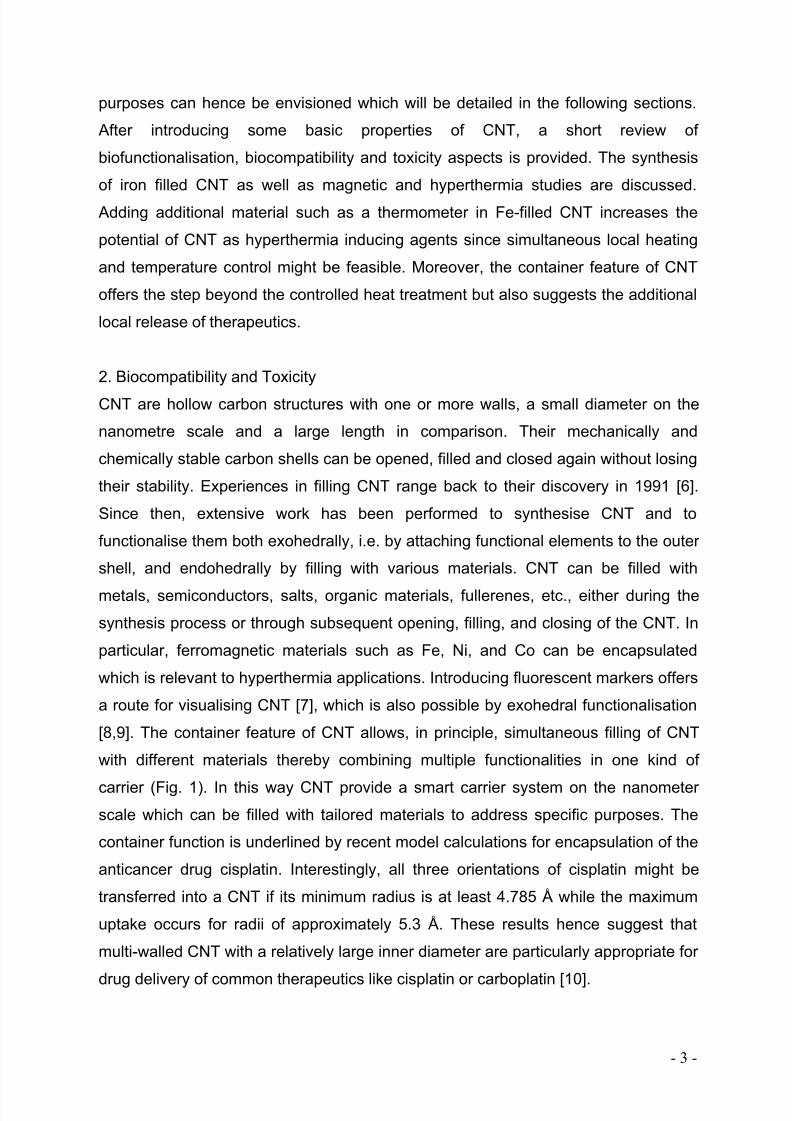
carrier (Fig. 1). In this way CNT provide a smart carrier system on the nanometer scale which can be filled with tailored materials to address specific purposes. The
container function is underlined by recent model calculations for encapsulation of the
anticancer drug cisplatin. Interestingly, all three orientations of cisplatin might be
transferred into a CNT if its minimum radius is at least 4.785 Å while the maximum
uptake occurs for radii of approximately 5.3 Å. These results hence suggest that
multi-walled CNT with a relatively large inner diameter are particularly appropriate for
drug delivery of common therapeutics like cisplatin or carboplatin [10].
8/3/2019 Review Hyperthermia v11b
http://slidepdf.com/reader/full/review-hyperthermia-v11b 4/21
- 4 -
Beyond the shielding effect against a biological environment, the carbon coating
offers an interface for further exohedral functionalisation with suitable (bio-)
molecules. A major task of such functionalisation is the stable dispersion of the
nanoparticles in aqueous media which still is a major challenge. Both non-covalent
and covalent modification strategies can be applied among which the former
preserve the pristine CNT structure while covalent modification introduces partialdamage of the outer wall but in general yields better dispersion. Dispersion becomes
even more crucial if ferromagnetically filled CNT are envisaged which exhibit an
increased tendency to agglomerate due to magnetic interactions. In addition,
exohedral functionalisation is needed to get the highly symmetric carbon structures
compatible to actual biological environments. Nowadays, also functionalisation
aiming to perform dynamic tasks such as target recognition, target transformation,
transport, or electrical conduction in the living cell is addressed. These functions can
be provided by biomolecules, like DNA and enzyme proteins. Importantly for any
therapeutical or biomedical usage, functionalised CNT can cross effectively biological
barriers such as the cell membrane and penetrate the individual cell. Recently, it was
shown that DNA wrapped SWNT were enveloped by cancer cells and they were
used to deliver a lethal dose of microwave radiation to the cancer cells [11]. In
contrast to such a wrapping CNT with biomolecules, any chemical exohedral
functionalisation needed to improve the biocompatibility of CNT will structurally
perturbate the external wall which however in the case of multi-walled CNT does not
affect the overall carbon shielding. A variety of methods exist for the exohedral
Ferromagnet
Antibody
Therapeutics
Temperature sensor
Functional groups
Fig. 1: Sketch of a filled carbon nanotube serving as multi-functional container for in-
vivo applications. A ferromagnet can induce heat in AC magnetic fields and a
material with strongly temperature dependent nuclear magnetic resonance (NMR)
signal might serve as a thermometer. Additional drug delivery can be envisaged.
Exohedral functionalisation to achieve biocompatibility is sketched. Note, that single-
walled as well as multi-walled CNT can be realised.
8/3/2019 Review Hyperthermia v11b
http://slidepdf.com/reader/full/review-hyperthermia-v11b 5/21
- 5 -
functionalization of carbon nanotubes [12,13], some of which have been successfully
applied for the conjugation of proteins, drugs and fluorescent dyes [14,15,16,17] and
active tumor targeting in-vivo [18]. Successful biofunctionalisation is e.g.
demonstrated by Pantarotto et al. who observed that functionalized CNT can cross
the cell membrane and accumulate in the cytoplasm or reach the nucleus without
being toxic for the cell [15]. Soluble CNT can be coupled with amino acids and
bioactive peptides [19] for further derivatization. It was also shown that stable
complex formation of CNT and cationic lipid may accelerate the delivery of
nanotubes into the bladder cancer human cells [20]. For the actual mechanism of
internalization, i.e. endocytosis or phagocytosis, which has been discussed
contradictorily, the actual morphology of the CNT seems to be crucial [14,15,21].
Despite various industrial applications of CNT and their large scale synthesis only
little is known about the interaction of CNT with biological environments. CNT and
other nanomaterials when placed in contact with human (or other mammalian) body
fluids and tissues are recognised by the immune system complement proteins, and
may also interact with other systems, such as coagulation proteins and cell-surface
proteins. Binding of complement proteins activates the complement system via both
classical and alternative pathways and results in strong binding of several
complement proteins to the CNT [22]. Such complement activation influences
subsequent interaction of the CNT with cells and tissues and is a predictor of
potential toxicity in animal models. Salvador-Morales et al. report that such protein
binding to CNT is highly selective. In particular, fibrinogen and apolipoproteins were
the proteins that bound to CNT in greatest quantity. Among proteins contained in
lung surfactant, SP-A and SP-D selectively bind to CNT so that chronic level
exposure may result in sequestration these proteins [23].
Although a variety of toxicity studies has been published (for recent reviews see e.g.
[24,25]), no clear picture evolves for CNT in general. This is e.g. illustrated by
contrasting in-vitro studies showing CNT to be safe [26,27] or to induce significant
toxic effects [28,29,30]. One reason for this ambiguity is the fact that – similar to
other nanoparticles – a large number of specific factors govern the toxicity of CNT
such as their shape and size (diameter, length), the number of shells (single- or multi-walled CNT), agglomeration state and surface chemistry [31]. In particular, the
8/3/2019 Review Hyperthermia v11b
http://slidepdf.com/reader/full/review-hyperthermia-v11b 6/21
- 6 -
concentration of CNT is not necessarily a main parameter. On the other hand,
ambiguity also results from a lack of standardisation and of thorough characterisation
of, e.g., defects of the outer shell or potentially toxic contaminants. Such non-carbon
material originating from the synthesis process of pristine empty CNT might amount
to 5-10% of the total mass. This presumably accounts for much of the reported
differences so that details of synthesis, choice of catalyst particles, washing
procedures and dispersion methods have to be considered thoroughly.
In the following we hence concentrate on multi-walled Fe-filled CNT which synthesis
and properties are described in §3 and §4. We recall the feasibility of an efficient
delivery of Fe-filled CNT into human cancer cell lines which has been shown in cell
culture experiments [20]. A pre-treatment of Fe-filled CNT with cationic lipid was
found to cause a qualitative delivery of the complexes into the cytoplasm but not into
the nucleus. A recent study on cytotoxic effects of Fe-filled CNT in-vitro addressed
metabolic activity, cell proliferation, apoptosis and cell cycle distribution of a
malignant (PC-3) and a non malignant (fibroblasts) cell lineage. The data imply that
CNT strongly associate with cells within a short incubation period. The presence of
CNT in cells did not pose any significant toxic effect [32]. This observation is
corroborated by an animal study on mice which indicated no remarkable toxic or
adverse effects over a period of 440 days after the intraperitoneal or intravenous
injection of pristine Fe-filled CNT. In particular, based on TEM and histological
analyses after 6 weeks Mönch et al. [33] report the absence of any indication on
inflammation. In one case, CNT have been administered up to a total of >1g Fe-filled
MWCNTs/kg body weight over a period of 3 months. Interestingly, in agreement with
the fact that pristine CNT were used, large agglomerates have been observed in
various organs in case of the intravenous treatment. However, such agglomeratesseem to have no drastic effects. All animals survived for more than one year and
showed no abnormalities in behaviour or weight suggesting a general
biocompatibility of the CNT for the applied doses.
The long-term absence of significant toxicity effects in the mentioned animal study
also implies the expected insignificant degradation of CNT in the biological
environment. Regarding their fate in-vivo, excretion in feces and urine has beenreported after injection of empty CNT injected intravenously or intraperitonally in mice
8/3/2019 Review Hyperthermia v11b
http://slidepdf.com/reader/full/review-hyperthermia-v11b 7/21
- 7 -
[34,35]. Studies on the biodistribution and translocation pathways of empty CNT in
mice, however, indicate accumulation predominately in liver and retain for long time
while low acute toxicity is confirmed [36].
3. Synthesis of Ferromagnetic Filled Multi-walled Carbon Nanotubes
Encapsulation of iron nanowires in CNT realises highly anisotropic ferromagnetic
nanoparticles which are discussed for a wide range of potential applications [37].
Among a variety of methods to synthesize these filled carbon structures such as arc
discharge [38] and laser ablation [39], the chemical vapour deposition (CVD) is
applied when a high yield uniform multi-walled CNT is aimed [40].
A suitable method for synthesizing CNT filled with metals as Fe, Co or Ni is the so-
called ‘‘in situ method’’, a special CVD-method, in which the formation of CNT and
their filling with ferromagnetic elements or compounds take place simultaneously.
Here, relevant precursors are needed, which e.g. provide as well iron for the filling as
carbon for the shells. The most common precursors are metallocenes [Me(C5H5)2;
Me = Fe, Co, Ni] which supply a large scale of well-defined multi-walled (MW) CNT
with a high filling yield (about 50 %) of the ferromagnetic material. Many groups
world-wide use this method in different equipments. Sen et al. reported the first
experimental results for the synthesis of ferromagnetic MWCNT [41]. In a typical
experiment the ferromagnetically filled MWCNT are grown in a quartz tube reactor
inside a dual zone furnace system. The main parameters of the deposition process
are the sublimation temperature of the metallocene in the first furnace, the gas flow
rate, and the temperature of the second hot furnace zone where the pyrolysis of
metallocene and the deposition of filled tubes take place. CNT are formed on
oxidized silicon substrates uncoated or coated (2 nm layer of different materials such
as Fe, Co and Permalloy) placed inside the reaction zone. In this case they grow inperpendicular alignment to the substrate surface. Much more nanotubes are formed
on the wall of the quartz tube reactor (which is a mixture of bundles of well-aligned
Fe-filled MWCNT) [42,43, 44, 45].
An additional technique is the liquid source CVD (LS-CVD) [40]. This method is
characterized by a constant and reproducible transport of the precursor. The
metallocene, especially the ferrocene, is dissolved in cyclopentane and dropped onthe moving band continuously. In the first part of the system the solvent is vaporized

8/3/2019 Review Hyperthermia v11b
http://slidepdf.com/reader/full/review-hyperthermia-v11b 8/21
- 8 -
and only the ferrocene is transferred into the reactor at a defined temperature and
with a constant transport velocity. In the deposition reactor a second moving band,
populated with precoated substrates is positioned. This LSCVD method is highly
suited for the continuous production of defined ferromagnetically filled CNT. Wang et
al. have synthesized Fe-filled CNT with a high filling ratio by using ferrocene and
dichlorbenzene as precursor [46]. This solution is injected through a nozzle directly
into the reactor so that a spontaneous decomposition reaction occurs. Pichot et al.
applied an aerosol assisted CVD (AA-CVD) [47] device to produce Fe filled CNT.
Ultrasonicating a solution of ferrocene in cyclohexane produced an aerosol, which
was then carried for 15 min by an argon flow through a quartz reactor placed in a
tubular furnace at 850 °C. Eventually, MWCNT carpets were deposited on the
reactor walls [48].
Filled CNT can be also synthesised by a post-synthesis method. This technique
includes (1) the synthesis of empty CNT, (2) the opening of their ends, (3) a filling
step with various materials (metals, salts, therapeutics) and (4) the closing step of
the filled CNT. The opening procedure is mostly done by well established wet-
chemistry techniques [49] or by oxidation in air [50, 51]. The easiest way to
incorporate the material into the open ended CNT is over a gasphase reaction where
the CNT and the filling material are inserted together into glass ampoules sealed
under vacuum and heated beyond the sublimation temperature of the filling material
[52, 53]. But also a wet chemical approach can be applied where capillarity is the
driving force [54,55],49. The closure of the openended and filled CNT can be realised
by a redeposition with a polymer or other carbon-containing phases [56]. Various
techniques such as transmission electron microscopy and X-ray diffraction
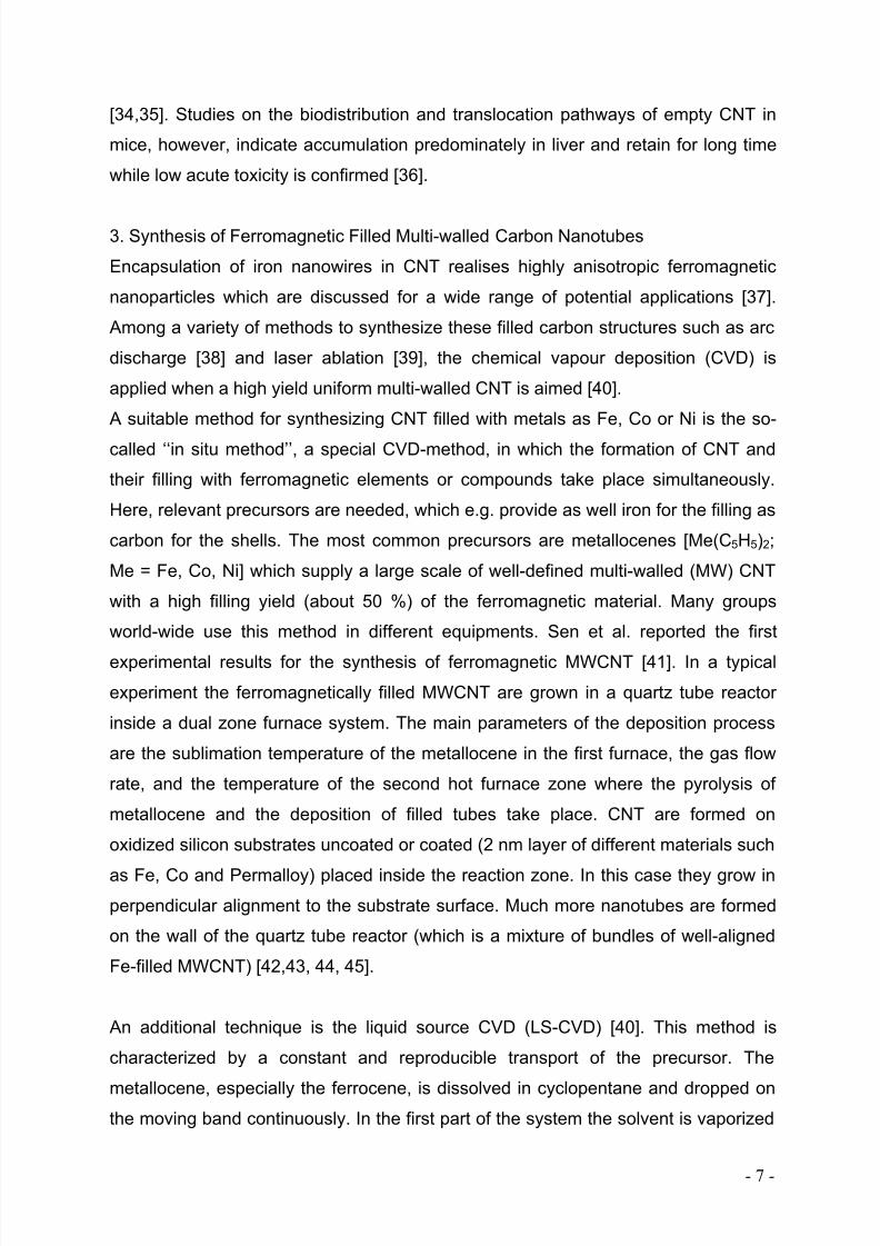
demonstrate the successful synthesis of Fe-filled CNT by our CVD technique[40,43,42,57,58,59,60]. E.g., the cross section shown in Fig. 2 clearly indicated the
single crystalline Fe filling as well as the carbon shell.
8/3/2019 Review Hyperthermia v11b
http://slidepdf.com/reader/full/review-hyperthermia-v11b 9/21
- 9 -
The length of CNT is in the micrometer range and outer diameters vary from 20-80
nm. The Fe-filling is discontinuous and consists of iron nanowires with a filling yield
of ~50%. Interestingly, by x-ray diffraction the existence of the body-centered
structure (α-Fe), the face-centered cubic structure ( γ-Fe) and in very low
concentration also of Fe3C could be verified. For a hyperthermia application a high
yield of the ferromagnetic α-Fe is requested. By changing details of the synthesis
process the relative yields of the different Fe-phases can be adjusted. The rate of the
ferromagnetic α-Fe is increased by an annealing process below the temperature of
the phase transition of γ-Fe to α-Fe which indeed yields a larger saturation
magnetization [59]. To get exact information about the morphology of the iron filled
CNT ( γ-Fe/α-Fe ratio), the 57Fe Mössbauer spectroscopy is an appropriate method.
Transmission Mössbauer spectroscopy (TMS) and backscattered conversion
electron Mössbauer spectroscopy (CEMS) were applied in order to distinguish
different Fe phases and their spatial distribution within the whole sample and along
the tubes’ height by the group of Ruskov. A characterization (on a large spatial scale)
of the aligned CNT samples was performed by obtaining TMS spectra for selected
spots positioned at different locations of the sample. While the total Fe content
changes considerably from one location to another, the γ-Fe/α-Fe phase ratio is
constant onto a relatively large area. Using TMS and CEMS for all aligned Fe-
MWCNT samples it is also shown that along the CNT axes, going to the top of the
nanotube the relative content of the γ-Fe phase increases. Going to the opposite
direction, i.e., towards the silicon substrate, the relative content of the Fe3C phaseincreases [61].
Fig. 2: Typical SEM and TEM images of well-aligned iron filled multi-walled carbon
nanotubes synthesised by LS-CV.

8/3/2019 Review Hyperthermia v11b
http://slidepdf.com/reader/full/review-hyperthermia-v11b 10/21
- 10 -
4. Magnetic properties and hyperthermia feasibility
Various studies have shown that, independently of the synthesis technique, the
carbon encapsulated iron is efficiently protected by the surrounding shells and its
magnetic properties are retained. Often, magnetic characterisation studies have
been performed for CNT fixed to a SiO2 substrate. For such studies, Fe-CNT are
grown directly on the substrate so that a rather dense coating of well aligned
nanowires is studied. For such aligned Fe-filled CNT, uniaxial magnetic anisotropy is
reported with the easy magnetic axis being parallel to the CNT axis. The anisotropy
can straightforwardly attributed to the shape anisotropy of the magnetic nanowires.
However, depending on the alignment [43] and density of CNT on the substrate
magnetic dipole-dipole interactions between the Fe-cores have to be considered, too.
Interestingly, enhanced magnetic coercive fields of H C >100 mT are observed in such
ensembles of Fe-filled CNT at room temperature [37,43] which significantly exceed
the value H C =9*10-3 mT observed in bulk Fe. The more representative data for
biomedical applications are achieved by powder measurements. Here, depending on
the filling ratio, diameter, etc. coercive fields in the range of H C ~ 20-50 mT have
been found. In a recent study the magnetisation of cells dried after incubation with
Fe-filled CNT exhibited a critical field of similar size (~23 mT) [33].
Magnetic coercivity is even more reduced when the magnetisation is measured in a
living cell culture [32]. For such a study, 2*105 PC-3 cells were incubated with
50µg/ml of Fe-filled CNT. After an incubation period of 4h, the cells were washed two
times with PBS, trypsinized and centrifuged at 4oC. This procedure in total yielded
approximately 1*106 cells which were resuspended in 100µl for media and
measured, at 4oC, in a maximum applied field of H = 0.5T. Interestingly, the data
imply only a vanishing coercivity of H C ~ (2±2) Oe.
The magnetisation data suggest the feasibility of Fe-filled CNT for by applying an
alternating (AC) magnetic field (see e.g. [62]). The heating effect is based on the
physical principle that applied AC magnetic fields induce magnetisation loops. If
these loops are not completely reversible (e.g. in the case of ferromagnetic particles
or for superparamagnetic particles below the blocking temperature), magnetic energy
is transformed into heat. We mention that residual ferromagnetic catalyst particlesappear in most synthesis processes which provide superparamagnetism in CNT (e.g.
8/3/2019 Review Hyperthermia v11b
http://slidepdf.com/reader/full/review-hyperthermia-v11b 11/21
- 11 -
[63]). Another route to superparamagnetic CNT employs the insertion of Gd ions to
form Gadonanotubes [64].
There are various physical mechanisms which yield non-reversible loops, i.e. which
are related to magnetic dissipation processes, and it should be emphasized that in
the case of Fe-filled CNT the mechanism has not yet been identified. In general,
different processes will be related to different energy scales and resonance
frequencies, so that the filling material, its size and its shape have a significant effect
on the heating properties. For example there will be strong differences between the
case of single-domain particles, where the magnetisation process competes with
shape and crystal anisotropies, and the motion of domain walls in multi-domain
particles.
The feasibility of Fe-filled CNT for hyperthermia has been demonstrated by a
calorimetric study in AC magnetic fields. Our test device consists of a high-frequency
generator with an impedance matching network and the magnetic coil system.
Water-cooled copper tubes are wound into a coil system (e.g., 4–10 turns, diameter
of bore 20–100mm) in which the sample is placed. Temperature is controlled by a
0 10 20 30 40 50 60 70
0
200
400
600
800
1000
H (kA/m)
S A R F e
( W / g F e
)
f = 230 kHzc
Fe~ 0.08 g/l
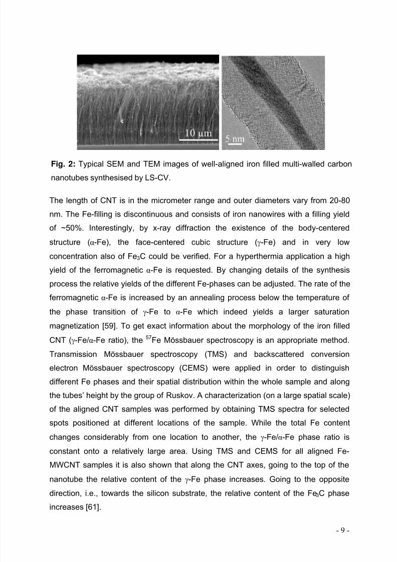
Fig. 3: Specific absorption rate SARFe of Fe-nanowires (~0.08 g Fe/l) encapsuled in
CNT. Measurements have been performed at f = 230 kHz after dispersing the CNT by
means of human albumin in PBS 4.2 /l .
8/3/2019 Review Hyperthermia v11b
http://slidepdf.com/reader/full/review-hyperthermia-v11b 12/21
- 12 -
fiber-optic temperature controller (LXT-Luxtron One). The generator can provide
alternating magnetic fields in the frequency range of 50-1200 kHz. The data shown in
Fig. 3 have been obtained at f = 230 kHz. Here, Fe-filled MWCNT have been
dispersed by human albumin in PBS. The concentration was 4.2 mg/ml so that about
0.08 mg Fe/ml can be estimated. This concentration is corroborated by analysis of
magnetization loops. The initial slope of temperature vs. time studies was used to
determine the specific absorption rate SAR. In agreement with previous data [33],
values of SARFe < 100 W/gFe were found in the field range below ~20 kA/m2 which is
applicable for medical treatment. We emphasize, however, that the presented first
studies did not yet elucidate the actual dissipative mechanism. Detailed information
about the magnetisation reversal will help to optimize e.g. the geometrical
dimensions of the Fe-nanowires in order to achieve improved switching behaviour.
In addition to a magnetic field induced thermal ablation described in detail above,
CNT are also feasible for near-infrad (NIR) light based hyperthermia. This method
applies the fact that biological systems are transparent to light in the NIR regime of
0.7-1.1 μm wavelength while the strong absorbance renders single-walled CNT for
hyperthermia agents in living cells [11,65,66]. Focused heat transduction and photo-
ablative destruction of kidney cancer cells has also been shown for multi-walled CNT
if being nitrogen doped [67]. Although NIR light is capable of passing through several
centimetres of tissue, its interaction with biological matter is by far larger than of
magnetic fields and the penetration depth is much smaller. For efficient heat transfer
to deep tissue and in order to avoid parasitic heating effects in surrounding tissue
magnetic agents hence seem to be advantageous.
5. Temperature control by NMR on filled CNT
Accurate control of the tissue temperature is mandatory in any hyperthermia
approach. Currently, in the clinical treatments, temperature is controlled by a
clinician's intervention by placing thermocouples or fiber-optical thermometers into
the tumor in combination with computer modelling later on. Any model, however,
demands estimates of the tissue properties, blood perfusion rate and other dynamic
properties [68]. Instead, a continuous non-invasive temperature monitoring appearsto be advantageous not only for hyperthermia treatment. Magnetic resonance (MR)
8/3/2019 Review Hyperthermia v11b
http://slidepdf.com/reader/full/review-hyperthermia-v11b 13/21
- 13 -
thermometry based on a temperature-dependent proton resonance frequency shift of
the water molecule provides temperature control in addition to a good spatial
localization, thereby allowing for accurate target identification in ultrasound thermal
therapy [69]. However, large doses of unshielded magnets which are present in
nanoparticle-based hyperthermia introduce magnetic field inhomogeneities that
reduce MRI contrastivity based on the proton relaxation-weighted image and prevent
proton-based MR thermometry.
A promising approach for a non-invasive in-vivo temperature control relies on the use
of a nanoscaled thermometer, which consists of a CNT and a filling material with
strongly temperature dependent NMR parameters. In particular, the filling material
might exhibit strong T-dependencies of the spin-lattice or the spin-spin relaxation,
resonance frequency, dipolar or scalar couplings, and electrical quadrupole coupling
at 310-350 K (ca. 20-60 °C) so that temperature detection is possible with high
accuracy (<0.1 degree). Due to the protecting carbon shell, the number of materials
which can be used for temperature sensoring without toxic adverse effects strongly
increases. On the other hand, the container feature of CNT might, in principle, allow
simultaneous filling of CNT with a temperature sensor and another probe such as a
ferromagnet (=heater), thereby combining different functionalities in one kind of CNT
(Fig. 1).
Up to know, the more simple system has been studied, where only the temperature
sensor is encapsulated in CNT [53]. Many alkali and cuprous halides are known to
show pronounced temperature dependencies of NMR parameters. From a family of
these compounds monovalent cuprous iodide (CuI) turned out to be most suited.
Here, both the copper and iodine nuclei have NMR active isotopes with a high
natural abundance.
The synthesis of CuI filled CNT was based on pristine nanotubes consisting from 10
to 40 carbon layers with inner diameters between 5 and 20 nm. For filling with CuI,
CNT were opened using thermal and acid treatment in combination with sonication.
The opened CNT were grinded in a mortar gently and put in a silica glass ampoule
together with CuI (Aldrich 99.99%) in excess. The ampoule was sealed under
vacuum (10-3
Torr) and heated at 600°C for 24h. At this temperature CuI iscompletely sublimated and transported into the opened CNT thanks to the capillarity
8/3/2019 Review Hyperthermia v11b
http://slidepdf.com/reader/full/review-hyperthermia-v11b 14/21
- 14 -
effect. The resultant material was examined by transmission electron microscopy
(TEM and HRTEM), X-ray diffraction analysis and energy dispersive X-ray analysis
(EDX) and identified as CNT filled to 80 % with single crystalline cuprous iodide.
Although both 63Cu and 127I nuclear isotopes possess a quadrupole moment, in CuI
copper and iodine atoms each are surrounded tetrahedrally by four atoms of the
opposite kind. This leads to a vanishing quadrupolar coupling. Therefore only the
resonance frequencies, linewidths and relaxation times can be considered as
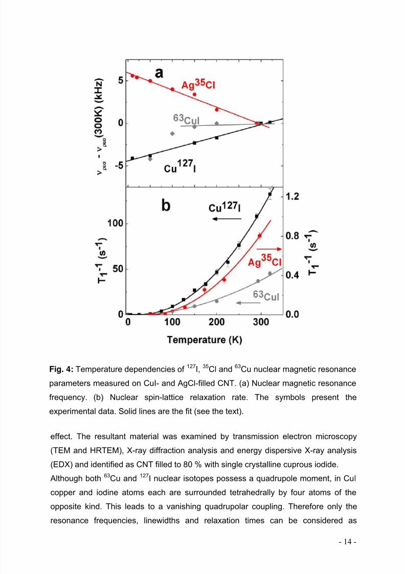
Fig. 4: Temperature dependencies of 127I, 35Cl and 63Cu nuclear magnetic resonance
parameters measured on CuI- and AgCl-filled CNT. (a) Nuclear magnetic resonance
frequency. (b) Nuclear spin-lattice relaxation rate. The symbols present the
experimental data. Solid lines are the fit (see the text).
8/3/2019 Review Hyperthermia v11b
http://slidepdf.com/reader/full/review-hyperthermia-v11b 15/21
- 15 -
temperature indicators. The NMR measurements were done in a standard solid state
NMR spectrometer in the external magnetic field of 7.05 T. Both 63Cu and 127I NMR
spectra obtained from the Fourier transformed echo signals represented a single
resonance line. The spin-lattice relaxation T1 was measured employing an inversion
recovery pulse sequence. The obtained decay of 63Cu and 127I longitudinal
magnetisation was analyzed following a standard equation for the spin I = 3/2 and I =
5/2, correspondingly. In both cases the magnetisation curves followed a simple
exponential form characterized by a single spin-lattice relaxation time.
The analysis of the 63Cu NMR spectra reveals insignificant changes in the resonance
frequency in the relevant temperature range, while the resonance frequencies
measured on the 127I nucleus indicate a stronger dependence on temperature (Fig.
4a). Such behaviour is explained by lattice effects which include both the lattice
vibration and the lattice expansion. The 127I NMR frequency data are well fitted with a
linear function over the whole temperature range studied. Thus, at first glance, the127I resonance frequency might be used as a measure for the temperature
determination. A relative small slope of this function leads, however, to an error of 15
K in temperature determination. Therefore the usage of this parameter for an
accurate temperature control is not reasonable. Furthermore, the spectral linewidths
at half maximum have been analyzed for both nuclei and were found to be constant
at temperatures below 320 K. Thus, also the linewidths could be ruled out to serve as
a temperature control parameter.
The temperature dependence of the 127I spin-lattice relaxation rates T1-1 is presented
in Fig. 4b. The 63Cu T1-1 dependence is similar but much smaller compared to the 127I
data. The T1-1 dependencies for both nuclei are found to be in very good agreementwith the law T1
-1 ~ T2 that is expected for a Raman two-phonon quadrupolar process.
[70]. This behaviour is observed over the entire temperature range implying no
contributions from impurities which might appear at low temperatures and from ionic
diffusion which might be observed at high temperatures. This is consistent with the
view that relaxation is driven by a quadrupolar mechanism in this compound [71].
The 127I experimental data are well fitted with a quadratic function T1-1 = a + bT + cT2,
where fitting coefficients are a = 1, b = (7±1)*10-2
and c = (1.49±0.05)*10-3
. Themean squared errors of the fitting coefficients provide an estimate of the accuracy of
8/3/2019 Review Hyperthermia v11b
http://slidepdf.com/reader/full/review-hyperthermia-v11b 16/21
- 16 -
the temperature determination. In the temperature range of biological interest (i.e.
290 to 320 K) the CuI-CNT nanothermometer can indicate the temperature with an
accuracy of 2 K by means of the spin-lattice relaxation measurement. Other Cu-
halides filled in CNT demonstrate a qualitatively similar behaviour but exhibit less
temperature sensitivity as shown in table 1. These results, in particular those on CuI-
CNT, provide a good starting point to look for further filling materials of CNT in order
to increase the accuracy of temperature determination.
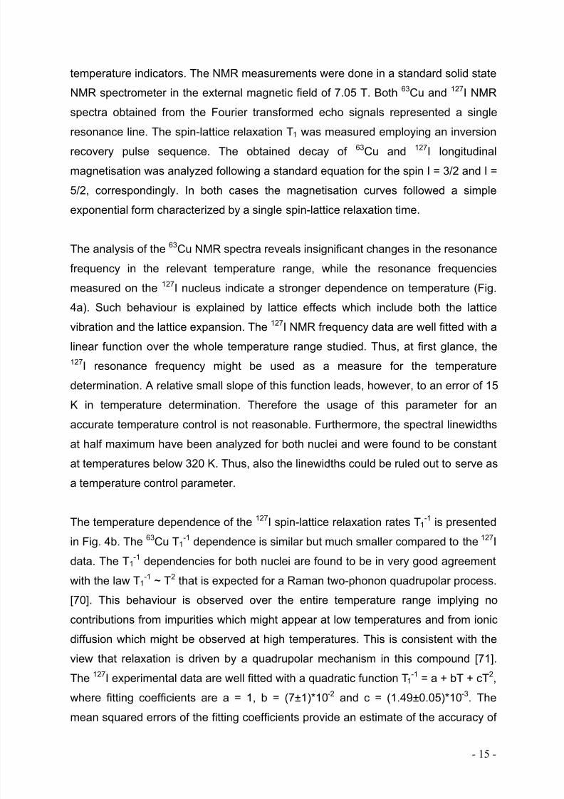
Material Nucleus dν res/dT (Hz/K) d(T 1-1)/dT (Hz/K)
CuI-CNT
63Cu
127I
-
14
0.27
0.86
CuBr-CNT
63Cu
81Br
19
7
0.15
0.75
CuCl2-CNT
63Cu
35Cl
15
1
0.23
-
AgCl-CNT 35Cl 21 0.006
Table 1: Temperature sensitivity parameters of several filled CNT. The table shows
the filling material, the respective nucleus as well as temperature dependence of
resonance frequency and T1-relaxation time in the temperature range of 300-320 K.
Conclusions
Due to their extraordinary physical and chemical properties carbon nanotubes reveal
a promising potential for applications on the cellular level. Upon filling, nanoscaled
containers are realised in which the active material is encapsulated by a protecting
carbon shell. We describe different synthesis routes and show the structural and
magnetic properties of CNT. In particular, the filling with magnetic material offers the
potential for hyperthermia applications while the insertion of NMR active substances
allows the usage as markers and sensors. This potential of carbon nanotubes for
biomedical applications is highlighted by hyperthermia studies which prove their
applicability for local in-situ heating. In addition we have shown that a non-invasive
8/3/2019 Review Hyperthermia v11b
http://slidepdf.com/reader/full/review-hyperthermia-v11b 17/21
- 17 -
temperature control by virtue of a carbon-wrapped nanoscaled thermometer is
possible.
A valuable extension would be spatially resolved NMR. The rapidly growing field of
cellular and molecular MR imaging enables to visualize cells and inserted CNT in
order to track cancer cells and to control therapies on the cellular level such as
magnetic hyperthermia. The container feature of CNT is extensively utilized if a
heating element (ferromagnet), a temperature sensor and a contrast agent are
confined within the same nanocontainer. Such a combination of different
functionalities on a nanoscale would provide simultaneous heating, temperature
control by means of MRI and high spatial resolution of the image. Furthermore, if
clinical usage of the static magnetic field is contra-indicated and MR imaging is no
longer suitable then the technology proposed here addresses materials with
temperature dependent nuclear quadrupolar resonance NQR (e.g. cuprous oxide) or
zero-field NMR (e.g. Co-based compounds) parameters that demonstrates versatility
of this approach for biomedical applications. The potential of CNT for biomedical
applications becomes even more evident if their container function is exploited for
drug delivery in magnetically functionalised and NMR labelled nanodevices. It has
been shown that anti-cancer drugs can be inserted in CNT [54] for which purpose the
multi-walled ones turn out to be particularly appropriate. Combing different
functionalities in well shielded containers hence seems to be the particular
advantage of carbon nanotubes.
Acknowledgements
This work was partly supported by the European Community through the Marie CurieResearch Training Network CARBIO under contract No. MRTN-CT-2006-035616.
The authors thank Diana Haase, Manfred Ritschel, Anastasia Vyalikh, Anja Wolter,
Kamil Lipert, Yulia Krupskaya, Christopher Mahn, Thomas Mühl, Dieter Elefant and
Hans-Jörg Uhlemann from IFW Dresden as well as Arthur Taylor and Kai Krämer
from the Department of Urology, Technical University Dresden. We particularly
appreciate Albrecht Leonhardt for support and valuable discussions. Scientific advice
by Sabine Achten (Boehringer Ingelheimer Fonds), Francois Rossi (European

8/3/2019 Review Hyperthermia v11b
http://slidepdf.com/reader/full/review-hyperthermia-v11b 18/21
- 18 -
Commission Joint Research Centre, Ispra) and Andreas Jordan (Magforce
Nanotechnologies AG) is gratefully acknowledged.
References
1 Jordan A, Scholz R, Wust P, Fahling H, Felix R. Magnetic fluid hyperthermia (MFH): Cancer treatment with AC magnetic field induced excitation of biocompatible superparamagneticnanoparticles. J. Magn. Magn. Mat. 1999;201:413-419
2 Johannsen M, Thiesen B, Jordan A, Taymoorian K, Gneveckow U, Waldöfner N, Scholz R,Koch M, Lein M, Jung K, Loening SA. Magnetic fluid hyperthermia (MFH) reduces prostatecancer growth in the orthotopic Dunning R3327 rat model. The Prostate 2005;64:283.
3 Matsuoka F, Shinkai M, Honda H, Kubo T, Sugita T, Kobayashi T. Hyperthermia usingmagnetite cationic liposomes for hamster osteosarcoma. Biomagnetic Research andTechnology 2004;2:3.
4 Johannsen M, Gneveckow U, Taymoorian K, Thiesen B, Waldöfner N, Scholz R, Jung K,Jordan A, Wust P, Loening SA. Morbidity and quality of life during thermotherapy usingmagnetic nanoparticles in locally recurrent prostate cancer: Results of a prospective phase Itrial. International Journal of Hyperthermia 2007;23:315-323.
5 Dale LH. Synthesis, Properties, and Applications of Iron Nanoparticles. Small 2005;1:4826 Iijima S. Nature 1991;359:707.7 Kim BM, Qian S, Bau HH. Filling Carbon Nanotubes with Particles. Nano Lett. 2005;5(5):873-
878.8 Prakash R, Washburn S, Superfine R, Cheney ER. Visualization of Individual Carbon
Nanotubes With fluorescence Microscopy Using Conventional Fluorophores. Appl. Phys. Lett.2003;83:1219-21.
9 Didenko VV, Moore VC, Baskin DS, Smalley RE. Visualization of Individual Single-WalledCarbon Nanotubes by Fluorescent Polymer Wrapping. NanoLett. 2005;5:1563-7.
10 Hilder TA, Hill JM. Modelling the encapsulation of the anticancer drug cisplatin into carbonnanotubes. Nanotechnology 2007;18:275704-12.
11 Kam NW, O'Connell M, Wisdom JA, Dai H. Proc. Natl. Acad. Sci. U.S.A 102, 11600 (2005)12 Bianco A, Prato M. Adv. Mat. 15 1765 (2003)13 Hirsch A, Vostrowsky O. Functionalization of Carbon Nanotubes. Topics in Current Chemistry
2005;245:193.14 Kostarelos K, Lacerda L, Pastorin G, Wu W, Wieckowski S, Luangsivilay J, Godefroy S,
Pantarotto D, Briand J-p, Muller S, Prato M, Bianco A. Cellular uptake of functionalized carbonnanotubes is independent of functional group and cell type. Nature Nanotechnology2007;2:108.
15 Pantarotto D, Briand J-P, Prato M, Bianco A. Translocation of bioactive peptides across cellmembranes by carbon nanotubes. Chemical Communications 2004;1:16-17.
16 Wu W, Wieckowski S, Pastorin G, Benincasa M, Klumpp C, Briand J-P, Gennaro R, Prato M,Bianco A. Targeted Delivery of Amphotericin B to Cells by Using Functionalized Carbon
Nanotubes. Angewandte Chemie International Edition 2005;44:6358.17 Kam NWS, Dai H. Carbon Nanotubes as Intracellular Protein Transporters: Generality andBiological Functionality. J. Am. Chem. Soc. 2005;127:6021.
18 Liu Z, Cai W, He L, Nakayama N, Chen K, Sun X, Chen X, Dai H. In vivo biodistribution andhighly efficient tumor targeting of carbon nanotubes in mice. Nat Nano 2007;2:47.
19 Wang S, Humphreys ES, Chung S-Y et al.: Nature Mater. 2003;2:196-200.20 Mönch I, Meye A, Leonhardt A, Krämer K, Kozhuharova R, Gemming T, Wirth MP, Büchner B.
Ferromagnetic filled carbon nanotubes and nanoparticles: synthesis and lipid-mediated deliveryinto human tumor cells. J. Magn. Magn. Mat. 2005;290-291:276-278.
21 Kam NWS, Liu Z, Dai H. J. Functionalization of carbon nanotubes via cleavable disulfide bondsfor efficient intracellular delivery of siRNA and potent gene silencing. J. Am. Chem. Soc.2005;127:12492–12493.
22 Salvador-Morales C, Flahaut E, Sim E, Sloan J, Green MLH, Sim RB. Complement activation
and protein adsorption by carbon nanotubes. Molec. Immun. 2006;43(3):193-201.23 Salvador-Morales C, Townsend P, Flahaut E, Venien-Bryan C, Vlandas A, Green MLH, SimRB. Binding of pulmonary surfactant proteins to carbon nanotubes; potential for damage to lungimmune defense mechanisms. Carbon 2007; 45(3): 607-617.
8/3/2019 Review Hyperthermia v11b
http://slidepdf.com/reader/full/review-hyperthermia-v11b 19/21
- 19 -
24 Smart SK, Cassady AI, Lu GQ, Martin DJ. The biocompatibility of carbon nanotubes. Carbon2006;44:1034-1047.
25 Donaldson K, Aitken R, Tran L, Stone V, Duffin R, Forrest G, Alexander A. Carbon nanotubes:a review of their properties in relation to pulmonary toxicology and workplace safety. ToxicolSci. 2006;92:5-22.
26 Kam NWS, Jessop TC, Wender PA, Dai H. Nanotube Molecular Transporters: Internalization of Carbon Nanotube-Protein Conjugates into Mammalian Cells. J. Am. Chem. Soc.2004;126:6850.
27 Worle-Knirsch JM, Pulskamp K, Krug HF. Oops They Did It Again! Carbon Nanotubes HoaxScientists in Viability Assays. Nano Lett. 2006;6:1261
28 Cui D, Tian F, Ozkan CS, Wang M, Gao H. Effect of single wall carbon nanotubes on humanHEK293 cells. Toxicology Letters 2005;155:73.
29 Tian F, Cui D, Schwarz H, Estrada GG, Kobayashi H. Cytotoxicity of single-wall carbonnanotubes on human fibroblasts. Toxicology in Vitro 2006;20:1202.
30 Bottini M, Bruckner S, Nika K, Bottini N, Bellucci S, Magrini A, Bergamaschi A, Mustelin T.Multi-walled carbon nanotubes induce T lymphocyte apoptosis. Toxicology Letters2006;160:121.
31 Oberdorster G, Maynard A, Donaldson K, Castranova V, Fitzpatrick J, Ausman K, Carter, J,
Karn B, Kreyling W, Lai D. Principles for characterizing the potential human health effects fromexposure to nanomaterials: Elements of a screening strategy. Part. Fibre Toxicol. 2005;2:8.
32 Taylor A, Lipert K, Krämer K, Hampel S, Füssel S, Meye A, Klingeler R, Ritschel M, Büchner B,Wirth MP. Biocompatibility of Iron Filled Carbon Nanotubes In Vitro, submitted 2007
33 Mönch I, MeyeA, Leonhardt A. In: Nanotechnologies for Life Sciences, ed. by C.S.S.R. Kumar,Vol. 6: Nanomaterials for Cancer Therapy and Diagnosis. Weinheim: Wiley-VCH, 2006, pp 259-337.
34 Singh R, Pantarotto D, Lacerda L, Pastorin G, Klumpp C, Prato M, Bianco A, Kostarelos K.Tissue biodistribution and blood clearance rates of intravenously administered carbon nanotuberadiotracers. PNAS 2006;103:3357.
35 Guo J, Zhang X, Li Q, Li W. Biodistribution of functionalized multiwall carbon nanotubes inmice. Nuclear Medicine and Biology 2007;34:579.
36 Deng X, Jia G, Wang H, Sun H, Wang X, Yang S, Wang T, Liu Y. Translocation and fate of
multi-walled carbon nanotubes in vivo. Carbon 2007;45:1419-1424.37 Grobert N, Hsu WK, Zhu YQ, Hare JP, Kroto HW, Terrones M, Terrones H, Redlich P, Rühle M,
Escudero R, Morales F. Appl. Phys. Lett. 1999;75:3363.38 Guerret-Piécourt C, Le Bouar Y, Loiseau A, Pascard H. Nature 1994; 372:761−765.39 Thess A, Lee R, Nikolaev P, Dai H, Petit P, Robert J, Xu C, Lee YH, Kim SG, Rinzler AG,
Colbert DT, Scuseria GE, Tománek D, Fischer JE, Smalley RE. Crystalline Ropes of MetallicCarbon Nanotubes. Science 1996;273:483.
40 Hampel S, Leonhardt A, Selbmann D, Biedermann K, Elefant D, Müller C, Gemming T, Büchner B. Growth and characterization of filled carbon nanotubes with ferromagnetic properties.Carbon 2006;44:2316-22.
41 Sen R, Govindaraj A, Roa CNR. Carbon nanotubes by the metallocene route. Chem. Phys.Lett. 1997; 267: 276-280.
42 Mueller C, Golberg D, Leonhardt A, Hampel S, Buechner B. Growth studies, TEM and XRD
investigations of iron-filled carbon nanotubes. Physica Status Solidi (a) 2006; 203: 1064-1068.43 Leonhardt A, Hampel S, Mueller C, Moench I, Koseva R, Ritschel M, Elefant D, Biedermann K,
Buechner B. Synthesis, properties and applications of ferromagnetic-filled carbon nanotubes.Chemical Vapor Deposition 2006; 12: 380-387.
44 Mueller C, Hampel S, Elefant D, Biedermann K, Leonhardt A, Ritschel M, Buechner B. Iron filledcarbon nanotubes grown on substrates with thin metal layers and their magnetic properties.Carbon 2006; 44: 1746-1753.
45 Mueller C, Leonhardt A, Hampel S, Buechner B. Diameter controlled growth of iron-filled carbonnanotubes. Physica Status Solidi (b) 2006; 243: 3091-3094.
46 Wang W, Wang K, Lv R, Wei J, Zhang X, Kang F, Chang J, Shu Q, Wang Y, Wu D. Synthesisof Fe-filled thin-walled carbon nanotubes with high filling ratio by using dichlorbenzene asprecursor. Carbon 2007; 45: 1105-1136.
47 Mayne M, Grobert N, Terrones M, Kamalakaran R, Rühle M, Kroto HW, Dalton DRM. Pyrolytic
production of aligned carbon nanotubes from homogeneously dispersed benzene-basedaerosols. Chem. Phys. Lett. 2001; 338: 101-107.

8/3/2019 Review Hyperthermia v11b
http://slidepdf.com/reader/full/review-hyperthermia-v11b 20/21
- 20 -
48 Pichot V, Launois P, Pinault M, Mayne-L´Hermite M, Reynaud C. Evidence of strong nanotubealignment and for iron preferential growth axis in multiwalled carbon nanotube carpets. Appl.Phys. Lett. 2004; 85: 243-245.
49 Tsang SC, Chen YK, Harris PJF, Green MLH. A simple chemical method of opening and fillingcarbon nanotubes. Nature 1994; 372: 159-162.
50 Ajayan PM, Ebbesen TW, Ichihashi T, Iijima S, Tanigaki K, Hiura H. Opening carbon nanotubeswith oxygen and implications for filling. Nature 1993; 362: 522-525.
51 Tsang SC, Harris PJF, Green MLH. Thinning and opening of carbon nanotubes by oxidationusing carbon dioxide. Nature 1993; 362: 520-522.
52 Dujardin E, Ebbesen TW, Hiura H, Tanigaki K. Capillarity and wetting ofcarbon nanotubes.Science 1994; 265: 1850-1852.
53 Vyalikh A, Klingeler R, Hampel S, Haase D, Ritschel M, Leonhardt A, Borowiak-Palen E,Rümmeli M, Bachmatiuk A, Kalenczuk RJ, Grafe HJ, Büchner B. A nanoscaled contactlessthermometer for biological systems. Physica Status Solidi b 2007; 244: 4092–4096.
54 Hampel S, Kunze D, Haase D, Rauschenbach M, Kraemer K, Ritschel M, Leonhardt A, ThomasJ, Oswald S, Hoffmann V, Buechner B. Carbon nanotubes filled with a chemotherapeutic agent – a nanocarrier mediates inhibition of tumor cell growth. submitted to Nanomedicine 2007
55 Moench I, Leonhardt A, Meye A, Hampel S, Kozhuharova-Koseva R, Elefant D, Wirth MP,
Buechner B. Synthesis and Characteristics of Fe-Filled Multi-Walled Carbon Nanotubes for Biomedical Application. Journal of Physics: Conference Series 2007; 61: 820–824.
56 Satishkumar BC, Govindaraj A, Mofokeng J, Subbanna GN, Rao CNR. Novel experiments withcarbon nanotubes: opening, filling, closing and functionalizing nanotubes. J.Phys. B: At. Mol.Opt. Phys. 1996; 29: 4925-4934.
57 Leonhardt A, Ritschel M, Kozhuharova R, Graff A, Muehl T, Huhle R, Moench I, Elefant D,Schneider CM. Synthesis and properties of filled carbon nanotubes. Diamond and RelatedMaterials 2003;12:790-793.
58 Muehl T, Elefant D, Graff A, Kozhuharova R, Leonhardt A, Moench I, Ritschel M, Simon P,Groudeva-Zotova S, Schneider CM. Magnetic properties of aligned Fe-filled carbon nanotubes.Journal of Applied Physics 2003;93:7894-7896.
59 Leonhardt A, Ritschel M, Elefant D, Mattern N, Biedermann K, Hampel S, Mueller C, GemmingT, Buechner B. Enhanced magnetism in Fe-filled carbon nanotubes produced by pyrolysis of
ferrocene. Journal of Applied Physics 2005;98:74315.60 Leonhardt A, Moench I, Meye A, Hampel S, Buechner B. Synthesis of ferromagnetic filled
carbon nanotubes and their biomedical application. Advances in Science and Technology2006;49:74-78.
61 Ruskov T, Spirov I, Ritschel M, Mueller C, Leonhardt A, Ruskov R. Moessbauer morphologicalanalysis of Fe-filled multiwalled carbon nanotube samples. Journal of Applied Physics 2006;100: 84326/1-8.
62 Pankhurst QA, Conolly J, Jones SK, Dobson J. Applications of magnetic nanoparticles inbiomedicine. J. Phys. D: Appl. Phys. 2003;36:R167-R181.
63 Ritschel M, Leonhardt A, Elefant D, Oswald S, Büchner B. J Phys. Chem. C 2007; 111: 8414-8417
64 Sitharaman B, Kissell K, Hartman K, Tran LA, Baikalov A, Rusakova I, Sun Y, Khant HA, LudtkeSJ, Chiu W, Laus S, Tóth E, Helm L, Merbach AE, Wilson LJ. Chem. Commun. 2005; 31: 3915-
3917.65 Panchapakesan B, Lu S, Sivakumar K, Teker K, Cesarone G, Wickstrom E. Nanobiotechnology
2005, 1: 133-14066 Gannon CJ, Cherukuri P, Yakobson BI, Cognet L, Kanzius JS, Kittrell C, Weisman RB, Pasquali
M, Schmidt HK, Smalley RE, Curley SA. Cancer 2007, 110: 2654.67 Torti SV, Byrne F, Whelan O, Levi N, Ucer B, Schmid M, Torti FM, Akman S, Liu J, Ajayan PM,
Nalamasu O, Carroll DL. Int J Nanomed. 2007, 2:68 Gneveckow U, Jordan A, Scholz R, Brüß V, Waldöfner N, Ricke J, Feussner A, Hildebrandt B,
Rau B, Wust P. Description and characterization of the novel hyperthermia- andthermoablation-system MFH (R) 300F for clinical magnetic fluid hyperthermia. Med. Phys.2004;31:1444-1451.
69 Smith NB, Merrilees NK, Hynynen K, Dahleh M. Control system for an MRI compatibleintracavitary ultrasound array for thermal treatment of prostate disease. Int. J. Hyperthermia
2001;17:271-282.70 Abragam A. Principles of Nuclear Magnetism. Oxford University Press, 1961.

8/3/2019 Review Hyperthermia v11b
http://slidepdf.com/reader/full/review-hyperthermia-v11b 21/21
71 Andrew ER, Hinshaw WS, Tiffen RS. Nuclear spin-lattice relaxation in solid cuprous halides. J.Phys. C: Solid State Phys. 1973;6:2217-2222.


![Malignant hyperthermia [final]](https://static.fdocuments.us/doc/165x107/58ceb1b71a28abb2218b5123/malignant-hyperthermia-final.jpg)