REVIEW Ev olution of carbohydr ate antigensÑmicr obial for ... · phylogen ies depicting the ev...
12
REVIEW Evolution of carbohydrate antigens—microbial forces shaping host glycomes? Joseph R. Bishop 2 and Pascal Gagneux 1,2 2 Glycobiology Research and Training Center, Cellular and Molecular Medicine-East, University of California, San Diego, 9500 Gilman Drive, La Jolla, CA 92093-0687 Received on December 5, 2006; revised on January 10, 2007; accepted on January 10, 2007 Many glycans show remarkably discontinuous distribution across evolutionary lineages. These differences play major roles when organisms belonging to different lineages interact as host – pathogen or host –symbiont. Certain lineage-specific glycans have become important signals for multicellular host organisms, which use them as mol- ecular signatures of their pathogens and symbionts through recognition by a toolkit of innate defense mol- ecules. In turn, pathogens have evolved to exploit host lineage-specific glycans and are constantly shaping the gly- comes of their hosts. These interactions take place in the face of numerous critical endogenous functions played by glycans within host organisms. Whether due to simple evol- utionary divergence or adaptive changes under natural selection resulting from endogenous functional require- ments, once different lineages elaborate on differential gly- comes these mutual differences provide opportunities for host exploitation and/or pathogen defense between lineages. Such phylogenetic molecular recognition mechan- isms will augment and likely contribute to the maintenance of lineage-specific differences in glycan repertoires. Key words: glycan/co-evolution/host-pathogen/animal lectin Introduction Carbohydrates makeup a substantial portion of the biomass on earth, mostly in the form of the two structural polysacchar- ides—cellulose from plants and chitin from arthropods and fungi. All known living organisms also display an array of free or covalently attached carbohydrates collectively known as glycans (Varki et al. 1999). Some of these complex mol- ecules decorate the surface of cells and are secreted into the surrounding environment where they function in a wide variety of processes required for life including structural support, protection, recognition, localization, and infor- mation/nutrient transfer. The precise compositions and combi- nations of different carbohydrates making up the glycan repertoire of each species can differ dramatically. The rapid development of glycomics methods (Raman et al. 2005) is bound to greatly increase our knowledge about natural glycan diversity, and evolutionary considerations will be crucial for interpreting glycan function within and between organisms (Varki 2006). Despite recent advances, we are yet to have a complete inven- tory of naturally occurring monosaccharides used to produce the glycan portion of these molecules, as many members of the Bacteria and Archaea domains synthesize a number of special- ized carbohydrates (Schaffer et al. 2001). In contrast, metazoan animals build most of their glycans from a very limited number of monosaccharide building blocks, allowing us to consider how these molecules might have evolved over time. Most metazoan glycoconjugates are built from six classes of monosaccharides including sialic acids, hexoses, hexosamines, deoxyhexoses, pentoses, and uronic acids (Varki et al. 1999) see Box 1. These monosaccharides can of course be modified to create greater complexity at the single monosaccharide level. Furthermore, the individual carbohydrate units can be attached via a variety of glycosidic linkages, into highly complex linear or branched structures. Thus in theory, there is virtually no limit to the number of different glycans that can be gener- ated. In practice though metazoan animals seem to generate only a limited range of these possibilities. It would be impossible to do justice to the overwhelming diversity of natural glycans and their functions in one review. Fortunately, a number of excellent recent reviews and texts address the biology of individual classes of glycans as well as their endogenous ligands, the glycan-binding animal lectins (Staudacher et al. 1999; Angata and Varki 2002; Esko and Selleck 2002; Spiro 2002; Lowe and Marth 2003; Varki and Angata 2006). The aim of this review is to address the taxo- nomic distribution of glycans and to reflect on the processes that are shaping this distribution. Our main focus will be on how interactions between multicellular animal hosts and their microbial or viral pathogens as well as symbionts may have contributed to the observed lineage-specific constellations of certain glycans, especially extracellular glycans. Distribution of glycans within the tree of life Glycans occur in a discontinuous and puzzling distribution across evolutionary lineages. Examples of discontinuously dis- tributed glycans are presented in Table I. The hypothetical evolutionary relationships of living organisms can be depicted in the form of phylogenetic trees. Figure 1 shows three 1 To whom correspondence should be addressed; e-mail: [email protected] # 2007 The Author(s) This is an Open Access article distributed under the terms of the Creative Commons Attribution Non-Commercial License (http://creativecommons.org/licenses/by-nc/2.0/uk/) which permits unrestricted non-commercial use, distribution, and reproduction in any medium, provided the original work is properly cited. 23R Glycobiology vol. 17 no. 5 pp. 23R–34R, 2007 doi:10.1093/glycob/cwm005 Advance Access publication on January 19, 2007
Transcript of REVIEW Ev olution of carbohydr ate antigensÑmicr obial for ... · phylogen ies depicting the ev...
REVIEW
Evolution of carbohydrate antigens—microbial forces shaping host glycomes?
Joseph R. Bishop2 and Pascal Gagneux1,2
2Glycobiology Research and Training Center, Cellular and MolecularMedicine-East, University of California, San Diego, 9500 Gilman Drive,La Jolla, CA 92093-0687
Received on December 5, 2006; revised on January 10, 2007; accepted onJanuary 10, 2007
Many glycans show remarkably discontinuous distributionacross evolutionary lineages. These differences play majorroles when organisms belonging to different lineagesinteract as host–pathogen or host–symbiont. Certainlineage-specific glycans have become important signalsfor multicellular host organisms, which use them as mol-ecular signatures of their pathogens and symbiontsthrough recognition by a toolkit of innate defense mol-ecules. In turn, pathogens have evolved to exploit hostlineage-specific glycans and are constantly shaping the gly-comes of their hosts. These interactions take place in theface of numerous critical endogenous functions played byglycans within host organisms. Whether due to simple evol-utionary divergence or adaptive changes under naturalselection resulting from endogenous functional require-ments, once different lineages elaborate on differential gly-comes these mutual differences provide opportunities forhost exploitation and/or pathogen defense betweenlineages. Such phylogenetic molecular recognition mechan-isms will augment and likely contribute to the maintenanceof lineage-specific differences in glycan repertoires.
Key words: glycan/co-evolution/host-pathogen/animal lectin
Introduction
Carbohydrates makeup a substantial portion of the biomass onearth, mostly in the form of the two structural polysacchar-ides—cellulose from plants and chitin from arthropods andfungi. All known living organisms also display an array offree or covalently attached carbohydrates collectively knownas glycans (Varki et al. 1999). Some of these complex mol-ecules decorate the surface of cells and are secreted into thesurrounding environment where they function in a widevariety of processes required for life including structuralsupport, protection, recognition, localization, and infor-mation/nutrient transfer. The precise compositions and combi-nations of different carbohydrates making up the glycan
repertoire of each species can differ dramatically. The rapiddevelopment of glycomics methods (Raman et al. 2005) isbound to greatly increase our knowledge about naturalglycan diversity, and evolutionary considerations will becrucial for interpreting glycan function within and betweenorganisms (Varki 2006).
Despite recent advances, we are yet to have a complete inven-tory of naturally occurring monosaccharides used to produce theglycan portion of these molecules, as many members of theBacteria and Archaea domains synthesize a number of special-ized carbohydrates (Schaffer et al. 2001). In contrast, metazoananimals build most of their glycans from a very limited numberof monosaccharide building blocks, allowing us to consider howthese molecules might have evolved over time. Most metazoanglycoconjugates are built from six classes of monosaccharidesincluding sialic acids, hexoses, hexosamines, deoxyhexoses,pentoses, and uronic acids (Varki et al. 1999) see Box 1.These monosaccharides can of course be modified to creategreater complexity at the single monosaccharide level.Furthermore, the individual carbohydrate units can be attachedvia a variety of glycosidic linkages, into highly complexlinear or branched structures. Thus in theory, there is virtuallyno limit to the number of different glycans that can be gener-ated. In practice though metazoan animals seem to generateonly a limited range of these possibilities.
It would be impossible to do justice to the overwhelmingdiversity of natural glycans and their functions in one review.Fortunately, a number of excellent recent reviews and textsaddress the biology of individual classes of glycans as well astheir endogenous ligands, the glycan-binding animal lectins(Staudacher et al. 1999; Angata and Varki 2002; Esko andSelleck 2002; Spiro 2002; Lowe and Marth 2003; Varki andAngata 2006). The aim of this review is to address the taxo-nomic distribution of glycans and to reflect on the processesthat are shaping this distribution. Our main focus will be onhow interactions between multicellular animal hosts and theirmicrobial or viral pathogens as well as symbionts may havecontributed to the observed lineage-specific constellations ofcertain glycans, especially extracellular glycans.
Distribution of glycans within the tree of life
Glycans occur in a discontinuous and puzzling distributionacross evolutionary lineages. Examples of discontinuously dis-tributed glycans are presented in Table I. The hypotheticalevolutionary relationships of living organisms can be depictedin the form of phylogenetic trees. Figure 1 shows three1To whom correspondence should be addressed; e-mail: [email protected]
# 2007 The Author(s)This is an Open Access article distributed under the terms of the Creative Commons Attribution Non-Commercial License(http://creativecommons.org/licenses/by-nc/2.0/uk/) which permits unrestricted non-commercial use, distribution, and reproduction in any medium,provided the original work is properly cited. 23R
Glycobiology vol. 17 no. 5 pp. 23R–34R, 2007doi:10.1093/glycob/cwm005Advance Access publication on January 19, 2007
phylogenies depicting the evolutionary relationships: betweenthe three domains of life (Figure 1A), among Eukarya(Figure 1B) and among the anthropoid primates (Figure 1C),respectively, along with the distribution patterns of selectedglycan across different evolutionary lineages. As seen, the dis-tribution patterns of glycans fall into four general patterns.
(1) Glycans conserved across many taxa. In contrast to riboso-mal RNA that is present in all living organisms, thusallowing the reconstruction of these phylogenies, nosingle glycan structure has been conserved to the sameextent. An example for a relatively conserved class ofglycan would be N-glycans found in organisms of all thethree primary lineages of life, albeit absent from manybacteria (Figure 1A).
(2) Glycans specific to a particular lineage, such as capsulemurein peptidoglycans in bacteria (Figure 1A) or ganglio-sides in vertebrates (Figure 1B).
(3) Glycans similar across distant taxa, examples include glyco-saminoglycans found in metazoans and bacteria (Figure 1A);cellulose in plants, bacteria and tunicates; sialic acids (longthought to be unique to metazoan animals) of the deuteros-tome lineage and also found in many bacteria and in cepha-lopod mollusks (squid and octopus); or Gal(Fuc alpha 1–4)N-acetylglucosamine (GlcNAc) (Lewis A) only found in pri-mates, some other vertebrates, plants, and few pathogenicbacteria (Figure 1B), and
(4) Glycans conspicuously absent from very restricted taxaonly (species, families, or higher units) within lineagesthat otherwise possess such glycans. Examples includeGal alpha 1–4Gal beta1–4GlcNAc present in most ver-tebrates but absent in mammals and some birds(Figure 1B); Gal alpha 1–3 Gal beta 1–4GlcNAc
(alpha-Gal) present in most mammals, but absent in OldWorld monkeys, apes and humans (Catarrhines), andN-glycolylneuraminic acid (Neu5Gc) present in most ver-tebrates but absent in humans (Figure 1C).
Why do glycans evolve?
Divergence
Like that of any biological molecule, glycan evolution is likelyto occur simply due to the divergence of evolutionary lineages.Phylogenies (literally: “history of lineages”) come aboutmostly by the successive bifurcation of lineages, as popu-lations derived from a common ancestor cease to exchangegenetic information (i.e., become reproductively isolated).See Box 2 for a list of some key evolution terminology. Thegenetic tool kits responsible for glycan synthesis and modifi-cation of different lineages are subsequently shaped by inde-pendent mutational histories, causing the glycan repertoires(glycomes) of different lineages to diverge as well. Anexample would be the use of cellulose in plants but not inmetazoans, with the exception of tunicates (Figure 1B).Divergence involves much historical contingency, whererandom changes in different lineages, such as the recruitmentof certain glycan types over others for specific functions, limitthe future evolution of their glycomes.
Natural selection
Selective pressures resulting from recognition processes dispro-portionately affect the glycans covering cell surfaces. Naturalselection acts on glycans, either by favoring the maintenanceof a particular glycan (stabilizing or purifying selection) or bydiminishing survival and/or reproductive success of organismscarrying a certain glycan (negative selection). Maintenance ofthe N-glycan synthesis pathway in all eukaryotes is anexample of stabilizing selection, since disruptions often leadto lethal consequences (Chui et al. 2001; Schachter 2002).Negative selection on glycans could occur whenever an import-ant pathogen exploits a particular glycan as a receptor for infec-tion. Positive selection would entail selection for rapid changein glycans e.g., to accommodate novel endogenous functions.
Convergence
Still another mechanism for generating diversity occurs whenorganisms belonging to distantly related lineages recruit or“reinvent” similar subsets of glycan repertoires. Such parallelevents may be due to particular demands of the environmentor be due to random recruitment of ancestral synthetic path-ways. The existence of the Lewis A antigen [Gal beta 1–4(Fuc alpha 1–4)GlcNAc] in Catarrhines and in plants couldbe such an example, as the enzymes involved in its synthesishave very different genomic sequences (Palma et al. 2001;Javaud et al. 2003) (Figure 1B). Alternatively, what appearsas convergent evolution could result from the differentialretention of ancestral enzymatic tool kits confined to a few dis-tantly related lineages.
Coevolution
When organisms belonging to different lineages repeatedlyinteract, as is the case in most natural ecological communities,
Table I. Some glycans with strikingly discontinuous taxonomic distribution
Peptidoglycans Bacteria (Koch 2000)
S-layer glycoproteins Archaeans (Kandler and Konig 1998)
Cellulose Plants, bacteria, and tunicates (Gibeautand Carpita 1994)
Chitin Fungi, arthropods, and mollusks, and oneteleost fish (Wagner 1994)
Hyaluronan Vertebrates, bacteria (DeAngelis 1999)
Glycosaminoglycans Metazoa and bacteria
Sulfated glycosaminoglycans Metazoa
Sialic acids Deuterostome metazoans, bacteria, fewmollusks (Angata and Varki 2002)
Neu5Gc sialic acid Vertebrates but not humans or birds (dataon brids incomplete) (Varki 2001)
Gangliosides Deuterostome metazoans and mollusks
Gal alpha 1–3 Gal Mammals but not Catarrhines (Galiliet al. 1988)
Gal alpha 1–4 Gal Vertebrates except mammals and somebird lineages (Suzuki et al. 2004)
Lactose secretion Mammals
Galactose beta 1–4(fucosealpha 1–4) GlcNAc
Catarrhines, Xenopus, plants, andpathogenic bacteria (H. pylori) (Oriolet al. 1999; Dupuy et al. 2002; Guerardelet al. 2003)
JR Bishop and P Gagneux
24R
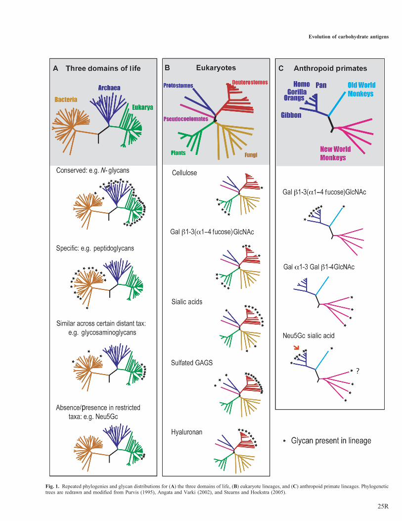
Fig. 1. Repeated phylogenies and glycan distributions for (A) the three domains of life, (B) eukaryote lineages, and (C) anthropoid primate lineages. Phylogenetictrees are redrawn and modified from Purvis (1995), Angata and Varki (2002), and Stearns and Hoekstra (2005).
Evolution of carbohydrate antigens
25R

then their glycomes can become involved in coevolutionary pro-cesses. Thus, the interactions of two distinct glycomes of theinteracting lineages directly influence their mutual evolution.There is ample evidence for coevolution in glycan diversity inthe interactions of microbes and their animal hosts. Thesecases of coevolution involve two distinct phenomena: (i) inde-pendent evolution of enzymatic tool kits for the production ofidentical molecules in microbes. Examples include glycansfound almost exclusively in multicellular hosts and in theirmicrobial pathogens such as glycosaminoglycans and sialicacids (Figure 1B), and (ii) synthesis of “mimic”, moleculesnot identical but very similar to hosts glycans such as polylegio-naminic acids by Legionella or pseudaminic acid byPseudomonas (Knirel et al. 1987; Kooistra et al. 2001). TheLewis A antigen is also found in certain strains ofHelicobacter pylori, which infect humans, likely reflecting coe-volution (Monteiro et al. 1998). Coevolution could also beoccurring via horizontal gene transfer between metazoans andtheir bacterial pathogens, as has been discussed for genesinvolved in sialic acid synthesis (Angata and Varki 2002).
Disclaimer about limitations of evolutionary research
While we would certainly agree with the statement that“nothing in glycobiology makes sense, except in the light ofevolution” (Varki 2006), we must also realize that evolutiononly occurred once and that evolution does not follow well-defined rules (Lewontin 2002). This situation is somewhatalleviated by the fact that after lineages diverge, more oftenthan not they remain separated for good and, thus provideresearchers with large numbers of iterations (“pseudosamples”) for which evolutionary processes have occurredindependently. The study of these divergent lineages providesa good opportunity to elucidate evolutionary mechanisms.
A further limitation arises with regard to glycan changes inrapidly evolving organisms such as microbes or viruses, as it isimpossible to gain information from long-extinct pathogens,which leave no fossils. The speed of evolution in pathogensmeans that the identity of past pathogens will never beknown and that many current pathogens may be descendentsof earlier innocuous microbes or even former symbionts.Rapid evolutionary rates are also associated with homoplasy,i.e., if the observed similarity between glycans is not necess-arily due to recent shared ancestry but could have evolvedindependently in different lineages (convergence). In the eraof genomics, the ability to investigate the genomic sequencesof the genes coding for enzymes that assemble and modifyglycans in different lineages provides a powerful means ofreconstructing the evolutionary history of glycosylation bydetermining key events in the establishment of glycan syn-thesis machinery.
Glycans in metazoan animals
In metzoan animals, cell surfaces are covered with an electrondense coating of glycoconjugates known as the glycocalyx.Further, glycans are directly secreted as polymers or attachedto proteins into the extracellular matrix and body fluids. Thisglycan landscape is often (for functional and historicalreasons) characteristic of both species and particular cell
types. (Paulson and Colley 1989; Roth 1996). Four basictypes of glycoconjugates are present in metazoans includingN-linked, O-linked, glycolipids, and proteoglycans (Varkiet al. 1999). These molecules play a large array of functionsrequired for life including support, signaling, protein folding,and protection (Table II).Why vertebrates use only such a small fraction of monosac-
charide types for the assembly of their glycans remains amystery (Box 1). For example, what is the reason why ver-tebrates, unlike plants do not carry terminal xylose on theirN-glycans or incorporate any trehalose in their glycan reper-toire? Absences of such structures are likely to representcases of lineage-specific evolutionary happenstance (contin-gency), whereby the independent mutational history of differ-ent lineages has led to differential evolution of glycanbiosynthesis enzymes along separate lineages. Paradoxically,however, even with their relatively reduced panel of monosac-charides (compared to bacteria for example), vertebrates gen-erate a staggering amount of structural variation bycombining just nine principal monosaccharides into chainsof varying lengths and degrees of branching on differentiallydecorated proteins and lipids (Manzi et al. 2000). It alsoappears that despite the relative small number of differentbuilding blocks (monosaccharides),vertebrates produce muchmore complex branched N-glycans than many other lineages(Varki et al. 1999).
Box 1. Principal building blocks of vertebrate glycans
Sialic acids: e.rg. N-acetylneuraminic acid (Neu5Ac. N-glycolylneurami-nic acid (Neu5Gc)Hexoses: Glucose, mannose, galactose (Gal)Hexosamines: N-acetylglucosamine (GlcNAc. N-acetylgalactoseamineGalNAcDeoxyhexoses: Fucose (Fuc)Pentoses: XyloseUronic acids: Iduronic acid, glucuronic acid
Table II. Endogenous functions of glycans in metazoans that go beyondstructural function
Protein folding/chaperone-assisted folding in ER
Protein subunit assembly
Cell–cell interactions
Cell-extracellular matrix interactions
Cell-complement interactions
Intra- and inter-cellular trafficking
Signaling
Protection from protease degradation
Exogenous functions of glycans in metazoans
Welcome signals for important symbionts
Long-term accommodation of symbionts
Arbitration of symbiotic microbial communities
Decoys against pathogen recognition
“Smoke-screen” against pathogen recognition
Detecting nonself based on absence of self-glycans
Detecting nonself based on conserved nonself-microbial glycans
Detecting nonself based on polymorphic nonself-glycans in the population
JR Bishop and P Gagneux
26R
Box 2. Glossary of evolution terminology
Antagonistic coevolution: “evolutionary arms race”, where changes inone lineage of a pair of host-parasite lineages are prompted by or promptchanges in the other lineage.Convergence: similarity between taxa despite independent evolutionaryhistories.Catarrhine: primates belonging to Old World monkeys, apes and humans.Demographic bottleneck: strong reduction in population size.Divergence: differences between taxa due to independent evolutionaryhistories.Domain: one of the three radiations of life including the Archaea,Bacteria, and Eukarya.Founder event: establishment of new populations by small numbers offounder individuals.Frequency dependent selection: when the fitness of a genotype dependson its frequency.Genetic drift: random variation in gene frequency from one generation toanother.Historical contingency: the effect of random event on the probability ofsubsequent events in a lineage.Homoplasy: similarities in character states for reasons other than inheri-tance from a common ancestor. These include convergence, parallelism,and reversal.Lineage: group of organisms sharing a common ancestor (monophyletic).Phylogeny: hypothetical history of related lineages based on DNAsequences or any other heritable derived traits.Purifying (stabilizing) selection: a type of selection that removes indi-viduals from both ends of a phenotypic distribution thus maintaining thesame distribution mean.Trade-off: the balancing of different selection pressures especially whenthese have opposing directions.
Variation in animals glycan antigens in time and space
Transient glycan variation in animals has been documentedduring key processes such as pregnancy, lactation, infection,or acute phase response, whereas ontogenetic glycan variationplays key roles in the regulation of metazoan development(Haltiwanger and Lowe 2004). Glycans can also vary inspace, as different compartments and adjacent tissues inmany animal species carry different glycan repertoires. In agiven metazoan, one could detect different glycan distributionfrom the outside as the secreted mucins and bound mucins ofthe mucous membranes, the epithelia, the basal layers, thestroma, the endothelia of blood vessels, the different types ofimmune cells, the cells of the peripheral and central nervoussystem, and the reproductive systems all usually vary withrespect to their surface glycans (Ohtsubo and Marth 2006).Such variation and its distribution might reflect trade-offsbetween the needs for endogenous function and adaptation toexternal selective pressures from pathogens or accommodationof important symbionts.
Genes coding for glycan biosynthetic enzymes haveundergone substantial expansion contributingto glycan diversity in metazoans
A substantial fraction (1–2%) of animal genes function inglycan biosynthesis and modification. Unlike genes codingfor a single protein product, these enzymes work in an “assem-bly-line” like system of glycan synthesis pathways (Lowe andMarth 2003). These pathways allow organisms to generaterapid phenotypic changes based on posttranslational modifi-cation of their glycoconjugates. Glycosyltransferases, whichcatalyze the addition of sugars to growing glycan chains and
proteins, have been subject to multiple lineage-specific expan-sions via gene duplication (Lespinet et al. 2002). In mammals,for example, there are 9 fucosyl transferases, compared to 4 inDrosophila and up to 18 in Caenorhabdiitis elegans (Javaudet al. 2003). In the case of fucosyl transfereases, genomic ana-lyses have determined that the more ancient genes in mammalshad multiple exons and typically encode enzymes that transferfucose near the base of the N- or O-peptidic sequences,whereas more recent genes are monoexonic and encode trans-ferases acting at the periphery (termini) of glycans (Oriol et al.1999). A variety of genetic mechanisms caused this expansion,including duplication, exon shuffling, point mutations, andtransposition (Javaud et al. 2003). Similar examples are seenin a host of enzymes including sialyltransferases (19 inmammals) and heparan sulfate proteoglycan modificationenzymes (15 in mammals) (Esko and Lindahl 2001; Harduin-Lepers et al. 2005). As the majority of these enzymes residein the endoplasmic reticulum (ER)–Golgi secretory system,these organelles have rightly been called the “evolvability”module of animal cells providing organisms with machineryfor generating variation through combinatorial modificationof expressed proteins (Kirschner and Gerhart 1998).
Genetic studies in model organisms with null mutations inbiosynthesis genes have proved that many glycans are requiredfor proper metazoan development, as these mutations producephenotypes ranging from embryonal lethality to growth defectsto impaired morphogenesis and cognitive function—but somecan also have no obvious effects under laboratory conditions(Natsuka and Lowe 1994; Kotani et al. 2001; Lowe andMarth 2003; Kudo et al. 2006). It is conspicuous that the con-sequences of experimental abolition of many glycans are oftennot evident in animal cell cultures, even when these prove tobe lethal as early as the embryonic stage in the whole organismfrom which the cells are cultured (Grobe et al. 2002). Thesefindings point to key functions of glycans for multicellulardevelopment, but they also leave open the possibility that acertain fraction of animal glycans can be selectively neutral,i.e., these can be altered without incurring major fitnesscosts to the organism. Laboratory studies looking at conse-quences of experimental glycan alteration for individuals areunlikely to shed light on population-level effects of glycanpolymorphism, such as the proposed protective effects in pre-venting the rapid spread of pathogens due to herd immunity-related mechanisms (Gagneux and Varki 1999). This idearemains untested in part because such effects would bebased on populations rather than individuals.
Animal lectin intractions and glycan evolution
In many cases, the endogenous function of glycans requiresinteraction with proteins, and recent decades have seen the dis-covery of a growing list of animal lectins with specific carbo-hydrate recognition domains (CRD) (Drickamer and Taylor1993; Gabius 1997; Probstmeier and Pesheva 1999; Rini andLobsanov 1999). Binding is usually highly specific forglycan type, as defined by its monosaccharide compositionand the nature of the glycosidic linkages by which these areconnected (Drickamer and Taylor 1993; Kaltner andStierstorfer 1998; Kilpatrick 2002). Lectin–glycan interactionscan mediate a variety of cell–cell recognition events includinginterspecies (host–pathogen), intraspecies (fertilization and
Evolution of carbohydrate antigens
27R
gestation), and intercellular (development and immune regu-lation) interactions (Gabius et al. 2002). Much of the diversityof metazoan glycans is found at the periphery of the glycansinvolving terminal portions capped by sialic acids, fucose,galactose or GalNac or, in the case of proteoglycans, bydirected modification of linear glycan chains (Varki 1993;Esko and Lindahl 2001). This increase in diversity towardsthe exterior termini of glycans on the surfaces of mammaliancells (Dennis et al. 1999; Varki 2006), combined with the factthat animal lectins often recognize specific glycan structuresfound on the termini, strongly suggests that recognition pro-cesses rather than simple divergence are driving this diversity.It is no surprise therefore, that microbial and viral pathogens ofmetazoan hosts have evolved their own sets of lectins toexploit these molecules for host recognition, attachment andtissue tropism (Karlsson 1995; Sharon 1996; Rostand andEsko 1997).
The evolutionary glycan arms race
The ubiquitous presence of species-specific glycans on hostcells and secretions predispose these as convenient receptorsto be exploited by microbes for host recognition, attachment,and invasion by way of a wide array of microbial and virallectins including adhesins, pili, fimbriae, and hemagglutinins(Gilboa-Garber and Garber 1989; Wadstrom and Ljungh1999). For specific examples of glycan-mediated host–pathogen interactions, we refer the reader to several excellentreviews (Sharon 1996; Rostand and Esko 1997; Hooper andGordon 2001; Olofsson and Bergstrom 2005). Host invasionoften occurs via the large epithelial layers lining the externalcavities of vertebrates, which participate in gas exchange,olfaction, nutrient uptake, secretion, and reproduction. Outerepithelia are characterized by mucous covered membranesderived from glycoconjugates (mucins, proteoglycans, etc)that form an important interface between these animals andtheir environments replete with ubiquitous microbes. Ratherthan providing convenient points of invasion, mucin secretionsmay act as efficient decoys or smokescreens absorbing intrud-ing pathogens before they can invade (Perrier et al. 2006).Apart from host recognition and invasion, microbes canfurther exploit multicellular host glycans in a variety ofways: they can (i) scavenge host glycans and use these ascarbon source (Sonnenburg et al. 2005). (ii) engage in hostmimicry by synthesizing glycans identical or nearly identicalto those of the host (Martin et al. 1997; Bersudsky et al.2000; Harvey et al. 2001; Vimr et al. 2004), and (iii) modulatehost glycans by expressing glycosidases to destroy host decoyglycans or to expose more appropriate underlying saccharidesfor lectin interaction (Dwarakanath et al. 1995; Vimr et al.2004). The intracellular parasite Trypanosoma cruzii evenexpresses an enzyme that transfers host sialic acids to itsown cell surface as a type of camouflage (Colli 1993).
It may seem that rapid glycan structural change in responseto pathogenic microbes would be the best route to evade infec-tion, however, change of glycans has the potential of nega-tively affecting critical endogenous functions or jeopardizingsuccessful interaction with symbionts (see Hostsymbiontcoevolution). Given this, we speculate that microbe drivenalteration in host glycan structure is more likely if thechange minimally affects endogenous function(s) or if the
selection is strong enough to outweigh the impaired endogen-ous function. Similarly, pathogens can evolve to counter-adaptto changes in host glycan structure by altering ligand/receptorspecificity. Such antagonistic coevolution (also called “evol-utionary arms race”) is known to lead to rapid evolutionarychange (Buckling and Rainey 2002). It appears that theongoing arms race between microbes and their animal hostsis constantly shaping the makeup of glycans of both sides,and such glycan changes must be considered against the back-ground of “normal” glycan variation.Owing to the observed glycome differences in distant
lineages, it is tempting to speculate that glycome differencesoften represent insurmountable barriers for pathogens of onedistant lineage for infecting hosts of another lineage. Forexample, plants and animals share few terminal glycans and,with one possible exception (Gibbs and Weiller 1999), thereseem to be no plant pathogens that also infect animals orvice versa.
Glycans as innate markers of nonself
Many microbes seem to be affected by lineage-dependent con-straints such as the glycan composition of their cell walls. Thehighly conserved capsule glycans in pathogenic microbes canbe exploited by multicellular hosts as “pathogen associatedmolecular patterns” (PAMPs) or “microbial motifs” and usedas target molecules for (Kawabata and Tsuda 2002) pathogenrecognition receptors of the innate immune system (Weiset al. 1998). Typical structures associated with bacteriallineages and used as PAMPs by multicellular host lectins arelipopolysaccharides of the outer membrane of gram-negativebacteria, lipoteichoic acid of gram-positive bacteria, highmannose glycan and betaglucans of fungi. Multicellularhosts have been able to exploit such PAMPs to the extentwhere their recognition is encoded in the germ line of thehost in the form of innate immune receptors such as toll-likereceptors, DC-SIGN, or mannose-binding lectins of dendriticcells (Cherayil et al. 1990; Akira et al. 2001; Appelmelket al. 2003). These glycan recognition molecules are essentialfor survival and some are even shared by metazoa and plants(Toll-like receptors). Innate immune systems of invertebratesseem to compensate for the absence of an adaptive immunesystem by having special lectins with divergent ligand specifi-cities for recognizing different polysaccharides of pathogenmembranes as well (Zhu et al. 2006). Given the multitude oflectin-based innate immune recognition mechanisms, itappears that glycans have formed a substantial part of thebasis for lineage–specific recognition of prevailing pathogensvia innate immune systems (Janeway and Medzhitov 2002).Further, by expanding terminal glycan structures which are
absent from pathogen lineages, metazoan hosts can recruitthese same structures as innate determinants of self.Mammals have evolved 19 sialic acid glycosyltranseferasesand utilize the absence of this terminal glycan for the detectionof nonself. Lack of sialic acid on any cell surface perturbsfactor H binding and allows complement molecules to bedeposited on the surface leading to an immune attack(Pangburn et al. 2000). Simultaneously, a family of endogen-ous mammalian lectins called Siglecs mediate immune cellfunctions based on the presence of sialic acid (Crocker andVarki 2001). Thus, host innate immune systems directly
JR Bishop and P Gagneux
28R
target microbe glycans and readily detect the absence of self-glycans as well (Janeway and Medzhitov 2002).
Nonself glycan and adaptive immunity
Jawed vertebrates have the capacity to generate virtuallyunlimited variation of receptors with their adaptive immunesystems. This important evolutionary innovation providesthese animals with a flexible system, capable of learning (affi-nity maturation) and experienced-based memory. This inno-vation is also double edged, as antibodies targeting foreignpeptides may cross-react with host epitopes includingglycans (Hedrick 2004), such as infection withCampylobacter, which can result in the autoimmune diseaseGuillain–Barre syndrome (Ang et al. 2004). Ironically, theadaptive immune system itself became possible by recruitmentof recombination-associated genes (RAG), which are them-selves of viral origin (Du Pasquier 2004). Some glycanshave the capacity to elicit immune responses when intro-duced into animals, which do not possess the same structureas part of their glycoprofile/glycan portfolio (Schauer 1988).More importantly, perhaps, glycans often provide crucialparts of antigenic epitopes found on glycolipids or glyco-proteins. Recent studies have shown, for example, thatlineage-specific glycans on plant glycoproteins are majorantigens and are responsible for human allergies to plants(Bardor et al. 2003). Humans also produce anti-Neu5Gc anti-bodies against this otherwise very common mammalian sialicacid (Nguyen et al. 2005). Also, vaccines such as theHaemophilus influnezae b (Hib) vaccine take advantage ofthe fact that when conjugated to bacterial proteins (such astoxins) glycan antigens generate a strong T-cell dependentimmune response (Kelly et al. 2004). Finally, some of themost potent adjuvants in mammals are glycans from very dis-tantly related taxa such as the mollusk keyhole limpet orhorseshoe crab (Jennemann et al. 1994). Thus, it seemslikely that vertebrates can generate specific antibodies topathogen glycoproteins, however, these must be limited tothose that fail to recognize self-glycans.
Host symbiont coevolution
Metazoans must tolerate huge numbers of microbial (nonself )symbionts. Thus, host immune systems must accommodate“a vast consortium of symbiotic bacteria” and all theirsurface glycans, while distinguishing them from pathogens(Cash et al. 2006) (Ironically, one of the reasons such microbesare essential is that vertebrates can extract valuable nutrientsfrom the abundant, but biochemically inaccessible, plant struc-tural polysaccharides only with microbial enzymatic help.).Host glycans appear to play crucial roles for providing symbio-tic microbes with attractive niches (“welcome mats”) whilediscriminating against pathogens. For example, mammalianhosts have microbe-binding lectins lacking complementrecruitment domains for gut symbionts (Cash et al. 2006).Gut micro flora can specifically modulate the gut glycosylationpattern (Freitas et al. 2002), and in mammals some of theseeffects are important for the establishment of proper host gly-cosylation after weaning (Bry et al. 1996). In addition, hostsneed to have mechanisms for monitoring symbiotic microbecommunities that are capable of turning into “pathologic
communities”, given the wrong circumstances (Ley et al.2006). Successful sequestration of important microbes is a pre-requisite for successful symbiosis and avoidance of invasion/infection. As such, symbiont management by hosts could beconsidered a stepping-stone for control of pathogenic microbese.g., via secretion of antimicrobial peptides. In setting the tol-erance appropriately from zero against pathogens and to sub-stantial for well-sequestered symbiotic microbes, one role ofglycans has been termed “legislators of host–microbialinteractions” and may have played a role in the distributionof glycans among divergent lineages (Hooper and Gordon2001).
Adaptation by glycan loss
A drastic mechanism for hosts to alter the glycan compositionof their cell surfaces is to abolish the expression of a terminalglycan structure in order to curtail pathogen interaction. Thecomplete loss of a particular glycan usually involves inactivat-ing mutations of one or more genes involved in assembly fol-lowed by the fixation of the inactive allele across thepopulation. Fixation of such mutations can come about dueto selection for absence of the glycan or by genetic drift dueto small population size (founder events or demographic bot-tlenecks). The complete loss a glycan modification, which isotherwise very common in many closely related lineages(e.g., alpha-Gal in Catarrhines or Neu5Gc in humans) has atleast two advantages: (i) the loss quickly prevents recognitionby pathogens using structure as a receptor and (ii) it opens thepossibility of adding the abolished glycan to the panel ofnonself-glycans recognized by adaptive immunity. Forexample, in the human and other primate blood groups, theabsence of a glycan type is also accompanied by the presenceof antibody against the missing glycan (Clausen and Hakomori1989).
Of course, there is a potential cost to such an adaptiveglycan loss. If the nonfunctional allele responsible for theloss becomes fixed in the population, the lost glycan willlikely be lost forever, as random mutations are much morelikely to further incapacitate a gene rather than to revive itsfunction. A further cost will result when a glycan with import-ant endogenous functions is lost (e.g., due to very strong nega-tive selection by a pathogen), as this will require subsequentcompensatory changes in the endogenous lectins. It followsthat the set of endogenous lectins of each lineage can beexpected to closely mirror that lineage’s glycan repertoire asfar as endogenous function is concerned. The human specificchanges in several siglec genes might be an example forsuch compensatory changes, as humans have lost the abilityto make Neu5Gc and some of their sialic-acid-bindingsiglecs have shifted from binding both Neu5Gc and Neu5Acto a strong preference for binding Neu5Ac (Brinkman-Vander Linden et al. 2000). The potential costs associated withsuch radical glycan remodeling are illustrated by the manydifferent forms of congenital disorders of glycosylation invol-ving deficiencies in N-glycan synthesis (even if each particularform is rare) (Aebi and Hennet 2001). It has been suggestedthat selection for altered levels of N-glycan synthesis couldbe linked to an inhibitory effect on viral replication (Freezeand Westphal 2001). Most animal populations are likely in apermanent process of striking a trade-off between glycan
Evolution of carbohydrate antigens
29R
changes in response to pathogens and conservation ofendogenous functions based on the same glycans.
Invariably, before a glycan can be lost from a particularlinage, the loss will only occur in certain individuals withinthe populations of a given species (the carriers of the inactivat-ing mutation). If the loss conveys selective advantages due tofrequency-dependent selection (under which the absence ofthe glycan is only selected for while it is relatively rarewithin the population, but becomes selected against whentoo common), this will produce polymorphisms within thespecies. Theoretically, any polymorphic system involving thepresence or absence of a particular glycan should be viewedas a candidate for eventual loss of the glycan due to negativeselection, genetic drift, or a combination of both. Because ofthe irreversible nature of loss, populations that maintain poly-morphisms retain more plasticity for future evolution thanthose that completely abolish a glycan.
Adaptation by glycan gain
A second way to evade pathogens is to create a new glycanstructure either by way of synthesis or modification. Thereseem to exist few documented cases of “neo-glycans” orglycan “inventions” in vertebrates. However, one apparentexample is Neu5Gc with its narrow distribution exclusivelyamong vertebrates and “higher” invertebrates (Angata andVarki 2002). While several nonhuman pathogens exploitNeu5Gc as a receptor (Kyogashima et al. 1989; Suzuki et al.2000), the lack of reports on microbes carrying Neu5Gcwould indicate that this vertebrate-specific sialic acid mayhave provided vertebrate hosts with some freedom frommicrobial mimicry (see Appreciating complex microbialglycan strategies). Another example is found in the glycosami-noglycan (GAG) chain modifications of vertebrates.Vertebrates, particularly mammals, have evolved elaborateenzyme sets for the modification of sugars on the GAGchains of proteoglycans (Grobe et al. 2002). In this system,modifications, which include N-sulfation of GlcNAc residues,O-sulfation of both GlcNAc and uronic acids, and epimeriza-tion uronic acids, have played a major role in the creation ofligand-binding sites, while they also distinguish mammalianGAGs from “mimics” produced by bacteria that are not modi-fied (Esko and Lindahl 2001). In turn, pathogens have evolvedmodified GAG-specific lectins to recognize permissive celltypes for invasion (Spear et al. 1992).
Is there neutral variation in glycans?
As an adaptation to a world filled with pathogens, multicellularhost populations would ideally maintain large and flexibleglycan repertoires, which include numerous neutral or near-neutral glycan structures encoded by genes with intermediatefrequencies in functional and nonfunctional alleles.Polymorphisms in humans involving “natural knock outs”with no discernable disadvantages do exist. Examplesinclude those lacking A and B blood-group glycans, theBombay blood group, and Lewis A negative individuals.While of no immediate adaptive value to an individual undernormal conditions, some of these polymorphisms seem topersist in populations with surprisingly stable frequencies, anindication that these are maintained by natural selection
effectively making them “non-neutral” (Marionneau et al.2001). These histo-blood groups also include variation intissue/cell type-specific glycan expression, as exemplified bythe secretor/nonsecretor status for ABO and Lewis antigens(Marionneau et al. 2001). How much of such tissue-specificmicroheterogeneity and variation of glycans on adjacenttissues and cell types could be attributed to neutral variation isan equally important yet unanswered question. Unfortunately,the current protocols for glycomic studies run the risk ofmissing much of this type of variation, as these generally relyon extractions of glycans from homogenized tissues, whereever cell types cannot be easily isolated (such as blood cells).
Glycans as markers of viral “microtransplantation”—protective host glycan diversity?
Viruses are of unknown evolutionary origin and still falloutside the three domains of life. They are characterized byminute genomes even when compared to bacteria and (withthe notable exception of glycosidases such as neuraminidases)they do not have genes for glycan synthesis or modification.Because glycosylation of infected host cells is carried out byhost enzymatic machinery in the ER–Golgi of the host,viruses will inherit host cell glycans after each round of repli-cation in a new host, a process especially noteworthy for envel-oped viruses, which also inherit entire host membraneglycoconjugates such as major histocompatibility complexmolecules (Liedtke et al. 1994). This means that the hostcell in which the virus last replicated generated the glycanson viral glycoproteins. Tissue graft rejection (a human inven-tion) is frequently mentioned as one of the costs of the adap-tive immune system in the medical literature, as if this was afailure of the adaptive immune system. Enveloped virus infec-tion can actually be considered as the oldest form of “micro”graft, as most enveloped viruses infecting a new host arrivecarrying glycoconjugates derived from the cells of the pre-vious host from which they last emanated. We had suggesteda few years ago that host cell surface glycan diversity couldact as protective diversity with respect to enveloped virusinfection (Gagneux and Varki 1999). Two studies have sincefound evidence for such a protective effect due to the presenceof mismatched ABO histo-blood group glycans on envelopedviruses. One involved in vitro studies with measles virus(Preece et al. 2002) and the other epidemiology of humanimmunodeficiency virus-1 (Neil et al. 2005). The alpha-Galepitope is present in millions of epitopes per cell in allmammals except Catarrhines. Anti-alpha-Gal antibodies arethe most abundant natural antibodies in human serum. It hasbeen suggested that the loss of alpha-Gal has allowedCatarrhines to severely reduce infection risks from envelopedviruses emanating from other mammalian species in their eco-system by abolishing an abundant surface glycan used byviruses as a receptor, and by carrying a preformed immunereaction in the form of circulating anti-alpha-Gal antibodies(Repik et al. 1994; Rother and Squinto 1996). Severalauthors have commented on the importance of keeping suchprotective mechanisms against cross-species viral transmissionin mind when considering the multiple efforts to rid pigs ofglycan antigens in order to provide sources of xeno-transplan-table organs (Weiss 2000; Yoo and Giulivi 2000; Magre et al.2003). A recent study using modeling has proposed a selective
JR Bishop and P Gagneux
30R
mechanism for the maintenance of ABO polymorphisms basedon the selection pressure from intracellular enveloped virusesincorporating host glycans versus those of extracellularbacteria exploiting glycans as receptors (Seymour et al.2004). In this regard, it is interesting that several primatelineages have independently evolved the O blood group bydifferent loss of function mutations in their respective (ortho-logous) transferase gene (Doxiadis et al. 1998; Kermarrecet al. 1999).
Appreciating complex microbial glycan strategies
In the course of the evolutionary arms race between microbesand their hosts, some microbes have evolved to exploit thegerm line encoded (and thus relatively inflexible) pathogenrecognition receptor (PRR) by letting themselves be recognizedonly to then infect the dendritic and other immune cells (effec-tively using their own PAMPs as Trojan horses in order to gainentry by exploiting host PRR). Neisseria gonorrhoeae uses theasialo lipooligosaccharide receptor on sperm to hitch a rideinto the female reproductive tract (Harvey et al. 2000). DC–SIGN are dendritic cell (DC) specific intercellular adhesionmolecule-grabbing nonintegrins (CD209). DC-SIGNs are C-type lectins that recognize mannose and fucose containingglycans on microbes such as Mycobacterium tuberculosis,Helicobacter pylori, and Leishmania (Appelmelk et al.2003). These dendritic cell lectins recognize helminth andmicrobial glycans but also certain similar plant glycans, e.g.,the major peanut antigen (PNAg) peptide nucleic acids glyco-protein in a glycan-dependent fashion (Shreffler et al. 2006).This illustrates the dangers of innate pathogen receptorsdepending on a type of molecule, which may also be encoun-tered in innocuous sources such as diet. Microbial glycans arealso used as first steps in “antigen trapping”, for example, bymacrophage mannose receptor (Stahl and Ezekowitz 1998).The human macrophage mannose receptor binds mannose,fucose, and GlcNAc via multiple CRD’s. Certain macrophagescarry galactose-recognizing lectins, which bind to terminalglycans lacking sialic acid (Cherayil et al. 1990). Evadingthese innate immune receptors of the host forms part of theglycan strategies of numerous pathogens.Several important pathogens have independently evolved
synthetic pathways for host terminal glycan analogs such asPseudomonas’ pseudaminic acid and Legionella’s legionami-nic acid, both 9-carbon sugar mimics of sialic acid (Knirelet al. 1986; Luneberg et al. 2000). Interestingly, these analogmonosaccharides are added to the same underlying sugars assialic acid is attached to in the host, effectively resulting inthe mimicry of the entire terminal branch of highly abundanthost glycans. Certain pathogens are capable of modulatingthese terminal glycans further to produce strains with particu-larly virulent and/or invasive properties (Lewis et al. 2006).Another important escape mechanism for microbial pathogensis “phase variation”, where bacterial populations, includinghighly clonal populations can undergo radical changes insurface glycans based on the high mutation rate of repeatelements in their genomes (Appelmelk et al. 1998). As we dis-cover novel aspects of endogenous glycan function, one of theobvious questions will have to concern the undiscovered waysby which pathogenic microbes are exploiting these hostprocesses.
Fundamental asymmetries in evolvability betweenmicrobes and metazoans with respect to glycans
There is a fundamental asymmetry between the manyendogenous constraints existing for glycan evolution in multi-cellular hosts and the relative lack thereof for most of their uni-cellular microbial pathogens. Naturally, microbial glycomesare also subject to certain constraints, such as the function oftheir capsule, the capacity to form biofilms or their suscepti-bility to infection by phages. Relatively speaking, however,bacteria probably can afford to undergo more rapid and dra-matic glycan changes than their hosts. This asymmetry addsto the uneven playing field between multicellular hosts andtheir microbial pathogens with respect to their respective evol-utionary rates. Microbes can evolve thousand to millions oftimes faster than their hosts. Furthermore, microbes canexchange genetic material across very distant lineages(Ochman et al. 2005), while such exchange in the form ofsexual reproduction is limited to within species for multicellu-lar host organisms. These long-distance genetic exchanges alsomean that microbes, unlike their vertebrate hosts, are morelikely to re-acquire glycan biosynthetic enzymatic capabilitiesthat have previously been lost. The reoccurring distributionpattern of glycans restricted to vertebrates and their bacterialpathogens (Figure 1) (sialic acids, GAGs, and hyaluronan)could well result from such bacterial evolutionary plasticity.Sexual reproduction also incurs the cost of loss of optimalgene combination in each generation (Maynard Smith 1978)and multicellular hosts are capable of transiently alteringglycan patterns, such as during the acute phase reaction(Brinkman-Van der Linden et al. 1996) or during lactation(Chaturvedi et al. 2001). Systemic changes in the glycome,however, are based on changes in allele frequencies ofglycan biosynthesis genes and such changes take muchlonger for host populations than for their rapidly evolvingmicrobial pathogens. Given these disadvantages of the hosts,one wonders why microbial pathogens have not already elimi-nated their hosts. Among the potential answers must be thatdifferent microbes compete for the same hosts, pathogens areforced to specialize for different hosts or cell types within agiven host, and that levels of virulence often diminish overtime as over-virulent pathogens run out of susceptible hosts.
Future directions and conclusions
The discontinuous distribution of glycan antigens across taxaof the living world is likely due to a combination of processes.Random events in the mutational history of each lineage mayremove, add, or expand the capacity for the biosynthesis ofparticular glycans and sometimes, entire glycan classes.Subsequent interactions between organisms from differentlineages in the form of host–pathogen or host–symbiont inter-actions can exert powerful selective pressure on the compo-sition of lineage-specific glycomes. These lineage-specificglycomes can then be exploited for host recognition, pathogenrecognition, and self-recognition. Their involvement in evol-utionary arms races has the potential to both dramaticallyaccelerate the rate of glycan evolution and to contribute tothe maintenance of lineage-specific glycomes.
We would like to end this review by highlighting some keyquestions and proposing some testable hypothesis. The
Evolution of carbohydrate antigens
31R
pressing questions are as follows: (i) How much ofthe observed glycan variation in hosts is selectively neutral?(ii) What are the evolutionary time periods necessary forglycans to become targets of innate immunity? (iii) Howmuch does the need to accommodate crucial symbionts con-strain the adaptation of metazoan hosts away from pathogenexploitation? and (iv) Why are there not more pathogens pre-tending to be symbionts?
The following testable hypotheses come to mind: (i) thereare likely many more cases of loss of particular glycan anti-gens in only isolated taxa of any given lineage (analogous toalpha-Gal and Neu5Gc loss); (ii) there are probably lineages,which have given up certain modified proteoglycans forsimilar reasons; (iii) loss of a particular glycan antigen ismore likely when fewer important endogenous functionsdepend on such a glycan; (iv) following the loss of a particularglycan antigen, there are likely to be a series of compensatorychanges in the endogenous lectins of the host species toaccommodate for the loss of endogenous functions; (v) fewpathogens can exploit many different glycans, i.e., mostmicrobial pathogen are “specialists” in their “glycoecology”,and (vi) species suffering from higher loads of envelopedviruses should maintain more glycan-based polymorphism.
Finally, we hope this review will motivate muchneeded research into the role of glycan diversity duringhost–pathogen/symbiont interactions at the individual leveland at the level of entire host populations.
Acknowledgments
We thank Ajit Varki and Sebastien Gagneux for their manyhelpful comments and suggestions.
Conflict of interest statement
None declared.
Abbreviations
CRD, carbohydrate recognition domains; ER, endoplasmicreticulum; GAGs, glycosaminoglycans; GlcNAc, N-acetylglu-cosamine; Hib, Haemophilus influenzae b; PAMPs, pathogenassociated molecular patterns; PRR, pathogen recognitionreceptor.
References
Aebi M, Hennet T. 2001. Congenital disorders of glycosylation: genetic modelsystems lead the way. Trends Cell Biol. 11:136–141.
Akira S, Takeda K, Kaisho T. 2001. Toll-like receptors: critical proteinslinking innate and acquired immunity. Nat Immunol. 2:675–680.
Ang CW, Jacobs BC, Laman JD. 2004. The Guillain–Barre syndrome: a truecase of molecular mimicry. Trends Immunol. 25:61–66.
Angata T, Varki A. 2002. Chemical diversity in the sialic acids and relatedalpha-keto acids: an evolutionary perspective. Chem Rev. 102:439–470.
Appelmelk BJ, et al. 1998. Phase variation in Helicobacter pylori lipopolysac-charide. Infect Immun. 66:70–76.
Appelmelk BJ, van Die I, van Vliet SJ, Vandenbroucke-Grauls CM,Geijtenbeek TB, van Kooyk Y. 2003. Cutting edge: carbohydrate profilingidentifies new pathogens that interact with dendritic cell-specific ICAM-3-grabbing nonintegrin on dendritic cells. J Immunol. 170:1635–1639.
Bardor M, Faveeuw C, Fitchette AC, Gilbert D, Galas L, Trottein F, Faye L,Lerouge P. 2003. Immunoreactivity in mammals of two typical plant
glyco-epitopes, core a(1,3)-fucose and core xylose. Glycobiology. 13:427–434.
Bersudsky M, Rosenberg P, Rudensky B, Wirguin I. 2000.Lipopolysaccharides of a Campylobacter coli isolate from a patient withGuillain-Barre syndrome display ganglioside mimicry. NeuromusculDisord. 10:182–186.
Brinkman-Van der Linden ECM, Van OECR, Van DW. 1996. Glycosylationof (alpha)1-acid glycoprotein in septic shock: changes in degree of branch-ing and in expression of sialyl Lewis x groups. Glycoconj J. 13:27–31.
Brinkman-Van der Linden ECM, Sjoberg ER, Juneja LR, Crocker PR, Varki N,Varki A. 2000. Loss of N-glycolylneuraminic acid in human evolution—implications for sialic acid recognition by siglecs. J Biol Chem. 275:8633–8640.
Bry L, Falk PG, Midtvedt T, Gordon JI. 1996. A model of host-microbial inter-actions in an open mammalian ecosystem. Science. 273:1380–1383.
Buckling A, Rainey PB. 2002. Antagonistic coevolution between a bacteriumand a bacteriophage. Proc Biol Sci. 269:931–936.
Cash HL, Whitham CV, Behrendt CL, Hooper LV. 2006. Symbiotic bacteriadirect expression of an intestinal bactericidal lectin. Science. 313:1126–1130.
Chaturvedi P, Warren CD, Altaye M, Morrow AL, Ruiz-Palacios G,Pickering LK, Newburg DS. 2001. Fucosylated human milk oligosacchar-ides vary between individuals and over the course of lactation.Glycobiology. 11:365–372.
Cherayil BJ, Chaitovitz S, Wong C, Pillai S. 1990. Molecular cloning of ahuman macrophage lectin specific for galactose. Proc Natl Acad SciUSA. 87:7324–7328.
Chui D, Sellakumar G, Green RS, Sutton-Smith M, McQuistan T, Marek KW,Morris HR, Dell A, Marth JD. 2001. Genetic remodeling of protein glyco-sylation in vivo induces autoimmune disease. Proc Natl Acad Sci USA. 98:1142–1147.
Clausen H, Hakomori S. 1989. ABH and related histo-blood group antigens;immunochemical differences in carrier isotypes and their distribution.Vox Sang. 56:1–20.
Colli W. 1993. Trans-sialidase: a unique enzyme activity discovered in the pro-tozoan Trypanosoma cruzi. FASEB J. 7:1257–1264.
Crocker PR, Varki A. 2001. Siglecs, sialic acids and innate immunity. TrendsImmunol. 22:337–342.
De Angelis PL. 1999. Hyaluronan synthases: fascinating glycosyltransferasesfrom vertebrates, bacterial pathogens, and algal viruses. Cell Mol LifeSci. 56:670–682.
Dennis JW, Granovsky M, Warren CE. 1999. Protein glycosylation in develop-ment and disease. BioEssays. 21:412–421.
Doxiadis GG, Otting N, Antunes SG, de GNG, Harvey M, Doxiadis II, Jonker M,Bontrop RE. 1998. Characterization of the ABO blood group genes in maca-ques: evidence for convergent evolution. Tissue Antigens. 51:321–326.
Drickamer K, Taylor ME. 1993. Biology of animal lectins. Annu Rev CellBiol. 9:237–264.
Du Pasquier L. 2004. Speculations on the origin of the vertebrate immunesystem. Immunol Lett. 92:3–9.
Dupuy F, Germot A, Marenda M, Oriol R, Blancher A, Julien R, Maftah A.2002. 1,4-Fucosyltransferase activity: a significant function in theprimate lineage has appeared twice independently. Mol Biol Evol. 19:815–824.
Dwarakanath AD, Tsai HH, Sunderland D, Hart CA, Figura N, Crabtree JE,Rhodes JM. 1995. The production of neuraminidase and fucosidase byHelicobacter pylori: their possible relationship to pathogenicity. FEMSImmunol Med Microbiol. 12:213–216.
Esko JD, Lindahl U. 2001. Molecular diversity of heparan sulfate. J ClinInvest. 108:169–173.
Esko JD, Selleck SB. 2002. Order out of chaos: assembly of ligand bindingsites in heparan sulfate. Annu Rev Biochem. 71:435–471.
Freeze HH, Westphal V. 2001. Balancing N-linked glycosylation to avoiddisease. Biochimie. 83:791–799.
Freitas M, Axelsson LG, Cayuela C, Midtvedt T, Trugnan G. 2002. Microbial-host interactions specifically control the glycosylation pattern in intestinalmouse mucosa. Histochem Cell Biol. 118:149–161.
Gabius HJ. 1997. Animal lectins. Eur J Biochem. 243:543–576.
Gabius HJ, Andre S, Kaltner H, Siebert HC. 2002. The sugar code: functionallectinomics. Biochim Biophys Acta Gen Subj. 1572:165–177.
JR Bishop and P Gagneux
32R
Gagneux P, Varki A. 1999. Evolutionary considerations in relating oligosac-charide diversity to biological function. Glycobiology. 9:747–755.
Galili U, Shohet SB, Kobrin E, Stults CL, Macher BA. 1988. Man, apes, andold world monkeys differ from other mammals in the expression of alpha-galactosyl epitopes on nucleated cells. J Biol Chem. 263:17755–17762.
Guerardel Y, et al. 2003. Identification of the blood group Lewis (a) determi-nant in the oviducal mucins of Xenopus tropicalis. FEBS Lett. 554(3):330–336.
Gibbs MJ, Weiller GF. 1999. Evidence that a plant virus switched hosts toinfect a vertebrate and then recombined with a vertebrate-infecting virus.Proc Natl Acad Sci USA. 96:8022–8027.
Gibeaut DM, Carpita NC. 1994. Biosynthesis of plant cell wall polysacchar-ides. FASEB J. 8:904–915.
Gilboa-Garber N, Garber N. 1989. Microbial lectin cofunction with lyticactivities as a model for a general basic lectin role. FEMS MicrobiolRev. 5:211–221.
Grobe K, Ledin J, Ringvall M, Holmborn K, Forsberg E, Esko JD, Kjellen L.2002. Heparan sulfate and development: differential roles of the N-acetyl-glucosamine N-deacetylase/N-sulfotransferase isozymes. BiochimBiophys Acta Gen Subj. 1573:209–215.
Haltiwanger RS, Lowe JB. 2004. Role of glycosylation in development. AnnuRev Biochem. 73:491–537.
Harduin-Lepers A, Mollicone R, Delannoy P, Oriol R. 2005. The animalsialyltransferases and sialyltransferase-related genes: a phylogeneticapproach. Glycobiology. 15:805–817.
Harvey HA, Porat N, Campbell CA, Jennings M, Gibson BW, Phillips NJ,Apicella MA, Blake MS. 2000. Gonococcal lipooligosaccharide is aligand for the asialoglycoprotein receptor on human sperm. MolMicrobiol. 36:1059–1070.
Harvey HA, Swords WE, Apicella MA. 2001. The mimicry of human glyco-lipids and glycosphingolipids by the lipooligosaccharides of pathogenicneisseria and haemophilus. J Autoimmun. 16:257–262.
Hedrick SM. 2004. The acquired immune system: a vantage from beneath.Immunity. 21:607–615.
Hooper LV, Gordon JI. 2001. Glycans as legislators of host-microbial inter-actions: spanning the spectrum from symbiosis to pathogenicity.Glycobiology. 11:1R–10R.
Janeway CA Jr, Medzhitov R. 2002. Innate immune recognition. Annu RevImmunol. 20:197–216.
Javaud C, Dupuy F, Maftah A, Julien R, Petit JM. 2003. The fucosyltransferasegene family: an amazing summary of the underlying mechanisms of geneevolution. Genetica. 118:157–170.
Jennemann R, Gnewuch C, Bosslet S, Bauer BL, Wiegandt H. 1994. Specificimmunization using keyhole limpet hemocyanin-ganglioside conjugates.J Biochem (Tokyo). 115:1047–1052.
Kaltner H, Stierstorfer B. 1998. Animal lectins as cell adhesion molecules.Acta Anat (Basel). 161:162–179.
Kandler O, Konig H. 1998. Cell wall polymers in Archaea (Archaebacteria).Cell Mol Life Sci. 54:305–308.
Karlsson KA. 1995. Microbial recognition of target-cell glycoconjugates. CurrOpin Struct Biol. 5:622–635.
Kawabata S, Tsuda R. 2002. Molecular basis of non-self recognition by thehorseshoe crab tachylectins. Biochim Biophys Acta Gen Subj. 1572:414–421.
Kelly DF, Moxon ER, Pollard AJ. 2004. Haemophilus influenzae type b con-jugate vaccines. Immunology. 113:163–174.
Kermarrec N, Roubinet F, Apoil PA, Blancher A. 1999. Comparison of alleleO sequences of the human and non-human primate ABO system.Immunogenetics. 49:517–526.
Kilpatrick DC. 2002. Animal lectins: a historical introduction and overview.Biochim Biophys Acta Gen Subj. 1572:187–197.
Kirschner M, Gerhart J. 1998. Evolvability. Proc Natl Acad Sci USA. 95:8420–8427.
Knirel YA, Vinogradov EV, Shashkov AS, Dmitriev BA, Kochetkov NK,Stanislavsky ES, Mashilova GM. 1986. Somatic antigens ofPseudomonas aeruginosa. The structure of O-specific polysaccharidechains of P. aeruginosa O10 (Lanyi) lipopolysaccharides. Eur JBiochem. 157:129–138.
Knirel YA, Kocharova NA, Shashkov AS, Dmitriev BA, Kochetkov NK,Stanislavsky ES, Mashilova GM. 1987. Somatic antigens ofPseudomonas aeruginosa. The structure of O-specific polysaccharide
chains of the lipopolysaccharides from P. aeruginosa O5 (Lanyi) andimmunotype 6 (Fisher). Eur J Biochem. 163:639–652.
Koch AL. 2000. The exoskeleton of bacterial cells (the sacculus): still a highlyattractive target for antibacterial agents that will last for a long time. CritRev Microbiol. 26:1–35.
Kooistra O, Luneberg E, Lindner B, Knirel YA, Frosch M, Zahringer U. 2001.Complex O-acetylation in Legionella pneumophila serogroup 1 lipopoly-saccharide. Evidence for two genes involved in 8-O-acetylation of legiona-minic acid. Biochemistry. 40:7630–7640.
Kotani N, Asano M, Iwakura Y, Takasaki S. 2001. Knockout of mouse beta1,4-galactosyltransferase-1 gene results in a dramatic shift of outer chainmoieties of N-glycans from type 2 to type 1 chains in hepatic membraneand plasma glycoproteins. Biochem J. 357:827–834.
Kudo T, et al. 2006. Mice lacking a1,3-fucosyltransferase IX demonstrate dis-appearance of Lewis x structure in brain and increased anxiety-like beha-viors. Glycobiology. 17:1–9.
Kyogashima M, Ginsburg V, Krivan HC. 1989. Escherichia coli K99 binds toN-glycolylsialoparagloboside and N-glycolyl-GM3 found in piglet smallintestine. Arch Biochem Biophys. 270:391–397.
Lespinet O, Wolf YI, Koonin EV, Aravind L. 2002. The role of lineage-specific gene family expansion in the evolution of eukaryotes. GenomeRes. 12:1048–1059.
Lewis AL, Hensler ME, Varki A, Nizet V. 2006. The group B Streptococcalsialic acid O-acetyltransferase is encoded by neuD, a conserved componentof bacterial sialic acid biosynthetic gene clusters. J Biol Chem. 281:11186–11192.
Lewontin RC. 2002. Directions in evolutionary biology. Annu Rev Genet. 36:1–18.
Ley RE, Peterson DA, Gordon JI. 2006. Ecological and evolutionary forcesshaping microbial diversity in the human intestine. Cell. 124:837–848.
Liedtke S, Adamski M, Geyer R, Pfutzner A, Rubsamen-Waigmann H, Geyer H.1994. Oligosaccharide profiles of HIV-2 external envelope glycoprotein:dependence on host cells and virus isolates. Glycobiology. 4:477–484.
Lowe JB, Marth JD. 2003. A genetic approach to mammalian glycan function.Annu Rev Biochem. 72:643–691.
Luneberg E, Zetzmann N, Alber D, Knirel YA, Kooistra O, Zahringer U,Frosch M. 2000. Cloning and functional characterization of a 30 kb genelocus required for lipopolysaccharide biosynthesis in Legionella pneumo-phila. Int J Med Microbiol. 290:37–49.
Magre S, Takeuchi Y, Bartosch B. 2003. Xenotransplantation and pigendogenous retroviruses. Rev Med Virol. 13:311–329.
Manzi AE, Norgard-Sumnicht K, Argade S, Marth JD, van HH, Varki A.2000. Exploring the glycan repertoire of genetically modified mice by iso-lation and profiling of the major glycan classes and nano-NMR analysis ofglycan mixtures. Glycobiology. 10:669–689.
Marionneau S, Cailleau-Thomas A, Rocher J, Le M-VB, Ruvoen N, Clement M,Le PJ. 2001. ABH and Lewis histo-blood group antigens, a model for themeaning of oligosaccharide diversity in the face of a changing world.Biochimie. 83:565–573.
Martin SL, Edbrooke MR, Hodgman TC, Van dEDH, Bird MI. 1997. Lewis Xbiosynthesis in Helicobacter pylori—molecular cloning of an alpha (1,3)-fucosyltransferase gene. J Biol Chem. 272:21349–21356.
Maynard Smith J. 1978. The evolution of sex. Cambridge University Press,Cambridge, UK.
Monteiro MA, et al. 1998. Simultaneous expression of type 1 and type 2 Lewisblood group antigens by Helicobacter pylori lipopolysaccharides. J BiolChem. 273:11533–11543.
Natsuka S, Lowe JB. 1994. Enzymes involved in mammalian oligosaccharidebiosynthesis. Curr Opin Struct Biol. 4:683–691.
Neil SJ, McKnight A, Gustafsson K, Weiss RA. 2005. HIV-1 incorporatesABO histo-blood group antigens that sensitize virions to complement-mediated inactivation. Blood. 105:4693–4699.
Nguyen DH, Tangvoranuntakul P, Varki A. 2005. Effects of natural humanantibodies against a nonhuman sialic acid that metabolically incorporatesinto activated and malignant immune cells. J Immunol. 175:228–236.
Ochman H, Lerat E, Daubin V. 2005. Examining bacterial species under thespecter of gene transfer and exchange. Proc Natl Acad Sci USA. 102Suppl 1:6595–6599.
Ohtsubo K, Marth JD. 2006. Glycosylation in cellular mechanisms of healthand disease. Cell. 126:855–867.
Evolution of carbohydrate antigens
33R
Olofsson S, Bergstrom T. 2005. Glycoconjugate glycans as viral receptors.Ann Med. 37:154–172.
Oriol R, Mollicone R, Cailleau A, Balanzino L, Breton C. 1999. Divergentevolution of fucosyltransferase genes from vertebrates, invertebrates, andbacteria. Glycobiology. 9:323–334.
Palma AS, Vila-Verde C, Pires AS, Fevereiro PS, Costa J. 2001. A novel plantalpha 1,4-fucosyltransferase (Vaccinium myrtillus L.) synthesises theLewis(a) adhesion determinant. FEBS Lett. 499:235–238.
Pangburn MK, Pangburn KL, Koistinen V, Meri S, Sharma AK. 2000.Molecular mechanisms of target recognition in an innate immunesystem: interactions among factor H, C3b, and target in the alternativepathway of human complement. J Immunol. 164:4742–4751.
Paulson JC, Colley KJ. 1989. Glycosyltransferases. Structure, localization, andcontrol of cell type-specific glycosylation. J Biol Chem. 264:17615–17618.
Perrier C, Sprenger N, Corthesy B. 2006. Glycans on secretory componentparticipate in innate protection against mucosal pathogens. J Biol Chem.281:14280–14287.
Preece AF, Strahan KM, Devitt J, Yamamoto F, Gustafsson K. 2002.Expression of ABO or related antigenic carbohydrates on viral envelopesleads to neutralization in the presence of serum containing specificnatural antibodies and complement. Blood. 99:2477–2482.
Probstmeier R, Pesheva P. 1999. I-type lectins in the nervous system. ProgNeurobiol. 58:163–184.
Purvis A. 1995. A composite estimate of primate phylogeny. Philos Trans RSoc Lond B Biol Sci. 348:405–421.
Raman R, Raguram S, Venkataraman G, Paulson JC, Sasisekharan R. 2005.Glycomics: an integrated systems approach to structure-function relation-ships of glycans. Nat Methods. 2:817–824.
Repik PM, Strizki JM, Galili U. 1994. Differential host-dependent expressionof alpha-galactosyl epitopes on viral glycoproteins: a study of easternequine encephalitis virus as a model. J Gen Virol. 75:1177–1181.
Rini JM, Lobsanov YD. 1999. New animal lectin structures. Curr Opin StructBiol. 9:578–584.
Rostand KS, Esko JD. 1997. Microbial adherence to and invasion throughproteoglycans. Infect Immun. 65:1–8.
Roth J. 1996. Protein glycosylation in the endoplasmic reticulum and the Golgiapparatus and cell type specificity of cell surface glycoconjugateexpression: analysis by the protein A-gold and lectin-gold techniques.Histochem Cell Biol. 106:79–92.
Rother RP, Squinto SP. 1996. The alpha-galactosyl epitope: a sugar coatingthat makes viruses and cells unpalatable. Cell. 86:185–188.
Schachter H. 2002. The role of the GlcNAcbeta1,2Manalpha- moiety inmammalian development. Null mutations of the genes encoding UDP-N-acetylglucosamine: alpha-3-D-mannoside beta-1,2-N-acetylglucosami-nyltransferase I and UDP-N-acetylglucosamine: alpha-D-mannosidebeta-1,2-N-acetylglucosaminyltransferase I.2 cause embryonic lethalityand congenital muscular dystrophy in mice and men, respectively.Biochim Biophys Acta Gen Subj. 1573:292–300.
Schaffer C, Graninger M, Messner P. 2001. Prokaryotic glycosylation.Proteomics. 1:248–261.
Schauer R. 1988. Sialic acids as antigenic determinants of complex carbo-hydrates. Adv Exp Med Biol. 228:47–72.
Seymour RM, Allan MJ, Pomiankowski A, Gustafsson K. 2004. Evolution ofthe human ABO polymorphism by two complementary selective pressures.Proc Biol Sci. 271:1065–1072.
Sharon N. 1996. Carbohydrate-lectin interactions in infectious disease. AdvExp Med Biol. 408:1–8.
Shreffler WG, Castro RR, Kucuk ZY, Charlop-Powers Z, Grishina G, Yoo S,Burks AW, Sampson HA. 2006. The major glycoprotein allergen fromArachis hypogaea, Ara h 1, is a ligand of dendritic cell-specific ICAM-grabbing nonintegrin and acts as a Th2 adjuvant in vitro. J Immunol.177:3677–3685.
Sonnenburg JL, Xu J, Leip DD, Chen CH, Westover BP, Weatherford J,Buhler JD, Gordon JI. 2005. Glycan foraging in vivo by an intestine-adapted bacterial symbiont. Science. 307:1955–1959.
Spear PG, Shieh MT, Herold BC, WuDunn D, Koshy TI. 1992. Heparin sulfateglycosaminoglycans as primary cell surface receptors for herpes simplexvirus. Adv Exp Med Biol. 313:341–353.
Spiro RG. 2002. Protein glycosylation: nature, distribution, enzymatic for-mation, and disease implications of glycopeptide bonds. Glycobiology.12:43R–56R.
Stahl PD, Ezekowitz RA. 1998. The mannose receptor is a pattern recognitionreceptor involved in host defense. Curr Opin Immunol. 10:50–55.
Staudacher E, Altmann F, Wilson IBH, Marz L. 1999. Fucose in N-glycans:from plant to man. Biochim Biophys Acta Gen Subj. 1473:216–236.
Stearns SC, Hoekstra R. 2005. Evolution, an introduction. 2nd ed. OxfordUniversity Press, Oxford, UK.
Suzuki N, Laskowski M Jr, Lee YC. 2004. Phylogenetic expression of Galalpha1–4Gal on avian glycoproteins: glycan differentiation inscribed inthe early history of modern birds. Proc Natl Acad Sci USA. 101:9023–9028.
Suzuki Y, Ito T, Suzuki T, Holland REJ, Chambers TM, Kiso M, Ishida H,Kawaoka Y. 2000. Sialic acid species as a determinant of the host rangeof influenza A viruses. J Virol. 74:11825–11831.
Varki A. 1993. Biological roles of oligosaccharides: all of the theories arecorrect. Glycobiology. 3:97–130.
Varki A, Cummings R, Esko JD, Freeze HF, Hart GW, Marth JD. 1999.Essentials of glycobiology. Cold Spring Harbor Laboratory Press,Plainview, NY.
Varki A. 2001. Loss of N-glycolylneuraminic acid in humans: mechanisms,consequences and implications for hominid evolution. Am J PhysAnthropol. Suppl 33:54–69.
Varki A, Angata T. 2006. Siglecs—the major subfamily of I-type lectins.Glycobiology. 16:1R–27R.
Varki A. 2006. Nothing in glycobiology makes sense, except in the light ofevolution. Cell 126:841–845.
Vimr ER, Kalivoda KA, Deszo EL, Steenbergen SM. 2004. Diversity ofmicrobial sialic acid metabolism. Microbiol Mol Biol Rev. 68:132–153.
Wadstrom T, Ljungh A. 1999. Glycosaminoglycan-binding microbial proteinsin tissue adhesion and invasion: key events in microbial pathogenicity.J Med Microbiol. 48:223–233.
Wagner GP. 1994. Evolution and multi-functionality of the chitin system.EXS. 69:559–577.
Weis WI, Taylor ME, Drickamer K. 1998. The C-type lectin superfamily in theimmune system. Immunol Rev. 163:19–34.
Weiss RA. 2000. Certain promise and uncertain peril. The debate on xeno-transplantation. EMBO Rep. 1:2–4.
Yoo D, Giulivi A. 2000. Xenotransplantation and the potential risk ofxenogeneic transmission of porcine viruses. Can J Vet Res. 64:193–203.
Zhu Y, Ng PM, Wang L, Ho B, Ding JL. 2006. Diversity in lectins enablesimmune recognition and differentiation of wide spectrum of pathogens.Int Immunol. 18:1671–1680.
JR Bishop and P Gagneux
34R