Review Epidemiology and management of interstitial lung ...
11
S-221 Clinical and Experimental Rheumatology 2020 1 Rheumatology Unit, University of Modena and Reggio Emilia, Azienda Ospedaliero-Universitaria Policlinico di Modena, Modena; 2 PhD program in Clinical and Experimental Medicine, University of Modena and Reggio Emilia; 3 Rheumatology Unit, Santa Maria Hospital, IRCCS, Reggio Emilia; 4 Department of Medicine and Surgery, University of Milan Bicocca, Respiratory Unit, San Gerardo Hospital, ASST Monza; 5 Pathology Unit, AUSL/IRCCS, Reggio Emilia; 6 Section of Radiology, Unit of Surgical Sciences, Department of Medicine and Surgery (DiMeC), University of Parma, Italy. Marco Sebastiani, MD Andreina Manfredi, MD Caterina Vacchi, PhD Giulia Cassone, PhD Paola Faverio, MD Alberto Cavazza, MD Nicola Sverzellati, Prof Carlo Salvarani, Prof Fabrizio Luppi, Prof Please address correspondence to: Marco Sebastiani Dipartimento di Reumatologia, Università degli Studi di Modena e Reggio Emilia, Via del Pozzo, 71, 41121 Modena, Italy. E-mail: [email protected] Received on March 8, 2020; accepted in revised form on April 16, 2020. Clin Exp Rheumatol 2020; 38 (Suppl. 124): S221-S231. © Copyright CLINICAL AND EXPERIMENTAL RHEUMATOLOGY 2020. Key words: interstitial lung disease, ANCA-associated vasculitis, microscopic polyangiitis, alveolar haemorrhage, usual interstitial pneumonia Competing interests: none declared. ABSTRACT Antineutrophil cytoplasmic antibodies (ANCA)-associated vasculitis (AAV) is a group of systemic vasculitides that predominantly affect small vessels, in- cluding granulomatosis with polyangii- tis (GPA), microscopic polyangiitis (MPA), and eosinophilic granulomato- sis with polyangiitis (EGPA). Pulmonary involvement is frequently observed in AAV patients, with various possible phenotypes in the different dis- eases. In the last years, among the pos- sible types of lung involvement, a grow- ing interest has been addressed to the interstitial lung disease (ILD). Prevalence of ILD is higher in MPA than in GPA; in fact, ILD has been re- ported in up to 45% of MPA patients and in 23% of GPA. Anti-MPO antibod- ies are the main ANCA subtype associ- ated to ILD, in about 46-71% of cases, while anti-PR3 antibodies are reported in 0-29% of patients. High resolution computed tomography (HRCT) frequently detects interstitial lung abnormalities in AAV, up to 66% of patients with MPA, even if with an XQFOHDU FOLQLFDO UHOHYDQFH VSHFLÀFDOO\ in asymptomatic patients. Ground glass opacities, mainly consistent with diffuse alveolar haemorrhage (DAH), are the PRVW IUHTXHQW ÀQGLQJ LQ 03$ SDWLHQWV but reticulations, interlobular septal thickening and honeycombing are also reported. ,/' VLJQLÀFDQWO\ DIIHFWV TXDOLW\ RI OLIH and survival, with mortality increased 2 to 4 times, particularly higher in MPA SDWLHQWV ZLWK SXOPRQDU\ ÀEURVLV Currently, immunosuppressive therapy is considered also as a possible treat- ment of ILD. However, a careful evalu- ation of progression and severity of lung involvement, should guide the treatment decision in the single patient. In this review, we discuss the available evidence on clinical features, diagnos- tic work-up, prognosis and manage- ment of AAV-ILD. ANCA-associated vasculitis: LQWURGXFWLRQ DQG FODVVLÀFDWLRQ criteria Antineutrophil cytoplasmic antibodies (ANCA)-associated vasculitis (AAV) is a heterogeneous group of systemic vasculitides that predominantly affect small vessels, including granulomatosis with polyangiitis (GPA), microscop- ic polyangiitis (MPA), together with renal-limited vasculitis, and eosino- philic granulomatosis with polyangiitis (EGPA) (1). AAV is associated with myeloperoxidase (MPO) or proteinase 3 (PR3) ANCA. However, cases of AN- CA-negative AAV can occur, especially in EGPA but also in GPA (2). Although PDQ\ FODVVLÀFDWLRQ FULWHULD DUH DYDLO- able for AAV, a differential diagnosis between GPA and MPA is sometimes unreliable, and a diagnosis of unclassi- ÀHG IRUP LV RFFDVLRQDOO\ PDGH The different types of ANCA have been associated to different disease extent and severity, with regard to re- lapse risk, response to therapy, and pa- tient outcome; therefore, some authors proposed to classify AAV on the basis RI WKH ´DXWRDQWLERG\ SURÀOHµ DV ´35 $1&$µ ´032$1&$µ RU ´VHURQ- HJDWLYHµ GLVHDVH 1HYHUWKHOHVV QRQH RI WKH $1&$ VSHFLÀFLWLHV LV SDWKRJQR- monic for any clinical feature (2, 4). A careful clinical assessment of pa- tients, a screening for all potentially affected organs and a categorisation of disease severity remain the best ap- proach to predict disease prognosis and to tailor the treatment, considering the FRPSOHPHQWDU\ UROH RI $1&$ VSHFLÀ- cities and clinical phenotypes (3, 5). Review Epidemiology and management of interstitial lung disease in ANCA-associated vasculitis M. Sebastiani 1 , A. Manfredi 1 , C. Vacchi 1,2 , G. Cassone 2,3 , P. Faverio 4 , A. Cavazza 5 , N. Sverzellati 6 , C. Salvarani 1,3 , F. Luppi 4
Transcript of Review Epidemiology and management of interstitial lung ...

S-221Clinical and Experimental Rheumatology 2020
1Rheumatology Unit, University of Modena and Reggio Emilia, Azienda Ospedaliero-Universitaria Policlinico di Modena, Modena; 2PhD program in Clinical and Experimental Medicine, University of Modena and Reggio Emilia; 3Rheumatology Unit, Santa Maria Hospital, IRCCS, Reggio Emilia; 4Department of Medicine and Surgery, University of Milan Bicocca, Respiratory Unit, San Gerardo Hospital, ASST Monza; 5Pathology Unit, AUSL/IRCCS, Reggio Emilia; 6Section of Radiology, Unit of Surgical Sciences, Department of Medicine and Surgery (DiMeC), University of Parma, Italy.Marco Sebastiani, MDAndreina Manfredi, MDCaterina Vacchi, PhDGiulia Cassone, PhDPaola Faverio, MDAlberto Cavazza, MDNicola Sverzellati, ProfCarlo Salvarani, ProfFabrizio Luppi, ProfPlease address correspondence to:Marco SebastianiDipartimento di Reumatologia, Università degli Studi di Modena e Reggio Emilia, Via del Pozzo, 71, 41121 Modena, Italy.E-mail: [email protected] on March 8, 2020; accepted in revised form on April 16, 2020.Clin Exp Rheumatol 2020; 38 (Suppl. 124): S221-S231.© Copyright CLINICAL AND EXPERIMENTAL RHEUMATOLOGY 2020.
Key words: interstitial lung disease, ANCA-associated vasculitis, microscopic polyangiitis, alveolar haemorrhage, usual interstitial pneumonia
Competing interests: none declared.
ABSTRACTAntineutrophil cytoplasmic antibodies (ANCA)-associated vasculitis (AAV) is a group of systemic vasculitides that predominantly affect small vessels, in-cluding granulomatosis with polyangii-tis (GPA), microscopic polyangiitis (MPA), and eosinophilic granulomato-sis with polyangiitis (EGPA).Pulmonary involvement is frequently observed in AAV patients, with various possible phenotypes in the different dis-eases. In the last years, among the pos-sible types of lung involvement, a grow-ing interest has been addressed to the interstitial lung disease (ILD). Prevalence of ILD is higher in MPA than in GPA; in fact, ILD has been re-ported in up to 45% of MPA patients and in 23% of GPA. Anti-MPO antibod-ies are the main ANCA subtype associ-ated to ILD, in about 46-71% of cases, while anti-PR3 antibodies are reported in 0-29% of patients.High resolution computed tomography (HRCT) frequently detects interstitial lung abnormalities in AAV, up to 66% of patients with MPA, even if with an XQFOHDU� FOLQLFDO� UHOHYDQFH�� VSHFLÀFDOO\�in asymptomatic patients. Ground glass opacities, mainly consistent with diffuse alveolar haemorrhage (DAH), are the PRVW� IUHTXHQW�ÀQGLQJ�LQ�03$�SDWLHQWV��but reticulations, interlobular septal thickening and honeycombing are also reported.,/'�VLJQLÀFDQWO\�DIIHFWV�TXDOLW\�RI� OLIH�and survival, with mortality increased 2 to 4 times, particularly higher in MPA SDWLHQWV�ZLWK�SXOPRQDU\�ÀEURVLV�Currently, immunosuppressive therapy is considered also as a possible treat-ment of ILD. However, a careful evalu-ation of progression and severity of lung involvement, should guide the treatment decision in the single patient.
In this review, we discuss the available evidence on clinical features, diagnos-tic work-up, prognosis and manage-ment of AAV-ILD.
ANCA-associated vasculitis: LQWURGXFWLRQ�DQG�FODVVLÀFDWLRQ�criteriaAntineutrophil cytoplasmic antibodies (ANCA)-associated vasculitis (AAV) is a heterogeneous group of systemic vasculitides that predominantly affect small vessels, including granulomatosis with polyangiitis (GPA), microscop-ic polyangiitis (MPA), together with renal-limited vasculitis, and eosino-philic granulomatosis with polyangiitis (EGPA) (1). AAV is associated with myeloperoxidase (MPO) or proteinase 3 (PR3) ANCA. However, cases of AN-CA-negative AAV can occur, especially in EGPA but also in GPA (2). Although PDQ\� FODVVLÀFDWLRQ� FULWHULD� DUH� DYDLO-able for AAV, a differential diagnosis between GPA and MPA is sometimes unreliable, and a diagnosis of unclassi-ÀHG�IRUP�LV�RFFDVLRQDOO\�PDGH������The different types of ANCA have been associated to different disease extent and severity, with regard to re-lapse risk, response to therapy, and pa-tient outcome; therefore, some authors proposed to classify AAV on the basis RI�WKH�´DXWRDQWLERG\�SURÀOHµ��DV�´35��$1&$µ�� ´032�$1&$µ�� RU� ´VHURQ-HJDWLYHµ� GLVHDVH�� 1HYHUWKHOHVV�� QRQH�RI�WKH�$1&$�VSHFLÀFLWLHV�LV�SDWKRJQR-monic for any clinical feature (2, 4).A careful clinical assessment of pa-tients, a screening for all potentially affected organs and a categorisation of disease severity remain the best ap-proach to predict disease prognosis and to tailor the treatment, considering the FRPSOHPHQWDU\�UROH�RI�$1&$�VSHFLÀ-cities and clinical phenotypes (3, 5).
Review
Epidemiology and management of interstitial lung disease in ANCA-associated vasculitis
M. Sebastiani1, A. Manfredi1, C. Vacchi1,2, G. Cassone2,3, P. Faverio4, A. Cavazza5, N. Sverzellati6, C. Salvarani1,3, F. Luppi4

S-222 Clinical and Experimental Rheumatology 2020
Interstitial lung disease in ANCA-associated vasculitis / M. Sebastiani et al.
Pulmonary involvement is frequently observed in AAV patients, and in the last years a growing number of evidence has been published on the interstitial lung involvement in these conditions.In this paper, we review clinical fea-tures, diagnostic work-up, prognosis and management of interstitial lung disease (ILD) associated to AAV. The role of ANCA in patients with idi-opathic interstitial pneumonias (IIPs) without an AAV will be also evaluated.
Pulmonary features of granulomatosis with polyangiitisGPA is a systemic vasculitis character-ised by granulomatous lesions and ne-crotising vasculitis with an incidence estimated as 4 to 21 cases/million. The peak incidence is in the fourth through seventh decades of life, without gen-der predominance (6, 7). Although any organs may be affected, the up-per and lower respiratory tract, along with kidney, are the most frequently organs involved in GPA. Moreover, GPA represents the most common pul-monary vasculitis (8, 9). About 90% of patients affected by GPA presents het-erogeneous pulmonary manifestations at high resolution computed tomogra-phy (HRCT), including lung nodules, segmental bronchial wall thickening, septal lines, consolidations, lobar bron-chial wall thickening, and bronchiecta-sis (10).Non-cavitated nodules, consolidations, SXOPRQDU\�LQÀOWUDWHV��DQG�JURXQG�JODVV�opacities (GGO) are usually consid-ered as features of mild parenchymal disease, while other lung complica-tions, such as alveolar haemorrhage or cavitated nodules can be life-threaten-ing and require an early diagnosis and prompt therapeutic approach (11).Nodules are the most frequent pulmo-QDU\�ÀQGLQJ�LQ�*3$��WKH\�DUH�ELODWHUDO��without regional predisposition, with different amount of granulomatous in-ÁDPPDWLRQ� DQG� QHFURWLF� WLVVXH� DW� KLV-tology. Nodules usually show a good response to treatment; however, they can be complicated by cavitation, es-pecially in case of nodules larger than 2 cm with irregular margins, that can sometimes become infected (12). Bilateral irregular consolidations can
also be detected and generally repre-VHQW�JUDQXORPDWRXV�LQÁDPPDWLRQ�ZLWK�necrosis and organising pneumonia. They are more frequently detected in wedge-shaped areas of peripheral con-solidation abutting the pleura and mim-icking pulmonary infarction (13). Alveolar haemorrhage, with or without capillaritis, rarely represents the clini-cal onset of GPA, but it is recognised to be another cause of GGO or consolida-tion in this type of vasculitis. Globally, GGO are described in about 25% of pa-tients (6, 14).Besides pulmonary parenchymal manifestations, both upper and lower respiratory tracts can be involved in the course of GPA (15, 16). airway in-volvement is described in 95% of GPA. Many studies report tracheobronchial involvement, namely bronchial wall thickening, bronchiectasis of the small airways, but also segmental and sub-segmental bronchial stenoses, expres-VLRQ� RI� LQÁDPPDWRU\� GDPDJH� ������Finally, pleural effusion is considered the most common pleural manifesta-tion in GPA patients; pleuritis, pleural nodules, and pneumothorax have also been described (15) (Table I; 6, 10-18).
Pulmonary features of eosinophilic granulomatosis with polyangiitisEGPA is the least common among AAV with an estimated incidence of 0.5 to 6.8 cases/million (19). EGPA usually shows a prodromal phase, including rhi-nosinusitis and asthma; an eosinophilic phase with blood and tissue eosinophil-ia, is usually followed by the vasculitic phase (20). Sometimes, the different stages of disease overlap among them and are not distinguishable.Asthma is the most common manifesta-tion in EGPA; it can precede the onset of vasculitis by 3–9 years and often ful-ÀOV� WKH� FULWHULD� IRU� VHYHUH� DVWKPD� ����23).Eosinophilic pneumonia is relatively frequent in patients with EGPA, but may be underdiagnosed because of the mild clinical manifestations and tran-VLHQW� SXOPRQDU\� LQÀOWUDWHV� UHVSRQVLYH�to corticosteroid therapy (24-26). Hy-pereosinophilic bronchiolitis may also be observed, and it is characterised by bronchiectasis and airway abnormali-
ties, such as centrilobular nodules and bronchial wall thickening (27).%RWK�YDVFXODU�LQÁDPPDWLRQ�DQG�HRVLQ-RSKLOLF� LQÀOWUDWLRQ� FRQWULEXWH� WR� RUJDQ�damage, but the clinical presentation is heterogeneous and it is commonly FKDUDFWHULVHG�E\�QRQ�VSHFLÀF�V\VWHPLF�symptoms, including fever, fatigue, ar-thralgia and weight loss. Frequently, in patients with a previous history of asthma, EGPA is suspected only after the onset of eosinophilia and vasculitic manifestations (i.e. multiple monon-euritis or purpura). Less frequently, asthma can be concurrent to vasculitis or can be absent (28). (Table II; 17-31).
Table I. Airway and pulmonary manifesta-tions in granulomatosis with polyangiitis.
Upper airway manifestationsSinusitis 61%Nasal mucosa ulcers/crusting up to 70%Saddle nose 20-50%Nasal mass rareOther (bone deformity)
Lower airway manifestationsStrictures and stenosis (usually 15%
subglottic)Bronchiectasis 13-20%2WKHU��XOFHUV��LQÁDPHG�PXFRVD�
Pulmonary manifestationsCavitated and noncavitated nodules 40-89%Consolidations 30%Ground glass opacities 25-50%Diffuse alveolar haemorrhage 5-10%
Pleural manifestationsPleural nodules rarePneumothorax rarePleural effusion 12-20%
Table II. Airway and pulmonary manifes-tations in eosinophilic granulomatosis with polyangiitis.
Upper airway manifestations % Nasal polyposis 50-76% Eosinophilic rhinitis Chronic/recurrent rhinosinusitis 14-73%
Lower airway manifestations % Asthma 95-100% Stenosis Bronchiectasis 15-20% Hypereosinophilic bronchiolitis
Pulmonary manifestations % Eosinophilic pneumonia 38-75% Consolidations/Nodules 11-89% Ground glass opacities 39-99% diffuse alveolar haemorrhage 3-8%
Pulmonary manifestations % Pleural effusion 12-22%

S-223Clinical and Experimental Rheumatology 2020
Interstitial lung disease in ANCA-associated vasculitis / M. Sebastiani et al.
Pulmonary features of microscopic polyangiitis MPA mainly affects pulmonary and re-nal small-size vessels, and it is charac-WHULVHG�E\�QHFURWLVLQJ�LQÁDPPDWLRQ�RI�blood vessels, circulating ANCA and absence of necrotising parenchymal in-ÁDPPDWLRQ�RQ�KLVWRSDWKRORJ\������The prevalence of MPA varies between countries, with an annual incidence es-timated as 18.2 cases/million in Japan and 6.5 cases/million in Europe. The mean age at diagnosis is 69 years in Japan and 60 years in the UK, without gender predominance (33, 34).Although MPA shares several clini-cal features with GPA, systemic signs, such as weight loss, fever, and arthral-gias are less frequent or mild (35). Moreover, differently by GPA, granu-lomas are always lacking (8).Diffuse alveolar haemorrhage (DAH) secondary to pulmonary capillaritis is the main lung manifestation of MPA, UDQJLQJ� IURP� LQFLGHQWDO� ÀQGLQJ� E\�imaging or bronchoalveolar lavage (BAL) to life-threatening acute respira-tory failure (Fig. 1). A variable degree of DAH may rep-resent the only lung manifestation of MPA in many patients, with acute or subacute onset; occasionally, a chronic occult DAH at imaging is observed, with siderophages found at BAL (17, 18) (Fig. 2).Most patients with DAH show dyspnoea progressing over a few days and nonspe-FLÀF� V\PSWRPV�� VXFK� DV� FRXJK� DQG�RU�chest pain (18). Haemoptysis is a typical feature when present, but it is lacking in about one-third of cases (36, 37).Chest-X-ray and HRCT are usually QRQVSHFLÀF��EXW�WKH�DSSHDUDQFH�RI�OXQJ�opacities at chest-X-ray and a reduction of haemoglobin or haematocrit over a few days are highly suggestive of DAH, even without haemoptysis (18).Chest X-Ray may be normal or show patchy or diffuse bilateral airspace opacities and consolidation, usually widespread, sometimes prevalent in the peri-hilar areas and in the mid and lower lung zones (18, 38, 39).GGO are the key feature at HRCT, without a characteristic distribution, and with patchy or uniform opacities. The presence of dense consolidations
UHSUHVHQWV� FRPSOHWH� ÀOOLQJ� RI� WKH� DO-veoli with blood (Fig. 3) (40, 41).'LDJQRVLV�RI�'$+�LV�XVXDOO\�FRQÀUPHG�E\� %$/� ÁXLG� H[DPLQDWLRQ�� VKRZLQJ�erythrocytes, siderophages and exclud-ing a concomitant infection (Fig. 2). An
increasingly haemorrhagic BAL after VHTXHQWLDO�VDPSOLQJ�LV�VSHFLÀF�IRU�'$+�and it is the best diagnostic test; when DAH began more than 2 days before, the presence of haemosiderin-laden alveolar macrophages (siderophages)
Fig. 1. Acute pulmonary haemorrhage in a patient with microscopic polyangiitis, consisting in the LQWUD�DOYHRODU� DFFXPXODWLRQ� RI� IUHVK� EORRG� DQG� ÀEULQ�ZLWK� D� IHZ� KDHPRVLGHULQ�ÀOOHG�PDFURSKDJHV��Notice the neutrophils in the alveolar septa (capillaritis). Haematoxylin-eosin, 100X.
Fig. 2.�%URQFKR�DOYHRODU�ODYDJH�ÁXLG�LQ�D�SDWLHQW�ZLWK�$1&$�DVVRFLDWHG�YDVFXOLWLV�SUHVHQWLQJ�ZLWK�alveolar haemorrhage, showing macrophages with coarse haemosiderin granules. Papanicolaou stain-ing, 400X.

S-224 Clinical and Experimental Rheumatology 2020
Interstitial lung disease in ANCA-associated vasculitis / M. Sebastiani et al.
�����FRQÀUPV� WKH�GLDJQRVLV�RI�'$+�(17); the Golde score allows to quan-tify siderophages and a score >100 is pathognomonic for DAH (42). Trans-bronchial biopsy is not mandatory for diagnosis, but, when performed, it shows a variable combination of blood, acute lung injury (i.e.�ÀEULQ��RUJDQLVLQJ�pneumonia), haemosiderin deposition and capillaritis. Finally, surgical lung biopsy is no longer used for its relative high risk and because it rarely helps to ÀQG�WKH�FDXVH�RI�EOHHGLQJ������DAH should be always categorised as a severe complication of MPA because of the risk of a rapid progression to a life-threatening condition with a high mor-tality rate of 10% to 25% (37, 44, 45).An oxygen saturation measured by pulse oximetry (SpO2) to fraction of inspired oxygen (FiO2) ratio of less than 450 at the disease onset, a C-reactive protein >25 mg/L, and a neutrophils count > 30% on the BAL ÁXLG�KDYH�EHHQ�LGHQWLÀHG�DV�LQGHSHQG-ent risk factors for the progression to respiratory failure (44, 45). Therefore,
the SpO2:FiO2 ratio should be evalu-ated in any MPA patient presenting with dyspnoea or with a pulmonary in-ÀOWUDWH�� HYHQ� LI� KDHPRSW\VLV� LV� DEVHQW�(45). (Table III; 17, 18, 41, 46-48).
Interstitial lung disease in ANCA-associated vasculitisEpidemiology of interstitial lung disease in ANCA-associated vasculitisIn 1990 Nada et al��UHSRUWHG�IRU�WKH�ÀUVW�time the presence of ILD in 2 patients with ANCA-associated MPA (49). Subsequently, in 1994, the association EHWZHHQ�,/'�DQG�$$9�ZDV�FRQÀUPHG�by Arinuma et al. in a Japanese study, showing that 43% of 46 MPO-ANCA-positive patients with connective tissue disease or glomerulonephritis showed ILD (50).Geographic differences have also been described: ILD has been reported more frequently in Japanese cohorts with AAV compared to Western patients (46, 51-55). An increased frequency of alveolar haemorrhage and a higher prevalence of MPO-ANCA antibodies in Japanese patients have been pro-posed as possible explanation for these differences (56-58). However, consid-ering only MPA patients the prevalence of ILD seems to be similar worldwide (46, 51, 52).Prevalence of ILD is higher in MPA than in GPA (52, 59, 60); in fact, ILD has been reported in 23% of GPA pa-tients (61) and up to 45% of MPA pa-tients. Anti-MPO antibodies are the
main ANCA subtype associated to ILD, in about 46-71% of cases, while anti-PR3 antibodies are reported in 0-29% of patients (53, 54, 61, 62, 63, 64).Patients with MPA-ILD show an age at onset similar to idiopathic pulmo-QDU\�ÀEURVLV��,3)���ZKLOH�03$�SDWLHQWV�without ILD are generally younger (66 vs. 55 years, for MPA patients with and without ILD, respectively). There are no conclusive data about gender pre-dominance, even if some series have re-ported a slight male predominance (60-65%) (46, 49, 51, 52, 59, 62, 63, 65-71).,Q�FRQWUDVW�WR�RWKHU�$$9��OXQJ�ÀEURVLV�is very rare in EGPA, and only one case has been described in 2006 (72).
Pathogenesis of interstitial lung disease in ANCA-associated vasculitisSeveral hypotheses have been proposed as possible pathogenetic pathways for OXQJ�ÀEURVLV�LQ�$$9�Repeated episodes of intra-alveolar haemorrhage have been suggested as a possible trigger for an exuberant repar-ative mechanism (73-75). Although this K\SRWKHVLV�KDV�QRW�\HW�EHHQ�FRQÀUPHG��markers of chronic alveolar bleeding have been found to be increased in BAL ÁXLG�DQG�KLVWRORJLF�VSHFLPHQV�RI�$$9�ILD subjects (76-77), unlike patients with ILD related to other autoimmune diseases (75).Furthermore, MPO-ANCA may play a direct role in the pathogenesis of lung ÀEURVLV� ��������� ZKLOH� 35��$1&$�seems not to be associated to ILD.MPO-ANCA may contribute to pul-monary tissue injury through the pro-duction of major oxidant products, resulting from the activation of MPO (80). Furthermore, ANCA-activated neutrophils locally release proteloytic enzymes, such as elastase (78), or neutrophil extracellular traps (NETs), produced during a distinct form of cell death, named NETosis (81). NETs are DEOH� WR� DFWLYDWH� OXQJ� ÀEUREODVWV� DQG�promote their differentiation into my-RÀEUREODVWV������Pulmonary damage could also be in-duced by eosinophils, as proven by the report of extensive eosinophilia in specimens of ANCA-IPF (83-85). 0RUHRYHU��OXQJ�ÀEUREODVWV�PLJKW�LQÁX-
Fig. 3. Microscopic polyangiitis on HRCT. Large patchy area of ground glass opacity in patient predominantly distributed in the right lung. HRCT features DUH� LQGHHG� QRW� VSHFLÀF�and diagnosis of pul-monary haemorrhage secondary to micro-scopic polyangiitis can be obtained only with corroboration from FOLQLFR�ODERUDWRU\� ÀQG-ings.
Table III. Airway and pulmonary manifes-tations in microscopic polyangiitis.
Upper airway manifestations %Sinusitis and nasal mucosa ulcers rare
Lower airway manifestations %Bronchiectasis 32%Hypereosinophilic bronchiolitis 55%
Pulmonary manifestations %Diffuse alveolar haemorrhage 10-55%8QL��RU�ELODWHUDO�SXOPRQDU\�LQÀOWUDWHV� ������/XQJ�ÀEURVLV�LQWHUVWLWLDO�DEQRUPDOLWLHV� ������

S-225Clinical and Experimental Rheumatology 2020
Interstitial lung disease in ANCA-associated vasculitis / M. Sebastiani et al.
ence function and survival of eosino-phils (84).On the other side, ILD itself could induce MPO-ANCA production, po-tentially explaining the appearance of ANCA after the onset of ILD. Tobacco toxicity and chronic lung parenchymal ischaemia could be other relevant fac-tors, stimulating MPO expression in epithelial cells (62, 86-88), while pro-LQÁDPPDWRU\� F\WRNLQHV� FRXOG� WULJJHU�an autoimmune response against MPO normally expressed by activated neu-trophils (87). In predisposed subjects, the consequent production of MPO-ANCA could lead to AAV.Finally, a Japanese study has recent-ly proposed an association between usual interstitial pneumonia (UIP) pat-tern in AAV and the single nucleotide polymorphism rs35705950(G/T) in the promotor region of MUC5B, encoding mucin 5B (89). An association between this polymorphism and UIP has been already described in IPF (90) and UIP related to rheumatoid arthritis (91), suggesting a similar pathogenetic path-way for UIP, independently by the as-sociated condition.
Clinical manifestations of interstitial lung disease in ANCA-associated vasculitisProgressive dyspnoea and non-produc-tive cough are the main symptoms of ILD-related to AAV (63, 65, 92, 93), such as in IIPs. Other possible manifestations are asso-FLDWHG�ZLWK�WKH�VSHFLÀF�W\SH�RI�YDVFX-litis, particularly alveolar haemorrhage and haemoptysis or constitutional symptoms, such as fever and weight loss (63, 86, 92).Vasculitic involvement is common in skin (8-31%), peripheral nervous sys-tem (8-53%), joints and muscles (23-31%) and kidney (57–100%) in pa-tients with MPA or GPA (Fig. 5) (51, 52, 60, 62, 94).Interestingly, some authors described less severe systemic involvement in patients with MPA-ILD compared to patients without, namely lower erythro-sedimentation rate, higher haemoglobin levels, a lower frequency of diffuse al-veolar haemorrhage, peripheral nerve and kidney involvement (46, 66).
Fig. 4. Surgical lung biopsy in a patient with IPF and p-ANCA positivity, developing a vasculitis with features of microscopic polyangiitis. The biopsy shows a classical UIP, consisting in the combination RI�SDWFK\�ÀEURVLV�DQG�ÀEUREODVWLF�IRFL��EDUHO\�YLVLEOH�DW�WKLV�PDJQLÀFDWLRQ���7KLV�VRUW�RI�KLVWRORJ\�LV�identical to UIP in patients with IPF and no clinical features of vasculitis. Haematoxylin-eosin, 40X.
Fig. 5. Surgical lung biopsy in a patient with the combination of IPF and granulomatosis with polyangiitis. Part of the biopsy shows D� ÀEURVLQJ� ,/'� ZLWK�UIP pattern, consisting in patchy scarring and ÀEUREODVWLF�IRFL��A). )RFDOO\�DQ�LQÁDPPDWR-ry nodule was present, with scattered suppura-tive foci with palisad-ing histiocytes (B). p-ANCA were positive and no microorganisms were found.A: haematoxylin-eosin, 20x. B: haematoxylin-eosin, 100x.

S-226 Clinical and Experimental Rheumatology 2020
Interstitial lung disease in ANCA-associated vasculitis / M. Sebastiani et al.
Among 33 MPA ANCA-positive pa-tients, Tzelepis et al. detected ILD in 12 (36%) at disease onset, while only one other developed it later (51, 95). All had renal involvement (necrotising, segmental glomerulonephritis) and, more importantly, patients with ILD had a worse prognosis (51, 95).
Imaging of interstitial lung disease in ANCA-associated vasculitisHRCT frequently detects interstitial lung abnormalities (ILAs) in AAV, in particular in MPA patients (96), even if the clinical meaning of ILAs occurring in asymptomatic patients is nowadays GLIÀFXOW�WR�EH�HVWDEOLVKHG������In 150 unselected, untreated MPA cases, HRCT showed at least one lung abnormality in 97% of patients, includ-ing ILAs in 66% (98). Interestingly, similar results have been reported in 62 MPO-ANCA positive patients with or without MPA. Among them, 77% showed HRCT abnormalities sugges-tive for interstitial involvement (99).**2�DUH�WKH�PRVW�IUHTXHQW�ÀQGLQJV�LQ�MPA patients (up to 90% of patients), but reticulations, interlobular septal thickening and honeycombing are also frequently reported (51, 52, 94, 98, 99). Moreover, airway abnormalities have been reported in 32-55% of cases, main-ly bronchiolitis, bronchial wall thicken-ing or bronchiectasis (Fig. 3) (51, 98).In particular, UIP pattern is described in almost half of patients, while non-VSHFLÀF� LQWHUVWLWLDO� SQHXPRQLD� �16,3��
is described in less of a third of cases and desquamative interstitial pneumo-nia in about 15% (46, 51, 52, 59, 60).In a recent French cohort of AAV and ILD, 38/62 patients (61%) showed an UIP pattern, while the other 24 (39%) a NSIP pattern at HRCT (Fig. 6) (100).Lung disease is usually symmetrical, predominantly involving the lower lobes and the peripheral areas of paren-chyma (52, 59, 94, 99).&RPELQHG�SXOPRQDU\�ÀEURVLV�ZLWK�HP-physema has also been reported in a few cases of MPA (60, 98, 101, 102). Finally, XS�WR�����RI�FDVHV�FDQQRW�EH�FODVVLÀHG�LQ� DQ\� VSHFLÀF�&7�SDWWHUQ� ����� ���� ����59, 60), also because of the coexistence of different patterns in the same patient.
Prognosis of interstitial lung diseaseMany studies comparing MPA patients with and without ILD, reported a re-duced survival for patients with ILD (51, 59, 62, 65, 103), with a mortality 2 to 4 times higher in MPA patients with SXOPRQDU\�ÀEURVLV����������FRPSDUHG�to AAV patients without ILD.Primary causes of death include infec-tions, progressive respiratory failure, and ILD-acute exacerbation (51, 52, 59, 60, 86). In a study by Fernandez Casares et al., respiratory failure was the main cause of death in all MPA-ILD patients (46).
ANCA-positive idiopathic interstitial pneumoniasIn 1999, Becker-Merok et al. described a case of IPF that developed in the fol-
lowing year a segmental pauci-immune glomerulonephritis and necrotising vasculitis of the peripheral nerves, in-cluding the presence of p-ANCA anti-ERGLHV��ZKLFK�DOORZHG� WR�PDNH�D�ÀQDO�diagnosis of MPA (105). In the next years, many other cases have been de-scribed (51, 52, 59, 62, 63, 65, 86, 93, 106-108), suggesting the possible as-sociation among IPF and MPO-ANCA with or without clinical manifestations for MPA (96). Patients positive for MPO-ANCA with idiopathic interstitial pneumonia (IIP) include individuals in whom ILD precedes MPA (51, 52, 59, 62, 63, 65, 86, 93, 106-108), but, also MPO-ANCA-positive patients that ap-parently never developed an AAV (64).ANCA can be frequently found in pa-tients with IIPs; MPO-ANCA have been reported in 4–36% of patients with IPF or other IIP, while PR3-AN-CA are rarer, being found only in 2–4% of cases (62-64, 86, 93, 94, 106-108). Patients with IPF can develop ANCA antibodies in 5–10% of patients nega-tive at diagnosis (62, 63, 86, 106, 107). In various studies including IPF pa-tients, MPA appeared ranging from 1.7 to 25.7% of patients during the follow-up (63, 86, 109).ILD occurs concurrently or before the onset of vasculitis in a large proportion of patients. In particular, ILD precedes vasculitis in 14–85% of patients, and appears simultaneously with other or-gan involvement in 36–67%. Onset of ANCA-associated vasculitis precedes the diagnosis of ILD only in 8–21% of
Fig. 6.�3XOPRQDU\�ÀEURVLV�LQ�$1&$�DVVRFLDWHG�YDVFXOLWV��$[LDO�&7�LPDJH��A) shows reticular abnormalities with traction bronchiectasis predominantly distributed in the right lower lobe. Both coronal (B) and sagittal (C) reformatted images more clearly depict the traction bronchiectasis and the widespread distribution of the reticular abnormalities throughout the lung (courtesy of Valentini A, IRRCS Fondazione Policlinico San Matteo, Pavia, Italy).
S-227Clinical and Experimental Rheumatology 2020
Interstitial lung disease in ANCA-associated vasculitis / M. Sebastiani et al.
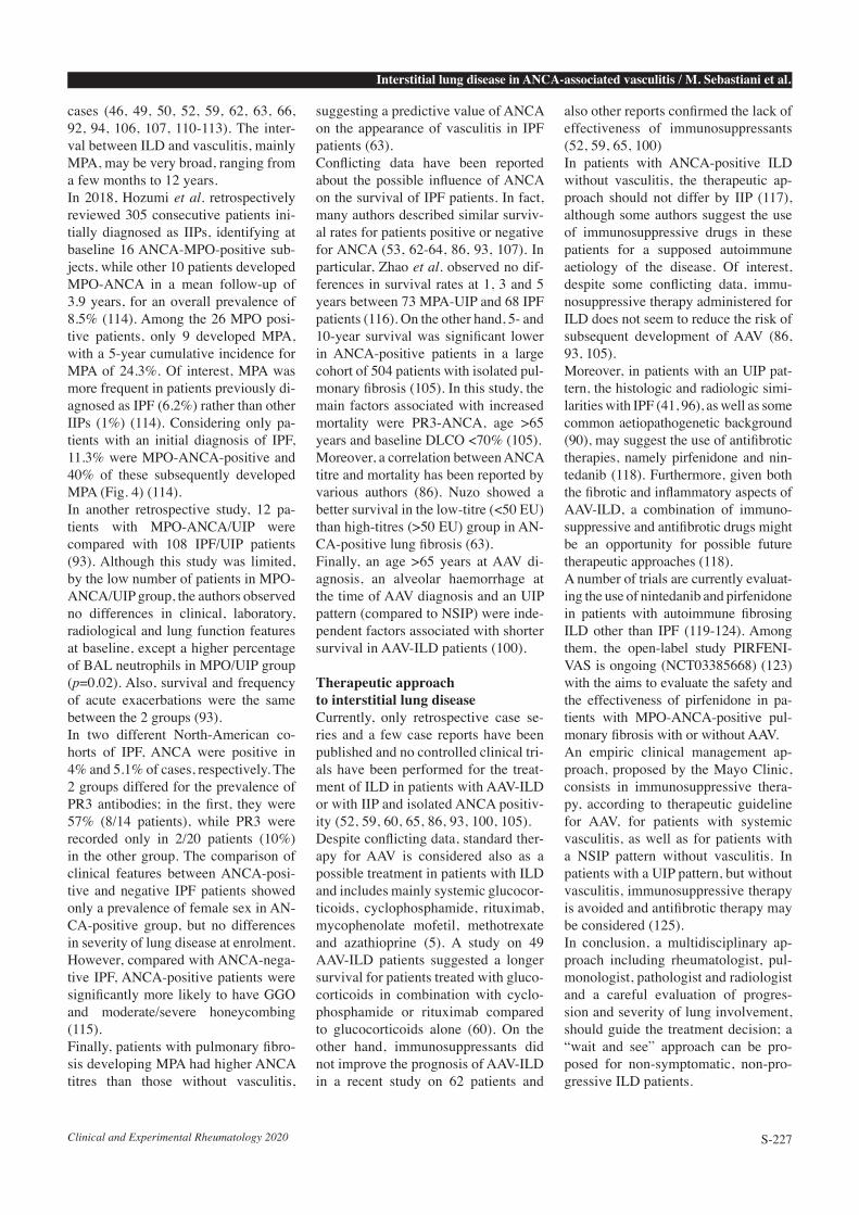
cases (46, 49, 50, 52, 59, 62, 63, 66, 92, 94, 106, 107, 110-113). The inter-val between ILD and vasculitis, mainly MPA, may be very broad, ranging from a few months to 12 years.In 2018, Hozumi et al. retrospectively reviewed 305 consecutive patients ini-tially diagnosed as IIPs, identifying at baseline 16 ANCA-MPO-positive sub-jects, while other 10 patients developed MPO-ANCA in a mean follow-up of 3.9 years, for an overall prevalence of 8.5% (114). Among the 26 MPO posi-tive patients, only 9 developed MPA, with a 5-year cumulative incidence for MPA of 24.3%. Of interest, MPA was more frequent in patients previously di-agnosed as IPF (6.2%) rather than other IIPs (1%) (114). Considering only pa-tients with an initial diagnosis of IPF, 11.3% were MPO-ANCA-positive and 40% of these subsequently developed MPA (Fig. 4) (114).In another retrospective study, 12 pa-tients with MPO-ANCA/UIP were compared with 108 IPF/UIP patients (93). Although this study was limited, by the low number of patients in MPO-ANCA/UIP group, the authors observed no differences in clinical, laboratory, radiological and lung function features at baseline, except a higher percentage of BAL neutrophils in MPO/UIP group (p=0.02). Also, survival and frequency of acute exacerbations were the same between the 2 groups (93).In two different North-American co-horts of IPF, ANCA were positive in 4% and 5.1% of cases, respectively. The 2 groups differed for the prevalence of 35��DQWLERGLHV�� LQ� WKH�ÀUVW�� WKH\�ZHUH�57% (8/14 patients), while PR3 were recorded only in 2/20 patients (10%) in the other group. The comparison of clinical features between ANCA-posi-tive and negative IPF patients showed only a prevalence of female sex in AN-CA-positive group, but no differences in severity of lung disease at enrolment. However, compared with ANCA-nega-tive IPF, ANCA-positive patients were VLJQLÀFDQWO\�PRUH�OLNHO\�WR�KDYH�**2�and moderate/severe honeycombing (115).)LQDOO\��SDWLHQWV�ZLWK�SXOPRQDU\�ÀEUR-sis developing MPA had higher ANCA titres than those without vasculitis,
suggesting a predictive value of ANCA on the appearance of vasculitis in IPF patients (63). &RQÁLFWLQJ� GDWD� KDYH� EHHQ� UHSRUWHG�DERXW� WKH�SRVVLEOH� LQÁXHQFH�RI�$1&$�on the survival of IPF patients. In fact, many authors described similar surviv-al rates for patients positive or negative for ANCA (53, 62-64, 86, 93, 107). In particular, Zhao et al. observed no dif-ferences in survival rates at 1, 3 and 5 years between 73 MPA-UIP and 68 IPF patients (116). On the other hand, 5- and ���\HDU� VXUYLYDO�ZDV� VLJQLÀFDQW� ORZHU�in ANCA-positive patients in a large cohort of 504 patients with isolated pul-PRQDU\�ÀEURVLV��������,Q�WKLV�VWXG\��WKH�main factors associated with increased mortality were PR3-ANCA, age >65 years and baseline DLCO <70% (105). Moreover, a correlation between ANCA titre and mortality has been reported by various authors (86). Nuzo showed a better survival in the low-titre (<50 EU) than high-titres (>50 EU) group in AN-&$�SRVLWLYH�OXQJ�ÀEURVLV�������Finally, an age >65 years at AAV di-agnosis, an alveolar haemorrhage at the time of AAV diagnosis and an UIP pattern (compared to NSIP) were inde-pendent factors associated with shorter survival in AAV-ILD patients (100).
Therapeutic approach to interstitial lung diseaseCurrently, only retrospective case se-ries and a few case reports have been published and no controlled clinical tri-als have been performed for the treat-ment of ILD in patients with AAV-ILD or with IIP and isolated ANCA positiv-ity (52, 59, 60, 65, 86, 93, 100, 105).'HVSLWH�FRQÁLFWLQJ�GDWD��VWDQGDUG�WKHU-apy for AAV is considered also as a possible treatment in patients with ILD and includes mainly systemic glucocor-ticoids, cyclophosphamide, rituximab, mycophenolate mofetil, methotrexate and azathioprine (5). A study on 49 AAV-ILD patients suggested a longer survival for patients treated with gluco-corticoids in combination with cyclo-phosphamide or rituximab compared to glucocorticoids alone (60). On the other hand, immunosuppressants did not improve the prognosis of AAV-ILD in a recent study on 62 patients and
DOVR�RWKHU�UHSRUWV�FRQÀUPHG�WKH�ODFN�RI�effectiveness of immunosuppressants (52, 59, 65, 100)In patients with ANCA-positive ILD without vasculitis, the therapeutic ap-proach should not differ by IIP (117), although some authors suggest the use of immunosuppressive drugs in these patients for a supposed autoimmune aetiology of the disease. Of interest, GHVSLWH� VRPH� FRQÁLFWLQJ� GDWD�� LPPX-nosuppressive therapy administered for ILD does not seem to reduce the risk of subsequent development of AAV (86, 93, 105). Moreover, in patients with an UIP pat-tern, the histologic and radiologic simi-larities with IPF (41, 96), as well as some common aetiopathogenetic background ������PD\�VXJJHVW�WKH�XVH�RI�DQWLÀEURWLF�therapies, namely pirfenidone and nin-tedanib (118). Furthermore, given both WKH�ÀEURWLF�DQG�LQÁDPPDWRU\�DVSHFWV�RI�AAV-ILD, a combination of immuno-VXSSUHVVLYH�DQG�DQWLÀEURWLF�GUXJV�PLJKW�be an opportunity for possible future therapeutic approaches (118).A number of trials are currently evaluat-ing the use of nintedanib and pirfenidone LQ� SDWLHQWV� ZLWK� DXWRLPPXQH� ÀEURVLQJ�ILD other than IPF (119-124). Among them, the open-label study PIRFENI-VAS is ongoing (NCT03385668) (123) with the aims to evaluate the safety and the effectiveness of pirfenidone in pa-tients with MPO-ANCA-positive pul-PRQDU\�ÀEURVLV�ZLWK�RU�ZLWKRXW�$$9��An empiric clinical management ap-proach, proposed by the Mayo Clinic, consists in immunosuppressive thera-py, according to therapeutic guideline for AAV, for patients with systemic vasculitis, as well as for patients with a NSIP pattern without vasculitis. In patients with a UIP pattern, but without vasculitis, immunosuppressive therapy LV�DYRLGHG�DQG�DQWLÀEURWLF�WKHUDS\�PD\�be considered (125).In conclusion, a multidisciplinary ap-proach including rheumatologist, pul-monologist, pathologist and radiologist and a careful evaluation of progres-sion and severity of lung involvement, should guide the treatment decision; a ´ZDLW� DQG� VHHµ� DSSURDFK� FDQ� EH� SUR-posed for non-symptomatic, non-pro-gressive ILD patients.

S-228 Clinical and Experimental Rheumatology 2020
Interstitial lung disease in ANCA-associated vasculitis / M. Sebastiani et al.
Proposal for patient management and treatmentThe treatment of AAV-ILD should be tailored for each patient and multidis-ciplinary approach, including at least rheumatologist, pulmonologist and radiologist, is mandatory to optimise therapy and follow-up strategies.Early diagnosis, functional and radio-logic follow-up of the lung involve-ment are necessary to identify patients with progressive disease (126, 127). In fact, the progression and the severity of ILD are the two main factors to be considered when a decision-making on treatment is requested. Patient’s age, ra-diologic or histo-pathologic pattern of ILD and subjective symptoms should also be carefully evaluated (128, 129).Moreover, when considering therapeu-tic options for patients with vasculitis, both pulmonary and extra-thoracic dis-ease manifestations need to be assessed
and taken into account (Fig. 7). Comor-bidities should be also considered for WKHLU�SRVVLEOH�LQÁXHQFH�RQ�WKH�VKRUW�DQG�long-term safety of the treatment (dia-betes mellitus, osteoporosis, etc.) (130).In such challenging condition, given the heterogeneity in disease presenta-tion, the multiple manifestations that may be present, and the broad range of disease severity, coordinated care is es-sential.In AAV-ILD patients, immunosuppres-VLYH�GUXJV�DUH�XVXDOO\�WKH�ÀUVW�FKRLFH��In particular, cyclophosphamide, my-cophenolate mofetil, rituximab or aza-thioprine have been demonstrated ef-ÀFDF\� RQ�$$9�DQG� KDYH� DOVR� VKRZHG�some evidence in ILD associated to rheumatic diseases (5, 131, 132).According to the results of INBUILD VWXG\�� LQ� SDWLHQWV� ZLWK� ÀEURWLF� SDWWHUQ�RI� ,/'� DQG� ZLWKRXW� VLJQLÀFDQW� DFWLY-ity of vasculitis, it is reasonable to pro-
pose the therapeutic strategies for IPF, QDPHO\�DQWLÀEURWLF�DJHQWV�VXFK�DV�SLUIH-nidone and nintedanib (117, 119, 133).,Q� SDWLHQWV� ZLWK� ÀEURWLF�� SURJUHVVLYH�ILD and active vasculitis, the results of INBUILD study and some previous ob-servations suggest the possibility of a combination therapy with both immu-QRVXSSUHVVDQWV�DQG�DQWL�ÀEURWLF�DJHQWV�(119, 134-136).Finally, in asymptomatic patients with mild, non-progressive ILD, a “wait and VHHµ�DSSURDFK�LV�XVXDOO\�DGRSWHG��������
ConclusionsThe association between AAV and ILD is well-known, but not always investi-gated in these patients. Moreover, even LI�,/'�VLJQLÀFDQWO\�LQÁXHQFH�WKH�SURJ-nosis and the quality of life of patients with AAV, ILD is not included in any activity or severity disease score pro-posed for AAV (namely BVAS, etc.), suggesting the need to review these scores in the light of the new knowl-edge on AAV-ILD (138).On the other hand, a search for ANCA is not usually included in the diagnos-tic work-up of IIP. Interestingly, ANCA have not been included in the research criteria for interstitial pneumonia with autoimmune features (IPAF), because its association with vasculitis rather than connective tissue diseases (139), and the current criteria do not allow to exclude an IPF in ANCA-positive pa-WLHQWV� ZLWK� SXOPRQDU\� ÀEURVLV� �������For all these reasons, ANCA should be investigated in all patients with IIP due to WKH�SRVVLEOH�VLJQLÀFDQW�SURJQRVWLF�LPSOL-cations, both on survival and on the risk of developing an AAV (46, 49, 51, 52, 59, 62, 63, 66, 92, 94, 104, 105, 108-111).Waiting for the results of ongoing tri-als, the treatment of AAV-ILD remains an unmet clinical need. The evaluation of the single patient with a multidisci-plinary approach including rheumatol-ogist, pulmonologist, pathologist and radiologist is mandatory for a correct FODVVLÀFDWLRQ�RI�WKHVH�SDWLHQWV�Finally, prospective ad hoc study should clarify the natural history of AAV-ILD and the role of ANCA posi-tivity in patients with IIP, other than the better therapeutic approach for the dif-ferent groups of patients.
Fig. 7. Proposed framework for the management and treatment of ANCA-associated vasculits pa-tients complicated by ILD. Therapeutic choice should derive by a multidisciplinary approach in-cluding rheumatologist, pulmonologist and radiologist. Use of immunosuppressants or, in selected SDWLHQWV��DQWLÀEURWLF�VKRXOG�EH�HYDOXDWHG�DFFRUGLQJ�WR�WKH�DFWLYLW\�DQG�VHYHULW\�RI�YDVFXOLWLF�PDQLIHV-tations and ILD.

S-229Clinical and Experimental Rheumatology 2020
Interstitial lung disease in ANCA-associated vasculitis / M. Sebastiani et al.
References 1. NAKAZAWA D, MASUDA S, TOMARU U,
ISHIZU A: Pathogenesis and therapeutic in-terventions for ANCA-associated vasculitis. Nat Rev Rheumatol 2019; 15: 91-101.
2. KALLENBERG CG: Key advances in the clin-ical approach to ANCA-associated vasculi-tis. Nat Rev Rheumatol 2014; 10: 484-93.
3. CORRAL-GUDINO L, GONZÁLEZ-VÁZQUEZ E, CALERO-PANIAGUA I et al.: The com-plexity of classifying ANCA-associated small-vessel vasculitis in actual clinical practice: data from a multicenter retrospec-tive survey. Rheumatol Int 2020; 40: 303-11.
4. LIONAKI S, BLYTH ER, HOGAN SL et al.: &ODVVLÀFDWLRQ�RI�DQWLQHXWURSKLO�F\WRSODVPLF�autoantibody vasculitides: the role of anti-neutrophil cytoplasmic autoantibody speci-ÀFLW\�IRU�P\HORSHUR[LGDVH�RU�SURWHLQDVH���LQ�disease recognition and prognosis. Arthritis Rheum 2012; 64: 3452-62.
5. YATES M, WATTS RA, BAJEMA IM et al.: EULAR/ERA-EDTA recommendations for the management of ANCA-associated vas-culitis. Ann Rheum Dis 2016; 75: 1583-94.
6. HOFFMAN GS, KERR GS, LEAVITT RY et al.: Wegener granulomatosis: an analysis of 158 patients. Ann Intern Med 1992; 116: 488-98.
7. WATTS RA, LANE SE, BENTHAM G et al.: Epidemiology of systemic vasculitis: a ten-year study in the United Kingdom. Arthritis Rheum 2000; 43, 414-9.
8. FRIES JF, HUNDER GG, BLOCH DA et al.: The American College of Rheumatology �����FULWHULD�IRU�WKH�FODVVLÀFDWLRQ�RI�YDVFX-litis: summary. Arthritis Rheum 1990; 33: 1135-6.
9. LYNCH JP: Granulomatosis with polyangiitis (Wegener’s Granulomatosis): evolving con-cepts in treatment. Semin Respir Crit Care Med 2018; 39: 434-58.
10. ANANTHAKRISHNAN L, SHARMA N, KANNE JP: Wegener’s granulomatosis in the FKHVW��KLJK�UHVROXWLRQ�&7�ÀQGLQJV��AJR Am J Roentgenol 2009; 192: 676-82.
11. THICKETT DR, RICHTER AG, NATHANI N et al.: Pulmonary manifestations of anti-neutrophil cytoplasmic antibody (ANCA)-positive vasculitis. Rheumatology (Oxford) 2006; 45: 261-8.
12. LEE K, KIM T, FUJIMOTO K et al.: Thoracic manifestation of Wegener’s granulomatosis: &7�ÀQGLQJV�LQ����SDWLHQWV��Eur Radiol 2003; 13: 43-51.
13. SEO J, IM JG, CHUNG J et al.: Pulmonary vas-FXOLWLV��WKH�VSHFWUXP�RI�UDGLRORJLFDO�ÀQGLQJV��Br J Radiol 2000; 73: 1224-31.
14. CORDIER JF, VALEYRE D, GUILLEVIN L et al.: Pulmonary Wegener’s granulomatosis: a clinical and imaging study of 77 cases. Chest 1990; 97: 906-13.
15. SHEEHAN RE, FLINT JD, MÜLLER NL: Computed tomography features of the tho-racic manifestations of Wegener granuloma-tosis. J Thorac Imaging 2003; 18: 34-41.
16. FAUCI AS, HAYNES BF, KATZ P et al.: Wegener’s granulomatosis: prospective clini-cal and therapeutic experience with 85 pa-tients for 21 years. Ann Intern Med 1983; 98: 76-85.
17. NASSER M, COTTIN V: The respiratory system in autoimmune vascular diseases.
Respiration 2018; 96: 12-28. 18. NASSER M, COTTIN V: Alveolar hemorrhage
in vasculitis (primary and secondary). Semin Respir Crit Care Med 2018; 39: 482-93.
19. WU EY, HERNANDEZ ML, JENNETTE JC et al.: Eosinophilic Granulomatosis with Poly-angiitis: Clinical Pathology Conference and Review. J Allergy Clin Immunol Pract 2018; 6: 1496-504.
20. ABRIL A: Churg-strauss syndrome: an up-date. Curr Rheumatol Rep 2011; 13: 489-95.
21. COTTIN V, BEL E, BOTTERO P et al.: Respira-tory manifestations of eosinophilic granulo-matosis with polyangiitis (Churg-Strauss). Eur Respir J 2016; 48: 1429-41.
22. REID AJ, HARRISON BD, WATTS RA et al.: Churg-Strauss syndrome in a district hospi-tal. QJM 1998; 91: 219-29.
23. CHUMBLEY LC, HARRISON EG JR, DERE-MEE RA: Allergic granulomatosis and an-giitis (Churg-Strauss syndrome). Report and analysis of 30 cases. Mayo Clin Proc 1977; 52: 477-84.
24. LANHAM JG, ELKON KB, PUSEY CD et al.: Systemic vasculitis with asthma and eo-sinophilia: a clinical approach to the Churg-Strauss syndrome. Medicine (Baltimore) 1984; 63: 65-81.
25. SIMON HU, ROTHENBERG ME, BOCHNER BS et al���5HÀQLQJ�WKH�GHÀQLWLRQ�RI�K\SHUH-osinophilic syndrome. J Allergy Clin Immu-nol 2010; 126: 45-9.
26. CHOI YH, IM JG, HAN BK et al.: Thoracic manifestation of Churg-Strauss syndrome: UDGLRORJLF�DQG�FOLQLFDO�ÀQGLQJV��Chest 2000; 117: 117-24.
27. CORDIER JF, COTTIN V, KHOUATRA C et al.: Hypereosinophilic obliterative bronchioli-tis: a distinct, unrecognised syndrome. Eur Respir J 2013; 41: 1126-34.
28. VALENT P, KLION AD, HORNY HP et al.: Contemporary consensus proposal on crite-ULD� DQG� FODVVLÀFDWLRQ� RI� HRVLQRSKLOLF� GLVRU-ders and related syndromes. J Allergy Clin Immunol 2012; 130: 607-12.e9.
29. COTTIN V: Eosinophilic Lung Diseases. Clin Chest Med 2016; 37: 535-56.
30. WU EY, HERNANDEZ ML, JENNETTE JC et al.: Eosinophilic Granulomatosis with Poly-angiitis: Clinical Pathology Conference and Review. J Allergy Clin Immunol Pract 2018; 6: 1496-504.
31. NGUYEN Y, GUILLEVIN L: Eosinophilic Granulomatosis with Polyangiitis (Churg-Strauss). Semin Respir Crit Care Med 2018; 39: 471-81.
32. JENNETTE JC, FALK RJ, BACON PA et al.: 2012 revised International Chapel Hill Con-sensus Conference Nomenclature of Vascu-litides. Arthritis Rheum 2013; 65: 1-11.
33. FUJIMOTO S, WATTS RA, KOBAYASHI S et al.: Comparison of the epidemiology of anti-neutrophil cytoplasmic antibody-associated vasculitis between Japan and the U.K. Rheu-matology (Oxford) 2011; 50: 1916-20.
34. NILSEN AT, KARLSEN C, BAKLAND G et al.: Increasing incidence and prevalence of ANCA-associated vasculitis in Northern Norway. Rheumatology (Oxford) 2019; 20: pii: kez597.
35. KARRAS A: Microscopic polyangiitis: new insights into pathogenesis, clinical features
and therapy. Semin Respir Crit Care Med 2018; 39: 459-64.
36. VILLIGER PM, GUILLEVIN L: Microscopic polyangiitis: clinical presentation. Autoim-mun Rev 2010; 9: 812-9.
37. DE PROST N, PARROT A, PICARD C et al.: Diffuse alveolar haemorrhage: factors asso-ciated with in-hospital and long-term mor-tality. Eur Respir J 2010; 35: 1303-11.
38. CASTAÑER E, ALGUERSUARI A, GALLAR-DO X et al.: When to suspect pulmonary vasculitis: radiologic and clinical clues. Radiographics 2010; 30: 33-53.
39. PRIMACK SL, MILLER RR, MÜLLER NL: Diffuse pulmonary hemorrhage: clinical, pathologic, and imaging features. AJR Am J Roentgenol 1995; 164: 295-300.
40. FERAGALLI B, MANTINI C, SPERANDEO M et al.: The lung in systemic vasculitis: radio-logical patterns and differential diagnosis. Br J Radiol 2016; 89: 20150992.
41. SUZUKI A, SAKAMOTO S, KUROSAKI A et al.: for Japan Research Committee of the Ministry of Health, Labour, and Welfare for Intractable Vasculitis and Research Commit-tee of Intractable Renal Disease of the Min-istry of Health, Labour, and Welfare of Ja-pan: Chest High-Resolution CT Findings of Microscopic Polyangiitis: A Japanese First Nationwide Prospective Cohort Study. AJR Am J Roentgenol 2019; 11: 1-11.
42. DE LASSENCE A, FLEURY-FEITH J, ESCUDI-ER E et al.: Alveolar hemorrhage. Diagnostic criteria and results in 194 immunocompro-mised hosts. Am J Respir Crit Care Med 1995; 151: 157-63.
43. CORDIER JF: A lung biopsy is unnecessary in the management of ANCA-positive patients with chest-roentgenographic abnormalities. Sarcoidosis Vasc Diffuse Lung Dis 1999; 13: 235-7.
44. CARTIN-CEBA R, DIAZ-CABALLERO L, AL-QADI MO et al.: Diffuse alveolar hemorrhage secondary to antineutrophil cytoplasmic antibody-associated vasculitis: predictors of respiratory failure and clinical outcomes. Arthritis Rheumatol 2016; 68: 1467-76.
45. THOMPSON GE, SPECKS U: Update on the management of respiratory manifestations of the antineutrophil cytoplasmic antibod-ies-associated vasculitides. Clin Chest Med 2019; 40: 573-82.
46. FERNANDEZ CASARES M, GONZALEZ A, FIELLI M et al.: Microscopic polyangiitis as-VRFLDWHG�ZLWK�SXOPRQDU\�ÀEURVLV��Clin Rheu-matol 2015; 34: 1273-7.
47. MOHAMMAD AJ, MORTENSEN KH, BABAR J et al.: Pulmonary involvement in antineu-trophil cytoplasmic antibodies (ANCA)-as-VRFLDWHG� YDVFXOLWLV�� WKH� LQÁXHQFH� RI�$1&$�subtype. J Rheumatol 2017; 44: 1458-67.
48. HRUSKOVA Z, CASIAN AL, KONOPASEK P et al.: Long-term outcome of severe alveolar haemorrhage in ANCA-associated vascu-litis: a retrospective cohort study. Scand J Rheumatol 2013; 42: 211-4.
49. NADA AK, TORRES VE, RYU JH et al.: 3XOPRQDU\� ÀEURVLV� DV� DQ� XQXVXDO� FOLQLFDO�manifestation of a pulmonary-renal vasculi-tis in elderly patients. Mayo Clin Proc 1990; 65: 847-56.
50. ARIMURA Y, MINOSHIMA S, TANAKA U et

S-230 Clinical and Experimental Rheumatology 2020
Interstitial lung disease in ANCA-associated vasculitis / M. Sebastiani et al.
al.: Pulmonary involvement in patients with P\HORSHUR[LGDVH� VSHFLÀF�DQWLQHXWURSKLO� F\-toplasmic antibody. Ryumachi 1995; 35: 46-55.
51. TZELEPIS GE, KOKOSI M, TZIOUFAS A et al.: 3UHYDOHQFH�DQG�RXWFRPH�RI�SXOPRQDU\�ÀEUR-sis in microscopic polyangiitis. Eur Respir J 2010; 36: 116-21.
52. ARULKUMARAN N, PERISELNERIS N, GASKIN G et al.: Interstitial lung disease and ANCA-associated vasculitis: a retrospective observational cohort study. Rheumatology (Oxford) 2011; 50: 2035-43.
53. KATSUMATA Y, KAWAGUCHI Y, YAMANAKA H: Interstitial lung disease with ANCA-as-sociated Vasculitis. Clin Med Insights Circ Respir Pulm Med 2015; 9: 51-6.
54. HIRAYAMA K, KOBAYASHI M, USUI J et al.: Pulmonary involvements of anti-neutrophil cytoplasmic autoantibody-associated renal vasculitis in Japan. Nephrol Dial Transplant 2015; 30: i83-93.
55. KATSUYAMA T, SADA KE, MAKINO H: Cur-rent concept and epidemiology of systemic vasculitides. Allergol Int 2014; 63: 505-13.
56. FURUTA S, CHAUDHRY AN, HAMANO Y et al.: Comparison of phenotype and outcome in microscopic polyangiitis between Europe and Japan. J Rheumatol 2014; 41: 325-33.
57. AZUMA A, HAGIWARA K, KUDOH S: Basis of acute exacerbation of idiopathic pulmonary ÀEURVLV� LQ� -DSDQHVH� SDWLHQWV�� Am J Respir Crit Care Med 2008; 177: 1397-8.
58. WATTS RA, SCOTT DG, JAYNE DR et al.: Re-nal vasculitis in Japan and the UK - are there differences in epidemiology and clinical phenotype? Nephrol Dial Transplant 2008; 23: 3928-31.
59. HERVIER B, PAGNOUX C, AGARD C et al.: 9DVFXOLWLV� VWXG\�� SXOPRQDU\� ÀEURVLV� DVVRFL-ated with ANCA-positive vasculitides. Retro-spective study of 12 cases and review of the literature. Ann Rheum Dis 2009; 68: 404-7.
60. COMARMOND C, CRESTANI B, TAZI A et al��� 3XOPRQDU\� ÀEURVLV� LQ� DQWLQHXWURSKLO�cytoplasmic antibodies (ANCA)-associated vasculitis: a series of 49 patients and review of the literature. Medicine (Baltimore) 2014; 93: 340-9.
61. CHEN M, YU F, ZHANG Y, ZHAO MH: Anti-neutrophil cytoplasmic autoantibody-asso-ciated vasculitis in older patients. Medicine (Baltimore) 2008; 87: 203-9.
62. FOULON G, DELAVAL P, VALEYRE D et al.: $1&$�DVVRFLDWHG� OXQJ�ÀEURVLV��DQDO\VLV�RI�17 patients. Respir Med 2008; 102: 1392-8.
63. NOZU T, KONDO M, SUZUKI K, TAMAOKI J, NAGAI A: A comparison of the clinical features of ANCA-positive and ANCA-neg-DWLYH�LGLRSDWKLF�SXOPRQDU\�ÀEURVLV�SDWLHQWV��Respiration 2009; 77: 407-15.
64. HOZUMI H, ENOMOTO N, OYAMA Y et al.: Clinical implication of proteinase-3-anti-neutrophil cytoplasmic antibody in patients with idiopathic interstitial pneumonias. Lung 2016; 194: 235-42.
65. HOMMA S, MATSUSHITA H, NAKATA K: 3XOPRQDU\�ÀEURVLV�LQ�P\HORSHUR[LGDVH�DQWL-neutrophil cytoplasmic antibody-associated vasculitides. Respirology 2004; 9: 190-6.
66. FLORES-SUÁREZ LF, RUIZ N, SALDAR-RIAGA RIVERA LM, PENSADO L: Reduced survival in microscopic polyangiitis patients
ZLWK� SXOPRQDU\� ÀEURVLV� LQ� D� UHVSLUDWRU\�referral centre. Clin Rheumatol 2015; 34: 1653-4.
67. ESCHUN GM, MINK SN, SHARMA S: Pul-PRQDU\� LQWHUVWLWLDO� ÀEURVLV� DV� D� SUHVHQWLQJ�manifestation in perinuclear antineutrophilic cytoplasmic antibody microscopic polyangi-itis. Chest 2003; 123: 297-301.
68. HIROMURA K, NOJIMA Y, KITAHARA T et al.: Four cases of anti-myeloperoxidase an-tibody-related rapidly progressive glomeru-lonephritis during the course of idiopathic SXOPRQDU\�ÀEURVLV��Clin Nephrol 2000; 53: 384-9.
69. NAKABAYASHI K, ARIMURA Y, YOSHIHARA K et al���&ODVVLÀFDWLRQ�RI�FOLQLFDO�VXEW\SHV��patient survival, kidney prognosis, and re-lapse in patients with MPO-ANCA-associ-ated vasculitis: a single-center experience. Mod Rheumatol 2009; 19: 420-6.
70. NAKABAYASHI K, FUJIOKA Y, NAGASAWA T et al.: Dual myeloperoxidase-antineutrophil cytoplasmic antibody- and antiglomerular base-ment membrane antibody-positive FDVHV�DVVRFLDWHG�ZLWK�SULRU�SXOPRQDU\�ÀEUR-sis: a report of four cases. Clin Exp Nephrol 2011; 15: 226-34.
71. SHIRAKI A, ANDO M, SHINDOH J et al.: Prevalence of myeloperoxidase-anti-neutro-phil cytoplasmic antibody (MPO-ANCA) in patients with interstitial pneumonia. Nihon Kokyuki Gakkai Zasshi 2007; 45: 921-6.
72. STREHO M, SABLE-FOURTASSOU R, BRION MC et al.: Churg-Strauss syndrome and SXOPRQDU\�ÀEURVLV��DQ�XQXVXDO�DVVRFLDWLRQ��Presse Med 2006; 35: 1259-62.
73. BIRNBAUM J, DANOFF S, ASKIN FB, STONE JH: Microscopic polyangiitis presenting as a ´SXOPRQDU\�PXVFOHµ�V\QGURPH��LV�VXEFOLQL-cal alveolar hemorrhage the mechanism of SXOPRQDU\� ÀEURVLV"�Arthritis Rheum 2007; 56: 2065-71.
74. PINETON DE CHAMBRUN M, NUNES H, BROCHERIOU I, HERTIG A: Idiopathic lung ÀEURVLV� DQG� DQWLP\HORSHUR[LGDVH� JORPHUX-lonephritis: the tree that hides the forest. BMC Pulm Med 2015; 15: 130.
75. SCHNABEL A, REUTER M, CSERNOK E, RICHTER C, GROSS WL: Subclinical alveo-lar bleeding in pulmonary vasculitides: cor-relation with indices of disease activity. Eur Respir J 1999; 14: 118-24.
76. BUSCHMAN DL, BALLARD R: Progressive PDVVLYH� ÀEURVLV� DVVRFLDWHG� ZLWK� LGLRSDWKLF�pulmonary hemosiderosis. Chest 1993; 104: 293-5.
77. CORRIN B, JAGUSCH M, DEWAR A et al.: Fine structural changes in idiopathic pulmo-nary haemosiderosis. J Pathol 1987; 153: 249-56.
78. FOUCHER P, HEERINGA P, PETERSEN AH et al.: Antimyeloperoxidase-associated lung disease. An experimental model. Am J Respir Crit Care Med 1999; 160: 987-94.
79. GUILPAIN P, CHEREAU C, GOULVESTRE C et al.: The oxidation induced by antimyeloper-R[LGDVH�DQWLERGLHV�WULJJHUV�ÀEURVLV�LQ�PLFUR-scopic polyangiitis. Eur Respir J 2011; 37: 1503-13.
80. FLAHERTY KR, THWAITE EL, KAZEROONI EA et al.: Radiological versus histological diagnosis in UIP and NSIP: survival impli-cations. Thorax 2003; 58: 143-8.
81. CHRYSANTHOPOULOU A, MITROULIS I, AP-OSTOLIDOU E et al.: Neutrophil extracellular traps promote differentiation and function of ÀEUREODVWV. J Pathol 2014; 233: 294-307.
82. YOSHIDA M, YAMADA M, SUDO Y et al.: Myeloperoxidase anti-neutrophil cytoplas-PLF�DQWLERG\�DIÀQLW\� LV�DVVRFLDWHG�ZLWK� WKH�formation of neutrophil extracellular traps in the kidney and vasculitis activity in my-eloperoxidase anti-neutrophil cytoplasmic antibody-associated microscopic polyangii-tis. Nephrology (Carlton) 2016; 21: 624-9.
83. TRAVIS WD, HOFFMAN GS, LEAVITT RY, PASS HI, FAUCI AS: Surgical pathology of the lung in Wegener’s granulomatosis. Re-view of 87 open lung biopsies from 67 pa-tients. Am J Surg Pathol 1991; 15: 315-33.
84. VANCHERI C, GAULDIE J, BIENENSTOCK J et al���+XPDQ� OXQJ�ÀEUREODVW�GHULYHG�JUDQ-ulocyte-macrophage colony stimulating fac-tor (GM- CSF) mediates eosinophil survival in vitro. Am J Respir Cell Mol Biol 1989; 1: 289-95.
85. PETERSON MW, MONICK M, HUNNING-HAKE GW: Prognostic role of eosinophils in SXOPRQDU\�ÀEURVLV��Chest 1987; 92: 51-6.
86. ANDO M, MIYAZAKI E, ISHII T et al.: Inci-dence of myeloperoxidase anti-neutrophil cytoplasmic antibody positivity and micro-scopic polyangitis in the course of idiopathic SXOPRQDU\� ÀEURVLV��Respir Med 2013; 107: 608-15.
87. BORIE R, CRESTANI B: Antineutrophil cyto-SODVPLF� DQWLERG\�DVVRFLDWHG� OXQJ� ÀEURVLV��Semin Respir Crit Care Med 2018; 39: 465-470.
88. CHURG A, ZAY K, SHAY S et al.: Acute cigarette smoke- induced connective tissue breakdown requires both neutrophils and macrophage metalloelastase in mice. Am J Respir Cell Mol Biol 2002; 27: 368-74.
89. NAMBA N, KAWASAKI A, SADA KE et al.: Association of MUC5B promoter polymor-phism with interstitial lung disease in my-eloperoxidase-antineutrophil cytoplasmic antibody-associated vasculitis. Ann Rheum Dis 2019; 78: 1144-6.
90. LEE MG, LEE YH: A meta-analysis exam-ining the association between the MUC-5Brs35705950 T/G polymorphism and sus-FHSWLELOLW\� WR� LGLRSDWKLF�SXOPRQDU\�ÀEURVLV��,QÁDPP�5HV 2015; 64: 463-70.
91. JUGE PA, LEE JS, EBSTEIN E et al.: MUC5B promoter variant and rheumatoid arthritis with interstitial lung disease. N Engl J Med 2018; 379: 2209-19.
92. YU AP, CHANG JX, LIU YJ, QU QR: Computed tomography image analysis before and after treatment of anti-neutrophil cytoplasmic antibody-associated pulmonary interstitial ÀEURVLV� LQ� �� SDWLHQWV��Clin Ther 2014; 36: 2064-71.
93. HOSODA C, BABA T, HAGIWARA E et al.: Clinical features of usual interstitial pneu-monia with anti-neutrophil cytoplasmic an-tibody in comparison with idiopathic pulmo-QDU\�ÀEURVLV��Respirology 2016; 21: 920-6.
94. HUANG H, WANG YX, JIANG CG et al.: A retrospective study of microscopic poly-angiitis patients presenting with pulmonary ÀEURVLV�LQ�&KLQD��BMC Pulm Med 2014; 14: 8.
95. HOMMA S, SUZUKI A, SATO K: Pulmonary involvement in ANCA-associated vasculitis

S-231Clinical and Experimental Rheumatology 2020
Interstitial lung disease in ANCA-associated vasculitis / M. Sebastiani et al.
from the view of the pulmonologist. Clin Exp Nephrol 2013; 17: 667-71.
96. ALBA MA, FLORES-SUÁREZ LF, HENDER-SON AG et al.: Interstital lung disease in ANCA vasculitis. Autoimmun Rev 2017; 16: 722-9.
97. HUNNINGHAKE GM: Interstitial lung abnor-malities: erecting fences in the path towards DGYDQFHG�SXOPRQDU\�ÀEURVLV��Thorax 2019; 74: 506-11.
98. YAMAGATA M, IKEDA K, TSUSHIMA K et al.: Prevalence and responsiveness to treatment of lung abnormalities on chest computed tomography in patients with microscopic polyangiitis: a multicenter, longitudinal, UHWURVSHFWLYH�VWXG\�RI�RQH�KXQGUHG�ÀIW\�FRQ-secutive hospital-based Japanese patients. Arthritis Rheum 2016; 68: 713-23.
99. ANDO Y, OKADA F, MATSUMOTO S, MORI H: Thoracic manifestation of myeloperoxidase-antineutrophil cytoplasmic antibody (MPO-$1&$��UHODWHG� GLVHDVH�� &7� ÀQGLQJV� LQ� ���patients. J Comput Assist Tomogr 2004; 28: 710-6.
100. MAILLET T, GOLETTO T, BELTRAMO G et al.: French Vasculitis Study Group (FVSG): Usual interstitial pneumonia in ANCA-asso-ciated vasculitis: A poor prognostic factor. J Autoimmun 2020; 106: 102338.
101. TZOUVELEKIS A, ZACHARIS G, OIKONO-MOU A et al���&RPELQHG�SXOPRQDU\�ÀEURVLV�and emphysema associated with microscopic polyangiitis. Eur Respir J 2012; 40: 505-7.
102. GOCHO K, SUGINO K, SATO K, HASEGAWA C, UEKUSA T, HOMMA S: Microscopic poly-angiitis preceded by combined pulmonary ÀEURVLV� DQG� HPSK\VHPD��Respir Med Case Rep 2015; 15: 128-32.
103. BOOTH AD, ALMOND MK, BURNS A et al.: Outcome of ANCA-associated renal vascu-litis: a 5-year retrospective study. Am J Kid-ney Dis 2003; 41: 776-84.
104. SCHIRMER JH, WRIGHT MN, VONTHEIN R et al.: Clinical presentation and long-term out-come of 144 patients with microscopic poly-angiitis in a monocentric German cohort. Rheumatology (Oxford) 2016; 55: 71-9.
105. BECKER-MEROK A, NOSSENT JC, RITLAND N: Fibrosing alveolitis predating microscopic polyangiitis. Scand J Rheumatol 1999; 28: 254-6.
106. KONO M, NAKAMURA Y, ENOMOTO N et al.: Usual interstitial pneumonia preceding col-lagen vascular disease: a retrospective case control study of patients initially diagnosed ZLWK� LGLRSDWKLF� SXOPRQDU\� ÀEURVLV�� PLoS One 2014; 9: e94775.
107. KAGIYAMA N, TAKAYANAGI N, KANAUCHI T, ISHIGURO T, YANAGISAWA T, SUGITA Y: Antineutrophil cytoplasmic antibody-posi-tive conversion and microscopic polyangii-tis development in patients with idiopathic SXOPRQDU\� ÀEURVLV��BMJ Open Respir Res 2015; 2: e000058.
108. TANAKA T, OTANI K, EGASHIRA R et al.: Interstitial pneumonia associated with MPO-ANCA: clinicopathological features of nine patients. Respir Med 2012; 106: 1765-70.
109. KANG BH, PARK JK, ROH JH et al.: Clini-FDO� VLJQLÀFDQFH� RI� VHUXP� DXWRDQWLERGLHV� LQ�idiopathic interstitial pneumonia. J Korean Med Sci 2013; 28: 731-7.
110. SOUID M, TERKI NH, NOCHY D, HILLION D:
Myeloperoxidase anti-neutrophil cytoplasmic autoantibodies (MPO-ANCA)-related rapidly progressive glomerulonephritis (RPGN) and SXOPRQDU\�ÀEURVLV��3)��ZLWK�GLVVRFLDWHG�HYR-lution. Clin Nephrol 2001; 55: 337-8.
111. ELEFTHERIOU D, KATSENOS S, ZORBAS S, GRIVEAS I, PSATHAKIS K�� 3XOPRQDU\� À-brosis presenting as an early manifestation of microscopic polyangiitis. Monaldi Arch Chest Dis 2012; 77: 141-4.
112. GAUDIN PB, ASKIN FB, FALK RJ, JENNETTE JC: The pathologic spectrum of pulmonary lesions in patients with anti-neutrophil cy-WRSODVPLF� DXWRDQWLERGLHV� VSHFLÀF� IRU� DQWL�proteinase 3 and anti-myeloperoxidase. Am J Clin Pathol 1995; 104: 7-16.
113. FLORES-SUAREZ LF: Limited pulmonary MPA, a new MPA entity? A rheumatolo-gist’s perspective. Clin Exp Nephrol 2013; 17: 672-5.
114. HOZUMI H, OYAMA Y, YASUI H et al.: Clini-FDO� VLJQLÀFDQFH� RI� P\HORSHUR[LGDVH�DQWL�neutrophil cytoplasmic antibody in idiopath-ic interstitial pneumonias. PLoS One 2018; 21: e0199659.
115. LIU GY, VENTURA IB, ACHTAR-ZADEH N et al��� 3UHYDOHQFH� DQG� FOLQLFDO� VLJQLÀFDQFH�of antineutrophil cytoplasmic antibodies in north american patients with idiopathic pul-PRQDU\�ÀEURVLV��Chest 2019; 156: 715-23.
116. ZHAO W, DAI H, LIU Y et al.: Clinical fea-tures and prognosis of microscopic poly-angiitis with usual interstitial pneumonia FRPSDUHG�ZLWK� LGLRSDWKLF�SXOPRQDU\�ÀEUR-sis. Clin Respir J 2019; 13: 460-6.
117. RAGHU G, ROCHWERG B, ZHANG Y et al.: $Q� 2IÀFLDO� $76�(56�-56�$/$7� &OLQLFDO�Practice Guideline: Treatment of Idiopathic Pulmonary Fibrosis. An Update of the 2011 Clinical Practice Guideline. Am J Respir Crit Care Med 2015; 192: e3-19.
118. MORISSET J, LEE JS: New trajectories in the treatment of interstitial lung disease: treat the disease or treat the underlying pattern? Curr Opin Pulm Med 2019; 25: 442-9.
119. FLAHERTY KR, WELLS AU, COTTIN V et al.: 1LQWHGDQLE�LQ�SURJUHVVLYH�ÀEURVLQJ�LQWHUVWL-tial lung diseases. N Engl J Med 2019; 381: 1718-27.
120. BEHR J, NEUSER P, PRASSE A et al.: Explor-LQJ� HIÀFDF\� DQG� VDIHW\� RI� RUDO� 3LUIHQLGRQH�IRU�SURJUHVVLYH��QRQ�,3)� OXQJ�ÀEURVLV� �5(-LIEF) - a randomized, double blind, place-bo-controlled, parallel group, multi-center, phase II trial. BMC Pulm Med 2017; 17: 122.
121. SOLOMON JJ, DANOFF SK, GOLDBERG HJ et al.: Trail Network. The design and rationale of the Trail1 trial: a randomized double-blind phase 2 clinical trial of pirfenidone in rheumatoid arthritis-associated interstitial lung disease. Adv Ther 2019; 36: 3279-87.
122. MAHER TM, CORTE TJ, FISCHER A et al.: 3LUIHQLGRQH� LQ� SDWLHQWV� ZLWK� XQFODVVLÀDEOH�SURJUHVVLYH� ÀEURVLQJ� LQWHUVWLWLDO� OXQJ� GLV-ease: design of a double-blind, randomised, placebo-controlled phase II trial. BMJ Open Resp Res 2018; 5: e000289.
123. LONDON J, AIT EL GHAZ S: Pilot study of SLUIHQLGRQH�LQ�SXOPRQDU\�ÀEURVLV�ZLWK�DQWL�myeloperoxydase antibodies (PIRFENI-VAS). https://clinicaltrials.gov/ct2/show/NCT03385668 Date last updated: March 19, 2018. Date last accessed: May 21, 2018.
124. BAUGHMAN RP, REEVES R: Pirfenidone for Progressive Fibrotic Sarcoidosis (Pi-rFS). https://clinicaltrials.gov/ct2/show/NCT03260556.
125. THOMPSON GE, SPECKS U: Update on the management of respiratory manifestations of the antineutrophil cytoplasmic antibod-ies-associated vasculitides. Clin Chest Med 2019; 40: 573-82.
126. KOLB M, VAŠÁKOVÁ M: The natural history RI�SURJUHVVLYH�ÀEURVLQJ�LQWHUVWLWLDO�OXQJ�'LV-eases. Respir Res 2019; 20: 57.
127. COTTIN V, HIRANI NA, HOTCHKIN DL et al.: Presentation, diagnosis and clinical course RI�WKH�VSHFWUXP�RI�SURJUHVVLYH�ÀEURVLQJ�LQ-terstitial lung diseases. Eur Respir Rev 2018; 27: 180076.
128. WONG AW, RYERSON CJ, GULER SA: 3URJUHVVLRQ�RI�ÀEURVLQJ�LQWHUVWLWLDO�OXQJ�GLV-ease. Respir Res 2020; 21: 32.
129. WUYTS WA, WIJSENBEEK M, BONDUE B et al.��,GLRSDWKLF�SXOPRQDU\�ÀEURVLV��EHVW�SUDF-tice in monitoring and managing a relentless ÀEURWLF� GLVHDVH��Respiration 2020; 99: 73-82.
130. MARGARITOPOULOS GA, ANTONIOU KM, WELLS AU: Comorbidities in interstitial lung diseases. Eur Respir Rev 2017; 26: 160027.
131. VACCHI C, SEBASTIANI M, CASSONE G et al.: Therapeutic options for the treatment of interstitial lung disease related to connective tissue diseases. A narrative review. J Clin Med 2020; 9: E407.
132. CASSONE G, MANFREDI A, VACCHI C et al.: Treatment of rheumatoid arthritis-associated interstitial lung disease: lights and shadows. J Clin Med 2020; 9: E1082.
133. COLLINS BF, RAGHU G��$QWLÀEURWLF�WKHUDS\�IRU� ÀEURWLF� OXQJ� GLVHDVH� EH\RQG� LGLRSDWKLF�SXOPRQDU\� ÀEURVLV�� Eur Respir Rev 2019; 28: 190022.
134. VACCHI C, MANFREDI A, CASSONE G et al.: Combination therapy with nintedanib and sarilumab for the management of rheuma-toid arthritis related interstitial lung disease. Case Rep Med 2020; 2020: 6390749.
135. Scleroderma Lung Study III - Combining Pirfenidone With Mycophenolate (SLSIII). &OLQLFDO7ULDOV�JRY�,GHQWLÀHU��1&7����������Available online: https://clinicaltrials.gov/ct2/show/NCT03221257 (accessed on 22 February 2020)
136. MONTANTE A, LE BRAS A, PAGNOUX C et al.: Cost-effectiveness of rituximab versus azathioprine for maintenance treatment in antineutrophil cytoplasmic antibody-associ-ated vasculitis. Clin Exp Rheumatol 2019; 37 (Suppl. 117): S137-43.
137. ANTONELOU M, PEREA ORTEGA L, HARVEY J, SALAMA AD: Anti-myeloperoxidase anti-body positivity in patients without primary systemic vasculitis. Clin Exp Rheumatol 2019; 37 (Suppl. 117): S86-9.
138. LUQMANI RA, BACON PA, MOOTS RJ et al.: Birmingham Vasculitis Activity Score (BVAS) in systemic necrotizing vasculitis. QJM 1994; 87: 671-8.
139. FISCHER A, ANTONIOU KM, BROWN KK et al��� $Q� RIÀFLDO� (XURSHDQ� 5HVSLUDWRU\� 6o-ciety/American Thoracic Society research statement: interstitial pneumonia with au-toimmune features. Eur Respir J 2015; 46: 976-87.