Review C erebrovascular requirement for sealant, anti ...
15
Brain Research Reviews 43 (2003) 164–178 www.elsevier.com / locate / brainresrev Review Cerebrovascular requirement for sealant, anti-coagulant and remodeling molecules that allow for the maintenance of vascular integrity and blood supply a,b, c b b * Craig S. Atwood , Richard L. Bowen , Mark A. Smith , George Perry a School of Medicine, University of Wisconsin and William S. Middleton Memorial Veterans Administration, GRECC 11G, 2500 Overlook Terrace, Madison, WI 53705, USA b Institute of Pathology, Case Western Reserve University, 2085 Adelbert Road, Cleveland, OH 44106, USA c Voyager Pharmaceutical Corporation, 8450 Colonnade Center Drive, Suite 409, Raleigh, NC 27615, USA Accepted 17 June 2003 Abstract The integrity of the vasculature and the maintenance of the blood supply to the brain are crucial for the survival of higher vertebrates. However, peripheral mechanisms of sealing the vasculature that rely on the clotting of blood and platelet aggregation around the site of a ‘leak’ would lead to decreased cerebral perfusion and compromise the viability of terminally differentiated and irreplaceable neurons. Therefore, in higher organisms it is likely that a sealant / anti-coagulant system that maintains vascular supply has evolved as a necessity to life. We propose that one such system involves the amyloid-b precursor protein (AbPP) and its cleavage product Ab since (1) both AbPP/Ab are known to deposit in the media of the cerebrovasculature wall following localized injury, (2) Ab is generated from AbPP, a known acute phase reactant, (3) Ab’s physiochemical properties allow it to span between the extracellular matrix and the (endothelial) cell membrane and under inflammatory conditions aggregate to form an intracranial ‘scab’, thereby maintaining structural integrity of the blood brain barrier, (4) AbPP/Ab together act as an anti-coagulant, (5) Ab promotes vascular / neuronal remodeling, and (6) Ab deposits resolve after injury. These properties are consistent with the acute phase generation and rapid cortical deposition of AbPP/Ab following injury (either sustained by trauma or stresses associated with aging) that would be an important compensatory response aimed at limiting the loss of terminally differentiated neurons. Such a system would allow the maintenance of blood supply to the brain by sealing vascular lesions, preventing hemorrhagic stroke while at the same time inhibiting the coagulation cascade from blocking capillaries. Obviously, strategies to remove Ab would have serious consequences for the integrity of the blood–brain barrier. Indeed, recent in vivo evidence demonstrates that the removal of deposited Ab from the vasculature leads to increased cerebral microhemorrhage and strongly support the above mentioned functions of AbPP/Ab. These insights also explain the root cause of the encephalitis and meningitis suffered by individuals in immunotherapy trials as being directly associated with the removal of Ab from the vasculature, i.e. immunological responses to Ab vaccination do not discriminate between physiologically purposive deposits of Ab (vascular deposits) and pathological deposits of Ab (senile plaques). 2003 Elsevier B.V. All rights reserved. Theme: Cellular and molecular biology Topic: Blood–brain barrier Keywords: Amyloid-b; Anti-coagulant; Blood–brain barrier; Blood supply; Cerebrovasculature; Hemorrhage; Sealant; Vascular integrity Contents 1. Introduction ............................................................................................................................................................................................ 165 2. Amyloid-b: a candidate vascular sealant, anti-coagulant and remodeling molecule ....................................................................................... 166 *Corresponding author. Tel.: 11-608-256-1901, Ext. 11664; fax: 11-608-280-7248. E-mail address: [email protected] (C.S. Atwood). 0165-0173 / 03 / $ – see front matter 2003 Elsevier B.V. All rights reserved. doi:10.1016 / S0165-0173(03)00206-6
Transcript of Review C erebrovascular requirement for sealant, anti ...

Brain Research Reviews 43 (2003) 164–178www.elsevier.com/ locate/brainresrev
Review
C erebrovascular requirement for sealant, anti-coagulant and remodelingmolecules that allow for the maintenance of vascular integrity and
blood supplya,b , c b b*Craig S. Atwood , Richard L. Bowen , Mark A. Smith , George Perry
aSchool of Medicine, University of Wisconsin and William S. Middleton Memorial Veterans Administration, GRECC 11G, 2500 Overlook Terrace,Madison, WI 53705,USA
bInstitute of Pathology, Case Western Reserve University, 2085 Adelbert Road, Cleveland, OH 44106,USAcVoyager Pharmaceutical Corporation, 8450 Colonnade Center Drive, Suite 409, Raleigh, NC 27615,USA
Accepted 17 June 2003
Abstract
The integrity of the vasculature and the maintenance of the blood supply to the brain are crucial for the survival of higher vertebrates.However, peripheral mechanisms of sealing the vasculature that rely on the clotting of blood and platelet aggregation around the site of a‘leak’ would lead to decreased cerebral perfusion and compromise the viability of terminally differentiated and irreplaceable neurons.Therefore, in higher organisms it is likely that a sealant /anti-coagulant system that maintains vascular supply has evolved as a necessity tolife. We propose that one such system involves the amyloid-b precursor protein (AbPP) and its cleavage product Ab since (1) bothAbPP/Ab are known to deposit in the media of the cerebrovasculature wall following localized injury, (2) Ab is generated from AbPP, aknown acute phase reactant, (3) Ab’s physiochemical properties allow it to span between the extracellular matrix and the (endothelial)cell membrane and under inflammatory conditions aggregate to form an intracranial ‘scab’, thereby maintaining structural integrity of theblood brain barrier, (4) AbPP/Ab together act as an anti-coagulant, (5) Ab promotes vascular /neuronal remodeling, and (6) Ab depositsresolve after injury. These properties are consistent with the acute phase generation and rapid cortical deposition of AbPP/Ab followinginjury (either sustained by trauma or stresses associated with aging) that would be an important compensatory response aimed at limitingthe loss of terminally differentiated neurons. Such a system would allow the maintenance of blood supply to the brain by sealing vascularlesions, preventing hemorrhagic stroke while at the same time inhibiting the coagulation cascade from blocking capillaries. Obviously,strategies to remove Ab would have serious consequences for the integrity of the blood–brain barrier. Indeed, recent in vivo evidencedemonstrates that the removal of deposited Ab from the vasculature leads to increased cerebral microhemorrhage and strongly support theabove mentioned functions of AbPP/Ab. These insights also explain the root cause of the encephalitis and meningitis suffered byindividuals in immunotherapy trials as being directly associated with the removal of Ab from the vasculature, i.e. immunologicalresponses to Ab vaccination do not discriminate between physiologically purposive deposits of Ab (vascular deposits) and pathologicaldeposits of Ab (senile plaques). 2003 Elsevier B.V. All rights reserved.
Theme: Cellular and molecular biology
Topic: Blood–brain barrier
Keywords: Amyloid-b; Anti-coagulant; Blood–brain barrier; Blood supply; Cerebrovasculature; Hemorrhage; Sealant; Vascular integrity
Contents
1 . Introduction ............................................................................................................................................................................................ 1652 . Amyloid-b: a candidate vascular sealant, anti-coagulant and remodeling molecule ....................................................................................... 166
*Corresponding author. Tel.:11-608-256-1901, Ext. 11664; fax:11-608-280-7248.E-mail address: [email protected](C.S. Atwood).
0165-0173/03/$ – see front matter 2003 Elsevier B.V. All rights reserved.doi:10.1016/S0165-0173(03)00206-6

165C.S. Atwood et al. / Brain Research Reviews 43 (2003) 164–178
2 .1. Localization of Ab deposition within the cerebrovasculature .............................................................................................................. 1662 .2. Physiochemical properties of Ab are consistent with that required for a sealant molecule ..................................................................... 1662 .3. Ab deposition prevents the coagulation cascade ................................................................................................................................ 1682 .4. Sources of Ab for sealing ................................................................................................................................................................ 1692 .5. In vivo evidence for Ab as a cerebrovascular sealant ......................................................................................................................... 1702 .6. In vivo evidence for Ab-induced hemorrhage.................................................................................................................................... 1712 .7. Neuronal /vascular remodeling ......................................................................................................................................................... 171
3 . Resolution of amyloid deposition during repair of injury ............................................................................................................................ 1723 .1. Microglia........................................................................................................................................................................................ 1723 .2. Plasmin system ............................................................................................................................................................................... 1723 .3. Other proteases ............................................................................................................................................................................... 1723 .4. Receptor for glycation end-products ................................................................................................................................................. 173
4 . Amyloid removal as a therapy for amyloid diseases?.................................................................................................................................. 1735 . Summary ................................................................................................................................................................................................ 173Acknowledgements ...................................................................................................................................................................................... 173References................................................................................................................................................................................................... 174
1 . Introduction the coagulation cascade leading to the clotting of bloodand platelet aggregation around the site of a ‘leak’,
Nowhere in the body is the integrity of the vasculature together with macrophage activation providing for a seal to(the blood–brain barrier (BBB)) and the maintenance of prevent blood components entering the brain. An obviousthe blood supply more important than in the brain, both for question is why platelet-induced sealing of a wound, suchmaintaining its nourishment and in preventing hemorrhage as in the periphery, could not fulfill such a role in thethat would compromise nutrient transfer into the brain. vasculature of the brain? The brain unlike other tissues, hasSince cerebrovascular hemorrhage has serious conse- a limited ability to replace the largely terminally differen-quences for neuronal and organismal survival, it would tiated neurons. Thus, unlike the periphery, where vascularseem likely that mechanisms to prevent and/or limit blockage and macrophage activation lead to the death ofvascular rupture must have evolved, particularly in higher cells surrounding the lesion, such blockage would lead tovertebrates of greater lifespan in which there is an in- ischemic stroke and substantive, permanent neuronal deathcreased time-related potential for vascular rupture. Indeed, in the brain. Any partial blockage of the vasculature thatit is difficult to understand how the integrity of such an leads to decreased cerebral blood flow and the supply ofextensive network that meanders more than600 km the major brain fuels, glucose and oxygen, also wouldthrough the human brain[74] could be maintained without greatly compromise the functioning and viability of neigh-such a system (Fig. 1; [81]). boring neurons. Clearance of such a blockage would then
Sealant systems do of course exist in the periphery, with lead to neurodegeneration such as occurs with ischemic-reperfusion injury.
Normal clotting involves platelet-receptor glycoprotein Iba (GPIba) and immobilized von Willebrand factor
(VWF) that mediate the rolling of platelets at sites ofvascular damage[52]. Rolling reduces platelet velocity andprolongs the contact time with reactive components of thecell matrix which facilitates platelet activation and sub-sequent integrin-mediated firm attachment[114]. Exposureof VWF and collagen facilitate the adhesion of circulatingplatelets via GPIb-IX-V and integrina2b1, respectively, tothe damaged vessel wall. This process activates plateletsand leads to a conformational change of a second integrinaIIbb3 that facilitates fibrinogen binding and plateletaggregation. Thrombin generated at the blood–plaqueinterface converts fibrinogen to fibrin, which stabilizesthrombus growth. Given our argument that such a systemmay promote neurodegeneration, we predict that in the
Fig. 1. Cerebrovascular network of the brain. India ink staining of the ratbrain, unlike the periphery, there are decreased levels ofbrain illustrates the extensive vascular network required to nourish theVWF and GPIba.brain (from Ref.[12]), but also the requirement for an efficient vascular
sealant system. Although injury is well known to induce vascular
C.S. Atwood et al. / Brain Research Reviews 43 (2003) 164–178166
hemorrhage, changes in the cerebrovasculature with aging and maintain vascular blood supply.include decreased microvascular density, loss of endo-thelium, increased tortuosity, twisted/string vessels, frag-mentation of the microvasculature, loss of the fine 2 . Amyloid-b: a candidate vascular sealant, anti-perivascular neural plexus and lumpy vessels. Such coagulant and remodeling moleculechanges are far more pronounced in the neurodegenerativecondition of Alzheimer’s disease (AD) With these thoughts in mind, it is likely that a mecha-[25,97,27,61,19,88,86,63,13,38,53,54].Cerebral capillary nism has evolved to quickly seal any tear or rupture in thedistortions caused by these changes create ‘disturbed’ vasculature so as to limit damage associated with hemor-rather than ‘laminar’ blood flow that impair normal rhage. Perhaps the best insights into identifying candidatedelivery of essential nutrients to brain neurons as well as neurovascular sealants come from studies of acute injuryimpede catabolic outflow of waste products[34]. Vascular (i.e. head trauma,Fig. 2), known to significantly alterchanges also occur following ischemia[99], chronic vascular structure and blood–brain barrier permeabilityhypoxia[46,14], chronic ethanol intoxication[68], chronic (e.g. Ref.[87]). Acute phase molecules involved in thehypoperfusion[33], and behavioral treatments[122]. initiation, clearance and subsequent tissue rebuilding pro-
The following sections will focus, not on the causes of cesses after acute injury[2,10] are prime candidates forthese age-related changes in vasculature (e.g. Ref.[25]), such a function. The amyloid-b precursor protein (AbPP)but rather how the brain responds to acute (head trauma) is one such acute phase response proteinand chronic (aging, AD) stresses to prevent hemorrhage[137,132,136,135,134],from which the Ab protein is
Fig. 2. Head trauma induces cerebrovascular damage (rupture) and a sequence of biochemical changes that promote healing. These changes include Ab
deposition and its clearance during the healing process.
167C.S. Atwood et al. / Brain Research Reviews 43 (2003) 164–178
cleaved and rapidly deposits in a diffuse, non-congophilic hydrophilic (N-terminus) regions that can span the plasmamanner, following head trauma[106,108,28,49].The gene- membrane and bind extracellular matrix (ECM) moleculesration of Ab from AbPP and its deposition following head [4,66,90].Ab has been shown to bind to heparan sulfate ofinjury strongly implicate Ab in having a compensatory the ECM (via residues 12–17 of the N-terminus,response to acute injury. Indeed, the rapid deposition of VHHQKL;[90,18,125]) (Fig. 4), and can therefore form aAb following trauma (within minutes) raises the question mesh between the basal lamina (containing heparan sulfateas to why this molecule would lay down so quickly, and proteoglycan, laminin, and collagen IV) and endothelialwhat microenvironmental changes would induce its deposi- cells to regulate adhesive events such as endotheliumtion ([9,7]; see Section 2.2). integrity, neurite outgrowth and synaptogenesis. Ab de-
Similar compensatory responses (Ab deposition) might posits have been shown to be tightly anchored to the basalbe expected with vascular compromise associated with lamina since repeated washings of capillaries with 15% failaging and disease (i.e. AD, vascular dementia). Changes in to detach them[110].vascular architecture with aging (described in the above These properties make Ab an excellent candidate mole-section) would place additional (chronic) stress on the cule that could form an intracranial ‘scab’, thereby sealingendothelium to maintain vascular integrity, and may or maintaining structural integrity. This ‘sealing reaction’explain such changes as amyloid deposition but also appears to be mediated by metal ions, particularly Zn andcapillary basement thickening[86] and collagen type IV Cu, which have been shown to bind Ab with high affinityaccumulation (fibrosis)[17]. It is perhaps not surprising both in vitro and ex vivo via histidine residues (6, 13 andthat vascular amyloid deposition has been identified in 14)[4,6,35]. Metal ions and mildly acidotic conditionsvirtually all AD patients and is indicative of age-related have been shown to induce Ab aggregation[4,6,22,23,50],loss of vascular integrity in such stressed individuals[138]. but not its fibrillization [149], in vitro. Indeed, recent
evidence indicating that the insertion of Ab into lipidbilayers is dependent upon Zn and Cu binding, pH and the
2 .1. Localization of Ab deposition within the cholesterol content of the membrane[31] indicates ancerebrovasculature important relationship between these molecules. The brain
maintains high concentrations of both Cu (|70 mM) andThat Ab could operate as a sealant of the vasculature is Zn (|350 mM; [75]). Therefore, with the release of these
of course suggested by its deposition within the tunica metal ions during injury, Ab could be rapidly assembledmedia of the cerebral vessel wall only within the brain (e.g. between the cell membrane and the extracellular space byRefs. [110,85]) (Fig. 3). Histochemical and immuno- Zn and/or Cu that are known to be mobilized to sites ofhistochemical analyses reveal the major component of injury and inflammation, and by the decrease in pH alsovascular and perivascular Ab deposits is Ab1–40, while known to be associated with injury and inflammationconfocal laser scanning microscopy has demonstrated that (reviewed in Refs.[4,5]). Therefore, metal ion and heparanAb1–40 deposits occur in and around blood vessels[85]. binding to Ab likely promote Ab assembly at sites ofSuch localization of diffuse amyloid to capillaries during damage (between the ECM and the cell membrane), whiletimes of vascular compromise, like that observed in head metal ion release would perhaps signal for the clearance ofinjury, AD, vascular dementia and stroke, strongly sup- Ab from membrane structures.ports a role for Ab in maintaining tight junctions. It could The binding and aggregation of Ab is mediated by metalbe visualized that Ab forms a mesh or lattice that ion binding to histidine and tyrosine residues in the N-maintains the integrity of the endothelium and the BBB, terminus[4,84] at concentrations expected to be present atdetected as diffuse amyloid deposits following head injury sites of inflammation or injury[5]. The role of histidine[8]. Incomplete sealing, perhaps due to a breakdown in the residues in the aggregation (and ECM binding) of Ab issystems generating or clearing Ab, or during a major supported by the fact that the loss of histidine residues,lesion that might result as we age, would result in vascular such as in rat Ab which contains three amino acidhemorrhage that presents as strokes and mini-strokes substitutions (Arg→Gly, Tyr→Phe and His→Arg at posi-(hemorrhagic stroke, particularly in the elderly). tions 5, 10 and 13, respectively)[60] or following histidine
modification, results in greatly diminished aggregation ofAb by Cu(II), Zn(II) or Fe(III) [4,73]. These results
2 .2. Physiochemical properties of Ab are consistent with indicate that histidine residues are essential for metal- andthat required for a sealant molecule heparan-mediated assembly of Ab between the membrane
and extracellular space and may explain why cerebral Ab
A number of the physiochemical properties of Ab deposition is not a feature of aged rats[60] even thoughsupport the idea that it acts as a vascular sealant[9,7]. Ab soluble Ab1–40 is produced by rat neuronal tissue[20].is a small protein that aggregates under inflammatory Although the rodent form of Ab does deposit in the rodentconditions of mild acidosis and high metal ion concen- brain after traumatic brain injury, it is mainly detectedtrations, and contains both hydrophobic (C-terminus) and within damaged axons[57,58], again suggesting that the
C.S. Atwood et al. / Brain Research Reviews 43 (2003) 164–178168
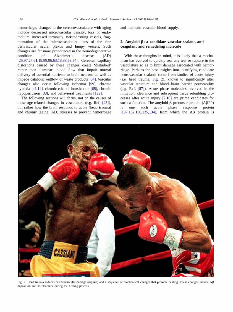
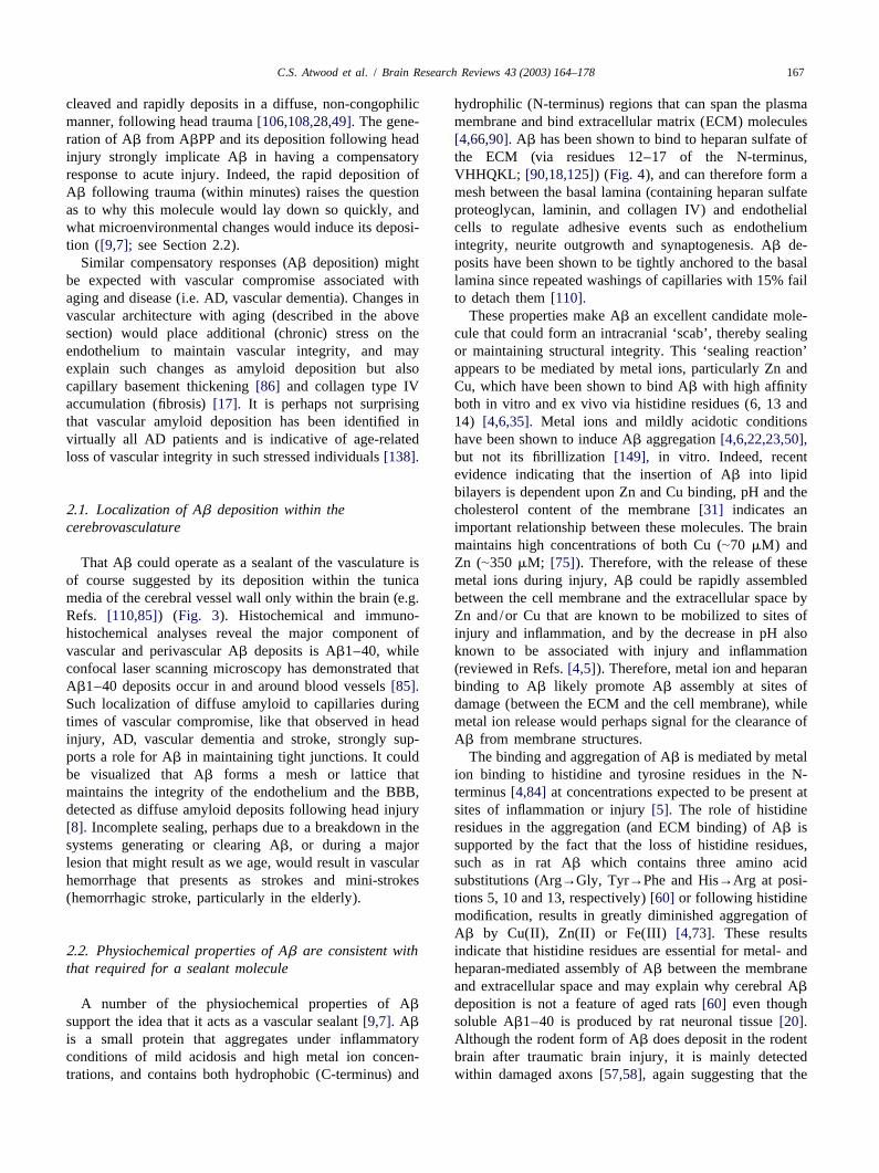
Fig. 3. Cerebrovascular amyloid deposition in the media of the vessel wall. Cross-section of a pair of cerebral arteries stained with Congo red (marked witharrows) showing characteristic amyloid-b deposition (Adapted from Ref.[65].
amino acid substitutions within the heparan proteoglycan conditions produces aggregated Cu,Zn-Ab that wouldbinding site of Ab may diminish binding to the ECM. This serve as a superoxide-scavenging, solid-phase matrix,diminished binding of AbPP/Ab may be less important in which disassembles when Zn and Cu levels lower as tissuean animal that can successfully reproduce within a few damage resolves[101]. In this respect, we have found thatmonths of life compared with an animal that must survive the anti-oxidant activity of Ab is modulated by Cudecades in order to successfully complete reproduction, i.e. concentration[26]. Recently, the antioxidant properties ofthe chances of brain trauma leading to compromised Ab were confirmed by Kontush et al.[66], showing thatvascular supply are less likely to impact the ability of Ab prevents lipoprotein oxidation in cerebral spinal fluidrodents as a species to survive compared to a long lived (CSF), and by Zou et al.[151], who showed that mono-mammal. Long lived animals, and those that have a long meric Ab1–40 inhibits the reduction of Fe(III) induced by
2?period between birth and successfully raising young to vitamin C and the generation of O .2
reproductive age (e.g. bear, dog, rabbit, monkey, etc.), are These properties of Ab explain the acute phase gene-those species that express the ‘human’ form of Ab [60] ration and rapid cortical deposition of Ab following headand which deposits with aging. That AbPP knockout mice trauma[107], an important physiological response thathave no apparent pathology (and live in the protected would limit the loss of terminally differentiated neuronsenvironment of cages their whole life) is consistent with following head injury.the idea that AbPP is required only during times ofinjury /degeneration. 2 .3. Ab deposition prevents the coagulation cascade
We have previously shown that Ab, unlike most pro-teins, binds Cu under acidotic conditions, in keeping with a Human cerebrovascular smooth muscle (HCSM) cellrole of Ab as a molecule involved in maintaining structural associated Ab fibrils serve as a site for the tight binding ofintegrity and/or as an antioxidant required under stress cell-secreted anticoagulant AbPP [133]. The increasedconditions [4,26]. In addition, and given the protein’s secretion of secreted AbPP appears to be a response to Ab
redox properties, Ab can dampen oxidative insults by deposition. AbPP is a potent inhibitor of key proteinasesbinding excess or loosely bound redox active metal ions of the coagulation cascade, and so its enhanced localization[123,115], which at the same time act to switch on its on Ab deposits provides a strong anticoagulant environ-neurotrophic properties (antioxidant activity). We have ment[132,136,119,118,79].In addition, HCSM cell-sur-proposed that the binding of Zn and Cu under these face Ab fibrils are potent stimulators of tissue plasminogen

169C.S. Atwood et al. / Brain Research Reviews 43 (2003) 164–178
Fig. 4. Schematic diagram of early vascular lesion: Ab binding to the extracellular components such as heparan sulfate proteoglycans allow for the sealingof vessels in order to prevent hemorrhage. The lesion is drawn from the epithelial layer, but also may arise from the basement membrane.
activator (tPA) creating a profibrinolytic milieu[64]. These do stresses such as energy shortage and Ca(II) dysregula-findings indicate that Ab fibril assembly on the HCSM cell tion promote AbPP expression, but they also route thesurface results in both a strong anticoagulant and fib- metabolism of AbPP from the non-amyloidogenic to therinolytic environment. The modulation of the deposition amyloidogenic pathway. Inhibition of mitochondrialversus the degradation of Ab (e.g. by tPA, see below) will energy metabolism alters the processing of AbPP todetermine whether there is a loss of vessel wall integrity generate amyloidogenic derivatives[44,80], while oxida-that could lead to hemorrhagic stroke. tive stress (likely associated with apoptosis) has been
shown to increase the generation of Ab [40,83,95].Con-2 .4. Sources of Ab for sealing sistent with this response AbPP and Ab have been
detected in the human brain a couple of days afterSources of Ab for sealing of vessels or maintenance of traumatic brain injury[107,94,45].In addition, Emmerling
regional integrity include neurons, astrocytes, glia and/or et al.[104] found a sharp increase in the concentrations ofvascular smooth muscle cells in close proximity to the Ab1–40 and Ab1–42, but not AbPP, in the CSF oflesion. AbPP is an acute phase reactant upregulated in individuals with severe traumatic brain injury. The releaseresponse to inflammation and a multitude of associated of Ab into the CSF, together with other markers ofdegenerative cellular stresses (reviewed in[10]). Not only traumatic brain injury indicates a neuronal source of Ab
C.S. Atwood et al. / Brain Research Reviews 43 (2003) 164–178170
required during structural damage[104]. Evidence for a tion of tPA and induce Ab, and perhaps also laminin (andneuronal origin is further suggested by the fact that an fibrin?) degradation/clearance, leading to a breakdown inAbPP23 transgenic mouse model of cerebral amyloid the integrity of the vascular media and rupture of vessels.angiopathy (CAA) on an AbPP-null background develops Which protease is responsible for Ab degradation (i.e.a similar degree of both plaques and CAA, suggesting that plasmin, neprilysin, insulin-degrading enzyme, endothelin-a neuronal source of AbPP/Ab is sufficient to induce converting enzyme, etc) is not clear (see below). Nonethe-cerebrovascular amyloid[24]. less, that removal of deposited Ab leads to the loss of
There also is evidence that Ab may originate from vascular integrity has been confirmed by the recent find-vessels that contain CSM cells. Given the extensive ings that immunotherapy promotes cerebral microhemor-network of vessels in the brain, it seems improbable that rhage in a mouse model of CAA[98].neuritic plaques are not associated with such vessels. It has Further experimental evidence indicating that Ab playsbeen suggested that extensive deposition of Ab, i.e. a role in maintaining structural integrity comes fromemboli, may lead to the degeneration and disappearance of studies showing thatChlamydia pneumoniae, an insidiousthe capillary, leaving cores of amyloid free in the paren- intracellular bacterium, when sprayed into the noses ofchyma [110]. Thus, Ab might originate primarily in the young wild-type BALB/c mice can cause progressivevascular tissue and either escape into the parenchyma from deposition of amyloid plaques, in essence creating a partialthe Ab-laden blood vessels or simply remain there after model of AD without using any transgenes[72]. While itdegeneration of the vessel to become cores of neuritic cannot be ruled out that the neuronal inflammation inducedplaques. This is supported by the finding of increased byChlamydia pneumoniae promotes amyloid deposition, itlevels of post-translational modifications in neuritic core is interesting to note that the blood monocytes harboringAb1–42 compared with vascular Ab1–42, indicating the pathogen appear to penetrate the BBB by altering tightneuritic core Ab is older than vascular Ab1–42 [110]. junctions [11,77]. If Ab does act to maintain structuralFurthermore, the pattern of deposition of Ab, perpen- integrity, then such a breach of the BBB would result indicular to the main axis of the vessel, appears to follow the the rapid deposition of Ab in the brain. Such a mechanismpattern of the ECM associated with the smooth muscle would explain why in these mice no neurofibrillary tanglescells of the arteriole, suggesting a vascular tissue source of develop, and whyChlamydia pneumoniae does not induceAb at least in the larger arterioles[110]. AD per se, but rather amyloidosis. Not surprisingly, no
Another source of Ab may be directly from platelets and major alterations in the BBB were observed in APP/PS1leukocytes in the bloodstream, which carry more than 90% double transgenic mice[102].of the circulating amyloid [21,32]. Reactive oxygen Since vascular compromise is less likely to occur in thespecies (ROS) stimulate the tumbling and margination of large vessels leading into the brain than in smaller vesselsplatelets on the endothelium, mediated by P-selectin[41]. of the cortical regions of the brain, the sealant properties ofDuring degranulation of the platelets, Ab could be released Ab are more likely to be observed in small capillariesand deposited in walls of arterioles[132,119,21,117]. rather than the larger penetrating vessels[142]. Indeed,
deposition of Ab is not seen in larger cerebral vessel walls2 .5. In vivo evidence for Ab as a cerebrovascular nor in extracranial vessels. Smaller vessels, such assealant intracortical arterioles and capillaries, that are more likely
prone to rupture (and participate in the BBB) display moreStrong in vivo evidence that Ab acts as a vascular extensive Ab deposition than stronger, thicker walled
sealant is shown by the fact that intravenous administration arterioles[110]. Histological and immunocytochemicalof tPA to a mouse model of CAA leads to an increase in studies have shown that Ab accumulates five times moremicrohemorrhages and can result in parenchymal and frequently around arteries than around veins, with selectivesubarachnoidal hematomas[96,146]. In this vein, aggre- involvement of smaller arteries[142]. The increasedgated, but not nonaggregated Ab increases the expression deposition of Ab in arteries also is consistent with the factof mRNA encoding tPA and uPA[129]. Moreover, Ab that arteries are more likely to develop hemorrhages, aaggregates, in addition to fibrin aggregates and laminin, result of increased arterial blood pressure in arteriescan activate tPA post-translationally[64,147,131]leading compared with veins. It has been suggested that Ab mayto the cleavage of plasminogen to the active protease accumulate in periarterial interstitial fluid drainage path-plasmin[127,128].This activity of Ab is specific for tPA ways of the brain, and that this contributes significantly to[147]. Importantly, a number of studies have shown that CAA in AD[142]. Thus, the capillaries leading to thepurified plasmin degrades Ab at multiple sites and with a hippocampus and cortex, those regions of the brain mostphysiologically relevant efficiency, thereby blocking Ab prone to AD neuropathology, might be expected to haveneurotoxicity in vitro [131,127,128,37].Modulation of the least cerebral perfusion, i.e. being of smaller diameterplasmin levels and activation would therefore dictate and/or more clogged. Over time, the compensatory in-whether amyloid deposits or not. The conversion of diffuse crease in cerebral blood perfusion becomes insufficient toamyloid deposits into fibrillar Ab may mediate the activa- provide adequate perfusion and nutrient supply to localized

171C.S. Atwood et al. / Brain Research Reviews 43 (2003) 164–178
regions within the hippocampal and cortical regions of the the overexpression of AbPP, also are involved. As sug-brain. gested by these authors, ‘although several factors may
The proteolytic processing of AbPP has been linked to contribute to CAA in humans, the neuronal origin ofsphingolipid–cholesterol microdomains (rafts). Protease transgenic AbPP, high levels of Ab in cerebrospinal fluid,plasmin is restricted to rafts of cultured hippocampal and regional localization of CAA in APP23 mice suggestneurons and increases the processing of human AbPP transport and drainage pathways rather than local pro-preferentially at thea-cleavage site, but also efficiently duction or blood uptake of Ab as a primary mechanismdegrades secreted amyloidogenic and non-amyloidogenic underlying cerebrovascular amyloid formation’. SinceAbPP fragments[71]. Thus, physiologic brain plasmin these clearance mechanisms would normally functionappears to play a preventive role in AbPP during adult life to resolve Ab deposits during the healingamyloidogenesis. Interestingly, brain tissue from AD pa- process following head trauma, such toxic deposition maytients contains reduced levels of plasmin[71], implying better model amyloid deposition in disease states such asthat there are signals to limit the removal of deposited AD and vascular dementias. Thus, it is likely that only theamyloid and hence promote amyloid deposition. This may excessive deposition of amyloid fibrils in the cerebrovas-be a result of chronic stresses upon the vasculature and the culature will induce toxicity and microhemorrhages[55].requirement for continuous amyloid deposition required to Indeed, it is likely that the first report of CAA followingmaintain vascular integrity. Indeed, we have found that the head trauma to a 74-year-old female, thought to lead toseverity of AD neuropathology is positively correlated progressive multiple intracerebral hemorrhages as deter-with soluble amyloid load[39]. Excessive signaling for the mined by CT scan (and death in this patient), was not duedeposition of Ab or capillary basement membrane thicken- to amyloid angiopathy (as determined by biopsy anding might however lead to the exact problem these pathological examination)[140], but rather an insufficientmechanisms have evolved to prevent, decreasing cerebro- amyloid response that did not prevent hemorrhage in thevascular perfusion. severely damaged vasculature.
2 .6. In vivo evidence for Ab-induced hemorrhage2 .7. Neuronal /vascular remodeling
Abnormal deposition of Ab, such as in amyloid an-giopathy [96,70] and certain related disorders including That Ab is involved in cell mobility and the extensionhereditary cerebral hemorrhage amyloidosis-Dutch type of neurites through the ECM was first suggested more than(HCHWA-D) (e.g. Ref. [15]) results in cerebrovascular a decade ago as part of a reactive plasticity response to thehemorrhage. In HCHWA-D, where individuals carry a neuronal loss associated with AD[143]. Ab /AbPP is aGlu→Gln substitution at position 22 of Ab, massive developmental protein as shown by its presence in cyto-vascular deposition of Ab results in recurrent strokes in the plasmic processes of astrocytes in the subpial layer andfourth and fifth decades of life that eventually leads to white matter of the developing human brain[126]. Thedeath. It is not clear if the hemorrhage is due to excessive increased deposition of Ab in AD indicates massiveAb deposition that leads to smooth muscle cell death (see remodeling within the diseased brain. Such an idea hasbelow) and eventual endothelium rupture, or protease- been proposed to explain the increased deposition of Ab
mediated clearance of formed amyloid deposits leading associated with neuronal remodeling and the cognitiveway to hemorrhage as recently demonstrated in a mouse deficits associated with repetitive mild traumatic brainmodel of CAA [146,98]. injury of AbPP-transgenic mice (Tg 2576)[124,130];
Studies have suggested that excessive cerebral amyloid Ab-mediated remodeling likely results in neurite anddeposition leads to the degeneration of vascular smooth synapse withdrawal and hence the loss of function.muscle cells of the large penetrating vessels as well as the Secreted AbPP also has been shown to increase neuro-cerebral capillaries that represent the BBB nal survival and growth by mediating nerve growth factor-[138,29,62,139,103].This degeneration of smooth muscle induced neurite extension[82,1], increasing synaptic den-cells likely leads to a cycle of increased expression of sity[109], having synaptotrophic properties[89], regulat-AbPP and Ab peptide generation and deposition[62,119] ing cell growth [113] and showing general trophic re-as a response to maintain regional integrity, resulting in sponses[3,148]. Both NGF and neuronal differentiationmassive Ab accumulation in the media of vessel walls. regulate AbPP expression[150,43,30].A direct role forDegeneration has best been demonstrated in a transgenic AbPP in wound healing has been shown in a MDCK cellmouse model of CAA (APP23) that leads to cerebrovascu- wound-healing assay where overexpression of AbPP accel-lar amyloidosis, smooth muscle cell loss, microhemor- erates cell migration and wound closure[111]. Coexpres-rhages, and in old age, spontaneous cerebral hematomas sion of AbPP and FE65 dramatically enhances the effect[24,145]. That spontaneous microhemorrhages were not of AbPP on cell movement. Therefore, it is likely thatobserved in these mice until after 20 months of age[145] AbPP is involved in the initial clearance and subsequentindicates other stress and/or age-related factors, other than rebuilding of tissue after injury[3]. The prominent growth
C.S. Atwood et al. / Brain Research Reviews 43 (2003) 164–178172
response induced by sAbPP may reflect attempted regene- 3 .2. Plasmin systemration of viable, healthy neurons following synaptic dis-connection due to death of other neurons, a situation that As mentioned earlier, tPA likely plays an important rolemight be expected during the course of AD and following in the degradation/clearance of Ab. Animal studies sup-head injury. porting this show that tPA knockout mice develop amyloid
deposits at 12–14 months and display increased brainedema compared with wild-type mice after controlledcortical impact brain injury, indicating a decreased clear-
3 . Resolution of amyloid deposition during repair of ance of ECM molecules promotes increased fluid buildupinjury [141]. That Ab aggregates can substitute for fibrin aggre-
gates in activating tPA[64] suggests the potential for theAcute phase molecules signal the pro-inflammatory removal of Ab deposits from the vasculature. Such remov-
mechanisms of wound healing[2]. Simultaneous signaling al also will be dependent upon the ratio of plasminfrom other molecules controls and assuages inflammation inhibitors to proteases, regulation of transcription and thetowards the end of the wound healing process. For localization of plasmin system components. Interestingly,example, transforming growth factorb1 (TGF-b1) has uPA and tPA have been localized to the vicinity of senilebeen implicated in the alleviation of inflammation and the plaques in AD tissue[105].tissue rebuilding process whilea2-macroglobulin (a2M),together with its protease inhibitory and removal activity, 3 .3. Other proteaseswhen bound to LRP, acts as a clearance system fornumerous inflammatory proteins[16] such as apolipo- Clearance also might be mediated by circulating (e.g.protein E (ApoE), AbPP, Ab [91], lactoferrin, tPA, uPA, neprilysin) and cellular (e.g. insulin degrading enzymePAI-1, lipoprotein lipase, receptor associated protein (IDE), endothelin-converting enzyme) proteases[56,69].[144,67],IL-1b, TGF-b, platelet derived growth factor and At least in AD, the decreased clearance of Ab deposits byfibroblast growth factor[16,36,51]. Indeed, it has been such proteases has been postulated to promote the accumu-demonstrated in rats that there is a time-dependent clear- lation of amyloid deposits. Recent data indicating IDEance of diffuse amyloid deposits from brain regions knockout mice have elevated brain Ab levels (Dr Suzanne
´affected by traumatic brain injury[57,58] and by ischemia Guenette, personal communication), and in vitro data(see Ref.[101]). The resolution of amyloid deposition in indicating that decreases in the cellular activity of IDEhumans is indicated from studies showing that Ab deposits promote increased Ab accumulation[121], support thisare not more common among survivors of head injury possibility. Neprilysin also has been shown to degrade Ab,when compared to controls[78]. The mechanisms that and in neprilysin heterozygous (1 /2) knockout mice,might resolve amyloid deposition are discussed below. there is a 50% increase in soluble Ab but not insoluble Ab
levels [112]. These findings suggests that different Ab-degrading proteases act preferentially on different pools of
3 .1. Microglia Ab (soluble, deposited, fibrillar). Neprilysin protein andmRNA expression and activity decrease with age especial-
It is known that damage to the brain, such as following ly after menopause/andropause, particularly in the termi-amyloid deposition and cerebral infarcts (strokes), leads to nal zones of the mossy fiber and perforant path. Thisan inflammatory response that includes the activation of decrement is associated with particular brain regions (e.g.microglial cells that clear away damaged tissue and CA3, terminal zones of perforant path and entorhinalstructures formed to maintain regional integrity. The cortex)[57,58,42], suggesting that neprilysin deficiencyinterplay between metal ions, ECM proteins and microglia, plays a role in the age-associated increase in amyloidthat all bind to the VHHQKL region of Ab, has important deposition. Further support for this comes from the factconsequences for the recruitment and activation of mi- that mutant Ab is degraded more slowly by neprilysin.croglia and clearance of the amyloid deposit. The release Moreover, neprilysin mRNA and protein levels are sig-of molecules from the VHHQKL region (i.e. as inflamma- nificantly elevated in AbPP transgenic mice for as long astion resides and pH increases) would be expected to 30 weeks following a single intracranial injection of Ab1–promote (Ab-induced) microglial and/or macrophage acti- 42. The rise in neprilysin levels is associated with thevation and the engulfing and removal of the vascular or prevention of plaque formation and reduced astrogliosisparencyhmal ‘scab’, respectively. Indeed, in the AD brain [93]. Clearance of Ab also likely depends upon ApoEat least, increased phagocytic activity does appear to clear isoform, since amyloid plaques are more likely to be founddiffuse amyloid deposits and the diffuse halo surrounding in the brains of individuals with the ApoE4 allele than withdense core plaques (Dickson, 2002). However, the conver- the ApoE3 allele[78,92], indicating either that depositionsion of Ab deposits into fibrillar hard cores appears to is greater and/or that catabolism of Ab is decreased inprevent microglial mediated clearance. individuals with the E4 allele.

173C.S. Atwood et al. / Brain Research Reviews 43 (2003) 164–178
3 .4. Receptor for glycation end-products cause of the encephalitis and meningitis suffered by theseindividuals has not been addressed. This cause is likely
In addition to the removal of aggregated non-modified associated with the direct removal of amyloid depositsAb, post-translationally modified proteins that result from from the parenchyma and vasculature, the proposed actionthe damage induced by the lesion environment also must of the vaccine. That is the immunotherapy was successfulbe cleared from the brain. In this respect, the upregulation in removing Ab, but a disaster for the health of theof receptor for glycation end-products (RAGE) during individuals. Put another way, amyloid vaccinations wereinflammation[48] associated with the lesion[120] likely not able to discriminate between physiologically purposivepromotes the clearance of oxidized and fibrillized proteins deposits of Ab (i.e. vascular and diffuse deposits) andfrom the damaged/ inflamed area during resolution of the pathological Ab (senile plaques, oligomers). Removal ofinjury. Indeed, one of the most important functions of vascular Ab would therefore alter the structural integrity ofRAGE may be to help clear from sites of injury post- that region and lead to leakage of serum components intotranslationally modified proteins. It is likely that all of the the brain (hemorrhagic stroke) resulting in an immune (orabove clearance mechanisms operate at some level to autoimmune) response characterized by inflammation. Thisresolve Ab deposits. breakdown of the blood–brain barrier is consistent with the
development of encephalitis and meningitis, and perhapsthe presence of viruses within the CSF as reported in some
4 . Amyloid removal as a therapy for amyloid of the patients under investigation. Indeed, neuronal in-diseases? flammation is not a feature of other autoimmune diseases.
Activation of the plasmin system that prevents theearlyThe prominence of the ‘amyloid hypothesis’ as the deposition of Ab might however be of therapeutic value in
cause of AD has driven researchers to develop therapeutic diseases of amyloid angiopathy (e.g. HCHWA-D) bystrategies targeted at the removal of Ab in order to preventing early cell loss, endothelial rupture and hemor-stabilize and/or reverse cognitive deficits. This idea has rhagic stroke. Activation of plasmin would both degradebeen spurred on by experiments showing that amyloid Ab and prevent its formation since plasmin in lipid raftsdeposits and cognitive deficits can be resolved following cleaves AbPP at the alpha-secretase site[71]. Conversely,immunization of transgenic mice overexpressing mutant in individuals with marked amyloid angiopathy, the use ofhuman AbPP[116]. This unfortunate model—the removal tPA inhibitors to prevent amyloid removal by plasminwith no apparent side-effects of a foreign human protein might actually prevent cerebral hemorrhage (providingthat had been greatly overexpressed in a mouse—led to the amyloid deposits are not toxic) and prolong lifespan inidea that such a course of action would benefit AD individuals with HCHWA-D.patients. It could be argued that removal of amyloid mightnot be expected to be problematic given that AbPP andBACE knockout mice[47,76] have no phenotype. How- 5 . Summaryever, given that Ab may only be required during injury,and as mentioned earlier, cerebrovascular sealant mecha- The acute phase generation of Ab has evolved as anisms may not be essential for the survival of rodent sealant, anti-coagulation, secondary antioxidant defensespecies, indicates these transgenics are a poor model of system and remodeling molecule required during times ofAD. In addition, rodents may be less dependent on Ab for remodeling and excessive ROS generation and/or traumaremodeling or neuronal development compared with other [4,5]. A large body of literature supports these activities ofmammals. Ab. The deposition of Ab immediately following head
That Ab does deposit in AD strongly indicates that the injury, its resolution during the healing process and itsintegrity of the parenchyma and vasculature are under deposition in the compromised elderly and AD vasculatureattack. It is clear, however, from our basic understanding are consistent with such roles for Ab. In this way,of the protein that the deposition of Ab appears to be a hemorrhage can be prevented while allowing for anpurposive response, one that acts to maintain structural uninterrupted blood supply. It remains to be determined ifintegrity of the BBB and parenchymal structures, prevent similar mechanisms have evolved in other tissues. Thesecoagulation, promote remodeling and reduce oxidative beneficial properties of Ab and its precursor protein dostress. These activities of Ab would provide an important however bring into question the validity of the therapeuticphysiological response to limit the loss of terminally efficacy of removing Ab from the brain.differentiated neurons after head trauma. Therefore, theremoval of amyloid from the brain via vaccination or othermeans after its deposition would be predicted to lead to A cknowledgementscerebral hemorrhage[9,7,8]. Indeed, the Phase IIA Abvaccination trials by Elan Pharmaceuticals were halted due This work is supported by funds from the Nationalto clinical signs of inflammation. Although much has been Institute of Health (RO1AG19356) and the Alzheimersaid and written about the suspension of the trials, the root Association.

C.S. Atwood et al. / Brain Research Reviews 43 (2003) 164–178174
[17] W .R. Brown, D.M. Moody, C.R. Thore,V.R. Challa, CerebrovascularR eferencespathology in Alzheimer’s disease and leukoaraiosis, Ann. NY Acad.Sci. 903 (2000) 39–45.
[1] C .A. Akar, W.C. Wallace, Amyloid precursor protein modulates the[18] K .R. Brunden, N.J. Richter-Cook, N. Chaturvedi, R.C. Frederickson,
interaction of nerve growth factor with p75 receptor and potentiatespH-dependent binding of synthetic beta-amyloid peptides to glycos-
its activation of trkA phosphorylation, Mol. Brain Res. 56 (1–2)aminoglycans, J. Neurochem. 61 (1993) 2147–2154.
(1998) 125–132.[19] L . Buee, P.R. Hof, A. Delacourte, Brain microvascular changes in[2] N euroinflammation Working Group, H. Akiyama, S. Barger, S.
Alzheimer’s disease and other dementias, Ann. NY Acad. Sci. 826Barnum, B. Bradt, J. Bauer, G.M. Cole, N.R. Cooper, P. Eikelen-(1997) 7–24.boom, M. Emmerling, B.L. Fiebich, C.E. Finch, S. Frautschy, W.S.
[20] J . Busciglio, D.H. Gabuzda, P. Matsudaira, B.A. Yankner, Gene-Griffin, H. Hampel, M. Hull, G. Landreth, L. Lue, R. Mrak, I.R.ration of beta-amyloid in the secretory pathway in neuronal andMackenzie, P.L. McGeer, M.K. O’Banion, J. Pachter, G. Pasinetti,nonneuronal cells, Proc. Natl. Acad. Sci. USA 90 (1993) 2092–C. Plata-Salaman, J. Rogers, R. Rydel, Y. Shen, W. Streit, R.2096.Strohmeyer, I. Tooyoma, F.L. Van Muiswinkel, R. Veerhuis, D.
[21] A .I. Bush, R.N. Martins, B. Rumble, R. Moir, S. Fuller, E. Milward,Walker, S. Webster, B. Wegrzyniak, G. Wenk, T. Wyss-Coray,J. Currie, D. Ames, A. Weidemann, P. Fischer et al., The amyloidInflammation and Alzheimer’s disease, Neurobiol. Aging 21 (2000)precursor protein of Alzheimer’s disease is released by human383–421.platelets, J. Biol. Chem. 265 (26) (1990) 15977–15983.[3] W . Araki, N. Kitaguchi, Y. Tokushima, K. Ishii, H. Aratake, S.
[22] A .I. Bush, W.H. Pettingell Jr., M.D. Paradis, R.E. Tanzi, ModulationShimohama, S. Nakamura, J. Kimura, Trophic effect of beta-of A beta adhesiveness and secretase site cleavage by zinc, J. Biol.amyloid precursor protein on cerebral cortical neurons in culture,Chem. 269 (1994) 12152–12158.Biochem. Biophys. Res. Commun. 181 (1991) 265–271.
[23] A .I. Bush, W.H. Pettingell, G. Multhaup, M. d Paradis, J.P.Vonsattel,[4] C .S. Atwood, X. Huang, R.D. Moir, N.M. Bacarra, D. Romano, R.E.J.F. Gusella, K. Beyreuther, C.L. Masters, R.E. Tanzi, RapidTanzi, A.I. Bush, Dramatic aggregation of Alzheimer Ab by Cu(II)induction of Alzheimer A beta amyloid formation by zinc, Scienceis induced by conditions representing physiological acidosis, J. Biol.265 (1994) 1464–1467.Chem. 273 (1998) 12817–12826.
[24] M .E. Calhoun, P. Burgermeister, A.L. Phinney, M. Stalder, M.[5] C .S. Atwood, X. Huang, R.D. Moir, R.E. Tanzi, A.I. Bush, The roleTolnay, K.H. Wiederhold, D. Abramowski, C. Sturchler-Pierrat, B.of free radicals and metal ions in the pathogenesis of Alzheimer’sSommer, M. Staufenbiel, M. Jucker, Neuronal overexpression ofdisease, in: A. Sigel, H. Sigel (Eds.), Interrelations between Freemutant amyloid precursor protein results in prominent deposition ofRadicals and Metal Ions in Life Processes, Metal Ions in Biologicalcerebrovascular amyloid, Proc. Natl. Acad. Sci. USA 23 (24) (1999)Systems, vol. 36, Marcel Dekker, New York, 1999, pp. 309–364.14088–14093.[6] C .S. Atwood, R.C. Scarpa, X. Huang, R.D. Moir, W.D. Jones, D.P.
[25] J . Cervos-Navarro, H.J. Gertz, V. Frydl, Cerebral blood vesselFairlie, R.E. Tanzi, A.I. Bush, Characterization of copper interac-changes in old people, Mech. Ageing Dev. 39 (1987) 223–231.tions with Alzheimer amyloid beta peptides: Identification of an
[26] C .-W. Chan, A. Dharmarajan, C.S. Atwood, X. Huang, R.E. Tanzi,attomolar-affinity copper binding site on amyloid beta1–42, J.A.I. Bush, R.N. Martins, Anti-apoptotic action of Alzheimer Ab,Neurochem. 75 (2000) 1219–1233.Alzheimer’s Rep. 2 (1999) 1–6.[7] C .S. Atwood, S.R. Robinson, M.A. Smith, Amyloid-beta: redox–
[27] L . Claudio, Ultrastructural features of the blood–brain barrier inmetal chelator and antioxidant, J. Alzheimer’s Dis. 4 (2002b) 203–biopsy tissue from Alzheimer’s disease patients, Acta Neuropathol.214.(Berl.) 91 (1996) 6–14.[8] C .S. Atwood, G. Perry, M.A. Smith, M. Pfeifer, S. Boncristiano, L.
[28] J . Clinton, M.W. Ambler, G.W. Roberts, Post-traumatic Alzheimer’sBondolfi, A. Stalder, T. Deller, M. Staufenbiel, P.M. Mathews, M.disease: preponderance of a single plaque type, Neuropathol. Appl.Jucker, Cerebral hemorrhage and amyloid-beta, Science 299 (2003)Neurobiol. 17 (1991) 69–74.1014.
[29] F . Coria, M. Larrondo-Lillo, B. Frangione, Degeneration of smooth[9] C .S. Atwood, G.M. Bishop, G. Perry, M.A. Smith, Amyloid-beta: amuscle cells in beta-amyloid angiopathies, J. Neuropathol. Exp.vascular sealant that protects against hemorrhage?, J. Neurosci. Res.Neurol. 48 (1989) 368–375.70 (2002a) 356.
[30] J .M. Cosgaya, M.J. Latasa, A. Pascual, Nerve growth factor and ras[10] C .S. Atwood, X. Huang, R.D. Moir, M.A. Smith, R.E. Tanzi, A.E.regulate beta-amyloid precursor protein gene expression in PC12Roher, A.I. Bush, G. Perry, Neuroinflammatory responses in thecells, J. Neurochem. 67 (1996) 98–104.Alzheimer’s disease brain promote the oxidative post-translational
[31] C .C. Curtain, F.E. Ali, D.G. Smith, A.I. Bush, C.L. Masters, K.J.modification of amyloid deposits, in: K. Iqbal, S.S. Sisodia, B.Barnham, Metal ions, pH, and cholesterol regulate the interactionsWinblad (Eds.), Alzheimer’s Disease: Advances in Etiology, Patho-of Alzheimer’s disease amyloid-beta peptide with membrane lipid, J.genesis and Therapeutics, Wiley, London, 2001, pp. 341–361.Biol. Chem. 278 (2003) 2977–2982.[11] B .J. Balin, H.C. Gerard, E.J. Arking, D.M. Appelt, P.J. Branigan,
[32] T .A. Davies, H.J. Long, K. Sgro, W.H. Rathbun, M.E. McMenamin,J.T. Abrams, J.A. Whittum-Hudson, A.P. Hudson, Identification andK. Seetoo, H. Tibbles, A.M. Billingslea, R.E. Fine, J.B. Fishman,localization of Chlamydia pneumoniae in the Alzheimer’s brain,C.A. Levesque, S.J. Smith, J.M. Wells, E.R. Simons, ActivatedMed. Microbiol. Immunol. (Berl). 187 (1998) 23–42.Alzheimer disease platelets retain more beta amyloid precursor[12] T . Bar, The vascular system of the cerebral cortex, Adv. Anat.protein, Neurobiol. Aging 18 (2) (1997) 147–153.Embryol. Cell Biol. 59 (1980) 1–62.
[33] G .I. De Jong, E. Farkas, C.M. Stienstra, J.R. Plass, J.N. Keijser, J.C.[13] T .M. Berzin, B.D. Zipser, M.S. Rafii, V. Kuo-Leblanc, G.D. Yan-de la Torre, P.G. Luiten, Cerebral hypoperfusion yields capillarycopoulos, D.J. Glass, J.R. Fallon, E.G. Stopa, Agrin and microvascu-damage in the hippocampal CA1 area that correlates with spatiallar damage in Alzheimer’s disease, Neurobiol. Aging 21 (2000)memory impairment, Neuroscience 91 (1999) 203–210.349–355.
[34] J .C. De la Torre, Hemodynamic consequences of deformed mi-[14] J .A. Boero, J. Ascher, A. Arregui, C. Rovainen, T.A. Woolsey,crovessels in the brain in Alzheimer’s disease, Ann. NY Acad. Sci.Increased brain capillaries in chronic hypoxia, J. Appl. Physiol. 86826 (1997) 75–91.(1999) 1211–1219.
[15] M . Bornebroek, J. Haan, R.A. Roos, Hereditary cerebral hemorrhage [35] J . Dong, C.S. Atwood, V.E. Anderson, S.L. Siedlak, G. Perry, M.A.with amyloidosis–Dutch type (HCHWA-D): a review of the variety Smith, P.R. Carey, Metal binding and oxidation of amyloid-b withinin phenotypic expression, Amyloid 6 (1999) 215–224. isolated senile plaque cores: Raman microscopic evidence, Bio-
[16] W . Borth, Alpha 2-macroglobulin, a multifunctional binding protein chemistry 42 (2003) 2768–2773.with targeting characteristics, FASEB J. 6 (1992) 3345–3353. [36] Y . Du, B. Ni, M. Glinn, R.C. Dodel, K.R. Bales, Z. Zhang, P.A.

175C.S. Atwood et al. / Brain Research Reviews 43 (2003) 164–178
Hyslop, S.M. Paul, alpha2-Macroglobulin as a beta-amyloid peptide- [54] O . Hunziker, S. Abdel’Al, H. Frey, M.J.Veteau, W. Meier-Ruge, Thebinding plasma protein, J. Neurochem. 69 (1997) 299–305. aging human cerebral cortex: a stereological characterization of
[37] C . Exley, O.V. Korchazhkina, Plasmin cleaves Abeta42 in vitro and changes in the capillary net, J. Gerontol. 34 (1979) 345–350.prevents its aggregation into beta-pleated sheet structures, Neurore-[55] S . Inoue, R. Kisilevsky, Beta-amyloid fibrils of Alzheimer’s disease:port 12 (2001) 2967–2970. pathologically altered, basement membrane-associated microfibrils?,
[38] E . Farkas, R.A. De Vos, E.N. Jansen Steur, P.G. Luiten, Are Ital. J. Anat. Embryol. 106 (2001) 93–102.Alzheimer’s disease, hypertension, and cerebrocapillary damage [56] N . Iwata, S. Tsubuki, Y. Takaki, K. Watanabe, M. Sekiguchi, E.related?, Neurobiol. Aging 21 (2000) 235–243. Hosoki, M. Kawashima-Morishima, H.J. Lee, E. Hama, Y. Sekine-
[39] J . Fonte, J. Miklossy, C. Atwood, R. Martins, The severity of Aizawa, T.C. Saido, Identification of the major Abeta1–42-degrad-cortical Alzheimer’s type changes is positively correlated with ing catabolic pathway in brain parenchyma: suppression leads toincreased amyloid-beta Levels: Resolubilization of amyloid-beta biochemical and pathological deposition, Nature Med. 6 (2000)with transition metal ion chelators, J. Alzheimer’s Dis. 1 (2001) 143–150.209–219. [57] N . Iwata, Y. Takaki, S. Fukami, S. Tsubuki, T.C. Saido, Region-
[40] P .H. Frederikse, D. Garland, J.S. Zigler, J. Piatigorsky, Oxidative specific reduction of A beta-degrading endopeptidase, neprilysin, instress increases production ofb-amyloid precursor protein and mouse hippocampus upon aging, J. Neurosci. Res. 70 (2002a)b-amyloid (A-beta) in mammalian lenses, and A-beta has toxic 493–500.effects on lens epithelial cells, J. Biol. Chem. 271 (1996) 10169–
[58] A . Iwata, X.H. Chen, T.K. McIntosh, K.D. Browne, D. Smith,10174.
Long-term accumulation of amyloid-beta in axons following brain[41] P .S. Frenette, R.C. Johnson, R.O. Hynes, D.D. Wagner, Platelets roll
trauma without persistent upregulation of amyloid precursor proteinon stimulated endothelium in vivo: an interaction mediated by
genes, J. Neuropathol. Exp. Neurol. 61 (2002b) 1056–1068.endothelial P-selectin, Proc. Natl. Acad. Sci. USA 92 (1995) 7450–[60] E .M. Johnstone, M.O. Chaney, F.H. Norris, R. Pascual, S.P. Little,7454.
Conservation of the sequence of the Alzheimer’s disease amyloid[42] S . Fukami, K. Watanabe, N. Iwata, J. Haraoka, B. Lu, N.P. Gerard,peptide in dog, polar bear and five other mammals by cross-speciesC. Gerard, P. Fraser, D. Westaway, P. St. George-Hyslop, T.C. Saido,polymerase chain reaction analysis, Mol. Brain Res. 10 (1991)Abeta-degrading endopeptidase, neprilysin, in mouse brain: synaptic299–305.and axonal localization inversely correlating with Abeta pathology,
[61] R .N. Kalaria, Cerebral vessels in ageing and Alzheimer’s disease,Neurosci. Res. 43 (2002) 39–56.Pharmacol. Ther. 72 (1996) 193–214.[43] K . Fukuchi, K. Kamino, S.S. Deeb, A.C. Smith, T. Dang, G.M.
[62] M . Kawai, R.N. Kalaria, P. Cras, S.L. Siedlak, M.E. Velasco, E.R.Martin, Overexpression of amyloid precursor protein alters itsShelton, H.W. Chan, B.D. Greenberg, G. Perry, Degeneration ofnormal processing and is associated with neurotoxicity, Biochem.vascular muscle cells in cerebral amyloid angiopathy of AlzheimerBiophys. Res. Commun. 182 (1992) 165–173.disease, Brain Res. 623 (1993) 142–146.[44] D . Gabuzda, J. Busciglio, L.B. Chen, P. Matsudaira, B.A. Yankner,
[63] J .I. Keuker, P.G. Luiten, E. Fuchs, Capillary changes in hippocampalInhibition of energy metabolism alters the processing of amyloidCA1 and CA3 areas of the aging rhesus monkey, Acta Neuropathol.precursor protein and induces a potentially amyloidogenic deriva-(Berl.) 100 (2000) 665–672.tive, J. Biol. Chem. 269 (1994) 13623–13628.
[64] I .B. Kingston, M.J. Castro, S. Anderson, In vitro stimulation of[45] S .M. Gentleman, M.J. Nash, C.J. Sweeting, D.I. Graham, G.W.tissue-type plasminogen activator by Alzheimer amyloid beta-pep-Roberts, Beta-amyloid precursor protein (beta APP) as a marker fortide analogues, Nature Med. 1 (1995) 138–142.axonal injury after head injury, Neurosci. Lett. 160 (1993) 139–144.
[65] R . Kisilevsky, Inflammation-associated amyloidogenesis. Lessons[46] S .I. Harik, M.A. Hritz, J.C. LaManna, Hypoxia-induced brainfor Alzheimer’s amyloidogenesis, Mol. Neurobiol. 8 (1994) 65–66.angiogenesis in the adult rat, J. Physiol. 485 (1995) 525–530.
[66] A . Kontush, C. Berndt, W. Weber, V. Akopyan, S. Arlt, S. Schip-[47] S . Heber, J. Herms, V. Gajic, J. Hainfellner, A. Aguzzi, T. Rulicke,pling, U. Beisiegel, Amyloid-beta is an antioxidant for lipoproteinsH. von Kretzschmar, C. von Koch, S. Sisodia, P. Tremml, H.P. Lipp,in cerebrospinal fluid and plasma, Free Radic. Biol. Med. 30 (2001)D.P. Wolfer, U. Muller, Mice with combined gene knock-outs reveal119–128.essential and partially redundant functions of amyloid precursor
[67] M .Z. Kounnas, E.B. Loukinova, S. Stefansson, J.A. Harmony, B.H.protein family members, J. Neurosci. 20 (2000) 7951–7963.Brewer, D.K. Strickland, W.S. Argraves, Identification of glycopro-[48] A . Heidland, K. Sebekova, R. Schinzel, Advanced glycation endtein 330 as an endocytic receptor for apolipoprotein J/clusterin, J.products and the progressive course of renal disease, Am. J. KidneyBiol. Chem. 270 (1995) 13070–13075.Dis. 38 (2001) S100–106.
[68] M . Kraszpulski, C. Tukaj, T. Wrzolkowa, Hippocampal capillaries in[49] K . Horsburgh, G.M. Cole, F. Yang, M.J. Savage, B.D. Greenberg,different age groups of chronically ethanol-intoxicated rats. Mor-S.M. Gentleman, D.I. Graham, J.A. Nicoll, Beta-amyloidphometrical studies, Folia Morphol. (Warsz.) 59 (2000) 121–129.(Abeta)42(43), abeta42, abeta40 and apoE immunostaining of
[69] I .V. Kurochkin, S. Goto, Alzheimer’s beta-amyloid peptide spe-plaques in fatal head injury, Neuropathol. Appl. Neurobiol. 26cifically interacts with and is degraded by insulin degrading enzyme,(2000) 124–132.FEBS Lett. 345 (1994) 33–37.[50] X . Huang, C.S. Atwood, R.D. Moir, M.A. Hartshorn, J.-P. Vonsattel,
[70] R . Leblanc, G. Haddad, Y. Robitaille, Cerebral hemorrhage fromR.E. Tanzi, A.I. Bush, Zinc-induced Alzheimer’s Ab1–40 aggrega-amyloid angiopathy and coronary thrombolysis, Neurosurgery 31tion is mediated by conformational factors, J. Biol. Chem. 272(1992) 586–590.(1997) 26464–26470.
[51] S .R. Hughes, O. Khorkova, S. Goyal, J. Knaeblein, J. Heroux, N.G. [71] M .D. Ledesma, J.S. Da Silva, K. Crassaerts, A. Delacourte, B. DeRiedel, S. Sahasrabudhe, Alpha2-macroglobulin associates with Strooper, C.G. Dotti, Brain plasmin enhances APP alpha-cleavagebeta-amyloid peptide and prevents fibril formation, Proc. Natl. Acad. and Abeta degradation and is reduced in Alzheimer’s disease brains,Sci. USA 95 (1998) 3275–3280. EMBO Rep. 1 (2000) 530–535.
[52] E .G. Huizinga, S. Tsuji, R.A. Romijn, M.E. Schiphorst, P.G. de [72] S . Little, C. Hammond, A. MacIntyre, D. Balin B Appelt, InductionGroot, J.J. Sixma, P. Gros, Structures of glycoprotein Ibalpha and its of Alzheimer’s disease-like pathology in the brains of young, non-complex with von Willebrand factor A1 domain, Science 297 (2002) transgenic, balb/c mice following infection with chlamydia pneu-1176–1179. moniae: a model for late onset /sporadic Alzheimer’s disease, in:
[53] O . Hunziker, S. Abdel’Al, H. Frey, M.J. Veteau, W. Meier-Ruge, Abstract No. 87 of the 8th International Conference on Alzheimer’sQuantitative studies in the cerebral cortex of aging humans, Geron- Disease and Related Disorders, 2002.tology 24 (1978) 27–31. [73] Y . Liu, Understanding the biological activity of amyloid proteins in

C.S. Atwood et al. / Brain Research Reviews 43 (2003) 164–178176
vitro: from inhibited cellular MTT reduction to altered cellular [92] J .A. Nicoll, G.W. Roberts, D.I. Graham, Apolipoprotein E epsilon 4cholesterol homeostatis, Prog. Neuropsychopharmacol. Biol. Psychi- allele is associated with deposition of amyloid beta-protein follow-atry 23 (1999) 377–395. ing head injury, Nature Med. 1 (1995) 135–137.
[74] A . Lokkegaard, J.R. Nyengaard, M.J. West, Stereological estimates [93] R .M. Nitsch, M.A. Wollmer, R. Poirier, H. Mohajeri, Role ofof number and length of capillaries in subdivisions of the human neprilysin in preventing amyloid formation, in: Abstract No. 554 ofhippocampal region, Hippocampus 11 (2001) 726–740. the 8th International Conference on Alzheimer’s Disease and
[75] M .A. Lovell, J.D. Robertson, W.J. Teesdale, J.L. Campbell, W.R. Related Disorders, 2002.Markesbery, Copper, iron and zinc in Alzheimer’s disease senile [94] N . Otsuka, M. Tomonaga, K. Ikeda, Rapid appearance of beta-plaques, J. Neurol. Sci. 158 (1998) 47–52. amyloid precursor protein immunoreactivity in damaged axons and
[76] Y . Luo, B. Bolon, S. Kahn, B.D. Bennett, S. Babu-Khan, P. Denis, reactive glial cells in rat brain following needle stab injury, BrainW. Fan, H. Kha, J. Zhang, Y. Gong, L. Martin, J.C. Louis, Q. Yan, Res. 568 (1991) 335–338.W.G. Richards, M. Citron, R. Vassar, Mice deficient in BACE1, the [95] D . Paola, C. Domenicotti, M. Nitti, A. Vitali, R. Borghi, D.Alzheimer’s beta-secretase, have normal phenotype and abolished Cottalasso, D. Zaccheo, P. Odetti, P. Strocchi, U.M. Marinari, M.beta-amyloid generation, Nature Neurosci. 4 (2001) 231–232. Tabaton, M.A. Pronzato, Oxidative stress induces increase in
[77] A . MacIntyre, S. Little, C. Hammond, D. Appelt, B. Balin, Effects intracellular amyloid beta-protein production and selective activationof chlamydia pneumoniae infection on the junctional complexes of of betaI and betaII PKCs in NT2 cells, Biochem. Biophys. Res.the blood–brain barrier: implications for the pathogenesis of Al-
Commun. 268 (2000) 642–646.zheimer’s disease, in: Abstract No. 1604 of the 8th International
[96] W .W. Pendlebury, E.D. Iole, R.P. Tracy, B.A. Dill, IntracerebralConference on Alzheimer’s Disease and Related Disorders, 2002.
hemorrhage related to cerebral amyloid angiopathy and t-PA treat-[78] D .P. Macfarlane, J.A. Nicoll, C. Smith, D.I. Graham, APOE epsilon4
ment, Ann. Neurol. 29 (1991) 210–213.allele and amyloid beta-protein deposition in long term survivors of[97] L .S. Perlmutter, H.C. Chui, Microangiopathy, the vascular basementhead injury, Neuroreport 10 (1999) 3945–3948.
membrane and Alzheimer’s disease: a review, Brain Res. Bull. 24[79] F . Mahdi, W.E. Van Nostrand, A.H. Schmaier, Protease nexin-2/(1990) 677–686.amyloid b-protein precursor inhibits Factor Xa in the prothrom-
[98] L .A. Pfeifer, L.R. White, G.W. Ross, H. Petrovitch, L.J. Launer,binase complex, J. Biol. Chem. 270 (1995) 23468–23474.Cerebral amyloid angiopathy and cognitive function: the HAAS[80] M .P. Mattson, Q. Guo, K. Furukawa, W.A. Pedersen, Presenilins, theautopsy study, Neurology 58 (2002) 1629–1634.endoplasmic reticulum, and neuronal apoptosis in Alzheimer’s
[99] P . Piotrowski, B. Gajkowska, H. Olszewska, M. Smialek, Electrondisease, J. Neurochem. 70 (1998) 1–14.microscopy studies on experimental diabetes and cerebral ischemia[81] G . Miller, Breaking down barriers, Science 297 (2002) 1116–1118.in the rat brain, Folia Neuropathol. 37 (1999) 256–263.[82] E .A. Milward, R. Papadopoulos, S.J. Fuller, R.D. Moir, D. Small, K.
[101] R . Pluta, M. Barcikowska, A. Misicka, A.W. Lipkowski, S.Beyreuther, C.L. Masters, The amyloid protein precursor of Al-Spisacka, S. Januszewski, Ischemic rats as a model in the study ofzheimer’s disease is a mediator of the effects of nerve growth factorthe neurobiological role of human beta-amyloid peptide. Time-on neurite outgrowth, Neuron 9 (1992) 129–137.dependent disappearing diffuse amyloid plaques in brain, Neurore-[83] H . Misonou, M. Morioshima-Kawashima, Y. Ihara, Oxidative stressport 10 (1999) 3615–3619.induces intracellular accumulation of Alzheimer amyloid-b in
[102] J .F. Poduslo, G.L. Curran, T.M. Wengenack, B. Malester, K. Duff,human neuroblastoma cells, Soc. Neurosci. Abstr. 25 (1999) 340.Permeability of proteins at the blood–brain barrier in the normal[84] T . Miura, K. Suzuki, N. Kohata, H. Takeuchi, Metal binding modesadult mouse and double transgenic mouse model of Alzheimer’sof Alzheimer’s amyloid beta-peptide in insoluble aggregates anddisease, Neurobiol. Dis. 8 (2001) 555–567.soluble complexes, Biochemistry 39 (2000) 7024–7031.
[103] R . Prior, D. D’Urso, R. Frank, I. Prikulis, G. Pavlakovic, Loss of[85] T . Miyakawa, T. Kimura, S. Hirata, N. Fujise, T. Ono, K. Ishizuka,vessel wall viability in cerebral amyloid angiopathy, Neuroreport 7J. Nakabayashi, Role of blood vessels in producing pathological(1996) 562–564.changes in the brain with Alzheimer’s disease, Ann. NY Acad. Sci.
[104] C .A. Raby, M.C. Morganti-Kossmann, T. Kossmann, P.F. Stahel,903 (2000) 46–54.M.D. Watson, L.M. Evans, P.D. Mehta, K. Spiegel, Y.M. Kuo, A.E.[86] D .M. Moody, W.R. Brown, V.R. Challa, H.S. Ghazi-Birry, D.M.Roher, M.R. Emmerling, Traumatic brain injury increases beta-Reboussin, Cerebral microvascular alterations in aging,amyloid peptide 1–42 in cerebrospinal fluid, J. Neurochem. 71leukoaraiosis, and Alzheimer’s disease, Ann. NY Acad. Sci. 826(1998) 2505–2509.(1997) 103–116.
[105] G .W. Rebeck, S.D. Harr, D.K. Strickland, B.T. Hyman, Multiple,[87] T . Mori, X. Wang, A.E. Kline, C.J. Siao, C.E. Dixon, S.E. Tsirka,diverse senile plaque-associated proteins are ligands of an apolipo-E.H. Lo, Reduced cortical injury and edema in tissue plasminogenprotein E receptor, the alpha 2-macroglobulin receptor / low-de-activator knockout mice after brain trauma, Neuroreport 12 (2001)nsity-lipoprotein receptor-related protein, Ann. Neurol. 37 (1995)4117–4120.211–217.[88] R .E. Mrak, S.T. Griffin, D.I. Graham, Aging-associated changes in
[106] G .W. Roberts, D. Allsop, C. Bruton, The occult aftermath ofhuman brain, J. Neuropathol. Exp. Neurol. 56 (12) (1997) 1269–boxing, J. Neurol. Neurosurg. Psychiatry 53 (1990) 373–378.1275.
[107] G .W. Roberts, S.M. Gentleman, A. Lynch, L. Murray, M. Landon,[89] L . Mucke, E. Masliah, W.B. Johnson, M.D. Ruppe, M. Alford, E.M.D.I. Graham, Beta amyloid protein deposition in the brain afterRockenstein, S. Forss-Petter, M. Pietropaolo, M. Mallory, C.R.severe head injury: implications for the pathogenesis of Alzheim-Abraham, Synaptotrophic effects of human amyloid beta proteiner’s disease, J. Neurol. Neurosurg. Psychiatry. 57 (1994) 419–425.precursors in the cortex of transgenic mice, Brain Res. 666 (1994)
[108] G .W. Roberts, S.M. Gentleman, A. Lynch, D.I. Graham, beta A4151–167.amyloid protein deposition in brain after head trauma, Lancet 338[90] S . Narindrasorasak, D. Lowery, P. Gonzalez-DeWhitt, R.A. Poor-(1991) 1422–1423.man, B. Greenberg, R. Kisilevsky, High affinity interactions be-
tween the Alzheimer’s beta-amyloid precursor proteins and the [109] J .M. Roch, E. Masliah, A.C. Roch-Levecq, M.P. Sundsmo, D.A.basement membrane form of heparan sulfate proteoglycan, J. Biol. Otero, I. Veinbergs, T. Saitoh, Increase of synaptic density andChem. 266 (1991) 12878–12883. memory retention by a peptide representing the trophic domain of
[91] M . Narita, D.M. Holtzman, A.L. Schwartz, G. Bu, Alpha2-macro- the amyloid beta/A4 protein precursor, Proc. Natl. Acad. Sci. USAglobulin complexes with and mediates the endocytosis of beta- 91 (1994) 7450–7454.amyloid peptide via cell surface low-density lipoprotein receptor- [110] A .E. Roher, J.D. Lowenson, S. Clarke, A.S. Woods, R.J. Cotter, E.related protein, J. Neurochem. 69 (1997) 1904–1911. Gowing, M.J. Ball, beta-Amyloid-(1–42) is a major component of

177C.S. Atwood et al. / Brain Research Reviews 43 (2003) 164–178
cerebrovascular amyloid deposits: implications for the pathology of patterns of beta-amyloid in the brains of normal and DownAlzheimer disease, Proc. Natl. Acad. Sci. USA 90 (1993) 10836– syndrome cases, Brain Dev. 12 (1990) 367–371.10840. [127] H .M. Tucker, M. Kihiko-Ehmann, S. Wright, R.E. Rydel, S. Estus,
Tissue plasminogen activator requires plasminogen to modulate[111] S .L. Sabo, A.F. Ikin, J.D. Buxbaum, P. Greengard, The Alzheimeramyloid-beta neurotoxicity and deposition, J. Neurochem. 75amyloid precursor protein (APP) and FE65, an APP-binding(2000a) 2172–2177.protein, regulate cell movement, J. Cell Biol. 153 (2001) 1403–
1414. [128] H .M. Tucker, M. Kihiko, J.N. Caldwell, S. Wright, T.Kawarabayashi, D. Price, D. Walker, S. Scheff, J.P. McGillis, R.E.[112] T .C. Saido, Identification and utilization of Ab-degrading peptid-Rydel, S. Estus, The plasmin system is induced by and degradesases, in: Abstract No. 553 of the 8th International Conference onamyloid-beta aggregates, J. Neurosci. 20 (2000b) 3937–3946.Alzheimer’s Disease and Related Disorders, 2002.
[129] H .M. Tucker, M. Kihiko, J.N. Caldwell, S. Wright, T.[113] T . Saitoh, M. Sundsmo, J.M. Roch, N. Kimura, G. Cole, D.Kawarabayashi, D. Price, D. Walker, S. Scheff, J.P. McGillis, R.E.Schubert, T. Oltersdorf, D.B. Schenk, Secreted form of amyloidRydel, S. Estus, The plasmin system is induced by and degradesbeta protein precursor is involved in the growth regulation ofamyloid-beta aggregates, J. Neurosci. 20 (2000) 3937–3946.fibroblasts, Cell 58 (1989) 615–622.
[130] K . Uryu, H. Laurer, T. McIntosh, D. Pratico, D. Martinez, S.[114] B . Savage, E. Saldivar, Z.M. Ruggeri, Initiation of plateletLeight, V.M. Lee, J.Q. Trojanowski, Repetitive mild brain traumaadhesion by arrest onto fibrinogen or translocation on von Wille-accelerates Abeta deposition, lipid peroxidation, and cognitivebrand factor, Cell 84 (1996) 289–297.impairment in a transgenic mouse model of Alzheimer[115] L .M. Sayre, G. Perry, P.L. Harris, Y. Liu, K.A. Schubert, M.A.amyloidosis, J. Neurosci. 22 (2002) 446–454.Smith, In situ oxidative catalysis by neurofibrillary tangles and
[131] W .E. Van Nostrand, M. Porter, Plasmin cleavage of the amyloidsenile plaques in Alzheimer’s disease: a central role for boundbeta-protein: alteration of secondary structure and stimulation oftransition metals, J. Neurochem. 74 (2000) 270–279.tissue plasminogen activator activity, Biochemistry 38 (1999)[116] D . Schenk, R. Barbour, W. Dunn, G. Gordon, H. Grajeda, T. Guido,11570–11576.K. Hu, J. Huang, K. Johnson-Wood, K. Khan, D. Kholodenko, M.
[132] W .E.Van Nostrand, A.H. Schmaier, J.S. Farrow, D.D. Cunningham,Lee, Z. Liao, I. Lieberburg, R. Motter, L. Mutter, F. Soriano, G.Protease nexin-II (amyloid beta-protein precursor): a platelet alpha-Shopp, N. Vasquez, C. Vandevert, S. Walker, M. Wogulis, T.granule protein, Science 248 (1990a) 745–748.Yednock, D. Games, P. Seubert, Immunization with amyloid-beta
attenuates Alzheimer-disease-like pathology in the PDAPP mouse, [133] W .E. Van Nostrand, J. Melchor, M. Wagner, J. Davis, Cerebrovas-Nature 400 (1999) 173–177. cular smooth muscle cell surface fibrillar A beta. Alteration of the
proteolytic environment in the cerebral vessel wall, Ann. NY Acad.[117] A .H. Schmaier, S. Amenta, T. Xiong, G.D. Heda, A.M. Gewirtz,Sci. 903 (2000) 89–96.Expression of platelet C1 inhibitor, Blood 82 (1993) 465–474.
[134] W .E.Van Nostrand, A.H. Schmaier, J.S. Farrow, D.D. Cunningham,[118] A .H. Schmaier, L.D. Dahl, A.A.K. Hasan, D.B. Cines, W.E. VanPlatelet protease nexin-2/amyloid beta-protein precursor. PossibleNostrand, Factor IXa inhibition by protease nexin-2/amyloidb-pathologic and physiologic functions, Ann. NY Acad. Sci. 640protein precursor on phospholipid vesicles and cell membranes,(1991b) 140–144.Biochemistry 34 (1995) 1171–1178.
[135] W .E. Van Nostrand, J.S. Farrow, S.L. Wagner, R. Bhasin, D.[119] A .H. Schmaier, L.D. Dahl, A.J.M. Rozemuller, R.A.C. Roos, S.L.Goldgaber, C.W. Cotman, D.D. Cunningham, The predominantWagner, R. Chung, W.E. Van Nostrand, Protease nexin-2/amyloidform of the amyloid beta-protein precursor in human brain isb-protein precursor: a tight binding inhibitor of coagulation factorprotease nexin 2, Proc. Natl. Acad. Sci. USA 88 (1991a) 10302–IXa, J. Clin. Invest. 92 (1993a) 2540–2545.10306.[120] A .M. Schmidt, M. Hasu, D. Popov, J.H. Zhang, J. Chen, S.D. Yan,
[136] W .E. Van Nostrand, S.L. Wagner, J.S. Farrow, D.D. Cunningham,J. Brett, R. Cao, K. Kuwabara, G. Costache et al., Receptor forImmunopurification and protease inhibitory properties of proteaseadvanced glycation end products (AGEs) has a central role innexin-2/amyloid beta-protein precursor, J. Biol. Chem. 265vessel wall interactions and gene activation in response to circulat-(1990b) 9591–9594.ing AGE proteins, Proc. Natl. Acad. Sci. USA 91 (1994) 8807–
8811. [137] W .E. Van Nostrand, S.L. Wagner, M. Suzuki, B.H. Choi, J.S.Farrow, J.W. Geddes, C.W. Cotman, D.D. Cunningham, Protease[121] D .J. Selkoe, The life cycle of Ab: production and degradation, in:nexin-II, a potent antichymotrypsin, shows identity to amyloidAbstract No. 552 of the 8th International Conference on Alzheim-beta-protein precursor, Nature 341 (1989) 546–549.er’s Disease and Related Disorders, 2002.
[138] H .V.Vinters, Cerebral amyloid angiopathy. A critical review, Stroke[122] A .M. Sirevaag, J.E. Black, D. Shafron, W.T. Greenough, Direct18 (1987) 311–324.evidence that complex experience increases capillary branching
[139] H .V. Vinters, D.L. Secor, S.L. Read, J.G. Frazee, U. Tomiyasy,and surface area in visual cortex of young rats, Brain Res. 471T.M. Stanley, J.A. Ferreiro, M.-A. Akers, Microvasculature in(1988) 299–304.brain biopsy specimens from patients with Alzheimer’s disease: an[123] M .A. Smith, P.L.R. Harris, L.M. Sayre, G. Perry, Iron accumula-immunohistochemical and ultrastructural study, Ultrastruct. Pathol.tion in Alzheimer disease is a source of redox-generated free18 (1994) 333–348.radicals, Proc. Natl. Acad. Sci. USA 94 (1997) 9866–9868.
[140] K . Wakui, K. Seguchi, T. Kuroyanagi, T. Sakai, Y. Tanaka, Y.[124] D .H. Smith, M. Nakamura, T.K. McIntosh, J. Wang, A. Rodriguez,Kamijoh, Z. Shiozawa, [Multiple intracerebral hemorrhages due toX.H. Chen, R. Raghupathi, K.E. Saatman, J. Clemens, M.L.cerebral amyloid angiopathy after head trauma], No Shinkei GekaSchmidt, V.M. Lee, J.Q. Trojanowski, Brain trauma induces16 (1988) 1287–1291.massive hippocampal neuron death linked to a surge in beta-
[141] T . Mori, X. Wang, A.E. Kline, C.J. Siao, C.E. Dixon, S.E. Tsirka,amyloid levels in mice overexpressing mutant amyloid precursorE.H. Lo, Reduced cortical injury and edema in tissue plasminogenprotein, Am. J. Pathol. 153 (1998) 1005–1010.activator knockout mice after brain trauma, Neuroreport 12 (2001)[125] A .D. Snow, R. Sekiguchi, D. Nochlin, P. Fraser, K. Kimata, A.4117–4120.Mizutani, M. Arai, W.A. Schreier, D.G. Morgan, An important role
[142] R .O. Weller, A. Massey, T.A. Newman, M. Hutchings, Y.M. Kuo,of heparan sulfate proteoglycan (Perlecan) in a model system forA.E. Roher, Cerebral amyloid angiopathy: amyloid beta accumu-the deposition and persistence of fibrillar A beta-amyloid in ratlates in putative interstitial fluid drainage pathways in Alzheimer’sbrain, Neuron 12 (1994) 219–234.disease, Am. J. Pathol. 153 (1998) 725–733.[126] S . Takashima, H. Kuruta, T. Mito, M. Nishizawa, T. Kunishita, T.
[143] J .S. Whitson, D.J. Selkoe, C.W. Cotman, Amyloid beta proteinTabira, Developmental and aging changes in the expression
C.S. Atwood et al. / Brain Research Reviews 43 (2003) 164–178178
enhances the survival of hippocampal neurons in vitro. Amyloid [148] K . Yamamoto, T. Miyoshi, T. Yae, K. Kawashima, H. Araki, K.beta protein enhances the survival of hippocampal neurons in vitro, Hanada, D.A. Otero, J.M. Roch, T. Saitoh, The survival of ratScience 243 (1989) 1488–1490. cerebral cortical neurons in the presence of trophic APP peptides,
[144] S .E. Williams, M.Z. Kounnas, K.M. Argraves, W.S. Argraves, D.K. J. Neurobiol. 25 (1994) 585–594.Strickland, The alpha 2-macroglobulin receptor / low density [149] Y . Yoshiike, K. Tanemura, O. Murayama, T. Akagi, M. Murayama,lipoprotein receptor-related protein and the receptor-associated S. Sato, X. Sun, N. Tanaka, A. Takashima, New insights on howprotein. An overview, Ann. NY Acad. Sci. 737 (1994) 1–13. metals disrupt amyloid beta-aggregation and their effects on
[145] D .T. Winkler, L. Bondolfi, M.C. Herzig, L. Jann, M.E. Calhoun, amyloid-beta cytotoxicity, J. Biol. Chem. 276 (2001) 32293–K.H. Wiederhold, M. Tolnay, M. Staufenbiel, M. Jucker, Sponta- 32299.neous hemorrhagic stroke in a mouse model of cerebral amyloid [150] K . Yoshikawa, T. Aizawa, K. Maruyama, Neural differentiationangiopathy, J. Neurosci. 21 (2001) 1619–1627. increases expression of Alzheimer amyloid protein precursor gene
[146] D .T. Winkler, L. Biedermann, M. Tolnay, P.R. Allegrini, M. in murine embryonal carcinoma cells, Biochem. Biophys. Res.Staufenbiel, C. Wiessner, M. Jucker, Thrombolysis induces cere- Commun. 171 (1990) 204–209.bral hemorrhage in a mouse model of cerebral amyloid angiopathy, [151] K . Zou, J.S. Gong, K. Yanagisawa, M. Michikawa, A novelAnn. Neurol. 51 (2002) 790–793. function of monomeric amyloid beta-protein serving as an anti-
[147] S . Wnendt, I. Wetzels, W.A. Gunzler, Amyloid beta peptides oxidant molecule against metal-induced oxidative damage, J.stimulate tissue-type plasminogen activator but not recombinant Neurosci. 22 (2002) 4833–4841.prourokinase, Thromb. Res. 85 (1997) 217–224.