REVIEW ARTICLES Magnetic Microspheres: A Novel Targeting...
16
FABAD J. Pharm. Sci., 42, 3, 209-223, 2017 Magnetic Microspheres: A Novel Targeting Delivery System Satinder KAKAR * ° , Anurekha JAIN * , Ramandeep SINGH ** REVIEW ARTICLES 209 * Jayoti Vidyapeeth Women University, Jaipur, Rajasthan, India ** Himachal Institute of Pharmacy, Paonta Sahib, Himachal Pradesh, India ° Corresponding Author; Department of Pharmacy, Jayoti Vidyapeeth Women University, Jaipur, Rajasthan, India E Mail: [email protected] Fax Number: +91- 01704-223736 Magnetic Microspheres: A Novel Targeting Delivery System SUMMARY Magnetic microspheres comprises the novel drug delivery systems. e major advantage is that reticuloendothelial clearance can be minimized and drug can reach the target site of action thus producing maximum efficacy and minimum side effects. Various techniques for preparation of magnetic microsphere, evaluation, application, marketed preparations are discussed in the paper. Keywords: Magnetic, reticuloendothelial, supramolecular, particles, target, novel, delivery Received: 16.05.2017 Revised: 13.06.2017 Accepted: 29.06.2017 Manyetik Mikroküreler: Yeni Bir Hedefleme Taşıyıcı Sistem ÖZET Manyetik mikroküreler yeni ilaç taşıyıcı sistemlerini içerir. En büyük avantajları retiküloendotelyal açıklığın en aza indirgenmesi ve ilacın hedef etki alanına ulaşabilmesi, böylece maksimum etkinlik ve minimum yan etki yaratmasıdır. Makalede manyetik mikrokürenin hazırlanması için çeşitli teknikler, değerlendirme, uygulama, piyasadaki müstahzarlar tartışılacaktır. Anahtar kelimeler: Manyetik, retiküloendotelyal, supramoleküler, partikül, hedefleme, yenilik, taşıyıcı
-
Upload
duongtuyen -
Category
Documents
-
view
214 -
download
0
Transcript of REVIEW ARTICLES Magnetic Microspheres: A Novel Targeting...
FABAD J. Pharm. Sci., 42, 3, 209-223, 2017
Magnetic Microspheres: A Novel Targeting Delivery System
Satinder KAKAR* °, Anurekha JAIN*, Ramandeep SINGH**
REVIEW ARTICLES
209
* Jayoti Vidyapeeth Women University, Jaipur, Rajasthan, India** Himachal Institute of Pharmacy, Paonta Sahib, Himachal Pradesh, India
° Corresponding Author;Department of Pharmacy, Jayoti Vidyapeeth Women University, Jaipur, Rajasthan, IndiaE Mail: [email protected] Number: +91- 01704-223736
Magnetic Microspheres: A Novel Targeting Delivery System
SUMMARY
Magnetic microspheres comprises the novel drug delivery systems. The major advantage is that reticuloendothelial clearance can be minimized and drug can reach the target site of action thus producing maximum efficacy and minimum side effects. Various techniques for preparation of magnetic microsphere, evaluation, application, marketed preparations are discussed in the paper.
Keywords: Magnetic, reticuloendothelial, supramolecular, particles, target, novel, delivery
Received: 16.05.2017Revised: 13.06.2017Accepted: 29.06.2017
Manyetik Mikroküreler: Yeni Bir Hedefleme Taşıyıcı Sistem
ÖZET
Manyetik mikroküreler yeni ilaç taşıyıcı sistemlerini içerir. En büyük avantajları retiküloendotelyal açıklığın en aza indirgenmesi ve ilacın hedef etki alanına ulaşabilmesi, böylece maksimum etkinlik ve minimum yan etki yaratmasıdır. Makalede manyetik mikrokürenin hazırlanması için çeşitli teknikler, değerlendirme, uygulama, piyasadaki müstahzarlar tartışılacaktır.
Anahtar kelimeler: Manyetik, retiküloendotelyal, supramoleküler, partikül, hedefleme, yenilik, taşıyıcı
210
Kakar, Jain, Singh
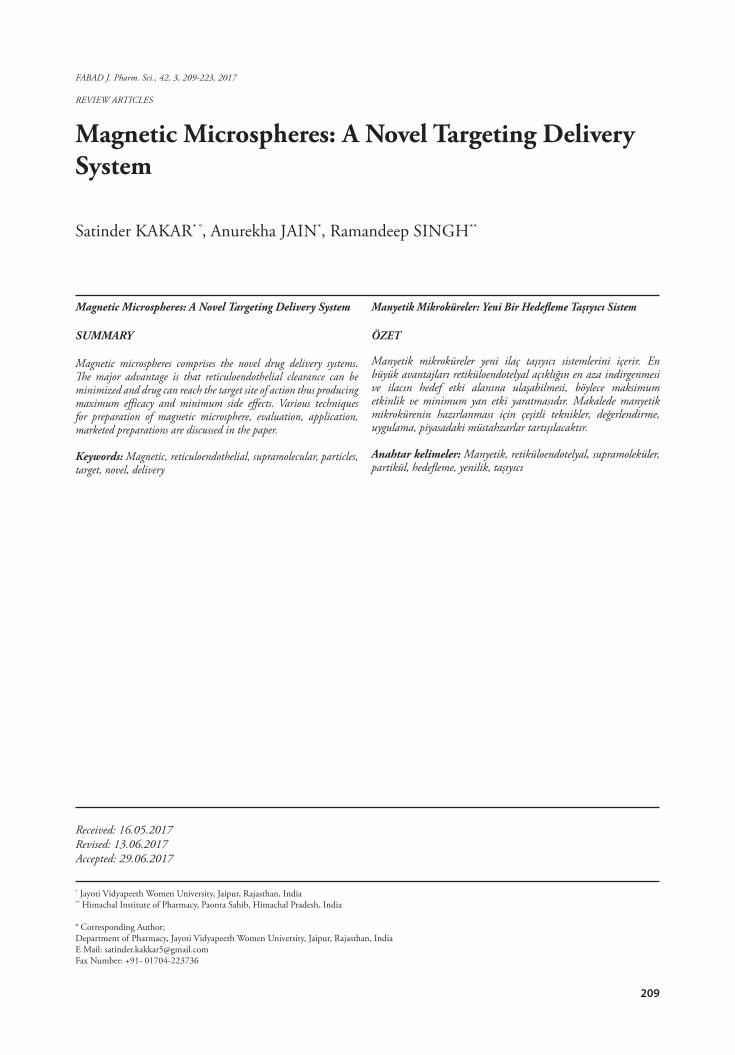
IntroductionMagnetic microspheres are the supramolecular
particles that are small enough to circulate through the capillaries but are sufficiently susceptible to be en-captured in microvessels by applying magnetic fields of 0.4 T-0.8 T. Targeted drug deliveries can be done by two modes i.e. magnetic drug delivery and non mag-netic drug delivery. Magnetic drug delivery by various novel carriers is an excellent method in which a drug is directly delivered to the diseased location/area. nonmagnetic drug delivery systems such as nanopar-ticles, microspheres and microparticles etc are suc-cessfully utilized for drug targeting but donot show a satisfactory site specificity and are rapidly cleared off by RES (reticuloendothelial system) (Widder et al. 1987). Magnetic polymer microspheres are usu-ally composed of magnetic cores to ensure a strong magnetic response and polymeric shells to protect from particle aggregation. These microspheres ex-hibit features such as small and proper size, different shapes, and various functional groups on the surface. They have therefore received much attention in recent years for wide potential applications such as immobi-lization of enzymes, protein separations, and various drug delivery processes. Thus magnetism plays an important role in living beings metabolism. For ex-ample, the haemoglobin is an iron complex present in blood and is magnetic in nature. Magnetite, Fe3O4, is a biocompatible structure and it has a cubic inverse spinal structure with oxygen forming a FCC closed packing and therefore it is one of the most commonly used biomaterials for biological and medical applica-tions from cell separation and drug delivery to hyper-thermia (Alexiou et al. 2001). Magnetically drug de-livery is a excellent way, in which a drug is binded to a small biocompatible magnetically active component, entrapped in the biodegradable polymeric matrix and pharmacologically active stable formulation is formu-lated , which is injected into the stream of blood and a high-gradient magnetic field is used to pull them out of suspension in the target region. Controlled drug release and further biodegradation are important for developing successful formulations. Mechanisms which involves Potential release are :
• Desorption of surface-bound /adsorbed drugs• Diffusion through the carrier matrix• carrier wall diffusion• Carrier matrix erosion; and• Combination of erosion /diffusion process
(Alagusundaram M et al. 2009).Principle of magnetic targeting1. A drug or therapeutic radioisotope is encap-
sulated in a magnetic compound; injected into pa-tient’s blood stream & powerful magnetic field in the target area is applied to stop it
2. Depending on the type of drug, it is then slowly released from magnetic, thus it reduces the loss of drug as freely circulating in body (Aggarwal A et al. 2012).
Figure 1,2 and 3 shows the drug targeting prin-ciple
Fig 1: Drug targeting via magnetic and non magnetic systems
Fig 2: Rationale of drug targeting
Fig 3: Mechanism of drug targeting
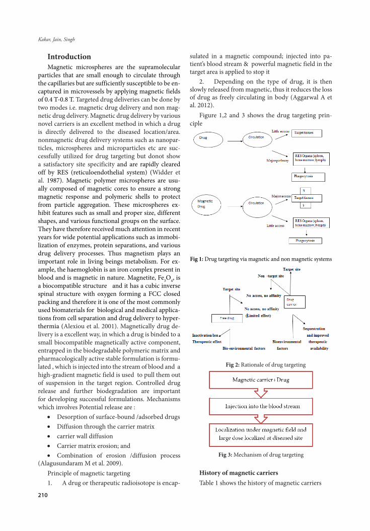
History of magnetic carriersTable 1 shows the history of magnetic carriers
211
FABAD J. Pharm. Sci., 42, 3, 209-223, 2017
Table 1: History of Magnetic Carriers
Year Scientist Discovery
1956 Gilchrist published a seminar paper in 1956 on the selective inductive heating of lymph nodes after injection of 20-100 nm sized magnetite particles into the lymph nodes near surgically removed cancer(Cleveland, 2004).
1960 Freeman, Arott, Watson
Transport of fine magnetic particles carrying radioactive materials or drugs through the vascular system with their subsequent focusing at localized parts of the body using magnetic fields (Freeman MW et al.1960)
1962 Meyers and co workers
Demonstrated the magnetic focusing of intra-arterially injected 1 to 3 µm radioactive iron (59Fe) particles in dogs(Meyers PH et al.1963; Meyers PH,1966).
1971 Nakamura et al
Reported the localized capture of magnetic particles introduced into the blood circulation of both rats and dogs (Nakamura T,1971).
1983 Ovadia et al pointed out that typical magnetic carriers are essentially saturated at 4000 Gauss (0.4 T) and that the magnitude of the field gradient then governs their focusing. This was consistent with their observation of the focusing of magnetic carriers at the edges of their magnet pole piece where the field gradients were highest, a phenomenon they referred to as the “edging effect.” Targeting an area with the face of a simple magnet could result in focusing of magnetic drug carriers more strongly to cells adjacent to the target. They proposed that the edge effect could be used to target specific areas by careful orientation and positioning of the magnet (Ovadia H,1983).
1984 Ishii et al described some in vitro studies using submicron magnetic liposome drug carriers(Ishii et al.1984).
1991 Goodwin et al observed extravasation of magnetic carriers (iron/carbon particles of diameter 0.5 to 5 µm, with 95% < 3 µm) in targeted areas of the liver in pigs. This, they claimed, was a mechanism for retention of the carriers at the targeted site after the removal of the magnetic field. The same carriers carrying doxorubicin were later used for a study of intravesical magnetic targeting of the bladder wall in healthy pigs using an externally applied neodymium-iron-boron (NdFeB) magnet (0.5 T). (Goodwin S et al. 1999; Goodwin SC et al.2001).
1997 Lübbe and co-workers
carried out the first Phase I clinical trial of magnetically targeted nanoparticles to deliver the anti-cancer drug 4’-epidoxorubicin to advanced solid tumors in 14 patients (Lubbe AS et al. 1983; Lubbe AS et al. 1996).
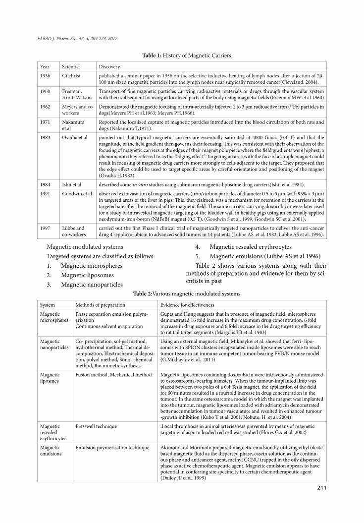
Magnetic modulated systemsTargeted systems are classified as follows:1. Magnetic microspheres2. Magnetic liposomes3. Magnetic nanoparticles
4. Magnetic resealed erythrocytes5. Magnetic emulsions (Lubbe AS et al.1996)Table 2 shows various systems along with their
methods of preparation and evidence for them by sci-entists in past
Table 2:Various magnetic modulated systems
System Methods of preparation Evidence for effectiveness
Magnetic microspheres
Phase separation emulsion polym-erizationContinuous solvent evaporation
Gupta and Hung suggests that in presence of magnetic field, microspheres demonstrated 16 fold increase in the maximum drug concentration, 6 fold increase in drug exposure and 6 fold increase in the drug targeting efficiency to rat tail target segments (Margolis LB et al. 1983)
Magnetic nanoparticles
Co- precipitation, sol-gel method, hydrothermal method, Thermal de-composition, Electrochemical deposi-tion, polyol method, Sono- chemical method, Bio mimetic synthesis
Using an external magnetic field, Mikhaylov et al. showed that ferri- lipo-somes with SPION clusters encapsulated inside liposomes were able to reach tumor tissue in an immune competent tumor-bearing FVB/N mouse model (G.Mikhaylov et al. 2011)
Magnetic liposmes
Fusion method, Mechanical method Magnetic liposomes containing doxorubicin were intravenously administered to osteosarcoma-bearing hamsters. When the tumour-implanted limb was placed between two poles of a 0.4 Tesla magnet, the application of the field for 60 minutes resulted in a fourfold increase in drug concentration in the tumour. In the same osteosarcoma model in which the magnet was implanted into the tumour, magnetic liposomes loaded with adriamycin demonstrated better accumulation in tumour vasculature and resulted in enhanced tumour -growth inhibition (Kubo T et al. 2001; Nobuto, H et al. 2004) .
Magnetic resealed erythrocytes
Presswell technique .Local thrombosis in animal arteries was prevented by means of magnetic targeting of aspirin loaded red cell was studied (Flores GA et al. 2002)
Magnetic emulsions
Emulsion poymerisation technique Akimoto and Morimoto prepared magnetic emulsion by utilizing ethyl oleate based magnetic fluid as the dispersed phase, casein solution as the continu-ous phase and anticancer agent, methyl CCNU trapped in the oily dispersed phase as active chemotherapeutic agent. Magnetic emulsion appears to have potential in conferring site specificity to certain chemotherapeutic agent (Dailey JP et al. 1999)
212
Kakar, Jain, Singh
Criteria for selection of drugs for formation of Magnetic microspheres
1. Magnetic microspheres are prepared when the drug is so dangerous that we cannot allow it to circulate freely into the blood stream.
2. When the agent is so expensive and we can-not afford to waste it.
3. Requires a selective regional effect to meet localized therapeutic objective.
4. Requires an alternative formulation essen-tial to continue treatment in patients whose systemic therapy must be temporarily discontinued due to life threatening toxicity directed at selective organs.
Methods of preparation of magnetic micro-spheres
1. Phase separation emulsion polymerization2. Continuous solvent evaporation3. Emulsion solvent extraction method4. Low temperature hydrothermal method5. Sonochemical method6. Chemical precipitation method7. Suspension polymerization method8. Swelling and penetration method9. Photopolymerisation method10. Emulsion solvent evaporation technique11. Vapour deposition technique12. Alkaline co-precipitation method13. Multiple emulsion method14. Crosslinking method15. Inverse phase suspension polymerization
method
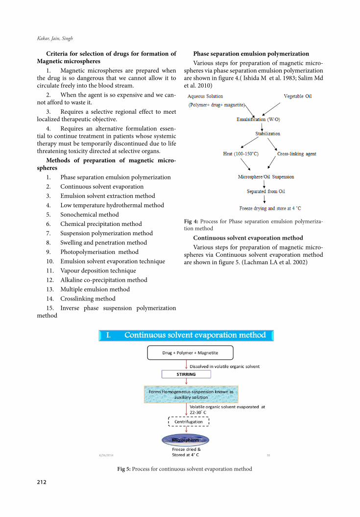
Phase separation emulsion polymerization Various steps for preparation of magnetic micro-
spheres via phase separation emulsion polymerization are shown in figure 4.( Ishida M et al. 1983; Salim Md et al. 2010)
Fig 4: Process for Phase separation emulsion polymeriza-tion method
Continuous solvent evaporation methodVarious steps for preparation of magnetic micro-
spheres via Continuous solvent evaporation method are shown in figure 5. (Lachman LA et al. 2002)
Fig 5: Process for continuous solvent evaporation method
213
FABAD J. Pharm. Sci., 42, 3, 209-223, 2017
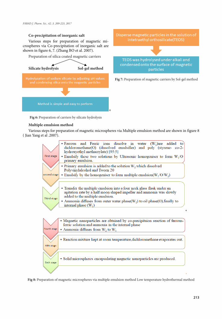
Co-precipitation of inorganic saltVarious steps for preparation of magnetic mi-
crospheres via Co-precipitation of inorganic salt are shown in figure 6, 7. (Zhang BO et al. 2007).
Preparation of silica coated magnetic carriers
Silicate hydrolysis Sol-gel method
Fig 6: Preparation of carriers by silicate hydrolysis
Fig 7: Preparation of magnetic carriers by Sol-gel method
Multiple emulsion methodVarious steps for preparation of magnetic microspheres via Multiple emulsion method are shown in figure 8
( Jian Yang et al .2007).
Fig 8: Preparation of magnetic microspheres via multiple emulsion method Low temperature hydrothermal method

214
Kakar, Jain, Singh
Various steps for preparation of magnetic microspheres via low temperature hydrothermal method are shown in figure 9 (Wei Jiang et al. 2013).
Fig 9: Low temperature hydrothermal method
Sonochemical methodVarious steps for preparation of magnetic microspheres via Sonochemical method are shown in figure 10
(Avivi S et al. 2001).
Fig 10: Sonochemical methodEmulsion polymerization methodVarious steps for preparation of magnetic microspheres via Emulsion polymerization method are shown in
figure 11 (E Pollert et al. 2006).
215
FABAD J. Pharm. Sci., 42, 3, 209-223, 2017
Fig 11: Emulsion polymerization method
Chemical precipitation method1. Polymer is added to aqueous solution of sodi-
um hydroxide depending on its solubility.2. Fe+2 and Fe+3(in molar ratio 1:2) are dissolved
in water and are added drop wise under continuous stirring at 25oC.
3. Hybrid material is separated by applying mag-netic field and washed several times with water and then in ethanol
4. Dry at 50o C to obtain magnetic microspheres for 24 hours
5. Magnetic microspheres are dispersed in mini-mum quantity of ultra-pure water and drug is added.
6. Finally dry at 40o C for 6 hours. (Carmen Mar-iana Chifiruic et al. 2012).
Suspension polymerization method1. Porous hydrophilic magnetic microspheres
are prepared by suspension polymerization. a satu-rated sodium chloride aqueous solution (50 ml) with the stabilizer soluble starch (2% of the total weight of the aqueous solution) as aqueous continuous phase is used, dropping a few methylene blue. The organic phase contained monomers GMA ( glycidyl methac-rylate), and VAc crosslinker EGDMA(ethylene glycol dimethacrylate (EGDMA) and vinyl acetate (VAc)), OA-Fe3O4(oleic acid coated) and n-heptane as the porogen.
2. The organic phase is placed overnight and vibrated with ultrasonic for 1 h, then initiator AIBN (azobisisobutyronitrile) is added before using.
3. Suspension polymerization is carried out in a 100 ml three-necked flask fitted with a nitrogen inlet, refluxing condenser and mechanical stirrer, the three-necked flask is placed in a water bath, stirred at 450 rpm under a nitrogen atmosphere.
4. The suspension polymerization is carried out at 50 .8oC for 1 h, 60.8oC for 2 h and then for 6 h at 75.8oC. After the reaction, the magnetic copolymer microspheres are isolated by magnetic decantation, and washed with heated distilled water several times, then are extracted in acetone for 48 h.
5. After that, the microspheres were alcoholysed
in 5% NaOH–methanol solution at room temperature for 12 h (Lei YL et al. 2001).
Swelling and penetration method1. PS (non porous polystyrene) particles are
mixed with NMP (Styrene, N-methyl-2-pyrrolidone)/water solution in a specific v/v NMP-to-water ratio.
2. SDS (sodium dodecyl Sulfate) is added to the NMP/water solution. The NMP/water mixture with PS spheres is left soaking for 24 h at room temperature while stirring (125 rpm).
3. Subsequently, the superparamagnetic nanopar-ticles dispersion is added to the mixture of PS sphere and NMP/ water solution at 30oC while shaking (at 140 rpm) for 1– 5 days to allow the magnetic nanopar-ticles to penetrate into the interior of the PS particles.
4. Afterwards, the polymer particles were sepa-rated from the solution by centrifugation.
5. Finally, particles were sequentially washed three times with methanol, three times with deion-ized water, and vacuum dried at room temperature for 1–2 days to yield the magnetic polymer microspheres. (T.H. Chung et al. 2008).
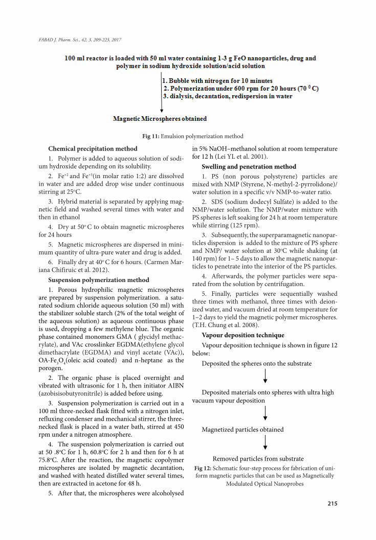
Vapour deposition techniqueVapour deposition technique is shown in figure 12
below:Deposited the spheres onto the substrate
Deposited materials onto spheres with ultra high vacuum vapour deposition
Magnetized particles obtained
Removed particles from substrateFig 12: Schematic four-step process for fabrication of uni-form magnetic particles that can be used as Magnetically
Modulated Optical Nanoprobes
Which production method is preferred for the preparation of the microparticles?
Production method depends on the type of drug used. Literature survey is done in order to find out which method is most compatible with our drug.
The most common methods used are Continuous solvent evaporation method and Phase separation emulsion polymerization method.
Phase separation may occur in case of Phase sep-aration emulsion polymerization which is its major disadvantage thus Continuous solvent evaporation method is preferred over it.FACTORS AFFECTING RATE OF DRUG DELIVERY
I. Amount and rate of drug delivery via magnet-ic microspheres can be regulated by changing size of microspheres, drug content, and content of magnetite and drug release of carrier.
• The size of microsphere is related to the drug content directly
• solubility characteristics of the drug and meth-od of preparation of microspheres affects the drug content
• Hydration state of microspheres affect drug release rate from the carrier
II. The retention of microspheres at the target sites is governed by the magnetic content and magnitude of applied field. If magnetic content is higher in micro-spheres, smaller magnetic fields are sufficient for ef-fective retention of microspheres at targeted sites. But in case of excessive magnetite in the microspheres the effective space available for drug in the microspheres is reduced appreciably. So balance between drug and magnetite content of microspheres is needed in order to design an efficient therapeutic system (Chopra KS et al. 1994).
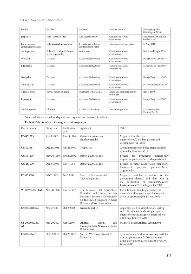
Various drugs which have been formulated as magnetic microspheres are shown in table 3.
Table 3:List of drugs,polymer,use and their respective methods for which Magnetic microspheres have been formulated
Drug Polymer Use Method Reference
Amphoterecin B Albumin Treatment of leishmaniasis Spray drying (Sánchez-Brunete JA, 2004)
Aztreonam Dextran Antimicrobial activity Continuous solvent evaporation
(Kang et al. 1987).
Adriamycin Albumin Cytotoxic effect on tumour cells
Heat stabilized protein methods
(Tao K et al., 1999).
Alpha chymotrypsin Titanium oxide hydrolysis of N-acetyl –L-tyrosine ethyl ester
Immobilization techniques (Izmaĭlov AF, 2000).
Aclarubicin Gelatin Intravascular tumour targeting
Water in oil emulsion polymerisation
(Kang et al. 1987).
Mesalamine Chitosan, Eudragit, Ethylcellulose
Ulcerative colitis Phase separation emulsion polymerisation
(Satinder Kakar et al. 2014).
dexamethasone Albumin Lymphocytic tumors Modified phase separation emulsion technique
(Sussan et al.1996).
azithromycin Dextran Potentiator effect on antimicrobial activity against S.aureus and P.aeruginosa reference strains
Continuous solvent evaporation
(Grumezesce et al. 2012).
Diclofenac sodium Gelatin Reduced joint swelling Emulsification and cross linking
(Saravanan M et al. 2008).
Oxantrazole Chitosan Cancer therapy Solvent evaporation (Hassan, EE,1992).
Vancomycin Starch Cytotoxic effect on cancer cells
Continuous solvent evaporation
(AM Grumezesce, 2012).
α- chymotrypsin Titanium oxide hydrolysis of N-acetyl-L-tyrosine ethyl ester
Immobilisation techniques (Izmaĭlov AF, 2000).
Penicillin Dextran Potentiator effect on antimicrobial activity
Continuous solvent evaporation
(Kang Choon Lee,1987)
Methotrexate Polyethylene glycol Anticancer activity Methotrexate reaction with amino-terminated magnetic microspheres
(Devineni, D,1995)
Doxorubicin Albumin Anticancer activity Crosslinking techniques (Kenneth, Widder, 1979).
Cisplatin Albumin Anticancer activity Phase separation emulsion polymerization
(Vyas MB 2012).
Sulforaphane Bovine serum albumin Cancer treatment Spray drying (Gerald G Enriquez, 2013)
217
FABAD J. Pharm. Sci., 42, 3, 209-223, 2017
Insulin Feridex diabetes Invasive method (Vijayaganapathy Vaithilingam,2016)
Etoposide Poly-(caprolactone) Anticancer activity Continuous solvent evaporation
(Vankayalu Devendiran Sundar, 2014)
Boron specific chelating substances
poly (glycidylmethacrylate) In treatment of boron comtaminated water
Dispersion polymerisation (D Yin, 2016)
6-thioguanine Polylactic acid-polyethylene glycol copolyester
anticancer Continuous solvent evaporation
(Kakar and Singh, 2014)
Ofloxacin Dextran Antimicrobial activity Continuous solvent evaporation
(Kang Choon Lee, 1987)
Rifampicin Dextran Antimicrobial activity Continuous solvent evaporation
(Kang Choon Lee, 1987)
Oxacyclin Dextran Antimicrobial activity Continuous solvent evaporation
(Kang Choon Lee, 1987)
Clindamycin Dextran Antimicrobial activity Continuous solvent evaporation
(AM Grumezesce, 2012)
5-Fluorouracil Bovine serum albumin Treatment of hepatomas Emulsion, heat stabilization technique
(Tao K,1999)
Piperacillin Dextran Antimicrobial activity Continuous solvent evaporation
(Kang Choon Lee, 1987)
Cephalosporins Chitosan Antibacterial activity Solvent evaporation (Carmen Mariana Chifiruic,2012).
Patents which are related to Magnetic microspheres are discussed in table 4.
Table 4: Patents related to magnetic microspheres
Patent number Filing date Publication date
Applicant Title
US4452773 Apr 5,1982 June 5,1984 Canadian patents and development ltd
Magnetic iron-dextran microspheres(Canadian patents and development ltd,1984)
US5032381 Dec 20,1988 July 16,1991 Tropix, Inc Chemiluminescence based static and flow cytometry (Tropix.,1991)
US5091206 May 30,1989 Feb 25,1992 Baxter diagnosis Inc Process for producing magnetically responsive particles(Baxter diagnosis Inc)
US5283079 Dec 14,1989 Feb 1,1994 Baxter diagnosis Inc Process to make magnetically responsive fluorescent polymer particles(Baxter diagnosis Inc)
US5855790 Feb 7,1995 Jan 5,1999 Selective Environmental Technologies, Inc.
Magnetic particles, a method for the preparation thereof and their use in the purification of solutions(Selective Environmental Technologies, Inc,1999)
WO1997020214A1 Nov 28,1996 June 5,1997 The Minister Of Agriculture, Fisheries and Food In her Britannic Majesty’s Government Of The United Kingdom Of Great Britain and Northern Ireland
Extraction and labelling of biological materials with magnetic and fluorescent beads or liposomes(US Patent,1997)
US20030186465 Nov 27,2001 Oct 2,2003 Kraus Robert H Apparatus used in identification, sorting and collection methods using magnetic microspheres and magnetic microsphere kits(Kraus Robert H,2003)
US 20090092837 A1
Mar 20,2007 Apr 9,2009 Andreas Axen, Eva Holmgren,Nils Norrman, Tobias E. Soderman
Magnetic beads(Andreas Axen,2009)
US8564776B2 Oct 12,2010 Oct 23,2013 Stevens W. Graves, Robert C. Habbersett
System and method for measuring particles in a sample stream of a flow cytometer using a low power laser source (Stevens W. Graves,2013)

218
Kakar, Jain, Singh
Applications of magnetic microspheres1. Magnetic microspheres in labelling and sep-
aration of cellsMagnetic and fluorescent properties of Probe(cell
surface) containing iron-containing polymeric mi-crospheres tagged with dyes which are fluorescent in nature and coupled chemically to antibodies or lec-tins have been used in the magnetic separation of red blood cells and in the identification of immunoglob-ulin receptors and wheat germ agglutinin (WGA) re-ceptors present on lymphocytes and Hela cells by SEM and fluorescent microscopy (RS Molday,1977).
2. Magnetic microspheres have widespread ap-plication in bioengineering, biomedicine, trends such as enzyme immobilization(Lea T,1985).
3. Deoxyribonucleic (DNA) research and ge-netic exploration
DNA makes each species different and unique. Nucleic acid isolation by means of magnetic beads is a good method of extracting DNA for analysis and genetic exploration. (Cax X,1977).
4. Magnetically induced Hyperthermia for treatment of cancer.
Reduction of viability of cancer cells on treat-ment of organs with heat to a 42–46 oC is termed as hyperthermia. It is based on the fact that tumor cells are more sensitive to temperature than normal cells. In hyperthermia a heat delivery system is established, such that the tumor cells are inactivated while normal ones are unaffected.( Burns MA,1985).
5. Human cholangiocarcinoma xenograftsCholangiocarcinoma, is a malignant disease af-
fecting the biliary tract, and the incidence ratio of
male to female is 1.46:1. Treatment includes mainly operation, and combined chemotherapy and radia-tion. But it is very difficult to diagnose it in early stag-es. the outcome of operation can be unsatisfactory, and the survival rate is very low. Single or combined application of chemotherapeutic drugs is usually less than 30% successful in the clinic. The targeting drug with magnetic microspheres to treat human cholan-giocarcinoma xenografts is a novel technique for it+ (Deore BV et al. 2009)
6. Delivery of chemotherapeutic drugs to liver tumors
Lubbe et al performed the first clinical cancer therapy trial using magnetic microspheres in Germa-ny for the treatment of advanced solid cancer in 14 patients. The phase I study showed the low toxicity of the method and the accumulation of the Magnetic Microspheres in the target area. MRI measurements were carried out which showed that more than 50% of the Magnetic Microspheres were ended up in the liv-er. This was likely due to the particles’ small size and low magnetic susceptibility which limited the ability to hold them at the target organ. (Kobayashi H, 1991; Matsunaga TJ, 1991; Lubbe et al. 1996)
7. BioassaysDrug, hormone, vitamins etc, are tested for de-
termining their activity by testing its potency on a living organism, and are measured in comparison to recognized standards. Magnetic beads provide high levels of chemical and physical stability for calculating changes of selected particles within a sample. (Mittag TW, 2000)
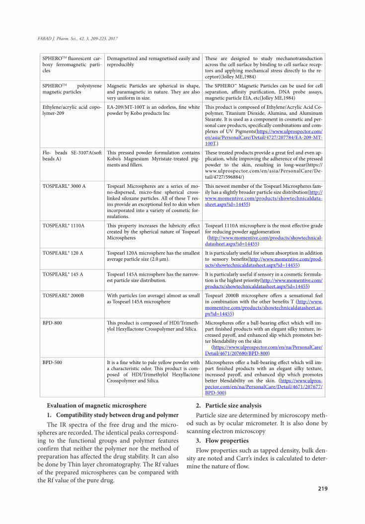
Market ProductsMarketed products are discussed below in table 5.
Table 5: Marketed products
Name Advantages Applications
NanoLink™ Streptavidin Magnetic Beads 0.8 μm
1. Highest Biotin Binding 2. Fast (<2 min) Response Time 3. Versatile nature
Magnetic Beads are suited for generating single-strand-ed PCR templates that can increase hybridization ef-ficiency to complementary probes by removal of the unbiotinylated, competing PCR strand (Fowler DM et al.2010)
AbraMagTM Magnetic beads 1.Superior binding capacity 2.Superior yield 3.Equal or superior purity of target analyte4.Small size, irregular surface but non-po-rous particles mean high
The AbraMag Genomic DNA Magnetic Beads Kit is de-signed to extract and purify genomic DNA from tissues and bacteria (gram-positive and -negative) ( Ahmed S,1980)
SPHEROTM carboxyl ferro-magnetic particles
Demagnetized and remagnetised easily and reproducibly
These are designed to study mechanotransduction across the cell surface by binding to cell surface recep-tors and applying mechanical stress directly to the re-ceptor (Jolley ME,1984)
SPHEROTM amino ferro-magnetic particles
Demagnetized and remagnetised easily and reproducibly
These are designed to study mechanotransduction across the cell surface by binding to cell surface recep-tors and applying mechanical stress directly to the re-ceptor(Jolley ME,1984)
219
FABAD J. Pharm. Sci., 42, 3, 209-223, 2017
SPHEROTM fluorescent car-boxy ferromagnetic parti-cles
Demagnetized and remagnetised easily and reproducibly
These are designed to study mechanotransduction across the cell surface by binding to cell surface recep-tors and applying mechanical stress directly to the re-ceptor((Jolley ME,1984)
SPHEROTM polystyrene magnetic particles
Magnetic Particles are spherical in shape, and paramagnetic in nature. They are also very uniform in size.
The SPHERO™ Magnetic Particles can be used for cell separation, affinity purification, DNA probe assays, magnetic particle EIA, etc(Jolley ME,1984)
Ethylene/acrylic acid copo-lymer-209
EA-209/MT-100T is an odorless, fine white powder by Kobo products Inc
This product is composed of Ethylene/Acrylic Acid Co-polymer, Titanium Dioxide, Alumina, and Aluminum Stearate. It is used as a component in cosmetic and per-sonal care products, specifically combinations and com-plexes of UV Pigments(https://www.ulprospector.com/en/asia/PersonalCare/Detail/4727/207784/EA-209-MT-100T.)
Flo- beads SE-3107A(soft beads A)
This pressed powder formulation contains Kobo’s Magnesium Myristate-treated pig-ments and fillers.
These treated products provide a great feel and even ap-plication, while improving the adherence of the pressed powder to the skin, resulting in long-wear(https://www.ulprospector.com/en/asia/PersonalCare/De-tail/4727/596884/)
TOSPEARL® 3000 A Tospearl Microspheres are a series of mo-no-dispersed, micro-fine spherical cross-linked siloxane particles. All of these T res-ins provide an exceptional feel to skin when incorporated into a variety of cosmetic for-mulations.
This newest member of the Tospearl Microspheres fam-ily has a slightly broader particle size distribution(http://www.momentive.com/products/showtechnicaldata-sheet.aspx?id=14455)
TOSPEARL® 1110A This property increases the lubricity effect created by the spherical nature of Tospearl Microspheres
Tospearl 1110A microsphere is the most effective grade for reducing powder agglomeration (http://www.momentive.com/products/showtechnical-datasheet.aspx?id=14455)
TOSPEARL® 120 A Tospearl 120A microsphere has the smallest average particle size (2.0 µm).
It is particularly useful for sebum absorption in addition to sensory benefits(http://www.momentive.com/prod-ucts/showtechnicaldatasheet.aspx?id=14455)
TOSPEARL® 145 A Tospearl 145A microsphere has the narrow-est particle size distribution.
It is particularly useful if sensory in a cosmetic formula-tion is the highest priority(http://www.momentive.com/products/showtechnicaldatasheet.aspx?id=14455)
TOSPEARL® 2000B With particles (on average) almost as small as Tospearl 145A microsphere
Tospearl 2000B microsphere offers a sensational feel in combination with the other benefits T (http://www.momentive.com/products/showtechnicaldatasheet.as-px?id=14455)
BPD-800 This product is composed of HDI/Trimeth-ylol Hexyllactone Crosspolymer and Silica.
Microspheres offer a ball-bearing effect which will im-part finished products with an elegant silky texture, in-creased payoff, and enhanced slip which promotes bet-ter blendability on the skin (https://www.ulprospector.com/en/na/PersonalCare/Detail/4671/207680/BPD-800)
BPD-500 It is a fine white to pale yellow powder with a characteristic odor. This product is com-posed of HDI/Trimethylol Hexyllactone Crosspolymer and Silica.
Microspheres offer a ball-bearing effect which will im-part finished products with an elegant silky texture, increased payoff, and enhanced slip which promotes better blendability on the skin. (https://www.ulpros-pector.com/en/na/PersonalCare/Detail/4671/207677/BPD-500)
Evaluation of magnetic microsphere1. Compatibility study between drug and polymerThe IR spectra of the free drug and the micro-
spheres are recorded. The identical peaks correspond-ing to the functional groups and polymer features confirm that neither the polymer nor the method of preparation has affected the drug stability. It can also be done by Thin layer chromatography. The Rf values of the prepared microspheres can be compared with the Rf value of the pure drug.
2. Particle size analysisParticle size are determined by microscopy meth-
od such as by ocular micrometer. It is also done by scanning electron microscopy
3. Flow propertiesFlow properties such as tapped density, bulk den-
sity are noted and Carr’s index is calculated to deter-mine the nature of flow.
220
Kakar, Jain, Singh
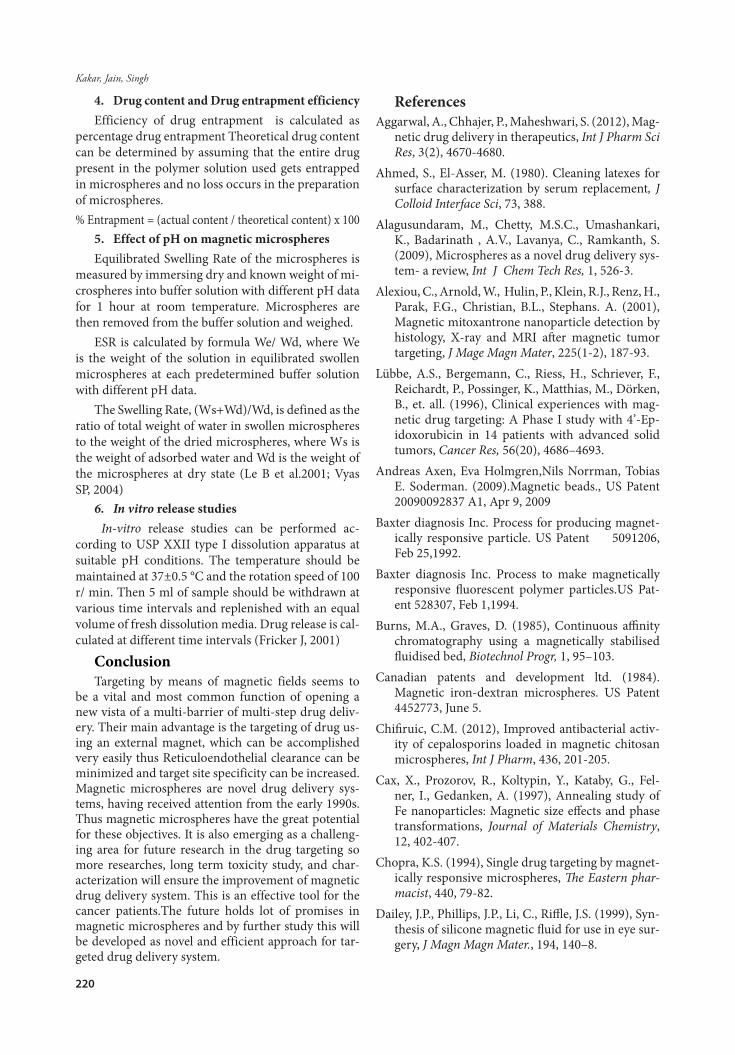
4. Drug content and Drug entrapment efficiencyEfficiency of drug entrapment is calculated as
percentage drug entrapment Theoretical drug content can be determined by assuming that the entire drug present in the polymer solution used gets entrapped in microspheres and no loss occurs in the preparation of microspheres. % Entrapment = (actual content / theoretical content) x 100
5. Effect of pH on magnetic microspheresEquilibrated Swelling Rate of the microspheres is
measured by immersing dry and known weight of mi-crospheres into buffer solution with different pH data for 1 hour at room temperature. Microspheres are then removed from the buffer solution and weighed.
ESR is calculated by formula We/ Wd, where We is the weight of the solution in equilibrated swollen microspheres at each predetermined buffer solution with different pH data.
The Swelling Rate, (Ws+Wd)/Wd, is defined as the ratio of total weight of water in swollen microspheres to the weight of the dried microspheres, where Ws is the weight of adsorbed water and Wd is the weight of the microspheres at dry state (Le B et al.2001; Vyas SP, 2004)
6. In vitro release studies In-vitro release studies can be performed ac-
cording to USP XXII type I dissolution apparatus at suitable pH conditions. The temperature should be maintained at 37±0.5 °C and the rotation speed of 100 r/ min. Then 5 ml of sample should be withdrawn at various time intervals and replenished with an equal volume of fresh dissolution media. Drug release is cal-culated at different time intervals (Fricker J, 2001)
ConclusionTargeting by means of magnetic fields seems to
be a vital and most common function of opening a new vista of a multi-barrier of multi-step drug deliv-ery. Their main advantage is the targeting of drug us-ing an external magnet, which can be accomplished very easily thus Reticuloendothelial clearance can be minimized and target site specificity can be increased. Magnetic microspheres are novel drug delivery sys-tems, having received attention from the early 1990s. Thus magnetic microspheres have the great potential for these objectives. It is also emerging as a challeng-ing area for future research in the drug targeting so more researches, long term toxicity study, and char-acterization will ensure the improvement of magnetic drug delivery system. This is an effective tool for the cancer patients.The future holds lot of promises in magnetic microspheres and by further study this will be developed as novel and efficient approach for tar-geted drug delivery system.
ReferencesAggarwal, A., Chhajer, P., Maheshwari, S. (2012), Mag-
netic drug delivery in therapeutics, Int J Pharm Sci Res, 3(2), 4670-4680.
Ahmed, S., El-Asser, M. (1980). Cleaning latexes for surface characterization by serum replacement, J Colloid Interface Sci, 73, 388.
Alagusundaram, M., Chetty, M.S.C., Umashankari, K., Badarinath , A.V., Lavanya, C., Ramkanth, S. (2009), Microspheres as a novel drug delivery sys-tem- a review, Int J Chem Tech Res, 1, 526-3.
Alexiou, C., Arnold, W., Hulin, P., Klein, R.J., Renz, H., Parak, F.G., Christian, B.L., Stephans. A. (2001), Magnetic mitoxantrone nanoparticle detection by histology, X-ray and MRI after magnetic tumor targeting, J Mage Magn Mater, 225(1-2), 187-93.
Lübbe, A.S., Bergemann, C., Riess, H., Schriever, F., Reichardt, P., Possinger, K., Matthias, M., Dörken, B., et. all. (1996), Clinical experiences with mag-netic drug targeting: A Phase I study with 4’-Ep-idoxorubicin in 14 patients with advanced solid tumors, Cancer Res, 56(20), 4686–4693.
Andreas Axen, Eva Holmgren,Nils Norrman, Tobias E. Soderman. (2009).Magnetic beads., US Patent 20090092837 A1, Apr 9, 2009
Baxter diagnosis Inc. Process for producing magnet-ically responsive particle. US Patent 5091206, Feb 25,1992.
Baxter diagnosis Inc. Process to make magnetically responsive fluorescent polymer particles.US Pat-ent 528307, Feb 1,1994.
Burns, M.A., Graves, D. (1985), Continuous affinity chromatography using a magnetically stabilised fluidised bed, Biotechnol Progr, 1, 95–103.
Canadian patents and development ltd. (1984). Magnetic iron-dextran microspheres. US Patent 4452773, June 5.
Chifiruic, C.M. (2012), Improved antibacterial activ-ity of cepalosporins loaded in magnetic chitosan microspheres, Int J Pharm, 436, 201-205.
Cax, X., Prozorov, R., Koltypin, Y., Kataby, G., Fel-ner, I., Gedanken, A. (1997), Annealing study of Fe nanoparticles: Magnetic size effects and phase transformations, Journal of Materials Chemistry, 12, 402-407.
Chopra, K.S. (1994), Single drug targeting by magnet-ically responsive microspheres, The Eastern phar-macist, 440, 79-82.
Dailey, J.P., Phillips, J.P., Li, C., Riffle, J.S. (1999), Syn-thesis of silicone magnetic fluid for use in eye sur-gery, J Magn Magn Mater., 194, 140–8.
221
FABAD J. Pharm. Sci., 42, 3, 209-223, 2017
Deore, B.V., Mahajan, H.S., Deore, U.V. (2009), Devel-opment and characterization of sustained release microspheres by quasi emulsion solvent diffusion method, International Journal of Chem Tech Re-search., 634-642.
Devineni, D., Blanton, C.D., Gallo, J.M. (1995), Prepa-ration and in vitro evaluation of magnetic micro-sphere-methotrexate conjugate drug delivery sys-tems, Bioconjug Chem, 6(2), 203-10.
Flores, G.A., Liu, J. (2002), In vitro blockage of a sim-ulated vascular system using magneto rheological fluids as a cancer therapy, Eur Cells Mater, 3, 9–11.
Fowler, D.M., Araya, C.L., Fleishman, S.J., Kel-logg, E.H., Stephany, J.J., Baker, D., Fields, S. (2010), High-resolution mapping of protein se-quence-function relationships, Nat Methods, 7(9), 741-6.
Freeman, M.W., Arrott, A., Watson, J.H.L. (1960), Magnetism in medicine, J Appl Phys, 31(5), S404–S405.
Fricker, J. (2001), Drugs with a magnetic attraction to tumours, Drug Discov Today, 6, 387-389.
Enriquez G. G., Rizvi, S.A., D’Souza M.J., Do, D.P. (2013), Formulation and evaluation of drug-load-ed targeted magnetic microspheres for cancer therapy, Int J Nanomedicine, 8, 1393–1402
Goodwin, S., Peterson, C., Hoh, C., Bittner, C. (1999), Targeting and retention of magnetic targeted car-riers (MTCs) enhancing intra-arterial chemother-apy, J Magn Magn Mater, 194 (1–3), 132–139.
Goodwin, S.C., Bittner, C.A., Peterson, C.L., Wong, G. (2001), Single-dose toxicity study of hepatic intra-arterial infusion of doxorubicin coupled to a novel magnetically targeted drug carrier, Toxicol Sci, 60(1), 177–183.
Grumezescu, A.M., Ficai, A., Ficai, D., Predan, G., Chifiriuc, M.C. (2012), Polymeric magnetic silica microspheres as a drug loader for antimicrobial delivery substances, Dig J Nanomater Biostruct, 7, 1891-1896.
Hassan, E.E., Parish, R.C., Gallo, J.M. (1992), Opti-mized formulation of magnetic chitosan micro-spheres containing the anticancer agent, oxantra-zole, Pharm Res, 9(3), 390-397.
http://www.momentive.com/products/showtechni-caldatasheet.aspx?id=14455. accessed on 20.9.16.
https://www.ulprospector.com/en/asia/PersonalCare/Detail/4727/207784/EA-209-MT-100T. accessed on 30.9.16.
https://www.ulprospector.com/en/asia/PersonalCare/Detail/4727/596884/PowderFoundation-with-Flo-Beads-SE-3107A-Softbeads-A-Formulation-KPP-022B. accessed on 30.9.16.
https://www.ulprospector.com/en/na/Personal-Care/Detail/4671/207677/BPD-500. accessed on 30.9.16.
https://www.ulprospector.com/en/na/Personal-Care/Detail/4671/207680/BPD-800. accessed on 20.9.16.
Ishida, M., Nambu, N., Nagai, T. (1983), Highly vis-cous gel ointment containing carbopol for appli-cation to oral mucosa, Chem Pharm Bull, 31, 4561.
Ishii, F., Takamura, A., Noro S. (1984), Magnetic microcapsules for in vitro testing as carrier for intravascular administration of anticancer drugs: preparation and physicochemical properties, Chem Pharm Bull, 32(2), 678–684.
Izmaĭlov, AF., Kiselev, MV., Vakurov, AV., Gladilin, AK., Levashov, AV. (2000), Alpha-chymotrypsin immobilized on ferromagnetic particles coat-ed with titanium oxide: production and catalytic properties, Prikl Biokhim Mikrobiol, 36(1), 68-73.
Yang, J., Bi, C.X., Su, Z.G. (2007), A novel process to prepare magnetic polymer microspheres, Chemis-try Letters, 36(8), 1062-1063
Jolley, M.E., Wang, C.H., Ekenberg S.J., Zuelke, M.S., Kelso, D.M. (1984), Particle concentration fluores-cence immunoassay (PCFIA): a new, rapid immu-noassay technique with high sensitivity, J Immunol Methods, 67(1), 21-35.
Kakar, S., and Singh, R. (2014), 6-Thioguanine load-ed magnetic microspheres as a new drug delivery system to cancer patients, Afr J Pharm Pharmacol, 8(31), 786-792.
Kang, C.L., Koh, I.B. (1987). Intravascular tumour tar-geting of aclarubicin-loaded gelatin microspheres: preparation, biocompatibility and biodegradabili-ty, Arch Pharm Res, 10(1), 42-49.
Widder, K., Flouret, G., Senyei A. (1979), Magnet-ic Microspheres: synthesis of a Novel Parenteral Drug Carrier, J Pharm Sci, 68( 1), 79-82
Kobayashi, H., Matsunaga, T.J. (1991), Applications of Nano-sized Magnetic Carriers in Biocatalysis and Bioseparation, Colloid Interface Sci, 141, 505–511
Kraus Robert H. “Apparatus used in identification, sorting and collection methods using magnetic microspheres and magnetic microsphere kits”, US Patent 20030186465,Oct 2,2003
Kubo T. (2001), Targeted systemic chemotherapy us-ing magnetic liposomes with incorporated adria-mycin for osteosarcoma in hamsters, Int J Oncol, 18, 121–125
Kuznetsov, A.A., Shlyakhtin, O.A., Brusentsov, N.A., Kuznetsov, O.A. (2002), Smart mediators for self-controlled inductive heating, Eur Cells Mater, 3, 75–7
222
Kakar, Jain, Singh
Lachman, L.A., Liberman, H.A., Kanig J.L. (2002), The theory and practice of industrial pharmacy, Mumbai (Hindistan): Varghese, p. 414-415.
Le, B., Shinkai, M., Kitade, T., Honda, H., Yoshida, J., Wakabayashi, T. (2001), Preparation of tumor-spe-cific magnetoliposomes and their application for hyperthermia, J Chem Eng Jpn, 34(1), 66-72.
Lea, T., Vartdal, F., Davies, C., Ugalsted, J. (1985)., Magnetic monosized polymeric particles for large and specific fraction at ion of human mononuclear cells scan, J Immunol, 22, 207-216
Lei, Y.L., Liu, Z.Z., Liu, Q.F., Wu, X.Y. (2001), Synthe-sis of a macroporous hydrophilic ternary copoly-mer and its application in boronate-affinity sepa-ration, React Funct Polym, 48, 159–67.
Lubbe, A.S., Bergemann, C., Huhnt, W., Fricke, T., Riess, H., Brock, J.W., Huhn, D. (1996), Preclinical experiences with magnetic drug targeting: toler-ance and efficacy, Cancer Res, 56(20), 4694–4701.
Lubbe, A.S., Bergemann, C., Riess, H., Schriever, F., Reichardt, P., Possinger, K., Matthias, M., Dörken, B., Herrmann, F., Gürtler, R., Hohenberger, P., Haas, N., Sohr, R., Sander, B., Lemke Margolis, L.B., Namiot, V.A., Kljkin, L.M. (1983), Biochim Biophysic Acta, 735, 193.
Meyers, P.H., Cronic, F., Nice, C.M. (1963), Exper-imental approach in the use of magnetic control of metallic iron particles in the lymphatic and vascular system of dogs as a contrast and isotopic agent, Am J Roentgenol Radium Ther Nucl Med, 90, 1068–1077.
Meyers, PH., Nice, CM., Jr, Meckstroth, G.R., Becker, H.C., Moser, P.J., Goldstein M. (1966), Patholog-ical studies following magnetic control of metal-lic iron particles in the lymphatic and vascular system of dogs as a contrast and isotropic agent, Am J Roentgenol Radium Ther Nucl Med, 96(4), 913–921.
Mittag, T.W., Danias, J., Pohorenec, G. (2000), Retinal damage after 3 to 4 months of elevated intraocular pressure in a rat glaucoma model, Invest Ophthal-mol Vis Sci, 41, 3451–3459.
Mikhaylov, G., Mikac, U., Magaeva, A.A., Iltin, V.I., Naiden, E.P., Psakhye, I., Babes, L., Reinheckel, T., et. all., (2011), Ferri-liposomes as an MRI-visible drug-delivery system for targeting tumours ISRN Nanomaterials and their microenvironment, Nat Nanotechnol, 6(9), 594–602
Nakamura, T., Konno, K., Moroné, T., Tsuya, N., Hat-ano, M. (1971), Magneto-medicine: biological as-pects of ferromagnetic fine particles, J Appl Phys, 42(4), 1320–1324.
Nobuto, H. (2004), Evaluation of systemic chemo-therapy with magnetic liposomal doxorubicin and a dipole external electromagnet, Int J Cancer, 109, 627–635
Ovadia, H., Paterson, PY., Hale, JR. (1983). Magnetic microspheres as drug carriers: Factors influencing localization at different anatomical sites in rats, Isr J Med Sci, 19(7), 631–637.
Pollert, E., Knizek, K., Marysko, M., Zaveta, K., Lan-cok, A., Bohacek, J., Horak, D., Babic, M. (2006), Magnetic poly(glycidyl methacrylate) micro-spheres containing maghemite prepared by emul-sion polymerization, J Magn Magn Mater, 306, 241-247.
Molday, R.S., Yen, S.P., Rembaum, A. (1977), Appli-cation of magnetic microspheres in labelling and separation of cells, Nature, 268 (5619), 437– 438.
Avivi, S., Felner, I., Novik, I., Gedanken, A. (2001), The preparation of magnetic proteinaceous micro-spheres using the sonochemical method, Biochim Biophys Acta., 1527 (3), 123-129.
Salim, M.D., Shukla, V.K., Bhardwaj, V., Garg, V.K., Sharma, P.K. (2010), Magnetic Microspheres as A Magnetically Targeted Drug Delivery System, Journal of Global Pharma Technology, 2(3), 36-46.
Sánchez-Brunete, J.A., Dea, M.A., Rama, S., Bolás, F., Alunda, J.M., Raposo, R. (2004), Treatment of ex-perimental visceral leishmaniasis with amphoter-icin B in stable albumin microspheres, Antimicrob Agents Chemother, 48(9), 3246-3252.
Saravanan, M., Anbu, J., Maharajan, G., Pillai, K.S. (2008), Targeted delivery of diclofenac sodium via gelatin magnetic microspheres formulated for intra-arterial administration, J Drug Target, 16(5), 366-378
Satinder, K., Ramandeep, S. (2014), Preparation of magnetic microspheres of mesalamine by phase separation emulsion polymerization technique, Afr J Pharm Pharmacol, 8(9), 246-258.
Selective Environmental Technologies, Inc., “Magnet-ic particles, a method for the preparation thereof and their use in the purification of solutions”,US Patent 5855790, Jan 5,1999.
Stevens W, Graves., Robert C,Habbersett. “System and method for measuring particles in a sample stream of a flow cytometer using a low power laser source”. US Patent 8564776B2, Oct 23,2013.
Sussan, G., Turaj, E., Forutan, S.M., Mortazavi, S.A. (1996), Dexamethasone loaded magnetic albumin microspheres, Int J Pharm, 130 (1), 49-55.
223
FABAD J. Pharm. Sci., 42, 3, 209-223, 2017
Chung, T.H., Lee, W.C. (2008), Preparation of sty-rene-based, magnetic polymer microspheres by a swelling and penetration process, React Funct Polym, 1441–1447.
Tao, K., Chen, D., Chen, J., Tian, Y., Wu, Z., Wang, X. (1999), Preparation of adriamycin magnetic albu-min microspheres and their experimental antitu-mor effects in vitro and in vivo, J Tongji Med Univ, 19, 295- 299.
Hafeli, U.O. The Cleveland Clinic Foundation (2004). Magnetically modulated therapeutic systems. Int J Pharm, 277 (1-2), 19-24.
The Minister Of Agriculture, Fisheries and Food In her Britannic Majesty’s Government Of The United Kingdom Of Great Britain and Northern Ireland, “Extraction and labelling of biological materials with magnetic and fluorescent beads or liposomes” ,US Patent WO 1997020214A1,June 5,1997
Tropix, “Chemiluminescence based static and flow cy-tometry”, US Patent 5032381, July 16,1991.
Sundar, V.D., Dhanaraju, M.D., Sathyamoorthy, N. (2014). Fabrication and characterization of etopo-side loaded magnetic polymeric microparticles. Int J Drug Delivery, 6,24-35.
Vaithilingam, V., Yim, M.M.W., Foster, J.L., Stait-Gard-ner, T., Oberholzer, J., Tuch, B.E. (2016). Noninva-sive Tracking of Encapsulated Insulin Producing Cells Labelled with Magnetic Microspheres by Magnetic Resonance Imaging, J Diabetes Res, 1-13.
Vyas, M.B., Shah, S.K. (2012), Design and character-ization of cisplatin magnetic microspheres, Int J Pharm Res Scholars, 1(4), 25-32.
Vyas, S.P., Khar, R.K, Targeted & Controlled Drug De-livery. Novel Carrier systems. CBS Publications; 2004, p. 458-483.
Jiang, W., Zhang, X., Sun, Z., Fang, Y., Li, F., Chen K., Huang, C. (2013), Preparation and mechanism of magnetic carbonaceous polysaccharide micro-spheres by low temperature hydrothermal meth-od, J Magn Magn Mater, 323, 2741-2747.
Widder, D.J., Greif, W.L., Widder, K.J., Edelman, R.R., Brady, T.J. (1987), Magnetite albumin micro-spheres: a new MR contrast material, AJR, Am J Roentgenol, 148 (2), 399-404.
Yin, D., Liu, H., Zhang, B., Geng., W. (2016), Magnet-ic boron specific chelating microsphere by disper-sion polymerisation for boron adsorption, Mater Technol Advance Perform Mat, 31(6), 352-357.
Zhang, B.O. (2007), Preparation and application of magnetic microsphere carriers, Front Chem Engi-neer China, 1(1), 96-101.