REVIEW ARTICLE Recent insights into the impairment of memory
21
REVIEW ARTICLE Recent insights into the impairment of memory in epilepsy: transient epileptic amnesia, accelerated long-term forgetting and remote memory impairment C. R. Butler 1 and A. Z. Zeman 2 1 Division of Clinical Neurosciences, University of Edinburgh, Edinburgh and 2 Department of Neurology, Peninsula Medical School, Exeter, UK Correspondence to: Prof. Adam Zeman, Professor of Cognitive and Behavioural Neurology, Peninsula Medical School, Barrack Road, Exeter, EX2 5DU, UK E-mail: [email protected] Complaints of memory difficulties are common among patients with epilepsy, particularly with temporal lobe epilepsy where memory-related brain structures are directly involved by seizure activity. However, the reason for these complaints is often unclear and patients frequently perform normally on standard neuropsychological tests of memory. In this article, we review the literature on three recently described and interrelated forms of memory impairment associated with epilepsy: (i) transient epileptic amnesia, in which the sole or main mani- festation of seizures is recurrent episodes of amnesia; (ii) accelerated long-term forgetting, in which newly acquired memories fade over days to weeks and (iii) remote memory impairment, in which there is loss of memories for personal or public facts or events from the distant past. Accelerated long-term forgetting and remote memory impairment are common amongst patients with transient epileptic amnesia, but have been reported in other forms of epilepsy. Their presence goes undetected by standard memory tests and yet they can have a profound impact on patients’ lives. They pose challenges to current theoretical models of memory. We discuss the evidence for each of these phenomena, as well as their possible pathophysiological bases, meth- odological difficulties in their investigation and their theoretical implications. Keywords: memory; epilepsy; transient epileptic amnesia; accelerated long-term forgetting; remote memory impairment Abbreviations: TLE = temporal lobe epilepsy; TEA = transient epileptic amnesia; ALF = accelerated long-term forgetting; RMI = remote memory impairment Received December 28, 2007. Revised April 2, 2008. Accepted May 20, 2008. Advance Access publication July 9, 2008 ‘A proper understanding of the amnesia accompanying epileptic attacks would contribute much to an understanding of the neurophysiological mechanisms of formation of memory patterns and of recall.’ Nisien JM. Memory and amnesia. Los Angeles: San Lucas Press, 1958. People with epilepsy frequently complain of memory difficulties. A community-based survey of over 1000 epi- lepsy patients in the United States revealed that cognitive difficulties ranked highest on a list of potential concerns (Fisher et al., 2000). In another study, 54% of over 700 people with epilepsy regarded memory problems as a moderate to severe nuisance (Thompson and Corcoran, 1992). Many interacting factors may affect memory function in patients with epilepsy including the underlying neuropathology (Lencz et al., 1992), seizure activity (Jokeit et al., 2005), anticonvulsant medication (Motamedi and Meador, 2004), surgery (Te ´llez-Zenteno et al., 2007), age (Lespinet et al., 2002), genetic background (Busch et al., 2007) and psychosocial factors (Elixhauser et al., 1999). Complaints of memory dysfunction are particularly widespread in temporal lobe epilepsy (TLE), the most common form of adult-onset epilepsy, in which memory- related brain structures including the hippocampus are directly involved by seizure activity. It is well recognized that TLE may cause deficits on neuropsychological tests of memory, which typically assess the ability to retain new information over a delay of about 30 min (Hermann et al., doi:10.1093/brain/awn127 Brain (2008), 131 , 2243^2263 ß The Author (2008). Published by Oxford University Press on behalf of the Guarantors of Brain. All rights reserved. For Permissions, please email: [email protected] by guest on February 7, 2012 http://brain.oxfordjournals.org/ Downloaded from
Transcript of REVIEW ARTICLE Recent insights into the impairment of memory
REVIEW ARTICLE
Recent insights into the impairment of memory inepilepsy: transient epileptic amnesia, acceleratedlong-term forgetting and remote memory impairmentC. R. Butler1 and A. Z. Zeman2
1Division of Clinical Neurosciences, University of Edinburgh, Edinburgh and 2Department of Neurology, Peninsula MedicalSchool, Exeter, UK
Correspondence to: Prof. Adam Zeman, Professor of Cognitive and Behavioural Neurology, Peninsula Medical School,Barrack Road, Exeter, EX2 5DU,UKE-mail: [email protected]
Complaints of memory difficulties are common among patients with epilepsy, particularly with temporal lobeepilepsy where memory-related brain structures are directly involved by seizure activity. However, the reasonfor these complaints is often unclear and patients frequently perform normally on standard neuropsychologicaltests of memory. In this article, we review the literature on three recently described and interrelated forms ofmemory impairment associated with epilepsy: (i) transient epileptic amnesia, in which the sole or main mani-festation of seizures is recurrent episodes of amnesia; (ii) accelerated long-term forgetting, in which newlyacquired memories fade over days to weeks and (iii) remote memory impairment, in which there is loss ofmemories for personal or public facts or events from the distant past. Accelerated long-term forgetting andremote memory impairment are common amongst patients with transient epileptic amnesia, but have beenreported in other forms of epilepsy. Their presence goes undetected by standard memory tests and yet theycan have a profound impact on patients’ lives.They pose challenges to current theoretical models of memory.We discuss the evidence for each of these phenomena, as well as their possible pathophysiological bases, meth-odological difficulties in their investigation and their theoretical implications.
Keywords: memory; epilepsy; transient epileptic amnesia; accelerated long-term forgetting; remote memory impairment
Abbreviations: TLE=temporal lobe epilepsy; TEA=transient epileptic amnesia; ALF=accelerated long-term forgetting;RMI=remote memory impairment
Received December 28, 2007. Revised April 2, 2008. Accepted May 20, 2008. Advance Access publication July 9, 2008
‘A proper understanding of the amnesia accompanying epilepticattacks would contribute much to an understanding of theneurophysiological mechanisms of formation of memorypatterns and of recall.’
Nisien JM. Memory and amnesia.Los Angeles: San Lucas Press, 1958.
People with epilepsy frequently complain of memorydifficulties. A community-based survey of over 1000 epi-lepsy patients in the United States revealed that cognitivedifficulties ranked highest on a list of potential concerns(Fisher et al., 2000). In another study, 54% of over 700people with epilepsy regarded memory problems as amoderate to severe nuisance (Thompson and Corcoran,1992). Many interacting factors may affect memory
function in patients with epilepsy including the underlyingneuropathology (Lencz et al., 1992), seizure activity (Jokeitet al., 2005), anticonvulsant medication (Motamedi andMeador, 2004), surgery (Tellez-Zenteno et al., 2007), age(Lespinet et al., 2002), genetic background (Busch et al.,2007) and psychosocial factors (Elixhauser et al., 1999).
Complaints of memory dysfunction are particularlywidespread in temporal lobe epilepsy (TLE), the most
common form of adult-onset epilepsy, in which memory-
related brain structures including the hippocampus are
directly involved by seizure activity. It is well recognized
that TLE may cause deficits on neuropsychological tests of
memory, which typically assess the ability to retain new
information over a delay of about 30 min (Hermann et al.,
doi:10.1093/brain/awn127 Brain (2008), 131, 2243^2263
� The Author (2008). Published by Oxford University Press on behalf of the Guarantors of Brain. All rights reserved. For Permissions, please email: [email protected]
by guest on February 7, 2012http://brain.oxfordjournals.org/
Dow
nloaded from
1997). The degree of impairment has been shown tocorrelate with pathological abnormalities (Oxbury andOxbury, 1989; Rausch and Babb, 1993; Pauli et al., 2006),hippocampal atrophy on brain MRI (Lencz et al., 1992;Kilpatrick et al., 1997; Reminger et al., 2004) and a numberof clinical variables such as the age of onset of epilepsy,seizure frequency and lifetime number of seizures (Dodrill,1992; Giovagnoli and Avanzini, 1999; Lespinet et al., 2002;Hendriks et al., 2004; Oyegbile et al., 2004). Furthermore,the laterality of the seizure focus has consistently beenfound to influence the type of material for which memory ismost affected, with left TLE causing more pronounceddeficits in verbal memory, and, less consistently, right TLEaffecting non-verbal memory (Hermann et al., 1997;Baxendale et al., 1998; Gleissner et al., 1998). However, theseverity of memory difficulty reported by patients withepilepsy, as with other types of neurological disease, correlatespoorly with objective measures, and many patients performat average or above-average levels (Piazzini et al., 2001).
Transient epileptic amnesia (TEA) is a form of TLE witha particularly intimate relation to memory (Kapur andMarkowitsch, 1990; Zeman et al., 1998; Butler et al., 2007).In this syndrome, the principle manifestation of seizures isepisodes of transient amnesia during which other cognitivefunctions remain intact. Patients also typically complain ofprominent interictal memory difficulties (Gallassi, 2006).However, performance on standard tests of memory isusually normal. Recent work has revealed two relativelynovel forms of memory impairment in TEA, which are notdetected by standard tests (Manes et al., 2005; Butler et al.,2007). In accelerated long-term forgetting (ALF), indi-viduals learn and initially retain information normally, butforget it at an unusually rapid rate over the following daysor weeks. Secondly, there is often a patchy loss of autobio-graphical memories extending back over many years. It ispossible that these phenomena also occur in other forms ofepilepsy, and they may go some way towards explaining thecommon mismatch between subjective complaints andobjective memory performance.
In this article, we review the available literature on thesethree ‘unconventional’ and interlinked forms of memorydeficit associated with epilepsy: transient amnesic episodes,ALF and remote memory impairment (RMI). We discussthe evidence for their existence, their possible causes, theirinterrelations and the methodological difficulties surround-ing their investigation.
Part I:Transient epileptic amnesiaMethodsWe performed an exhaustive search of the medical andpsychological literature for case reports and case series of transientamnesia associated with epilepsy. We looked for publicationsindexed in MEDLINE (from 1966), EMBASE (from 1980) andPSYCHINFO (from 1967) prior to the end of November 2007.The titles and available abstracts of pertinent papers were
scrutinized and further hand searching of reference lists wasused to identify un-indexed reports. For a case to be included, thefollowing diagnostic criteria (Butler et al., 2007) had to be met:
(1) a history of recurrent witnessed episodes of transient amnesia(2) cognitive functions other than memory judged to be intact
during typical episodes by a reliable witness(3) evidence for a diagnosis of epilepsy based on one or more of
the following:
(a) epileptiform abnormalities on electroencephalography(b) the concurrent onset of other clinical features of epilepsy
(e.g. lip-smacking, olfactory hallucinations)(c) a clear-cut response to anticonvulsant therapy
Using the same diagnostic criteria, 50 patients with TEA wererecruited from around the United Kingdom between August 2003and April 2005 as part of the TIME (The Impairment of Memory inEpilepsy) Project. A detailed clinical and neuropsychologicalevaluation was performed in each case. The methods used andresults obtained have previously been reported (Butler et al., 2007).
The cases were then studied for information about the fol-lowing: (i) demographics, (ii) aetiology, (iii) clinical features ofattacks, (iv) anatomical and physiological basis of the syndrome,(v) interictal memory deficits and the relation to other clinicalfeatures and (vi) prognosis.
Results and discussionOur literature search resulted in 93 case reports in 45papers. Of these we identified 54 cases in 28 papers meetingour diagnostic criteria. The remaining 39 cases were dis-carded due to incomplete description (21 cases), singleepisode (8 cases), no witness (5 cases), evidence of othercognitive impairment (3 cases), no evidence of epilepsy(1 case), pure anomia rather than typical amnesia (1 case).Of the 50 cases included in the TIME Study (Butler et al.,2007), 10 had been previously described [8 in Zeman et al.(1998), 2 in Manes et al. (2005)]. The final analysis there-fore included: 50 cases from the TIME study (‘TIME cases’)and 44 from elsewhere in the literature (‘literature cases’).These cases are summarised in Table 1.
TIME cases were prospectively recruited and assessed. Theavailable data on clinical features, past medical andpsychiatric history and neuropsychological test performanceare therefore comparatively consistent. In contrast, the relat-ive inconsistency of the data available from literature cases isreflected by variability in the denominator used when pro-portions are calculated below. Importantly, certain litera-ture case reports offer valuable insight from clinical andelectrophysiological data obtained during the amnesic attack.
Demographics of TEAThe mean age of onset of amnesic attacks was 57.2 years(literature cases = 55.1 years; TIME cases = 62.1 years). Therange was 11–82 years in the literature cases and 44–77 yearsin the TIME cases. Nine literature cases had an onset before40 years of age. From the available data, no other clinicalfeatures clearly distinguish this subpopulation. TEA istherefore usually a syndrome of middle to old age. This is
2244 Brain (2008), 131, 2243^2263 C. R. Butler and A. Z. Zeman
by guest on February 7, 2012http://brain.oxfordjournals.org/
Dow
nloaded from
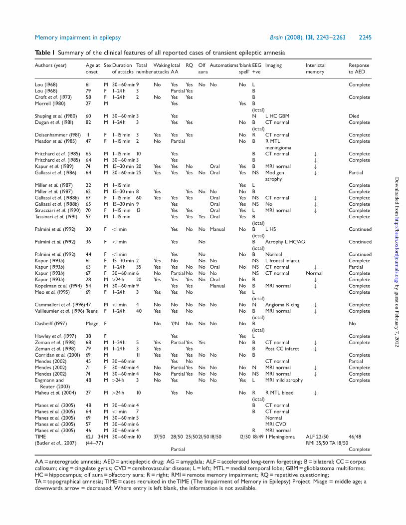
Table 1 Summary of the clinical features of all reported cases of transient epileptic amnesia
Authors (year) Age atonset
SexDurationof attacks
Totalnumber
Wakingattacks
IctalAA
RQ Olfaura
Automatisms‘blankspell’
EEG+ve
Imaging Interictalmemory
Responseto AED
Lou (1968) 61 M 30^60min9 No Yes Yes No No No L CompleteLou (1968) 79 F 1^24h 3 PartialYes BCroft et al. (1973) 58 F 1^24h 2 No Yes Yes B CompleteMorrell (1980) 27 M Yes Yes B
(ictal)Shuping et al. (1980) 60 M 30^60min3 Yes N L HCGBM DiedDugan et al. (1981) 82 M 1^24h 3 Yes Yes No B
(ictal)CT normal Complete
Deisenhammer (1981) 11 F 1^15min 3 Yes Yes Yes No R CT normal CompleteMeador et al. (1985) 47 F 1^15min 2 No Partial No B R MTL
meningiomaComplete
Pritchard et al. (1985) 65 M 1^15min 10 Yes B CT normal # CompletePritchard et al. (1985) 64 M 30^60min3 Yes B # CompleteKapur et al. (1989) 74 M 15^30min 20 Yes Yes No Oral Yes B MRI normal #
Gallassi et al. (1986) 64 M 30^60min25 Yes Yes Yes No Oral Yes NS Mod genatrophy
# Partial
Miller et al. (1987) 22 M 1^15min Yes L CompleteMiller et al. (1987) 62 M 15^30min 8 Yes Yes No No No B CompleteGallassi et al. (1988b) 67 F 1^15min 60 Yes Yes Yes Oral Yes NS CT normal # CompleteGallassi et al. (1988b) 65 M 15^30min 9 Yes Oral Yes NS No # CompleteStracciari et al. (1990) 70 F 1^15min 13 Yes Yes Oral Yes L MRI normal # CompleteTassinari et al. (1991) 57 M 1^15min Yes Yes Yes Oral Yes B
(ictal)Complete
Palmini et al. (1992) 30 F 51min Yes No No Manual No B(ictal)
L HS Continued
Palmini et al. (1992) 36 F 51min Yes No B(ictal)
Atrophy L HC/AG Continued
Palmini et al. (1992) 44 F 51min Yes No No B Normal ContinuedKapur (1993b) 61 F 15^30min 2 Yes No No No NS L frontal infarct CompleteKapur (1993b) 63 F 1^24h 35 Yes Yes No No Oral No NS CT normal # PartialKapur (1993b) 67 F 30^60min6 No PartialNo No No NS CT normal Normal CompleteKapur (1993b) 28 M 424h 20 Yes Yes Yes No Oral No B # CompleteKopelman et al. (1994) 54 M 30^60min9 Yes Yes Manual No B MRI normal # CompleteMeo et al. (1995) 69 F 1^24h 3 Yes Yes No Yes L
(ictal)Complete
Cammalleri et al. (1996)47 M 51min 4 No No No No No No N Angioma R cing # CompleteVuilleumier et al. (1996) Teens F 1^24h 40 Yes Yes No No B
(ictal)MRI normal # Complete
Dasheiff (1997) M/age F No Y/N No No No No B(ictal)
No
Hawley et al. (1997) 38 F Yes Yes L CompleteZeman et al. (1998) 68 M 1^24h 5 Yes PartialYes Yes No B CT normal # CompleteZeman et al. (1998) 79 M 1^24h 3 Yes Yes B Post CC infarct #
Corridan et al. (2001) 69 M 11 Yes Yes Yes No No No B CompleteMendes (2002) 45 M 30^60min Yes No CT normal PartialMendes (2002) 71 F 30^60min4 No PartialYes No No No N MRI normal # CompleteMendes (2002) 74 M 30^60min4 No PartialYes No No No NS MRI normal # CompleteEngmann andReuter (2003)
48 M 424h 3 No Yes No No Yes L MRI mild atrophy Complete
Maheu et al. (2004) 27 M 424h 10 Yes No No R(ictal)
R MTL bleed #
Manes et al. (2005) 48 M 30^60min4 B CT normalManes et al. (2005) 64 M 51min 7 B CT normalManes et al. (2005) 69 M 30^60min5 NormalManes et al. (2005) 57 M 30^60min6 MRI CVDManes et al. (2005) 46 M 30^60min4 R MRI normalTIME 62.1 34M 30^60min10 37/50 28/50 25/5021/5018/50 12/50 18/49 1Meningioma ALF 22/50 46/48(Butler et al., 2007) (44^77) RMI 35/50 TA18/50
Partial Complete
AA=anterograde amnesia; AED=antiepileptic drug; AG=amygdala; ALF=accelerated long-term forgetting; B=bilateral; CC=corpuscallosum; cing=cingulate gyrus; CVD=cerebrovascular disease; L= left; MTL=medial temporal lobe; GBM=glioblastoma multiforme;HC=hippocampus; olf aura=olfactory aura; R=right; RMI=remote memory impairment; RQ=repetitive questioning;TA=topographical amnesia; TIME=cases recruited in theTIME (The Impairment of Memory in Epilepsy) Project. M/age = middle age; adownwards arrow = decreased; Where entry is left blank, the information is not available.
Memory impairment in epilepsy Brain (2008), 131, 2243^2263 2245
by guest on February 7, 2012http://brain.oxfordjournals.org/
Dow
nloaded from
similar to the age range of Transient Global Amnesia (TGA),a syndrome which also results in transient impairment ofdeclarative memory and is attributed to neuronal dysfunctionin the medial temporal lobes, although the precise mechanismremains to be established. It is not clear why advancing ageshould predispose to transient dysfunction of the medialtemporal lobes. Given the sensitivity of this region to hypoxicdamage and its situation at a ‘watershed’ between anteriorand posterior circulation, it seems possible that cell damagesecondary to vascular insufficiency may act as an epilepticfocus. This is discussed later.
The sex ratio of TEA cases was approximately two males toone female in both the literature cases (27 males, 17 females)and the TIME cases (34 males, 16 females). The overallpercentage of males was 64.9% (95% CI = 55.3–74.6%).The reasons for this sex difference are not clear. A recent ofreview of 1333 published cases of TGA (Quinette et al., 2006)found no significant sex difference. The proportion ofmales with TGA was 46.4%, significantly different from theproportion in TEA patients (�2 = 3.62, P50.001).
DurationThe typical duration of amnestic attacks for each case wascategorized as follows: 51; 1–15; 15–30; 30–60 min; 1–24and 424 h. The median duration was 30–60 min forliterature cases, TIME cases and the group as a whole.This is briefer than the typical duration of TGA (4–10 h)and confirms previous observations (Zeman et al., 1998).Of note, however, 23 cases (24.5%) had attacks of similarduration to TGA, which could lead to diagnostic confusion.Very brief attacks (51 min) are less likely to be noticed bythe patient or observers. It remains to be explained why themajority of attacks are longer than would be expected fortemporal lobe seizures and why some persist for severaldays. The possibility that these are due to persistent epi-leptic activity (non-convulsive status epilepticus) is sup-ported by accounts of ongoing or intermittent automatisms(Kapur, 1993b) and the cases of Lee et al. (1992), Meo et al.(1995) and Vuilleumier et al. (1996). Lee et al. (1992)describe a 38-year-old woman who suffered a 12-dayepisode of pure amnesia accompanied by persistentepileptic activity isolated to the left temporal region. Meoet al. (1995) report a 69-year-old female with an amnesicepisode lasting several hours during which ictal dischargesoriginating from the right temporo-central region wererecorded. In both these cases, the patient’s behaviourduring bursts of ictal discharges was indistinguishable fromthat observed during interictal activity. Vuilleumier et al.(1996) describe a 41-year-old woman with persistent failureof recollection for 10 h during which EEG revealedcontinuous generalized epileptic activity with phase reversalin bilateral fronto-temporal regions. However some pro-longed TEA attacks may result from post-ictal amnesia or a‘Todd’s paralysis of memory’ (Morrell, 1980)—presumablyfollowing a brief or subclinical period of seizure activity—as
in the cases described by Morrell (1980), Tassinari et al.(1991) and Maheu et al. (2004). It is recognized that bothseizures and the post-ictal period can be prolonged inelderly patients (Rowan, 2000).
FrequencyBefore treatment, the overall mean frequency of attacks was14.8/year (literature cases = 19.7/year; TIME cases = 13.6/year). This is higher than previously reported (Zeman et al.,1998) and distinguishes TEA from transient global amnesiain which the recurrence rate is low [around 3% per year(Hodges and Warlow, 1990)]. However, the range acrossindividuals is wide (51 to 460 attacks per year) and a lowfrequency of attacks does not preclude a diagnosis ofepilepsy. There is, moreover, a likelihood that attacks willbe underreported, particularly those that are not witnessedby another person.
Amnesia on awakeningAn association between amnesic attacks and arousal fromsleep was noted in 70.4% of TEA cases (13/21 literaturecases and 37/50 TIME cases). In 11/50 TIME cases, amnesicattacks occurred exclusively upon waking. The closerelationship between sleep and epilepsy is well-recognized.Primary generalized epilepsies are particularly associatedwith sleep, with purely nocturnal seizures occurring in 45%of cases (Janz, 1962), and seizures upon or shortly afterwaking being a characteristic feature of both juvenile myo-clonic epilepsy (JME) and absence epilepsy. Among thefocal epilepsies, nocturnal seizures are particularly commonin frontal lobe epilepsy, whereas in temporal lobe epilepsythe percentage is much lower. In an electroencephalo-graphic study of patients with medically intractable focalepilepsy, 61% of frontal seizures but only 10.9% oftemporal lobe seizures were recorded during sleep(Crespel et al., 1998). In general, both seizures and interictalepileptiform discharges are predominantly associated withthe highly synchronized brain activity of non-rapid eyemovement sleep rather than the desynchronized state ofrapid eye movement sleep (Sammaritano et al., 1991). Thereason for the close relationship of TEA with sleep isunclear. It may be that the transition from sleep to wakingacts as a trigger to a seizure focus in the medial temporallobe. Alternatively, amnesia upon waking may reflectpersistent post-ictal dysfunction of medial temporal lobestructures following a seizure during sleep. In one TIMEcase, for example, morning amnesia was always preceded bya brief arousal at around 2 a.m. when the patient sat up inbed, staring and said ‘Oh, the smell, the smell’ before goingstraight back to sleep (case 83).
Ictal amnesiaAnterograde amnesia is usually understood as a deficit inmemory encoding or in storage mechanisms, whereas ret-rograde amnesia, as least insofar as it proves transient, can
2246 Brain (2008), 131, 2243^2263 C. R. Butler and A. Z. Zeman
by guest on February 7, 2012http://brain.oxfordjournals.org/
Dow
nloaded from
be thought of as a memory retrieval problem. The relativeimpairment of anterograde and retrograde memory duringTEA attacks, despite being difficult to ascertain from a ret-rospective patient or witness report, may therefore provideclues about the anatomy and degree of neuronal dysfunc-tion. In transient global amnesia, anterograde amnesia iscomplete and the patient later has no recollection of eventsthat occurred during the episode. In contrast, 44% ofpatients with TEA (10/36 literature cases; 28/50 TIMEcases) describe at least partial preservation of anterogradememory during attacks and afterwards may remember notbeing able to remember. A small number of TEA patients(2/36 literature cases; 3/50 TIME cases) are able toremember their attacks in rich subjective detail, suggestingminimal impairment of encoding. This later recollection ofictal events does not necessarily correlate with the apparentanterograde amnesia during the attack. The three TIMEcases referred to here displayed repetitive questioning ofwitnesses during their amnesic attacks. Vuilleumier et al.(1996) describe a patient who, during non-convulsive statusepilepticus, could not perform anterograde memory tasksbut who nevertheless encoded events into long-termmemory. This presumably reflects temporary impairmentof memory retrieval but preserved encoding and storage.In contrast, Dasheiff (1997) describes a patient who,during electrographic seizures recorded with both scalpand invasive EEG monitoring, performed normally onmemory tests but was subsequently amnesic for theseepisodes.
The degree of retrograde amnesia experienced during anepisode of TEA also varies across individuals. Of 22 clas-sifiable cases in the TIME study, 12 were associated withextensive ictal retrograde amnesia, stretching years into thepast, whereas 10 appeared to have an ictal retrogradeamnesia that was limited to events of the most recent daysor weeks. Five of 33 cases in the literature are described ashaving no apparent retrograde memory deficit during theattack, although none of them had formal retrograde memorytesting during the ictus. Such cases of apparently pureanterograde amnesia may go unnoticed as the patient willremain orientated and behave normally during the attackunless very recent memory is probed. The possibility thatattacks of TEA irreversibly disrupt retrograde memory is notmentioned in any of the literature case reports but threeTIME study patients volunteered that their TEA attackspermanently ‘erased’ memories of the preceding 24 h.
Repetitive questioningIn transient global amnesia, the patient will repeatedly askobservers questions such as: ‘Where am I?’ ‘What day is it?’or ‘What is happening to me?’ In TEA, however, this is afeature in just over 50% (17/30 literature cases; 25/50 TIMEcases). Interestingly, the presence of repetitive questioningdoes not predict of the degree of later recollection of ictalevents (�2 = 0.325, P = 0.569).
Additional featuresIn 65.8% of cases (16/29 literature cases, 36/50 TIMEcases), amnesic attacks were sometimes accompanied byadditional features. However, 46/50 TIME cases had at leastone attack with memory loss as the only feature.
Olfactory hallucinations. Amongst the TIME cases, olfac-tory/gustatory hallucinations were the most common addi-tional feature and were reported by 42% (21/50) of patientsupon direct questioning. Of these, 16 cases reported apurely olfactory sensation, 2 purely gustatory and 3 a mixedolfactory/gustatory sensation. The odour or taste wasdescribed as unpleasant in all but one case. In contrast,olfactory or gustatory hallucinations were described in only2/16 literature cases. Three additional TIME cases com-plained of a greatly decreased sense of smell since the onsetof their amnesic attacks. Olfactory and gustatory hallucina-tions are generally held to be a rare feature of epilepsy, andalmost always associated with a temporal lobe focus.Estimations of frequency vary. Penfield and Perot (1963)found 7 cases (0.6%) in 520 temporal lobe seizure patients.Acharya et al. (1998) found only 13 patients (0.9%) witholfactory auras among 1423 evaluated at the ClevelandClinic between 1991 and 1996. Higher prevalence rates havebeen reported by Chen et al. (2003) (5.5%), Ebner andKerder (2000) (6.3%) and Manford et al. (1996) (7.1%). Allstudies except Manford et al. (1996) investigated onlypatients with medically intractable focal epilepsy. Severalstudies have reported a particularly high incidence oftumours among patients with olfactory auras, includingJackson and Beevor (1890), Penfield and Jasper (1954) andAcharya et al. (1998). The latter found a neoplastic lesion in10 of the 13 patients in their study. Others, however, havedisputed this and maintain that hippocampal sclerosis is themost commonly associated pathological finding (Friedet al., 1995; Chen et al., 2003). There is greater consensusabout the approximate anatomical origin of epilepsy-associated olfactory hallucinations, with the majority ofpatients having a seizure focus in the anterior medialtemporal lobe. Hughlings–Jackson localized olfactory andgustatory functions to the uncus. Olfactory hallucinationshave also been provoked by direct cortical stimulation ofthe amygdala, rather than the hippocampus, in severalstudies (Jasper and Rasmussen, 1958; Fergusson et al., 1969;Andy et al., 1975; Gloor et al., 1982; Bartolomei et al.,2004). The hallucinations were, however, only rarelyelicited, and it remains uncertain whether spontaneousevents have the same precise anatomical origin.
Deja vu. Deja vu is a common experience amongst healthyindividuals as well as being associated with a variety ofneurological and psychiatric conditions, among themtemporal lobe epilepsy (Warren-Gash and Zeman, 2003)in which prevalence estimates vary widely [from 6% to 80%(Brown, 2003)]. Given the probable involvement of medialtemporal lobe structures in TEA, it is perhaps surprisingthat just five (10%) TIME cases described experiencing
Memory impairment in epilepsy Brain (2008), 131, 2243^2263 2247
by guest on February 7, 2012http://brain.oxfordjournals.org/
Dow
nloaded from
frequent deja vu, usually on occasions distinct from theiramnestic attacks. This symptom was not reported in any ofthe literature cases. Ictal deja vu may involve inappropriateactivation of brain circuits underlying familiarity. Corticalstimulation studies have elicited deja vu from stimulationin the anterior medial temporal lobes, more frequently onthe right than the left in right-handed patients (Gloor et al.,1982; Halgren et al., 1985; Bancaud et al., 1994). A recentstudy (Bartolomei et al., 2004) found deja vu to be morefrequently produced by stimulation of the entorhinal cortexthan the hippocampus or amygdala. The frequency of‘physiological’ deja vu declines with age, but it is otherwiseunclear why TEA, which is likely to arise from structures inwhich epileptic activity can give rise to deja vu, should sorarely be associated with it.
Automatisms. Automatisms (involuntary, semi-purposefulmovements) were reported to accompany some amnesicattacks in 40.6% of patients (10/19 literature cases, 18/50TIME cases). Oro-alimentary automatisms (chewing orlip smacking) were the most common (8/10 literaturecases, 14/18 TIME cases) and the remainder were manualautomatisms. Oral automatisms are significantly associatedwith a temporal rather than frontal seizure focus (Manfordet al., 1996) and with hippocampal rather than extra-hippocampal temporal lobe seizures (Gil-Nagel andRisinger, 1997).
Unresponsiveness. A brief period of unresponsiveness wassometimes associated with the amnesic episode in 28.2%of patients (10/28 literature cases, 12/50 TIME cases).Unresponsiveness, one of the hallmarks of ‘altered con-sciousness’ in complex partial seizures, is thought to repre-sent bilateral spread of seizure activity. When this occurs atthe beginning of the episode, it may indicate that the sub-sequent memory impairment is a post-ictal phenomenon(Morrell, 1980).
Other seizure types. Patients with TEA may also experienceseizures in which amnesia is not a prominent feature. Theseoccurred in 21.3% of cases (13/39 literature cases, 6/50TIME cases) and were almost always complex partial sei-zures. This observation raises the possibility that transientepisodes of amnesia are more widespread among TLEpatients than is generally recognized, but that they gounnoticed or unreported. Generalized tonic–clonic seizureswere only reported in 3/94 cases (3%).
ElectroencephalographyInterictal EEG. Interictal epileptiform abnormalities onelectroencephalography (EEG) were seen in 43.6% cases(23/44 literature cases, 18/49 TIME cases). Of these, 31.7%were left-sided, 12.2% were right-sided and 56.1% werebilateral. All were localized over the temporal or fronto-temporal region. The interictal EEG was entirely normal in19.4% of cases (3/44 literature cases, 15/49 TIME cases).Surface EEG lacks sensitivity to discharges originating in themedial temporal lobes and the detection rate in TEA, given
variable usage of routine or sleep EEG recording, is similarto that in other forms of temporal lobe epilepsy.
Ictal EEG. Surface EEG recording during an amnesic attackwas performed in nine literature cases and one TIME case.All recordings showed seizure activity, which in 8/10 casesinvolved both temporal lobes and in the others remainedunilateral (one left- and one right-sided). Amnesia wasobserved as an ictal phenomenon in six cases and as post-ictal in four cases.
Brain imagingBrain imaging results were available for 29/44 literaturecases (CT = 14, MRI = 15) and 49 TIME cases (CT = 2,MRI = 47). Focal lesions were detected in 7/78 cases ofwhich four were likely to have played an aetiological role inthe seizures (5.1%). These were: two right medial temporallobe meningiomas [(Meador et al., 1985) and TIME case40], haemosiderin deposition in the right medial temporallobe (Maheu et al., 2004) and a glioblastoma multiforme inthe left hippocampal region (Shuping et al., 1980). We haverecently observed medial temporal lobe high signal onT2-weighted MRI sequences in two patients with particu-larly frequent episodes of TEA, with unilateral hippocampalhypermetabolism on a PET scan in one of these, resolvingwhen the seizures subsided (Butler, in press). In general,therefore, TEA is not associated with clinically detectable,focal brain lesions, but where these are present they involvethe medial temporal lobes.
AetiologyBesides these rare structural abnormalities, the cause ofTEA is usually obscure. Zeman et al. (1998) pointed outthat a history of cardiac disease was common in their series,and hypothesized that cardiac-related hypoxic damage tomedial temporal lobe structures might have caused theepilepsy. Amongst the TIME cases, however, Butler et al.(2007) failed to find an excess of overt cardiovascular orcerebrovascular disease.
Interictal memory disturbanceComplaints of interictal memory dysfunction are commonin TEA and were noted in 80.6% patients (16/17 literaturecases, 38/50 TIME cases). Inconsistency of neuropsychologi-cal assessment across the literature cases precludes usefulanalysis of data. Amongst TIME patients, the mostcommon problems were (i) a patchy loss of memories forremote, personally experienced events (35/50 TIME cases),(ii) accelerated forgetting over days to weeks of newlyacquired memories (22/50 TIME cases) and (iii) topograph-ical memory deficits (18/50 TIME cases). Despite thesecomplaints, the majority of patients performed normally onstandard neuropsychological tests of memory, although thegroup as a whole showed a subtle but statistically significantimpairment relative to matched control subjects (Butleret al., 2007).
2248 Brain (2008), 131, 2243^2263 C. R. Butler and A. Z. Zeman
by guest on February 7, 2012http://brain.oxfordjournals.org/
Dow
nloaded from
In a series of papers, Gallassi (Gallasi et al., 1986, 1988a,b, 1990, 1992; Gallassi, 2006) has described the ‘epilepticamnesic syndrome’, in which patients present withcomplaints of severe, persistent memory impairment inthe context of usually subtle temporal lobe seizures. Insome cases, these seizures are followed by a period oftransient amnesia, which Gallassi, following Pritchard et al.(1985), has called ‘epileptic amnesic attacks’ (EAA). Clearly,the patients described by Gallassi have much in commonwith TEA patients and some of those for whom sufficientclinical data have been published have been included in thepresent review. The epileptic amnesic syndrome and TEAare not, however, coterminous as some patients with TEAdo not have pronounced interictal memory disturbance andsome patients with epilepsy and memory problems do nothave transient amnesic attacks.
Treatment responsivenessComplete cessation of transient amnesic episodes wasachieved with anticonvulsant therapy in 88.5% patients(25/32 literature cases, 46/48 TIME cases). The remaindershowed a substantial decrease in attack frequency. The reasonfor this excellent response rate is unknown but is generallyreported in late-onset epilepsy (Stephen and Brodie, 2000).
SummaryThis review reveals that the clinical features of TEA areconsistent across numerous independent reports publishedover the past 40 years, strengthening the view that it shouldbe regarded as a distinct syndrome of epilepsy. A numberof clinically important questions relating to the aetiology,epidemiology and prognosis of TEA remain unansweredand should be addressed in future work.
Part II: Accelerated long-term forgettingAccording to traditional models, information has beenencoded into long-term memory if it can be accuratelyretrieved after an interval during which active rehearsal isprevented. Thereafter, a process of ‘consolidation’ rendersthe memory trace progressively less vulnerable to disruption(Squire et al., 1984). This process, thought to involve agradual reorganization of the memory trace at a neural-systems level (Squire and Alvarez, 1995), may continue forweeks, months or even years but it is often assumed that itsefficacy can be assessed at relatively brief delays. Standardneuropsychological instruments, therefore, typically testmemory retention at intervals of up to 30 min, and littleis known about forgetting beyond that point. Over recentyears, a number of case reports have described a novel formof forgetting, apparent only over extended periods of time,providing evidence for a prolonged multiple-stage con-solidation process. These cases, together with group studiesevaluating very long-term forgetting in epilepsy, are thefocus of this section. Several of the studies included have
been summarized in a recent review paper by Bell andGiovagnoli (2007). Although other authors have called thisphenomenon long-term amnesia (LTA) (Kapur et al., 1996,1997; Mayes et al., 2003), we adopt the alternative ALF todistinguish the disorder from the amnesic syndrome and toinclude cases in which long-term memory may be deficientbut not completely absent.
MethodsUsing the search methods described in Part I, we first identifiedcase reports describing patients with epilepsy in whom learningand memory performance over delays of up to 1 h was consideredto be in the normal range (by comparison with published normsor a matched control group) but in whom testing over longerdelays revealed impairment. We then searched for studiescomparing memory over delays of 24 h or longer in groups ofpatients with epilepsy and healthy control subjects.
Case reportsSeven case reports of ALF meeting our inclusion criteriawere found (Kapur et al., 1996, 1997; O’Connor et al.,1997; Lucchelli and Spinnler, 1998; Mayes et al., 2003;Cronel-Ohayon et al., 2006; Manning et al., 2006). Threeadditional cases were described as showing ALF but did notundergo formal testing (Kapur et al., 1989, 1996) or weretested only at short delays (Maravita et al., 1995). Onereport demonstrated abnormal long-term forgetting in apatient with no documented seizures (De Renzi andLucchelli, 1993). The seven cases we consider are summa-rized in Table 2. Four patients were males and threefemales. Their ages ranged from 18 years to 65 years(mean = 48.1, SD = 15.6). In striking contrast with classicallyamnesic individuals, these patients maintained active,independent lives and several remained in employment.
The aetiology of the memory impairment included: closedhead injuries (Kapur et al., 1996; Mayes et al., 2003),paraneoplastic limbic encephalitis (O’Connor et al., 1997),neuronal dysplasia in the left amygdala and hippocampalsclerosis (Manning et al., 2006). Two patients had late-onsettemporal lobe epilepsy with no clear cause (Kapur et al., 1997;Lucchelli and Spinnler, 1998). In two cases (Kapur et al.,1996; Lucchelli and Spinnler, 1998) seizures resolvedimmediately with anticonvulsant medication. Structuralbrain imaging was abnormal in six patients. Damage wasrestricted to the temporal lobes in all cases except one (Mayeset al., 2003), in whom the right orbitofrontal cortex was alsoaffected. The hippocampus was the only abnormal region inone case (Kapur et al., 1997), whereas this was sparedin another (Mayes et al., 2003). Both medial and lateraltemporal cortices were damaged in the remaining four cases(Kapur et al., 1996; O’Connor et al., 1997; Cronel-Ohayonet al., 2006; Manning et al., 2006), two of whom hadundergone temporal lobectomy (Cronel-Ohayon et al., 2006;Manning et al., 2006). Only one patient had normal structuralbrain imaging (Lucchelli and Spinnler, 1998).
Memory impairment in epilepsy Brain (2008), 131, 2243^2263 2249
by guest on February 7, 2012http://brain.oxfordjournals.org/
Dow
nloaded from
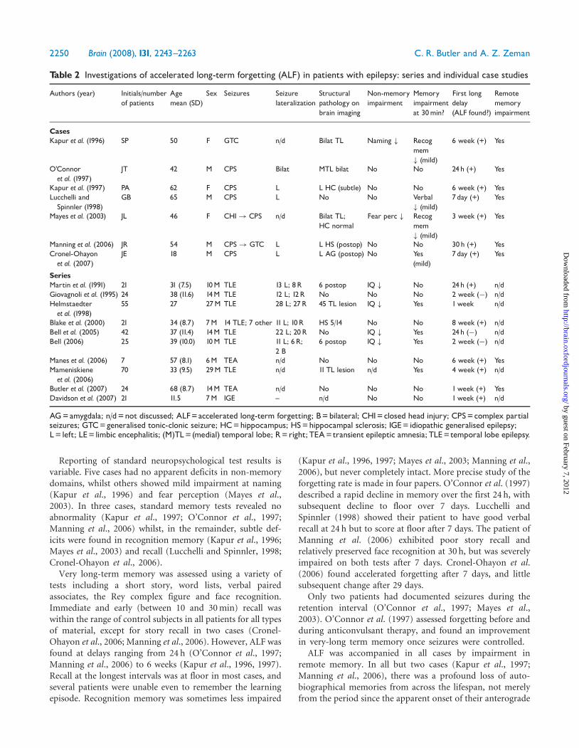
Reporting of standard neuropsychological test results isvariable. Five cases had no apparent deficits in non-memorydomains, whilst others showed mild impairment at naming(Kapur et al., 1996) and fear perception (Mayes et al.,2003). In three cases, standard memory tests revealed noabnormality (Kapur et al., 1997; O’Connor et al., 1997;Manning et al., 2006) whilst, in the remainder, subtle def-icits were found in recognition memory (Kapur et al., 1996;Mayes et al., 2003) and recall (Lucchelli and Spinnler, 1998;Cronel-Ohayon et al., 2006).
Very long-term memory was assessed using a variety oftests including a short story, word lists, verbal pairedassociates, the Rey complex figure and face recognition.Immediate and early (between 10 and 30 min) recall waswithin the range of control subjects in all patients for all typesof material, except for story recall in two cases (Cronel-Ohayon et al., 2006; Manning et al., 2006). However, ALF wasfound at delays ranging from 24 h (O’Connor et al., 1997;Manning et al., 2006) to 6 weeks (Kapur et al., 1996, 1997).Recall at the longest intervals was at floor in most cases, andseveral patients were unable even to remember the learningepisode. Recognition memory was sometimes less impaired
(Kapur et al., 1996, 1997; Mayes et al., 2003; Manning et al.,2006), but never completely intact. More precise study of theforgetting rate is made in four papers. O’Connor et al. (1997)described a rapid decline in memory over the first 24 h, withsubsequent decline to floor over 7 days. Lucchelli andSpinnler (1998) showed their patient to have good verbalrecall at 24 h but to score at floor after 7 days. The patient ofManning et al. (2006) exhibited poor story recall andrelatively preserved face recognition at 30 h, but was severelyimpaired on both tests after 7 days. Cronel-Ohayon et al.(2006) found accelerated forgetting after 7 days, and littlesubsequent change after 29 days.
Only two patients had documented seizures during theretention interval (O’Connor et al., 1997; Mayes et al.,2003). O’Connor et al. (1997) assessed forgetting before andduring anticonvulsant therapy, and found an improvementin very-long term memory once seizures were controlled.
ALF was accompanied in all cases by impairment inremote memory. In all but two cases (Kapur et al., 1997;Manning et al., 2006), there was a profound loss of auto-biographical memories from across the lifespan, not merelyfrom the period since the apparent onset of their anterograde
Table 2 Investigations of accelerated long-term forgetting (ALF) in patients with epilepsy: series and individual case studies
Authors (year) Initials/numberof patients
Agemean (SD)
Sex Seizures Seizurelateralization
Structuralpathology onbrain imaging
Non-memoryimpairment
Memoryimpairmentat 30min?
First longdelay(ALF found?)
Remotememoryimpairment
CasesKapur et al. (1996) SP 50 F GTC n/d Bilat TL Naming # Recog
mem# (mild)
6 week (+) Yes
O’Connoret al. (1997)
JT 42 M CPS Bilat MTL bilat No No 24h (+) Yes
Kapur et al. (1997) PA 62 F CPS L L HC (subtle) No No 6 week (+) YesLucchelli andSpinnler (1998)
GB 65 M CPS L No No Verbal# (mild)
7day (+) Yes
Mayes et al. (2003) JL 46 F CHI! CPS n/d Bilat TL;HC normal
Fear perc # Recogmem# (mild)
3 week (+) Yes
Manning et al. (2006) JR 54 M CPS! GTC L L HS (postop) No No 30h (+) YesCronel-Ohayonet al. (2007)
JE 18 M CPS L L AG (postop) No Yes(mild)
7day (+) Yes
SeriesMartin et al. (1991) 21 31 (7.5) 10M TLE 13 L; 8R 6 postop IQ # No 24h (+) n/dGiovagnoli et al. (1995) 24 38 (11.6) 14M TLE 12 L; 12R No No No 2 week (�) n/dHelmstaedteret al. (1998)
55 27 27M TLE 28 L; 27R 45 TL lesion IQ # Yes 1week n/d
Blake et al. (2000) 21 34 (8.7) 7M 14 TLE; 7 other 11 L; 10R HS 5/14 No No 8 week (+) n/dBell et al. (2005) 42 37 (11.4) 14M TLE 22 L; 20R No IQ # Yes 24h (�) n/dBell (2006) 25 39 (10.0) 10M TLE 11 L; 6R;
2 B6 postop IQ # Yes 2 week (�) n/d
Manes et al. (2006) 7 57 (8.1) 6M TEA n/d No No No 6 week (+) YesMameniskieneet al. (2006)
70 33 (9.5) 29M TLE n/d 11 TL lesion n/d Yes 4 week (+) n/d
Butler et al. (2007) 24 68 (8.7) 14M TEA n/d No No No 1week (+) YesDavidson et al. (2007) 21 11.5 7M IGE ^ n/d No No 1week (+) n/d
AG=amygdala; n/d=not discussed; ALF=accelerated long-term forgetting; B=bilateral; CHI=closed head injury; CPS=complex partialseizures; GTC=generalised tonic-clonic seizure; HC=hippocampus; HS=hippocampal sclerosis; IGE= idiopathic generalised epilepsy;L= left; LE= limbic encephalitis; (M)TL= (medial) temporal lobe; R=right; TEA=transient epileptic amnesia; TLE= temporal lobe epilepsy.
2250 Brain (2008), 131, 2243^2263 C. R. Butler and A. Z. Zeman
by guest on February 7, 2012http://brain.oxfordjournals.org/
Dow
nloaded from
memory problems. Memory for public events dating fromthe premorbid period was affected in every case.
Group studiesWe identified 10 studies addressing the issue of long-termanterograde memory in epilepsy (Martin et al., 1991;Helmstaedter et al., 1998; Blake et al., 2000; Bell et al., 2005;Manes et al., 2005; Mameniskiene et al., 2006; Bell, 2006; Belland Giovagnoli, 2007; Butler et al., 2007; Davidson et al., 2007).These are summarized in Table 2. Nine examined patientswith TLE, and one of these (Blake et al., 2000) also included agroup of seven patients with other focal epilepsies. Two studies(Manes et al., 2005; Butler et al., 2007) exclusively investigatedTEA. The mean patient age ranged from 27 to 39 years, exceptfor TEA patients who were older. One study investigated chil-dren with idiopathic generalized epilepsy (IGE) (Davidson et al.,2007), in whom the mean age was 11.5 years (range 8–16 years).In total, 138 male and 172 female patients were studied.
In contrast to several of the case reports described earlier,patients in these studies did not have epilepsy resultingfrom a specified brain injury. Neuroimaging revealedvarying proportions of hippocampal sclerosis, neoplasticand other lesions among patients, but no study quantita-tively assessed their relation to memory function. Post-operative patients were included in the studies by Martinet al. (1988) (six patients) and Bell (2006) (six patients).The TEA patients studied by Manes et al. (2005) and Butleret al. (2007) had no clinically apparent abnormalities onbrain MRI besides, in some cases, evidence of small vesseldisease. Davidson et al. (2007) do not report imagingfindings, but these are expected to be normal in IGE.
Memory retention over 30 min was found to be compa-rable in patients and controls in five studies (Martin et al.,1991; Giovagnoli et al., 1995; Blake et al., 2000; Butler et al.,2007; Davidson et al., 2007), and one (Manes et al., 2005)found normal verbal but impaired non-verbal memory.Four studies (Helmstaedter et al., 1998; Bell et al., 2005;Bell, 2006; Mameniskiene et al., 2006) identified impair-ment amongst patients even at this short delay. At extendedintervals ranging from 24 h (Martin et al., 1991; Bell et al.,2005) to 8 weeks (Blake et al., 2000), seven studies (Martinet al., 1991; Helmstaedter et al., 1998; Blake et al., 2000;Manes et al., 2005; Mameniskiene et al., 2006; Butler et al.,2007; Davidson et al., 2007) found patients to show a dis-proportionate degree of forgetting compared with controls.In contrast, three studies (Giovagnoli et al., 1995; Bell et al.,2005; Bell, 2006) found no difference in long-term forget-ting rate between patients and controls. Bell et al. (2005)and Bell (2006) also failed to identify a significant numberof individual patients in whom memory performance wasnormal at 30 min but impaired at an extended delay.
These results raise questions about the existence, scopeand prevalence of ALF amongst patients with epilepsy, thebest means of assessing very long-term memory, thepathophysiology of ALF, and its relation to RMI.
Discussion
Does ALF occur in epilepsy? If so, for which types of materialand is there a laterality effect?The seven case reports suggest that ALF is an identifiableclinical phenomenon closely linked to epilepsy. Several of thegroup studies identified ALF in patients with TLE and onein patients with IGE. However, three studies report negativeresults. The reason for this discrepancy is not clear, but itmay arise partly from methodological differences discussedlater. ALF may also be more prominent in certain subtypes ofTLE, such as TEA, in which 44% patients report symptomsof accelerated forgetting (Butler et al., 2007).
Patients were shown to learn at a normal rate in threestudies (Martin et al., 1991; Blake et al., 2000; Butler et al.,2007), whereas learning was found to be impaired byHelmstaedter et al. (1998), Giovagnoli et al. (1995) andDavidson et al. (2007). In the latter study, the learningimpairment showed by IGE patients accounted for most ofthe difference in very long-term forgetting, suggesting thatan encoding problem underlies the ALF seen in IGE.
The available data are inconclusive on the issue of whetherhemispheric differences cause material-specificity in ALF.Using a test of verbal memory, Blake et al. (2000) identifiedALF in patients with left but not right TLE. On the other hand,Martin et al. (1991), again only using verbal material, foundno hemispheric effect. Without identifying the seizure focus,two studies (Mameniskiene et al., 2006; Butler et al., 2007)found ALF for both verbal and non-verbal material. AmongstIGE patients, ALF was identified for verbal material and atrend towards ALF for non-verbal material.
The balance of evidence suggests that recognition memoryis also affected by ALF, but the findings are inconsistent. Fourstudies (Helmstaedter et al., 1998; Blake et al., 2000; Bell et al.,2005; Manes et al., 2005; Bell, 2006) found patients to showimpairment on recognition memory at extended delays.Martin et al. (1991), on the other hand, did not findrecognition memory impairment, although their delay wasjust 24 h. Davidson et al. (2007) also found no recognitionmemory deficit amongst IGE patients, even after a 7-daydelay, although patients and controls scored close to ceiling.
In summary, the data currently available suggest that, insome patients with epilepsy, anterograde memory perfor-mance is normal at standard test intervals but declinesabnormally rapidly thereafter—the phenomenon of ALF.The negative results indicate that this does not alwaysoccur. Some possible explanations for these mixed resultsare considered later.
Methodological issues in the assessment of ALFThe assessment of long-term forgetting encounters anumber of methodological problems:
(i) Patient and control groups should be matchedas carefully as possible for demographic and non-memory cognitive variables to isolate the phenomenon
Memory impairment in epilepsy Brain (2008), 131, 2243^2263 2251
by guest on February 7, 2012http://brain.oxfordjournals.org/
Dow
nloaded from
of interest. Thus it may be relevant that in thestudies by Martin et al. (1991), Helmstaedter et al.(1998), Bell (2006) and Bell et al. (2005) patientshad a significantly lower mean IQ than controlsubjects. Other groups (Blake et al., 2000; Maneset al., 2005; Butler et al., 2007) performed moreextensive testing and revealed no differences betweenpatients and controls on measures of language, visuo-spatial perception and executive function.
(ii) The choice of study material is likely to be important.Relative impairments in verbal and non-verbalmemory depend on the laterality of the seizurefocus. Also, forgetting may be different for semanti-cally related (e.g. a story) and unrelated (e.g. a wordlist) material (Isaac and Mayes, 1999a, b).
(iii) ALF is most convincingly demonstrated when patientsexhibit normal initial learning and 30-min recall,but clear impairment at longer delays (Martin et al.,1988; Blake et al., 2000; Manes et al., 2005; Butler et al.,2007). In cases where patients are already impairedover short delays, a number of techniques may beused to assess long-term forgetting rates:
(a) Using a variety of the technique introduced byHuppert and Piercy (1978), the experimenter canmodulate exposure to the study material to ensurethat patients and control subjects reach the sameinitial level of learning. In four studies (Martinet al., 1991; Blake et al., 2000; Butler et al., 2007;Davidson et al., 2007), all the material wasrepeatedly presented until the subject reached alearning criterion. As Bell has observed (Bell et al.,2005), this ‘over-learning’ method may mask earlyforgetting with a ceiling effect. However, in thestudies by Blake et al. (2000) and Butler et al.(2007), patients who showed ALF of ‘over-learned’material nonetheless performed normally on stan-dard memory tests. An alternative, employed byfour studies (Martin et al., 1991; Giovagnoli et al.,1995; Bell, 2006; Bell and Giovagnoli, 2007), is touse a ‘selective reminding’ technique, in which onlynon-remembered items are represented at eachlearning trial.
(b) Individual patients and controls may be matchedfor learning on a case-by-case basis. This method,however, risks producing non-representativeresults if ‘upper range’ patients are matchedwith ‘lower range’ control subjects.
(c) Differing acquisition levels may be accepted and theoverall shape of the forgetting curves compared.This method was employed by three studies (Bellet al., 2005; Bell, 2006; Mameniskiene et al., 2006),with mixed results. The problem which arises hereis that there is no widely accepted model of howvariations in initial learning level affect forgettingover time (Rubin and Wenzel, 1996).
(iv) Rehearsal of the material between test sessions mayconfound results. In some studies, subjects wereforewarned about the delayed tests, whereas in othersthey were not. The material used will also influencethis: a story is more likely to be rehearsed than a largenumber of meaningless visual designs.
(v) The length of the interval between testing sessionsmay determine whether or not ALF is found—theunderlying mechanisms may operate over 24 h orseveral weeks. An interval should be chosen at whichcontrol subjects perform neither at ceiling nor floor.
(vi) The nature of the retrieval task—free recall, cuedrecall or recognition—may be important. Davidsonet al. (2007) suggest that their failure to find a deficitin recognition memory at an extended delay impliesthat ALF in their patients was due to a problem withmemory retrieval rather than storage.
Aetiology and pathophysiology of ALFAccepting that ALF occurs in some patients with epilepsy,we may hypothesize several contributory mechanisms:(i) clinical or subclinical seizure activity; (ii) structuralor other underlying brain pathology; (iii) an adverseeffect of anticonvulsant medication or (iv) psychologicalmechanisms.
Seizures. Anecdotally, patients with TEA sometimes reportthat seizures seem to ‘wipe out’ memories of precedingevents, and feel that their memory abilities improve onceseizures are controlled with anticonvulsant therapy.O’Connor et al. (1997) document such an improvementin a single case of temporal lobe epilepsy. Mameniskieneet al. (2006) found a positive correlation between long-termforgetting and both (i) manifest seizures during theexperimental period and (ii) subclinical epileptiform EEGactivity. As in the study by Blake et al. (2000), however,long-term memory was not found to correlate with theaverage seizure frequency reported by patients. The closeassociation between amnesic attacks and waking in TEAraises the intriguing question of whether sub-clinical, noc-turnal epileptiform activity might disturb sleep-dependentmemory consolidation processes (Walker, 2005; Ellenbogenet al., 2006; Walker and Stickgold, 2006).
In a study directly addressing the question of whetherincident seizures accelerate forgetting, Bergin et al. (1995)tested immediate, 30 min and 48 h memory for verbal andnon-verbal material in 58 patients undergoing video telem-etry for the investigation of medically refractory partialseizures. No difference was found in long-term forgettingbetween patients who did and did not have seizures duringthe study period. This important result does not, however,rule out a negative influence of seizures upon anterogradememory. Features such as the timing, duration and anat-omical focus of seizures may play an important role. Jokeitet al. (2001) examined memory over 24 h for verbalmaterial in a small group of patients (n = 10) undergoing
2252 Brain (2008), 131, 2243^2263 C. R. Butler and A. Z. Zeman
by guest on February 7, 2012http://brain.oxfordjournals.org/
Dow
nloaded from
videotelemetry. They found a difference in long-term recallbetween days with and without seizures, but this wasrestricted to the group of patients with a left temporal lobeseizure focus. A further source of evidence comes fromstudies that document an improvement in verbal memoryscores in patients following right temporal lobectomy(Novelly, 1984; Martin et al., 1998). This suggests that aseizure focus in one hippocampus can negatively affectfunction in distant brain regions.
The question of whether transient impairment of neu-ronal function can disrupt very long-term memory has alsobeen addressed in patients undergoing electroconvulsivetherapy (ECT) for depression, a procedure known to induceanterograde and retrograde amnesia. Squire (1981) inves-tigated recognition memory for pictures and sentences atintervals of 10, 30 min and 30 h in patients on two occa-sions: 2 h and 4 months after ECT. The subjects thereforeacted as their own controls. Initial acquisition was matchedby using longer stimulus presentation on the earlier occa-sion. Picture forgetting was significantly more rapid whenthe subjects had recently received ECT. On the other hand,patients with Korsakoff’s syndrome (KS) (diencephalicamnesia) and a patient with chronic medial temporal lobeamnesia did not show accelerated forgetting when initialacquisition was matched to a group of healthy controlsubjects. These findings were replicated and extended byLewis and Kopelman (1998) who included a group ofdepressed patients not undergoing ECT. Accelerated forget-ting was again found solely in the post-ECT group and couldtherefore not be attributed to depression per se. Transientimpairment of brain function also underlies post-traumaticamnesia (PTA). Levin et al. (1988), investigating recognitionmemory for photographs, found accelerated forgetting over32 h in head injury patients in PTA, compared with headinjury patients who had recovered from PTA.
Structural lesions. Many of the case studies reviewedearlier also had radiological evidence of structural brainpathology. ALF might, therefore, represent a mild form ofthe amnesic syndrome, caused by subtle damage to themedial temporal lobes.
If so, forgetting should be dramatically accelerated inamnesics. A number of studies have addressed this issueand results have been mixed [see Isaac and Mayes (1999a)for a review]. The problems of matching initial acquisitionare of course much more acute here than in patients withnormal or near-normal immediate memory. One earlyinvestigation suggested that accelerated forgetting was afeature of amnesia caused by medial temporal lobe lesionsbut not diencephalic lesions (Huppert and Piercy, 1979).However, this finding was later found not to be replicable(Freed et al., 1987). Isaac and Mayes (Isaac and Mayes,1999a, b) conclude that forgetting in the amnesic syndromeis accelerated over the first 10 min but only for certaintypes of material—particularly free recall of prose andsemantically related words. They interpret this as
reflecting impairment of early consolidation processes dueto medial temporal lobe damage. Beyond 10 min, forgettingrates have been measured in patients with anoxic braindamage (McKee and Squire, 1992), Alzheimer’s Disease(Kopelman, 1985) and head injury after recovery frompost-traumatic amnesia (Levin et al., 1988), and have beenfound to be normal. Accelerated forgetting over longerperiods has been reported in healthy older subjects by someauthors (Huppert and Kopelman, 1989; Davis et al., 2003),but not others (Petersen et al., 1992).
If ALF is essentially due to a mild form of hippocampalamnesia one might also predict some mild degree ofimpairment over standard testing intervals. The observationthat some patients perform normally on standard tests yetexhibit ALF appears to argue against the existence of anydefect in acquisition and initial retention of declarativememories. It could be, however, that standard tests areinsufficiently sensitive to reveal a mild deficit in these stagesof memory processing. Techniques such as recording eventrelated EEG potentials at memory encoding could shedfurther light on this possibility.
Finally additional imaging techniques, including manualor automated volumetric measurement, MR spectroscopyand diffusion tensor imaging have the potential to revealsubtle regional pathology within or affecting the MTL thatmay elude less sophisticated studies.
Anticonvulsant medication. There is good evidence thatantiepileptic drugs (AEDs) can have a negative impact uponcognition, although the field is fraught with methodologicaldifficulties (Kwan and Brodie, 2001; Motamedi andMeador, 2004). The most commonly observed effects areslowed mental processing and reduced attention, and theseare most marked with high doses and polytherapy. How-ever, a specific impact on memory has been reported inseveral studies. Some newer drugs may have a better cog-nitive profile (Motamedi and Meador, 2003), althoughtopiramate may be a significant exception (Thompson et al.,2000). The specific question of whether anticonvulsants canaccelerate shorter term forgetting has been addressed in asingle, retrospective study (Jokeit et al., 2005). Amongst 162patients with medically refractory epilepsy, higher serumlevels of AED were associated with greater forgetting of bothverbal and visual material over a 30-min delay aftercontrolling for potentially confounding variables such as IQ,age, duration of epilepsy and seizure frequency.
Whilst it remains possible that the ALF observed in somestudies reviewed earlier is a direct result of treatment withanticonvulsants, it seems unlikely for a number of reasons:first, patients with TEA complain of ALF prior to initiationof therapy; second, patients with TEA usually report thattheir memory improves once treatment is started (Gallassiet al., 1988a; Zeman et al., 1998; Gallassi, 2006; Butler et al.,2007); third, the forgetting observed by Blake et al. (2000)was specific to the group of patients with left temporal lobeepilepsy; and fourth, the doses of anticonvulsants used in
Memory impairment in epilepsy Brain (2008), 131, 2243^2263 2253
by guest on February 7, 2012http://brain.oxfordjournals.org/
Dow
nloaded from
TEA patients, those who complain most profoundly of ALF,are generally low.
Psychosocial factors. The disparity between subjectivereports of memory difficulty amongst patients with epilepsyand their performance on neuropsychological tests(Corcoran and Thompson, 1992) has been attributed todisturbances of mood and poor self-esteem (Giovagnoliet al., 1997; Elixhauser et al., 1999). It is undoubtedlyimportant to take such factors into account when inves-tigating cognitive function in epilepsy. However, they areunlikely to play a major causal role in ALF. Three studies(Blake et al., 2000; Mameniskiene et al., 2006; Butler et al.,2007) assessed mood using the Hospital Anxiety andDepression Scale and found no correlation with verylong-term memory performance. In addition, as mentionedearlier, Lewis and Kopelman (1998) did not find acceleratedforgetting in a group of depressed patients after equatinginitial levels of learning.
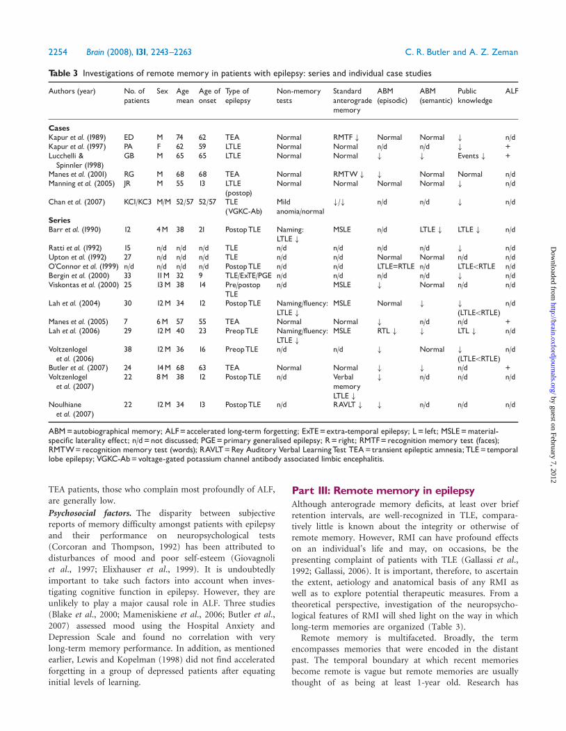
Part III: Remote memory in epilepsyAlthough anterograde memory deficits, at least over briefretention intervals, are well-recognized in TLE, compara-tively little is known about the integrity or otherwise ofremote memory. However, RMI can have profound effectson an individual’s life and may, on occasions, be thepresenting complaint of patients with TLE (Gallassi et al.,1992; Gallassi, 2006). It is important, therefore, to ascertainthe extent, aetiology and anatomical basis of any RMI aswell as to explore potential therapeutic measures. From atheoretical perspective, investigation of the neuropsycho-logical features of RMI will shed light on the way in whichlong-term memories are organized (Table 3).
Remote memory is multifaceted. Broadly, the termencompasses memories that were encoded in the distantpast. The temporal boundary at which recent memoriesbecome remote is vague but remote memories are usuallythought of as being at least 1-year old. Research has
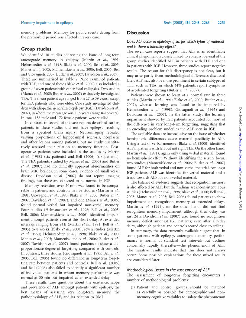
Table 3 Investigations of remote memory in patients with epilepsy: series and individual case studies
Authors (year) No. ofpatients
Sex Agemean
Age ofonset
Type ofepilepsy
Non-memorytests
Standardanterogradememory
ABM(episodic)
ABM(semantic)
Publicknowledge
ALF
CasesKapur et al. (1989) ED M 74 62 TEA Normal RMTF # Normal Normal # n/dKapur et al. (1997) PA F 62 59 LTLE Normal Normal n/d n/d # +Lucchelli &Spinnler (1998)
GB M 65 65 LTLE Normal Normal # # Events # +
Manes et al. (2001) RG M 68 68 TEA Normal RMTW # # Normal Normal n/dManning et al. (2005) JR M 55 13 LTLE
(postop)Normal Normal Normal Normal # n/d
Chan et al. (2007) KC1/KC3 M/M 52/57 52/57 TLE(VGKC-Ab)
Mildanomia/normal
#/# n/d n/d # n/d
SeriesBarr et al. (1990) 12 4M 38 21 PostopTLE Naming:
LTLE #MSLE n/d LTLE # LTLE # n/d
Ratti et al. (1992) 15 n/d n/d n/d TLE n/d n/d n/d n/d # n/dUpton et al. (1992) 27 n/d n/d n/d TLE n/d n/d Normal Normal n/d n/dO’Connor et al. (1999) n/d n/d n/d n/d PostopTLE n/d n/d LTLE=RTLE n/d LTLE5RTLE n/dBergin et al. (2000) 33 11M 32 9 TLE/ExTE/PGE n/d n/d n/d n/d # n/dViskontas et al. (2000) 25 13M 38 14 Pre/postop
TLEn/d MSLE # Normal n/d n/d
Lah et al. (2004) 30 12M 34 12 PostopTLE Naming/fluency:LTLE #
MSLE Normal # #
(LTLE5RTLE)n/d
Manes et al. (2005) 7 6M 57 55 TEA Normal Normal # n/d n/d +Lah et al. (2006) 29 12M 40 23 PreopTLE Naming/fluency:
LTLE #MSLE RTL # # LTL # n/d
Voltzenlogelet al. (2006)
38 12M 36 16 PreopTLE n/d n/d # Normal #
(LTLE5RTLE)n/d
Butler et al. (2007) 24 14M 68 63 TEA Normal Normal # # n/d +Voltzenlogelet al. (2007)
22 8M 38 12 PostopTLE n/d VerbalmemoryLTLE #
# n/d n/d n/d
Noulhianeet al. (2007)
22 12M 34 13 PostopTLE n/d RAVLT # # n/d n/d n/d
ABM=autobiographical memory; ALF=accelerated long-term forgetting; ExTE=extra-temporal epilepsy; L= left; MSLE=material-specific laterality effect; n/d=not discussed; PGE=primary generalised epilepsy; R=right; RMTF=recognition memory test (faces);RMTW=recognition memory test (words); RAVLT=Rey Auditory Verbal LearningTest TEA=transient epileptic amnesia; TLE= temporallobe epilepsy; VGKC-Ab=voltage-gated potassium channel antibody associated limbic encephalitis.
2254 Brain (2008), 131, 2243^2263 C. R. Butler and A. Z. Zeman
by guest on February 7, 2012http://brain.oxfordjournals.org/
Dow
nloaded from
concentrated on the declarative components of remotememory, which fall into several categories. Episodicmemories relate to personally experienced events and arepart of an individual’s broader autobiographical memory,which also includes knowledge about ‘personal semantics’such as where they went to school and what their first jobwas. Autobiographical memory can be distinguished fromgeneral knowledge—memory for public events, famouspeople, words and so forth. Deficits in remote memory maybe due in part to a long-standing anterograde amnesiawhereby neurological damage has impaired memoryaccumulation. However, there is evidence that retrogradeamnesia, with loss of, or loss of access to, previously well-established memories can also occur, albeit rarely, in theabsence of any anterograde impairment. Reports ofneurological disease causing such ‘focal retrograde amnesia’have generated considerable debate (Kapur, 1993a, 2000;Kopelman, 2000). If brain regions involved in the storage orretrieval of old memories are damaged, it is difficult—though not necessarily impossible (Evans et al., 2003)—toexplain how newly acquired information could be success-fully remembered over the long term. Moreover, a dispro-portionate affliction of retrograde memory is characteristicof psychogenic amnesia (Kopelman, 2002a), making a care-ful neuropsychiatric assessment mandatory in such cases.Examples of apparently focal retrograde amnesia have beenassociated with a range of aetiologies including traumaticbrain injury (Levine et al., 1998), herpes simplex encephalitis(Tanaka et al., 1999), cerebral vasculitis (Evans et al., 2003),stroke (Miller et al., 2001) and TLE (Manes et al., 2001).
In this section, we review the evidence that TLE may beassociated with remote memory deficits by examiningindividual case reports and larger series.
MethodsUsing the methods described in Part I, we searched the literaturefor studies of remote memory in patients with epilepsy. Inaddition, we examined all reported cases of focal or dispropor-tionate retrograde amnesia in which seizures were also a feature.As in Part II, we first discuss the case studies demonstrating thatRMI can be associated with epilepsy, and then review groupstudies to investigate whether it occurs more generally.
Case studiesEighteen case reports were found describing pronouncedRMI in the context of epilepsy. In several, including four ofthe seven cases of ALF described earlier, seizures resultedfrom extensive brain damage caused by trauma or enceph-alitis. We will restrict our considerations to seven cases(Kapur et al., 1989, 1997; Lucchelli and Spinnler, 1998;Manes et al., 2001; Manning et al., 2005; Chan et al., 2007)in which structural imaging was either normal or revealedonly very focal abnormalities, since it is well-recognizedthat structural pathology in a variety of brain regions can
cause retrograde amnesia in the absence of seizures(Kopelman, 2002b). There were six males and one femalepatient. The ages ranged from 52 to 74 years.
All seven patients had mesial temporal lobe epilepsy. Thisarose clearly from the left hemisphere in four cases, and wasprobably bilateral in three cases (Manes et al., 2001; Chanet al., 2007). One (Manning et al., 2005) had undergone arecent left temporal lobectomy for hippocampal sclerosis.Two cases had non-paraneoplastic limbic encephalitisassociated with antibodies against voltage-gated potassiumchannels (VGKC-Ab) and bilateral medial temporal lobedamage seen on MRI. In the other four cases, seizuresbegan in later life (range 58–69 years), there was no clearcause and neuroimaging was unremarkable except for asubtle left hippocampal lesion seen in one patient (Kapuret al., 1997). Two patients had TEA. Seizures were well-controlled on medication in all cases.
General neuropsychological assessment was normal inmost patients except for subtle recognition memoryimpairment in two cases (Kapur et al., 1989; Manes et al.,2001), and more general anterograde memory impairmentin the two cases of VGKC-Ab encephalitis (Chan et al.,2007). Very long-term memory tests revealed ALF in twopatients (Kapur et al., 1997; Lucchelli and Spinnler, 1998).The relative disturbance of autobiographical and publicmemory varies between cases. Public semantic memoryappears disproportionately impaired in two cases (Kapuret al., 1989; Manning et al., 2005), whereas detailed testingrevealed disturbance only in the autobiographical domainin one case (Manes et al., 2001). In six cases, the deficit wasshown to affect memories from the previous 30 to 40 years(Kapur et al., 1989; Lucchelli and Spinnler, 1998; Maneset al., 2001; Manning et al., 2005; Chan et al., 2007). In theother, it extended back just 5 years, and is interpreted bythe authors as an anterograde memory problem caused byaccelerated forgetting (Kapur et al., 1997).
Group studiesWe found 13 studies that investigated remote memory ingroups of patients with epilepsy (Barr et al., 1990; Rattiet al., 1992; Upton et al., 1992; O’Connor et al., 1999;Bergin et al., 2000; Viskontas et al., 2000; Lah et al., 2004,2006; Manes et al., 2005; Voltzenlogel et al., 2006;Noulhiane et al., 2007; Butler et al., 2007; Voltzenlogelet al., 2007). The patient population varies across thesestudies. As discussed later, these differences have majorimplications for the interpretation of the resulting data.The only study to include patients with extratemporal orprimary generalised epilepsy is that by Bergin et al. (2000),and these patients were found not to differ from controlson memory for remote public events. Five studies examinedonly patients who had undergone unilateral temporallobectomy (Barr et al., 1990; O’Connor et al., 1999; Lahet al., 2004; Noulhiane et al., 2007; Voltzenlogel et al.,2007), whilst Viskontas et al. (2000) had a patient group
Memory impairment in epilepsy Brain (2008), 131, 2243^2263 2255
by guest on February 7, 2012http://brain.oxfordjournals.org/
Dow
nloaded from

consisting of both pre- and post-operative patients. In twofurther studies (Lah et al., 2006; Voltzenlogel et al., 2006),the patients investigated were candidates for surgery.There is therefore an emphasis towards the study ofpatients with medically intractable seizures. Interestingly, inone study no significant differences in remote autobiogra-phical memory were found between the pre- and post-operative patients (Viskontas et al., 2000) and, in another,surgery was found to improve memory for recent events(Voltzenlogel et al., 2007). Of the remaining five studies,two (Manes et al., 2005; Butler et al., 2007) concentratedupon TEA, in which remote memory complaints arecommon. In total, over 262 patients were studied, andthe mean age was �42 years.
The detail in which the performance of patients and theirage- and sex-matched control subjects on standard neuro-psychological tests is reported varies considerably across thestudies. Non-memory deficits are reported in two studies(Lah et al., 2004, 2006), in which patients with left TLEwere impaired on verbal fluency and naming. With regardto anterograde memory, material-specific impairments werefound amongst patients depending upon the side of theirseizure focus in five studies (Barr et al., 1990; Viskontaset al., 2002; Lah et al., 2004, 2006; Noulhiane et al., 2007).
Twelve of the 13 studies found deficits in some form ofremote memory, episodic memory, personal semantics orpublic knowledge, amongst TLE patients. The results raiseseveral questions about RMI in epilepsy including its pre-valence and extent, the best methods for studying it, itsaetiology and its relation with anterograde amnesia.
DiscussionIs remote memory impaired in temporal lobe epilepsy? If so,for which types of material and is there a laterality effect?Nine studies compared autobiographical episodic memoryin TLE patients and healthy controls (Upton et al., 1992;Viskontas et al., 2000; Lah et al., 2004, 2006; Manes et al.,2005; Voltzenlogel et al., 2006, 2007; Butler et al., 2007;Noulhiane et al., 2007). Of these, two found no evidence ofimpairment in the epilepsy group. The abstract publishedby Upton et al. (1992) reports no autobiographical memoryimpairment in a group of 29 TLE patients with subjectivememory complaints. Lah et al. (2004) failed to find a deficitin the number of autobiographical events recalled by post-temporal lobectomy patients. This result is at odds with thatreported in their subsequent study of pre-operative epilepsypatients (Lah et al., 2006) in which impairment was found.The remaining seven studies report significant impairmentamongst TLE patients. One group (Voltzenlogel et al., 2006)found left TLE patients to be more impaired than right TLEpatients on memory for autobiographic episodes, whereastwo (Lah et al., 2006; Noulhiane et al., 2007) found greaterimpairment in the right TLE group.
Seven studies examined memory for personal semantics.Subtle differences were found in four (Barr et al., 1990;
Lah et al., 2004, 2006; Butler et al., 2007). These weregenerally less marked than the differences in episodicmemory. In the remaining three studies (Upton et al., 1992;Viskontas et al., 2000; Voltzenlogel et al., 2006), non-significant differences could be explained by a ceiling effect.
Five studies examined memory for public events (Barr et al.,1990; Bergin et al., 2000; Lah et al., 2004, 2006; Voltzenlogelet al., 2006). All found significant impairment amongstepilepsy patients compared with healthy controls. Deficitswere also found for other non-personal, semantic memoriesincluding famous faces, famous names, television pro-grammes and famous scenes. In four of these studies (Barret al., 1990; Lah et al., 2004, 2006; Voltzenlogel et al., 2006),patients with left temporal epilepsy were more severelyimpaired that those with right temporal lobe epilepsy, aresult also described in the abstract by O’Connor et al.(1999), who did not include a healthy control group.
Methodological issues in the assessment of remotememoryThe study of remote memory poses several methodologicalchallenges:
(i) The original acquisition of the memories in questionis beyond the control of the investigator. Thus it isdifficult to be certain whether apparently ‘missing’memories were ever actually acquired or retained forany length of time. Several factors may be in playhere. First, it is important to consider the possibilitythat a long-standing anterograde memory deficitmight explain an individual’s RMI, through defectiveinitial acquisition. In some patients, for example someof those with TEA, it is possible to be confident thatremote memories had been successfully acquired asthey were captured in family photographs that werereviewed regularly or had been repeatedly discussed.Second, even individuals with normal anterogradememory function differ markedly in their exposure toand interest in current events (Kapur et al., 1999).This is rarely taken into account in research studies.Third, the persistence of autobiographical eventmemories is closely linked to contextual factors suchas emotional arousal (‘flashbulb’ memories), currentgoals (Conway and Pleydell-Pearce, 2000) and lifeperiod (Rubin and Schulkind, 1997): these must beconsidered in interpreting variations in autobiogra-phical recollection.
(ii) The ‘age’ of a memory—often important in theorisingabout consolidation processes—cannot be straightfor-wardly equated with the time since its initial acquisition:re-exposure and rehearsal are important confoundingfactors.
(iii) There is no universally accepted method for elicitingand probing remote memories. For autobiographicalmemories, cuing may be minimal, for example: ‘tell me
2256 Brain (2008), 131, 2243^2263 C. R. Butler and A. Z. Zeman
by guest on February 7, 2012http://brain.oxfordjournals.org/
Dow
nloaded from
about something that happened to you in your30’s’; moderate: ‘tell me about an incident involv-ing a boat’ (Crovitz, 1973); or highly specific andderived from information obtained from a closefriend or relative. Cues may be verbal or non-verbal(e.g. photographs). Similarly, the third-party valida-tion of reported memories can be difficult, althoughfrank confabulation is thought to be rare (Tranel andJones, 2006).
(iv) There is a range of possible methods for scoring thesubject’s report ranging from simple true/false discrimi-nation, to detailed textual analysis of transcribed res-ponses (Levine et al., 2002). The third and fourthconsiderations are reflected in the variety of tools avail-able for the study of autobiographical memory, severalof which are mentioned in the following paragraph.
Four studies used the Autobiographical Memory Interview(AMI) (Kopelman et al., 1989) to investigate episodic andsemantic autobiographical memory (Upton et al., 1992;O’Connor et al., 1999; Viskontas et al., 2000; Voltzenlogelet al., 2006). Of these, none found a difference in personalsemantic memory and just two found a deficit in episodicmemory, amongst TLE patients. However, the AMI hasbeen criticised as being relatively insensitive to milder orpatchy retrograde memory deficiencies (Fujii et al., 2000;Manes et al., 2001). Two studies used an autobiographicalfluency test, whereby subjects are given a fixed amount oftime to generate as many names of acquaintances orpersonally experienced events as possible from a particularlife period (Lah et al., 2004, 2006). Notably, this test doesnot examine detailed episodic memories, but awards pointseven for vague memories of recurrent events and dependsheavily on intact executive control mechanisms. Finally, fivestudies used quite specific probes to prompt recall forevents from different life periods and scored memoriesaccording to the degree of experiential richness conveyed bythe subject (Manes et al., 2005; Voltzenlogel et al., 2006,2007; Noulhiane et al., 2007; Butler et al., 2007). These allfound impairment amongst TLE patients.
Particular challenges arise when investigating remotememory in epilepsy. First, the clinical onset of seizures,especially in TLE, often dates back many years to childhoodor adolescence, and the underlying pathological processesperhaps even to infancy. It can therefore be particularlydifficult to distinguish between the anterograde and retro-grade components of memory impairment. Second, thepresence of an active seizure focus in a young brain inducesremodelling of the neural pathways involved in memory sothat the cognitive and neural processes occurring in patientsand controls may differ. Third, a diagnosis of epilepsy oftenhas further consequences that may influence the formationand retrieval of memories, including the use of medication,interrupted schooling and restricted employment opportu-nities. Fourth, the interpretation of the effects of the siteand laterality of pathology upon remote memory is not
straightforward: even when structural imaging revealspathology only in one temporal lobe, there may be impair-ment of function remotely in both ipsilateral andcontralateral hemispheres (Vermathen et al., 2002; Diehlet al., 2003; Liu, 2003; Bernasconi et al., 2004).
The nature of the deficit: the relationship betweenanterograde and retrograde memory loss and thetemporal gradient of retrograde amnesiaRMI can, in principle, reflect a deficit at any stage ofmemory processing: acquisition, consolidation, storage orretrieval. Strong correlations between measures of antero-grade and retrograde memory would raise the possibilitythat an impairment of remote memory is due to failure ofacquisition. On the other hand, a lack of correlationbetween measures of anterograde and retrograde memory,or apparent loss of memories that were previously acces-sible, point to failure of storage or retrieval. However, asKopelman (2000) has pointed out, it is important toremember that, in assessments of anterograde and retro-grade memory impairment, ‘like’ is not always being com-pared with ‘like’. Tests of retrograde memory may, forexample, require greater effort or be more dependent onvisual imagery than tests of anterograde memory. Scoringmethods for remote memories [e.g. that described byLevine et al. (2002)] may be more exacting than thoseusually applied in anterograde tests.
Whilst the majority of single cases reviewed here showprofound retrograde loss of remote memories for eventsand facts learned decades before the onset of epilepsy butrelatively preserved new learning, the evidence bearing onthese matters in the group studies is mixed. Barr et al.(1990), examining post-surgical TLE patients, found sig-nificant correlations between two measures of remotelyacquired semantic memory—famous face naming andrecognition of television programs—and verbal anterogradememory, but only amongst left temporal lobectomypatients. Lah and colleagues, in studying both pre-surgical(Lah et al., 2006) and post-surgical (Lah et al., 2004)patients, found numerous correlations between remotememory measures and both anterograde memory and lan-guage tasks. Only autobiographical event memory failed tocorrelate with any anterograde memory task. In contrast,Butler et al. (2007) describe a group of 22 patients withTEA who performed normally on standard tests of anter-ograde memory but showed substantial impairment on atest of autobiographical memory across many decades.Finally, Voltzenlogel et al. (2007) found that patients whounderwent right temporal lobectomy showed improvementin their ability to remember recent events after surgery, andthat this change correlated with their improvement ondelayed recall of a short story. Summarizing these results, itappears that performance on standard anterograde memorytests can account for at least some of the impairment seenin remote memory amongst patients with TLE, both before
Memory impairment in epilepsy Brain (2008), 131, 2243^2263 2257
by guest on February 7, 2012http://brain.oxfordjournals.org/
Dow
nloaded from
and after surgery. Nevertheless, there remain cases, partic-ularly in the TEA literature, where other factors must play arole. ALF may be part of the explanation, but this cannotbe responsible for the loss of remote memories that hadpreviously been accessible for many years, a phenomenoncommonly reported by patients with TEA. Thus the impair-ment of remote memory that occurs in patients with epi-lepsy is likely to have a number of distinct causes, each withits own underlying pathophysiology.
Several studies have examined the distribution of RMIacross the life span—its ‘temporal gradient’ (Barr et al.,1990; Viskontas et al., 2000; Lah et al., 2004, 2006; Maneset al., 2005; Butler et al., 2007; Noulhiane et al., 2007). Ofthe six studies that investigated autobiographical episodicmemory, all except Lah et al. (2004) found a significantgroup effect: patients scored lower than controls across thelifetime. However, only Manes et al. (2005) found a groupby time interaction and, here, the patients were better atrecalling more recent memories: there was a reversedtemporal gradient. In three studies (Viskontas et al., 2000;Manes et al., 2005; Butler et al., 2007), memory was shownto be impaired for events that occurred prior to the clinicalonset of seizures. A notable feature of the RMI described bypatients with TEA is that it appears to be patchy, with‘islands’ of preserved memory (Zeman et al., 1998; Maneset al., 2005; Butler et al., 2007). This symptom remains tobe explored using appropriate objective measures, but mayprovide important clues with regard to the underlyingmechanism.
No study found a significant group by time interactionfor personal semantic memory. Memory for public eventsfrom different life periods was investigated in three studies(Barr et al., 1990; Lah et al., 2004, 2006). The only signi-ficant group by time interaction was found for patients whohad undergone a right temporal lobectomy, who wereworse at recognizing more recent public events (Lah et al.,2006). Since this difference was not found for pre-operativepatients with right-sided TLE, the authors suggest that itmay be a direct result of the surgery rather than theepilepsy itself.
Aetiology and pathophysiology of RMI in epilepsySix studies examined the possible causes of RMI in epilepsy(Bergin et al., 2000; Viskontas et al., 2000; Lah et al., 2004,2006; Voltzenlogel et al., 2006; Noulhiane et al., 2007). Datawere variously gathered on demographics, seizure variables(lifetime number of all or only generalized tonic–clonicseizures, age of onset, current seizure frequency, durationof epilepsy), structural variables (underlying pathology,extent of any temporal lobe excision) and anticonvulsanttherapy.
Seizures. No study identified correlations between seizure-related variables and autobiographical memory perfor-mance. However, several factors were found to correlatenegatively with remote memory for public events and
general knowledge including: a greater lifetime number ofgeneralized tonic–clonic seizures (Bergin et al., 2000), earlierage of epilepsy onset (Lah et al., 2006) and continuing post-operative seizures (Lah et al., 2004). However, contradictoryfindings are reported by other studies that found no effect ofgeneralized tonic–clonic seizures (Lah et al., 2004, 2006), ageof epilepsy onset (Lah et al., 2004; Voltzenlogel et al., 2006;Noulhiane et al., 2007), duration of epilepsy (Lah et al., 2004;Voltzenlogel et al., 2006) or seizure frequency (Voltzenlogelet al., 2006). Several factors may account for these incon-sistencies, including the difficulty of accurately estimatingseizure frequency, the emphasis on patients with medicallyintractable seizures and the possible contribution of otherfactors such as mood and general intelligence.
Structural lesions. The lack of firm evidence for acorrelation between seizure variables and remote memoryperformance led three groups (Bergin et al., 2000; Lah et al.,2004; Voltzenlogel et al., 2006) to conclude that temporallobe pathology, rather than the resulting seizures, wasresponsible for patients’ difficulty in remembering the past.Recently, Noulhiane et al. (2007), investigating a smallgroup (n = 22) of post-operative TLE patients, foundcorrelations between autobiographical memory scores andthe volumes of residual medial temporal lobe structures,particularly in the right hemisphere, as measured on MRIscans. In other studies, remote semantic memory impair-ment was found to vary with aetiology (Viskontas et al.,2000; Voltzenlogel et al., 2006), pre- or post-operativestatus (Viskontas et al., 2000) and the extent of operativeexcision (Lah et al., 2004). Two studies found an additionalcorrelation with age (Bergin et al., 2000; Viskontas et al.,2000). On the other hand, in the syndrome of TEA (Maneset al., 2001, 2005; Butler et al., 2007) and in the five casereports discussed earlier, extensive retrograde amnesiaoccurs with minimal or no clinically apparent structuralbrain damage. Kapur (2000) has suggested that, in suchcases, intermittent clinical or sub-clinical seizure activitybeginning in the medial temporal lobes may disruptneocortical networks that act as storage or retrieval sitesfor remote memory.
Functional brain imaging studies of activation patternsduring autobiographical memory recall [see Maguire(2001); Svoboda et al. (2006) for reviews] implicate theinvolvement of a wide network of brain regions, includingfrontal, temporal and posterior cortices, as well ascerebellum and sub-cortical structures. The relative impor-tance of these areas is likely to vary with factors such as theage of the memory, the emotional and perceptual contentand the degree of personal significance (Svoboda et al.,2006). Future studies of patients with disproportionateimpairments of autobiographical memory, such as thoseconsidered here, using both structural and functionalimaging in life, together with post-mortem examination,will help to clarify which brain regions and processescontribute to this deficit.
2258 Brain (2008), 131, 2243^2263 C. R. Butler and A. Z. Zeman
by guest on February 7, 2012http://brain.oxfordjournals.org/
Dow
nloaded from
Anticonvulsant medication. Two studies found a positivecorrelation between remote semantic memory loss and thenumber of anticonvulsants taken (Lah et al., 2004, 2006).Interpretation of this result is confounded by the closerelation between medication dose and the severity ofepilepsy. It would be possible to investigate the relationbetween remote memory and anticonvulsants more directlyby studying patients taking these drugs for conditions otherthan epilepsy. However, it seems unlikely that this is theprinciple cause of the problem since, for example, TEApatients complain of profound autobiographical memoryloss prior to starting treatment and are usually maintainedon low dose monotherapy (Butler et al., 2007).
Psychosocial factors. Mood disturbance is associated with‘overgeneral’ memory for public and autobiographicalevents (Warren and Haslam, 2007). It is thereforeimportant to consider the possibility that mood disturbanceassociated with epilepsy may be playing a part in producingRMI. Only one of the studies reports data pertaining tothis: Butler et al. (2007) found no evidence of a differencein past or present diagnoses of major depression orgeneralized anxiety, and no correlation between scores onthe Hospital Anxiety and Depression Scale and RMI or ALFin patients with TEA. In future explorations of themechanisms underlying RMI, detailed data on possiblemood disturbance will be essential.
Part IV:Theoretical aspects and futuredirectionsWe have reviewed three linked forms of memory impair-ment related to epilepsy which, collectively, may help toexplain the high prevalence of memory complaints inpeople with epilepsy who score normally on standardneuropsychological tests. The three phenomena co-occurregularly in the syndrome of TEA, but ALF and RMI arealso seen in other forms of epilepsy, and RMI can resultfrom a variety of neuropathologies.
In this final section we will (i) place these phenomena inthe context of current theories of memory consolidation;(ii) review the possible roles of seizures and structuralpathology in ALF and RMI occurring in epilepsy; (iii)briefly explore the association between amnesia onawakening and persistent memory disruption evident inthe syndrome of TEA.
(i) Theories of consolidationAccording to the ‘standard theory’ of consolidation(STC) (Squire, 1997), the medial temporal lobesfacilitate the gradual formation of direct connectionsbetween neocortical elements of declarative memorytraces. Thus, over time, these traces become indepen-dent of the medial temporal lobes. The theoryproposes that this process consolidates both semanticand episodic memory traces. The STC has beenchallenged by a rival view, ‘multiple trace theory’
(MTT) (Nadel and Moscovitch, 1997; Moscovitch andNadel, 1998). This suggests that certain types ofdeclarative memories, true ‘episodic’ memories,remain dependent on the hippocampus regardless oftheir age, and that their rehearsal and reactivation leadto the creation of multiple memory traces within themedial temporal lobe.Which of these theories better accommodates thelinked phenomena of TEA, ALF and RMI in epilepsy?At first sight, MTT appears a more promising model.It allows for the possibility that pathology restricted tothe medial temporal lobes may impair both antero-grade and remote episodic memory. Whether it wouldpredict the particular combination of featuresobserved in TEA—subtle hippocampal pathology,normal or near normal anterograde memory onstandard tests, marked accelerated forgetting andremote episodic memory loss—is less clear: computa-tional modelling of medial temporal lobe function,and its pathologies, may help to clarify whether thiscluster of phenomena is compatible with the predic-tions of MTT. The STC, on the other hand, canreadily explain the occurrence of ALF in the context ofhippocampal pathology. But it cannot so readilyexplain the occurrence of RMI, assuming (in keepingwith evidence cited earlier) that this is not the resultof impaired initial acquisition. To explain this, theSTC must invoke some other factor, perhaps disrup-tion of distributed neocortical memory traces byepileptiform activity, as suggested by Kapur (2000).
(ii) The roles of seizures and structural pathology in thegenesis of ALF and RMI in epilepsyThe case series and group studies discussed in sectionsII and III indicate that ALF and RMI occur in somepatients with epilepsy, particularly TLE. It is temptingto infer that seizures or subclinical seizure activity candisrupt memory consolidation, giving rise to ALF, anddegrade the neural representations of remote mem-ories, causing autobiographical amnesia and othervarieties of RMI. However, ALF and RMI often persistafter successful treatment of the associated seizures,and as indicated earlier, there are plausible alternativeexplanations for ALF and RMI, in particular thestructural pathology that is responsible for theepilepsy in the first place. The evidence summarizedin sections II and III suggests that both epileptiformactivity and structural pathology are likely to play apart. Teasing apart their contributions is an importanttask for future research, as it will have implications forthe prevention and treatment of these forms ofmemory loss, as well as for their theoreticalinterpretation.
(iii) The association between amnesia on awakening andmemory disruption in the syndrome of TEAThe occurrence of amnesia on awakening is a usefulclinical clue to a diagnosis of TEA. As discussed earlier it
Memory impairment in epilepsy Brain (2008), 131, 2243^2263 2259
by guest on February 7, 2012http://brain.oxfordjournals.org/
Dow
nloaded from
is unclear whether this reflects the occurrence ofepileptiform activity on awakening or a post-ictal state.In either case, it suggests that the epileptiform activityresponsible for TEA occurs commonly during thehours of sleep. As several recent studies have drawnattention to a possible role for sleep in memoryconsolidation [see for e.g. (Walker and Stickgold,2006)], subclinical epileptiform activity during sleepprovides one possible explanation for the persistentinterictal memory disturbances—ALF and RMI—thatcommonly occur in patients with TEA specifically andTLE more generally. This possibility merits furtherstudy using a combination of polysomnography andmemory measures in patients with TEA.
Part V: ConclusionWe have reviewed the existing evidence relating to threerelatively novel, linked, forms of memory impairmentoccurring among patients with epilepsy. Patients withtransient epileptic amnesia (TEA), who are usuallymiddle-aged or elderly, experience brief, recurrent episodesof anterograde and/or retrograde amnesia, that often occuron awakening, and reflect either ictal or post-ictaldisturbance of medial temporal lobe function. Patientswith TEA, and those with TLE more generally, often alsodescribe two persistent disorders of interictal memory: ALFis the unusually rapid decay of memories that were initiallyaccessible at a testing interval of 30 min over the followingdays and weeks; remote memory impairment (RMI) is theimpairment of memory for autobiographical and publicevents, often from life periods predating the onset ofepilepsy by many years. It is likely that both seizure activityand underlying structural pathology play a part in pro-ducing ALF and RMI but further work is required to definetheir respective contributions. The relationship betweensleep and attacks of TEA raises the possibility that dis-ruption of sleep-related processes of memory consolidationmay be involved. Further data from patients with theseforms of memory impairment occurring in epilepsy willboth help to guide the prevention and treatment of thesedisorders and test and inform theoretical models ofmemory.
AcknowledgementsThis work was supported by grants from the Patrick BerthoudCharitable Trust (C.R.B.) and The Health Foundation (A.Z.).The authors have no conflicts of interest.
ReferencesAcharya V, Acharya J, Luders H. Olfactory epileptic auras. Neurology
1998; 51: 56–61.
Andy OJ, Jurko MF, Hughes JR. The amygdala in relation to olfaction.
Confin Neurol 1975; 37: 215–22.
Bancaud J, Brunet-Bourgin F, Chauvel P, Halgren E. This paper is
dedicated to the memory of Jean B. Anatomical origin of deja vu and
vivid ‘memories’ in human temporal lobe epilepsy. Brain 1994; 117:
71–90.
Barr WB, Goldberg E, Wasserstein J, Novelly RA. Retrograde amnesia
following unilateral temporal lobectomy. Neuropsychologia 1990; 28:
243–55.
Bartolomei F, Barbeau E, Gavaret M, Guye M, McGonigal A, Regis J, et al.
Cortical stimulation study of the role of rhinal cortex in deja vu and
reminiscence of memories. Neurology 2004; 63: 858–64.
Baxendale SA, Paesschen W, Thompson PJ, Connelly A, Duncan JS,
Harkness WF, et al. The Relationship between quantitative MRI and
neuropsychological functioning in temporal lobe epilepsy. Epilepsia
1998; 39: 158–66.
Bell BD. WMS-III logical memory performance after a two-week delay in
temporal lobe epilepsy and control groups. J Clin Exp Neuropsychol
2006; 28: 1435–43.
Bell BD, Fine J, Dow C, Seidenberg M, Hermann BP. Temporal lobe
epilepsy and the selective reminding test: the conventional 30-minute
delay suffices. Psychol Assess 2005; 17: 103–9.
Bell BD, Giovagnoli AR. Recent tnnovative studies of memory in temporal
lobe epilepsy. Neuropsychol Rev 2007; 17: 455–76.
Bergin PS, Thompson PJ, Baxendale SA, Fish DR, Shorvon SD. Remote
memory in epilepsy. Epilepsia 2000; 41: 231–9.
Bergin PS, Thompson PJ, Fish DR, Shorvon SD. The effect of seizures on
memory for recently learned material. Neurology 1995; 45: 236–40.
Bernasconi N, Duchesne S, Janke A, Lerch J, Collins DL, Bernasconi A.
Whole-brain voxel-based statistical analysis of gray matter and white
matter in temporal lobe epilepsy. NeuroImage 2004; 23: 717–23.
Blake RV, Wroe SJ, Breen EK, McCarthy RA. Accelerated forgetting in
patients with epilepsy: evidence for an impairment in memory
consolidation. Brain 2000; 123: 472–83.
Brown AS. A review of the deja vu experience. Psychol Bull 2003; 129:
394–413.
Busch RM, Lineweaver TT, Naugle RI, Kim KH, Gong Y, Tilelli CQ, et al.
ApoE-4 is associated with reduced memory in long-standing intractable
temporal lobe epilepsy. Neurology 2007; 68: 409–14.
Butler CR, Graham KS, Hodges JR, Kapur N, Wardlaw JM, Zeman AZ.
The syndrome of transient epileptic amnesia. Ann Neurol 2007; 61:
587–98.
Butler CR, Zeman AZ. A case of transient epileptic amnesia with
radiological localization. Nat Clin Prac Neurol 2007 (in press).
Cammalleri R, Gangitano M, D’Amelio M, Raieli V, Raimondo D,
Camarda R. Transient topographical amnesia and cingulate cortex
damage: a case report. Neuropsychologia 1996; 34: 321–6.
Chan D, Henley SM, Rossor MN, Warrington EK. Extensive and
temporally ungraded retrograde amnesia in encephalitis associated
with antibodies to voltage-gated potassium channels. Arch Neurol
2007; 64: 404–10.
Chen C, Shih Y-H, Yen D-J, Lirng J-F, Guo Y-C, Yu H-Y, et al. Olfactory
auras in patients with temporal lobe epilepsy. Epilepsia 2003; 44:
257–60.
Conway MA, Pleydell-Pearce CW. The construction of autobiographical
memories in the self-memory system. Psychol Rev 2000; 107: 261–88.
Corcoran R, Thompson P. Memory failure in epilepsy: retrospective
reports and prospective recordings. Seizure 1992; 1: 37–42.
Corridan BJ, Leung SN, Jenkins IH. A case of sleeping and forgetting.
Lancet 2001; 357: 524.
Crespel A, Baldy-Moulinier M, Coubes P. The relationship between sleep
and epilepsy in frontal and temporal lobe epilepsies: practical and
physiopathologic considerations. Epilepsia 1998; 39: 150–7.
Croft PB, Heathfield KWG, Swash M. Differential diagnosis of transient
amnesia. Br Med J 1973; 593–6.
Cronel-Ohayon S, Zesiger P, Davidoff V, Boni A, Roulet E, Deonna T.
Deficit in memory consolidation (abnormal forgetting rate) in child-
hood temporal lobe epilepsy. Pre and postoperative long-term observa-
tion. Neuropediatrics 2006; 37: 317–24.
Crovitz HF. Frequency of episodic memories as a function of their age.
Bull Psychon Soc 1974; 4: 517–8.
2260 Brain (2008), 131, 2243^2263 C. R. Butler and A. Z. Zeman
by guest on February 7, 2012http://brain.oxfordjournals.org/
Dow
nloaded from
Dasheiff RM. Pure transient amnesia during nonconvulsive status
epilepticus. Neurology 1997; 48: 1736–7.
Davidson M, Dorris L, O’Regan M, Zuberi SM. Memory consolidation
and accelerated forgetting in children with idiopathic generalized
epilepsy. Epilepsy Behav 2007; 11: 394–400.
Davis HP, Small SA, Stern Y, Mayeux R, Feldstein SN, Keller FR.
Acquisition, recall, and forgetting of verbal information in long-term
memory by young, middle-aged, and elderly individuals. Cortex 2003;
39: 1063–91.
De Renzi E, Lucchelli F. Dense retrograde amnesia, intact learning
capability and abnormal forgetting rate: a consolidation deficit? Cortex
1993; 29: 449–66.
Deisenhammer E. Transient global amnesia as an epileptic manifestation. J
Neurol 1981; 225: 289–92.
Diehl B, LaPresto E, Najm I, Raja S, Rona S, Babb T, et al. Neocortical
temporal FDG-PET hypometabolism correlates with temporal lobe
atrophy in hippocampal sclerosis associated with microscopic cortical
dysplasia. Epilepsia 2003; 44: 559–64.
Dodrill CB. The relationship of neuropsychological abilities to seizure
factors and to surgery for epilepsy. Acta Neurol Scand Suppl 1992; 140:
106–10.
Dugan TM, Nordgren RE, O’Leary P. Transient global amnesia associated
with bradycardia and temporal lobe spikes. Cortex 1981; 17: 633–7.
Ebner A, Kerder MS. Electrical stimulation of the olfactory and gustatory
cortex. In: Luders HO, Noachtar S, editors. Epileptic seizures:
pathophysiology and clinical semiology. New York: Churchill
Livingston; 2000. p. 237–42.
Elixhauser A, Leidy NK, Meador K, Means E, Willian MK. The
relationship between memory performance, perceived cognitive
function, and mood in patients with epilepsy. Epilepsy Res 1999; 37:
13–24.
Ellenbogen JM, Hulbert JC, Stickgold R, Dinges DF, Thompson-Schill SL.
Interfering with theories of sleep and memory: sleep, declarative
memory, and associative interference. Curr Biol 2006; 16: 1290–4.
Engmann B, Reuter M. A case history of sudden memory dysfunction –
caused by transient epileptic amnesia. Aktuelle Neurologie 2003; 30:
350–353.
Evans JJ, Graham KS, Pratt KH, Hodges JR. The impact of disrupted
cortico-cortico connectivity: a long-term follow-up of a case of focal
retrograde amnesia. Cortex 2003; 39: 767–90.
Fergusson SM, Rayport M, Gardner E, Kass W, Weiner H, Reiser MF.
Similarities in mental content of psychotic states, spontaneous seizures,
dreams, and responses to electrical brain stimulation in patients with
temporal lobe epilepsy. Psychosom Med 1969; 31: 479–98.
Fisher RS, Vickrey BG, Gibson P, Hermann B, Penovich P, Scherer A,
et al. The impact of epilepsy from the patient’s perspective I.
Descriptions and subjective perceptions. Epilepsy Res 2000; 41: 39–51.
Freed DM, Corkin S, Cohen NJ. Forgetting in H.M.: a second look.
Neuropsychologia 1987; 25: 461–71.
Fried I, Spencer DD, Spencer SS. The anatomy of epileptic auras: focal
pathology and surgical outcome. J Neurosurg 1995; 83: 60–6.
Fujii T, Moscovitch M, Nadel L. Memory consolidation, retrograde
amnesia, and the temporal lobe. In: Boller F, Grafman J, editors.
Handbook of neuropsychology. Amsterdam: Elsevier; 2000. p. 223–50.
Gallassi R. Epileptic amnesic syndrome: an update and further considera-
tions. Epilepsia 2006; 47 (Suppl 2): 103–5.
Gallassi R, Morreale A, Di Sarro R. Epileptic amnesic syndrome: further
observations and considerations. Bollettino – Lega Italiana Contro
L’Epilessia 1990; Issue 70–71.
Gallassi R, Morreale A, Di Sarro R, Lugaresi E. Epileptic amnesic
syndrome. Epilepsia 1992; 33 (Suppl 6): S21–5.
Gallassi R, Morreale A, Lorusso S, Pazzaglia P, Lugaresi E. Epilepsy
presenting as memory disturbances. Epilepsia 1988a; 29: 624–9.
Gallassi R, Morreale A, Lorusso S, Pazzaglia P, Lugaresi E. Epileptic
transient amnesia. Ital J Neurol Sci 1988b; Suppl 9: 37–9.
Gallassi R, Pazzaglia P, Lorusso S, Morreale A. Neuropsychological
findings in epileptic amnesic attacks. Eur Neurol 1986; 25: 299–303.
Gil-Nagel A, Risinger MW. Ictal semiology in hippocampal versus
extrahippocampal temporal lobe epilepsy. Brain 1997; 120: 183–92.
Giovagnoli AR, Avanzini G. Learning and memory impairment in patients
with temporal lobe epilepsy: relation to the presence, type, and location
of brain lesion. Epilepsia 1999; 40: 904–11.
Giovagnoli AR, Casazza M, Avanzini G. Visual learning on a selective
reminding procedure and delayed recall in patients with temporal lobe
epilepsy. Epilepsia 1995; 36: 704–11.
Giovagnoli AR, Mascheroni S, Avanzini G. Self-reporting of everyday
memory in patients with epilepsy: relation to neuropsychological,
clinical, pathological and treatment factors. Epilepsy Res 1997; 28:
119–28.
Gleissner U, Helmstaedter C, Elger CE. Right hippocampal contribution to
visual memory: a presurgical and postsurgical study in patients with
temporal lobe epilepsy. J Neurol Neurosurg Psychiatry 1998; 65: 665–9.
Gloor P, Olivier A, Quesney LF, Andermann F, Horowitz S. The role of
the limbic system in experiential phenomena of temporal lobe epilepsy.
Ann Neurol 1982; 12: 129–44.
Halgren E, Wilson CL, Stapleton JM. Human medial temporal-lobe
stimulation disrupts both formation and retrieval of recent memories.
Brain Cogn 1985; 4: 287–95.
Hawley RJ, Cuenco N, Lastinger L. Pure transient amnesia during
nonconvulsive status epilepticus. Neurology 1997; 48: 1736–7.
Helmstaedter C, Hauff M, Elger CE. Ecological validity of list-learning tests
and self-reported memory in healthy individuals and those with
temporal lobe epilepsy. J Clin Exp Neuropsychol 1998; 20: 365–75.
Hendriks MPH, Aldenkamp AP, Alpherts WCJ, Ellis J, Vermeulen J, van
der Vlugt H. Relationships between epilepsy-related factors and memory
impairment. Acta Neurol Scand 2004; 110: 291–300.
Hermann BP, Seidenberg M, Schoenfeld J, Davies K. Neuropsychological
characteristics of the syndrome of mesial temporal lobe epilepsy. Arch
Neurol 1997; 54: 369–76.
Hodges JR, Warlow CP. Syndromes of transient amnesia: towards a
classification. A study of 153 cases. J Neurol Neurosurg Psychiatry 1990;
53: 834–43.
Huppert FA, Kopelman MD. Rates of forgetting in normal ageing: a
comparison with dementia. Neuropsychologia 1989; 27: 849–60.
Huppert FA, Piercy M. Dissociation between learning and remembering in
organic amnesia. Nature 1978; 275: 317–8.
Huppert FA, Piercy M. Normal and abnormal forgetting in organic
amnesia: effect of locus of lesion. Cortex 1979; 15: 385–90.
Isaac CL, Mayes AR. Rate of forgetting in amnesia: I. Recall and
recognition of prose. J Exp Psychol Learn Mem Cogn 1999a; 25: 942–62.
Isaac CL, Mayes AR. Rate of forgetting in amnesia: II. Recall and
recognition of word lists at different levels of organization. J Exp
Psychol Learn Mem Cogn 1999b; 25: 963–77.
Jackson JH, Beevor CE. Case of tumour of the right temporo-sphenoidal
lobe bearing on the localization of the sense of smell and on the
interpretation of a particular variety of epilepsy. Brain 1890; 12: 346–57.
Janz D. The grand mal epilepsies and the sleeping-waking cycle. Epilepsia
1962; 3: 69–109.
Jasper H, Rasmussen H. Studies of clinical and electrical responses to deep
temporal stimulation in man with some consideration of functional
anatomy. Res Publ Assoc Re Nerv Ment Dis 1958; 36: 316–34.
Jokeit H, Daamen M, Zang H, Janszky J, Ebner A. Seizures accelerate
forgetting in patients with left-sided temporal lobe epilepsy. Neurology
2001; 57: 125–6.
Jokeit H, Kramer G, Ebner A. Do antiepileptic drugs accelerate forgetting?
Epilepsy Behav 2005; 6: 430–2.
Kapur N. Focal retrograde amnesia in neurological disease: a critical
review. Cortex 1993a; 29: 217–34.
Kapur N. Transient epileptic amnesia–a clinical update and a reformula-
tion. J Neurol Neurosurg Psychiatry 1993b; 56: 1184–90.
Kapur N. Focal retrograde amnesia and the attribution of causality: an
exceptionally benign commentary. Cogn Neuropsychol 2000; 17:
623–37.
Memory impairment in epilepsy Brain (2008), 131, 2243^2263 2261
by guest on February 7, 2012http://brain.oxfordjournals.org/
Dow
nloaded from
Kapur N, Markowitsch HJ. Transient epileptic amnesia: a clinically distinct
form of neurological memory disorder. Transient global amnesia and
related disorders. New York: Hogrefe and Huber; 1990. p. 140–51.
Kapur N, Millar J, Colbourn C, Abbott P, Kennedy P, Docherty T.
Very long-term amnesia in association with temporal lobe epilepsy:
evidence for multiple-stage consolidation processes. Brain Cogn 1997;
35: 58–70.
Kapur N, Scholey K, Moore E, Barker S, Brice J, Thompson S. Long-term
retention deficits in two cases of disproportionate retrograde amnesia.
J Cogn Neurosci 1996; 8: 416–34.
Kapur N, Thompson P, Kartsounis LD, Abbott P. Retrograde amnesia:
clinical and methodological caveats. Neuropsychologia 1999; 37: 27–30.
Kapur N, Young A, Bateman D, Kennedy P. Focal retrograde amnesia: a
long term clinical and neuropsychological follow-up. Cortex 1989; 25:
387–402.
Kilpatrick C, Murrie V, Cook M, Andrewes D, Desmond P, Hopper J.
Degree of left hippocampal atrophy correlates with severity of
neuropsychological deficits. Seizure 1997; 6: 213–8.
Kopelman MD. Rates of forgetting in Alzheimer-type dementia and
Korsakoff’s syndrome. Neuropsychologia 1985; 23: 623–38.
Kopelman MD. Focal retrograde amnesia and the attribution of causality:
an exceptionally critical review. Cogn Neuropsychol 2000; 17: 585–621.
Kopelman MD. Disorders of memory. Brain 2002a; 125: 2152–90.
Kopelman MD. Retrograde amnesia. In: Baddeley AD, Kopelman MD,
Wilson BA, editors. The handbook of memory disorders. Chichester:
John Wiley & Sons Ltd; 2002b. p. 189–207.
Kopelman MD, Panayiotopoulos CP, Lewis P. Transient epileptic amnesia
differentiated from psychogenic "fugue": neuropsychological, EEG, and
PET findings. J Neurol Neurosurg Psychiatry 1994; 57: 1002–4.
Kwan P, Brodie MJ. Neuropsychological effects of epilepsy and
antiepileptic drugs. Lancet 2001; 357: 216–22.
Lah S, Grayson S, Lee T, Miller L. Memory for the past after temporal
lobectomy: impact of epilepsy and cognitive variables. Neuropsychologia
2004; 42: 1666–79.
Lah S, Lee T, Grayson S, Miller L. Effects of temporal lobe epilepsy on
retrograde memory. Epilepsia 2006; 47: 615–25.
Lee BI, Lee BC, Hwang YM, Sohn YH, Jung JW, Park SC, et al. Prolonged
ictal amnesia with transient focal abnormalities on magnetic resonance
imaging. Epilepsia 1992; 33: 1042–6.
Lencz T, McCarthy G, Bronen RA, Scott TM, Inserni JA, Sass KJ, et al.
Quantitative magnetic resonance imaging in temporal lobe epilepsy:
relationship to neuropathology and neuropsychological function. Ann
Neurol 1992; 31: 629–37.
Lespinet V, Bresson C, N’Kaoua B, Rougier A, Claverie B. Effect of age of
onset of temporal lobe epilepsy on the severity and the nature of
preoperative memory deficits. Neuropsychologia 2002; 40: 1591–600.
Levin HS, High WM Jr, Eisenberg HM. Learning and forgetting during
posttraumatic amnesia in head injured patients. J Neurol Neurosurg
Psychiatry 1988; 51: 14–20.
Levine B, Black SE, Cabeza R, Sinden M, McIntosh AR, Toth JP, et al.
Episodic memory and the self in a case of isolated retrograde amnesia.
Brain 1998; 121: 1951–73.
Levine B, Svoboda E, Hay JF, Winocur G, Moscovitch M. Aging and
autobiographical memory: dissociating episodic from semantic retrieval.
Psychol Aging 2002; 17: 677–89.
Lewis P, Kopelman MD. Forgetting rates in neuropsychiatric disorders.
J Neurol Neurosurg Psychiatry 1998; 65: 890–8.
Liu RSN. Progressive neocortical damage in epilepsy. Ann Neurol 2003; 53:
312–24.
Lou H. Repeated episodes of transient global amnesia. Acta Neurol Scand
1968; 44: 612–8.
Lucchelli F, Spinnler H. Ephemeral new traces and evaporated remote
engrams: a form of neocortical temporal lobe amnesia? A preliminary
case report. Neurocase 1998; 4: 447–59.
Maguire EA. Neuroimaging studies of autobiographical event memory.
Philos Trans R Soc Lond B Biol Sci 2001; 356: 1441–51.
Maheu G, Adam C, Hazemann P, Baulac M, Samson S. A case of postictal
transient anterograde and retrograde amnesia. Epilepsia 2004; 45:
1459–60.
Mameniskiene R, Jatuzis D, Kaubrys G, Budrys V. The decay of memory
between delayed and long-term recall in patients with temporal lobe
epilepsy. Epilepsy Behav 2006; 8: 278–88.
Manes F, Graham KS, Zeman A, de Lujan Calcagno M, Hodges JR.
Autobiographical amnesia and accelerated forgetting in transient
epileptic amnesia. J Neurol Neurosurg Psychiatry 2005; 76: 1387–91.
Manes F, Hodges JR, Graham KS, Zeman A. Focal autobiographical
amnesia in association with transient epileptic amnesia. Brain 2001; 124:
499–509.
Manford M, Fish DR, Shorvon SD. An analysis of clinical seizure patterns
and their localizing value in frontal and temporal lobe epilepsies. Brain
1996; 119: 17–40.
Manning L, Chassagnon S, Hirsch E, Kehrli P, Maitrot D. Selective
memory impairment for public events in a patient with left temporal
lobe epilepsy. Epilepsy Res 2005; 64: 45–7.
Manning L, Voltzenlogel V, Chassagnon S, Hirsch E, Kehrli P, Maitrot D.
Selective memory impairment for public events associated with
accelerated forgetting in a patient with left temporal lobe epilepsy.
Revue Neurologique 2006; 162: 222–8.
Maravita A, Spadoni M, Mazzucchi A, Parma M. A new case of retrograde
amnesia with abnormal forgetting rate. Cortex 1995; 31: 653–67.
Martin RC, Loring DW, Meador KJ, Lee GP. Differential forgetting in
patients with temporal lobe dysfunction. Arch Clin Neuropsychol 1988;
3: 351–8.
Martin RC, Loring DW, Meador KJ, Lee GP, Thrash N, Arena JG.
Impaired long-term retention despite normal verbal learning in patients
with temporal lobe dysfunction. Neuropsychology 1991; 5: 3–12.
Martin RC, Sawrie SM, Roth DL, Gilliam FG, Faught E, Morawetz RB,
et al. Individual memory change after anterior temporal lobectomy: a
base rate analysis using regression-based outcome methodology.
Epilepsia 1998; 39: 1075–82.
Mayes AR, Isaac CL, Holdstock JS, Cariga P, Gummer A, Roberts N.
Long-term amnesia: a review and detailed illustrative case study. Cortex
2003; 39: 567–603.
McKee RD, Squire LR. Equivalent forgetting rates in long-term memory
for diencephalic and medial temporal lobe amnesia. J Neurosci 1992; 12:
3765–72.
Meador KJ, Adams RJ, Flanigin HF. Transient global amnesia and
meningioma. Neurology 1985; 35: 769–71.
Mendes MH. Transient epileptic amnesia: an under-diagnosed phenom-
enon? Three more cases. Seizure 2002; 11: 238–42.
Meo R, Bilo L, Striano S, Ruosi P, Estraneo A, Nocerino C. Transient
global amnesia of epileptic origin accompanied by fever. Seizure 1995; 4:
311–7.
Miller JW, Yanagihara T, Petersen RC, Klass DW. Transient global amnesia
and epilepsy. Electroencephalographic distinction. Arch Neurol 1987; 44:
629–33.
Miller LA, Caine D, Harding A, Thompson EJ, Large M, Watson JD.
Right medial thalamic lesion causes isolated retrograde amnesia.
Neuropsychologia 2001; 39: 1037–46.
Morrell F. Memory loss as a Todd’s paralysis. Epilepsia 1980; 21: 185.
Moscovitch M, Nadel L. Consolidation and the hippocampal complex
revisited: in defense of the multiple-trace model. Curr Opin Neurobiol
1998; 8: 297–300.
Motamedi G, Meador K. Epilepsy and cognition. Epilepsy Behav 2003; 4:
25–38.
Motamedi GK, Meador KJ. Antiepileptic drugs and memory. Epilepsy
Behav 2004; 5: 435–9.
Nadel L, Moscovitch M. Memory consolidation, retrograde amnesia and
the hippocampal complex. Curr Opin Neurobiol 1997; 7: 217–27.
Noulhiane M, Piolino P, Hasboun D, Clemenceau S, Baulac M, Samson S.
Autobiographical memory after temporal lobe resection: neuropsycho-
logical and MRI volumetric findings. Brain 2007; 130: 3184–99.
2262 Brain (2008), 131, 2243^2263 C. R. Butler and A. Z. Zeman
by guest on February 7, 2012http://brain.oxfordjournals.org/
Dow
nloaded from
Novelly RA. Selective memory improvement and impairment in temporal
lobectomy for epilepsy. Ann Neurol 1984; 15: 64–7.
O’Connor M, Sieggreen MA, Ahern G, Schomer D, Mesulam M.
Accelerated forgetting in association with temporal lobe epilepsy and
paraneoplastic encephalitis. Brain Cogn 1997; 35: 71–84.
O’Connor M, Morin M, Verfaellie M, Greenblatt D, Doherty R, Cahn G,
et al. Performance of temporal lobectomy patients on tests of remote
memory. J Int Neuropsychol Soc 1999; 5: 117.
Oxbury JM, Oxbury SM. Neuropsychology, memory and hippocampal
pathology. In: Reynolds EH, Trimble MR, editors. The bridge between
neurology and psychiatry. New York: Churchill Livingstone Inc.; 1989.
Oyegbile TO, Dow C, Jones J, Bell B, Rutecki P, Sheth R, et al. The nature
and course of neuropsychological morbidity in chronic temporal lobe
epilepsy. Neurology 2004; 62: 1736–42.
Palmini AL, Gloor P, Jones-Gotman M. Pure amnestic seizures in
temporal lobe epilepsy. Definition, clinical symptomatology and
functional anatomical considerations. Brain 1992; 115: 749–69.
Pauli E, Hildebrandt M, Romstock J, Stefan H, Blumcke I. Deficient
memory acquisition in temporal lobe epilepsy is predicted by
hippocampal granule cell loss. Neurology 2006; 67: 1383–9.
Penfield W, Jasper HH. Epilepsy and the functional anatomy of the human
brain. Boston, MA: Little, Brown and Company; 1954.
Penfield W, Perot P. The brain’s record of auditory and visual experience:
a final summary and discussion. Brain 1963; 86: 595–696.
Petersen RC, Smith G, Kokmen E, Ivnik RJ, Tangalos EG. Memory
function in normal aging. Neurology 1992; 42: 396–401.
Piazzini A, Canevini MP, Maggiori G, Canger R. The perception of
memory failures in patients with epilepsy. Eur J Neurol 2001; 8: 613–20.
Pritchard PB III, Holmstrom VL, Roitzsch JC, Giacinto J. Epileptic
amnesic attacks: benefit from antiepileptic drugs. Neurology 1985; 35:
1188–9.
Quinette P, Guillery-Girard B, Dayan J, de la Sayette V, Marquis S,
Viader F, et al. What does transient global amnesia really mean? Review
of the literature and thorough study of 142 cases. Brain 2006; 129:
1640–58.
Ratti M, Galimberti C, Manni R, Tantara A. Remote memory impairment
in temporal-lobe epilepsy. Seizure 1992; 1: 11.
Rausch R, Babb TL. Hippocampal neuron loss and memory scores before and
after temporal lobe surgery for epilepsy. Arch Neurol 1993; 50: 812–7.
Reminger SL, Kaszniak AW, Labiner DM, Littrell LD, David BT, Ryan L,
et al. Bilateral hippocampal volume predicts verbal memory function in
temporal lobe epilepsy. Epilepsy Behav 2004; 5: 687–95.
Rowan AJ. Epilepsy and the elderly. Epilepsy Behav 2000; 1: S12–4.
Rubin DC, Schulkind MD. Distribution of important and word-cued
autobiographical memories in 20-, 35-, and 70-year-old adults. Psychol
Aging 1997; 12: 524–35.
Rubin DC, Wenzel AE. One hundred years of forgetting: a quantitative
description of retention. Psychol Rev 1996; 103: 734–60.
Sammaritano M, Gigli GL, Gotman J. Interictal spiking during wakefulness
and sleep and the localization of foci in temporal lobe epilepsy.
Neurology 1991; 41: 290–7.
Shuping JR, Toole JF, Alexander E Jr. Transient global amnesia due to
glioma in the dominant hemisphere. Neurology 1980; 30: 88–90.
Squire LR. Two forms of human amnesia: an analysis of forgetting.
J Neurosci 1981; 1: 635–40.
Squire LR. Amnesia, memory and brain systems. Phil Trans Roy Soc B:
Biol Sci 1997; 352: 1663–73.
Squire LR, Cohen N, Nadel L. The medial temporal lobe and memory
consolidation: a new hypothesis. In: Eingertner E, Parker E, editors.
Memory consolidation: psychobiology of cognition. Hillsdale, NJ:
Lawrence Erlbaum; 1984. p. 185–210.
Squire LR, Alvarez P. Retrograde amnesia and memory consolidation: a
neurobiological perspective. Curr Opin Neurobiol 1995; 5: 169–77.
Stephen LJ, Brodie MJ. Epilepsy in elderly people. Lancet 2000; 355:
1441–6.
Stracciari A, Ciucci G, Bianchedi G, Rebucci GG. Epileptic transient
amnesia. Eur Neurol 1990; 30: 176–9.
Svoboda E, McKinnon MC, Levine B. The functional neuroanatomy of
autobiographical memory: a meta-analysis. Neuropsychologia 2006; 44:
2189–208.
Tanaka Y, Miyazawa Y, Hashimoto R, Nakano I, Obayashi T.
Postencephalitic focal retrograde amnesia after bilateral anterior
temporal lobe damage. Neurology 1999; 53: 344–50.
Tassinari CA, Ciarmatori C, Alesi C, Cardinaletti L, Salvi F, Rubboli G,
et al. Transient global amnesia as a postictal state from recurrent partial
seizures. Epilepsia 1991; 32: 882–5.
Tellez-Zenteno JF, Dhar R, Hernandez-Ronquillo L, Wiebe S. Long-term
outcomes in epilepsy surgery: antiepileptic drugs, mortality, cognitive
and psychosocial aspects. Brain 2007; 130: 334–45.
Thompson PJ, Corcoran R. Everyday memory failures in people with
epilepsy. Epilepsia 1992; 33 (Suppl 6): S18–20.
Thompson PJ, Baxendale SA, Duncan JS, Sander JWAS. Effects of
topiramate on cognitive function. J Neurol Neurosurg Psychiatry
2000; 69: 636–41.
Tranel D, Jones R. Knowing what and knowing when. J Clin Exp Psychol
2006; 28: 43–66.
Upton D, Corcoran R, Fowler A, Thompson P. Autobiographical memory
in epilepsy. Seizure 1992; 1: 14.
Vermathen P, Laxer KD, Schuff N, Matson GB, Weiner MW. Evidence of
neuronal injury outside the medial temporal lobe in temporal lobe
epilepsy: N-acetylaspartate concentration reductions detected with
multisection proton mr spectroscopic imaging – initial experience.
Radiology 2002; 2226: 195–203.
Viskontas IV, McAndrews MP, Moscovitch M. Remote episodic memory
deficits in patients with unilateral temporal lobe epilepsy and excisions.
J Neurosci 2000; 20: 5853–7.
Viskontas IV, McAndrews MP, Moscovitch M. Memory for famous people
in patients with unilateral temporal lobe epilepsy and excisions.
Neuropsychology 2002; 16: 472–80.
Voltzenlogel V, Despres O, Vignal J-P, Kehrli P, Manning L. One-year
postoperative autobiographical memory following unilateral temporal
lobectomy for control of intractable epilepsy. Epilepsia 2007; 48: 605–8.
Voltzenlogel V, Despres O, Vignal JP, Steinhoff BJ, Kehrli P, Manning L.
Remote memory in temporal lobe epilepsy. Epilepsia 2006; 47: 1329–36.
Vuilleumier P, Despland PA, Regli F. Failure to recall (but not to
remember): pure transient amnesia during nonconvulsive status
epilepticus. Neurology 1996; 46: 1036–9.
Walker MP. A refined model of sleep and the time course of memory
formation. Behav Brain Sci 2005; 28: 51–64.
Walker MP, Stickgold R. Sleep, memory, and plasticity. Annu Rev Psychol
2006; 57: 139–66.
Warren Z, Haslam C. Overgeneral memory for public and autobiograph-
ical events in depression and schizophrenia. Cogn Neuropsychiatry
2007; 12: 301–21.
Warren-Gash C, Zeman A. Deja vu. Practical Neurol 2003; 3: 106–9.
Zeman AZJ, Boniface SJ, Hodges JR. Transient epileptic amnesia: a descrip-
tion of the clinical and neuropsychological features in 10 cases and a
review of the literature. J Neurol Neurosurg Psychiatry 1998; 64: 435–43.
Memory impairment in epilepsy Brain (2008), 131, 2243^2263 2263
by guest on February 7, 2012http://brain.oxfordjournals.org/
Dow
nloaded from