Review Article Prognostic Significance of Serum Alkaline...
12
Review Article Prognostic Significance of Serum Alkaline Phosphatase Level in Osteosarcoma: A Meta-Analysis of Published Data Hai-Yong Ren, Ling-Ling Sun, Heng-Yuan Li, and Zhao-Ming Ye Department of Orthopaedics, Second Affiliated Hospital, College of Medicine, Zhejiang University, No. 88, Jiefang Road, Hangzhou 310009, China Correspondence should be addressed to Zhao-Ming Ye; [email protected] Received 16 July 2015; Accepted 12 October 2015 Academic Editor: Dominic Fan Copyright © 2015 Hai-Yong Ren et al. is is an open access article distributed under the Creative Commons Attribution License, which permits unrestricted use, distribution, and reproduction in any medium, provided the original work is properly cited. Background. Serum alkaline phosphatase (SALP) is commonly elevated in osteosarcoma patients. A number of studies have investigated the prognostic role of SALP level in patients with osteosarcoma but yielded inconsistent results. Method. Systematic computerized searches were performed in PubMed, Embase, and Web of Science databases for relevant original articles. e pooled hazard ratios (HRs) and relative risks (RRs) with corresponding confidence intervals (CIs) were calculated to assess the prognostic value of SALP level. Results. Finally, 21 studies comprising 3228 patients were included. Overall, the pooled HRs of SALP suggested that elevated level had an unfavorable impact on osteosarcoma patients’ overall survival (OS) (HR = 1.82; 95% CI: 1.61–2.06; < 0.001) and event-free survival (EFS) (HR = 1.97; 95% CI: 1.61–2.42; < 0.001). Combined RRs of SALP indicated that elevated level was associated with presence of metastasis at diagnosis (RR = 5.55; 95% CI: 1.61–9.49; = 0.006). No significantly different results were obtained aſter stratified by variables of age range, cancer stage, sample size, and geographic region. Conclusion. is meta-analysis demonstrated that high SALP level is significantly associated with poor OS or EFS rate and presence of metastasis at diagnosis. SALP level is a convenient and effective biomarker of prognosis for osteosarcoma. 1. Introduction Osteosarcoma is the most common primary bone tumor in childhood and adolescence. It is the second highest cause of cancer-related death in these age groups due to development of oſten fatal metastasis, usually in the lungs [1]. Prior to the use of chemotherapy, 80–90% of patients with osteosarcoma developed metastatic disease, despite achieving local tumor control, and died of their diseases [2]. Although multidisciplinary management including neoadjuvant and adjuvant chemotherapy with aggressive surgical resection has improved clinical outcomes, the overall 5-year survival rate remains 60–70% [3]; the treatment of osteosarcoma is still unsatisfactory for the risk of local relapse and the develop- ment of metastasis [4, 5]. Osteosarcoma has a predilection for metastasizing to the lungs. Pulmonary metastases occur in approximately half of the osteosarcoma patients and are the main cause of death for patients with osteosarcoma. At the time of osteosarcoma diagnosis, fewer than 20% of patients present with identified metastatic diseases [6–8], while most patients with localized osteosarcoma are assumed to have undetectable micrometastases [9–12]. e 5-year OS rate for patients with metastatic spread is less than 30%, largely unchanged during the past 30 years [8, 13]. erefore, valuable prognostic factors should be found to identify the high-risk patients efficiently, and aggressive therapeutic reg- imens could be initiated earlier on these patients to improve prognosis. Alkaline phosphatases (ALPs) are a family of metalloen- zymes that catalyze the hydrolysis of organic phosphate esters at an alkaline environment with low substrate specificity [14]. Four genes encode ALP, including tissue-nonspecific ALP (TNAP) gene located on 1p36.12, which is expressed in various tissues such as osteoblasts, hepatocytes, kidney, and early placenta, and three tissue-specific ALP genes located on 2q37, which are expressed in intestine (IAP), placenta (PLAP), and germ cells (placental-like AP or GCAP), respectively [15]. In healthy individuals, SALP derives mostly from bone, hepatic tissues, or kidney [16]. It is known that patients with osteosarcoma are commonly detected with increased Hindawi Publishing Corporation BioMed Research International Volume 2015, Article ID 160835, 11 pages http://dx.doi.org/10.1155/2015/160835
-
Upload
duongtuyen -
Category
Documents
-
view
230 -
download
0
Transcript of Review Article Prognostic Significance of Serum Alkaline...

Review ArticlePrognostic Significance of Serum Alkaline Phosphatase Level inOsteosarcoma: A Meta-Analysis of Published Data
Hai-Yong Ren, Ling-Ling Sun, Heng-Yuan Li, and Zhao-Ming Ye
Department of Orthopaedics, Second Affiliated Hospital, College of Medicine, Zhejiang University, No. 88, Jiefang Road,Hangzhou 310009, China
Correspondence should be addressed to Zhao-Ming Ye; [email protected]
Received 16 July 2015; Accepted 12 October 2015
Academic Editor: Dominic Fan
Copyright © 2015 Hai-Yong Ren et al. This is an open access article distributed under the Creative Commons Attribution License,which permits unrestricted use, distribution, and reproduction in any medium, provided the original work is properly cited.
Background. Serum alkaline phosphatase (SALP) is commonly elevated in osteosarcoma patients. A number of studies haveinvestigated the prognostic role of SALP level in patients with osteosarcoma but yielded inconsistent results. Method. Systematiccomputerized searches were performed in PubMed, Embase, and Web of Science databases for relevant original articles. Thepooled hazard ratios (HRs) and relative risks (RRs) with corresponding confidence intervals (CIs) were calculated to assess theprognostic value of SALP level. Results. Finally, 21 studies comprising 3228 patients were included. Overall, the pooled HRs ofSALP suggested that elevated level had an unfavorable impact on osteosarcoma patients’ overall survival (OS) (HR = 1.82; 95% CI:1.61–2.06; 𝑝 < 0.001) and event-free survival (EFS) (HR = 1.97; 95% CI: 1.61–2.42; 𝑝 < 0.001). Combined RRs of SALP indicatedthat elevated level was associated with presence of metastasis at diagnosis (RR = 5.55; 95%CI: 1.61–9.49; 𝑝 = 0.006). No significantlydifferent results were obtained after stratified by variables of age range, cancer stage, sample size, and geographic region.Conclusion.Thismeta-analysis demonstrated that high SALP level is significantly associatedwith poorOS or EFS rate and presence ofmetastasisat diagnosis. SALP level is a convenient and effective biomarker of prognosis for osteosarcoma.
1. Introduction
Osteosarcoma is the most common primary bone tumorin childhood and adolescence. It is the second highestcause of cancer-related death in these age groups due todevelopment of often fatal metastasis, usually in the lungs [1].Prior to the use of chemotherapy, 80–90% of patients withosteosarcomadevelopedmetastatic disease, despite achievinglocal tumor control, and died of their diseases [2]. Althoughmultidisciplinary management including neoadjuvant andadjuvant chemotherapywith aggressive surgical resection hasimproved clinical outcomes, the overall 5-year survival rateremains 60–70% [3]; the treatment of osteosarcoma is stillunsatisfactory for the risk of local relapse and the develop-ment of metastasis [4, 5]. Osteosarcoma has a predilectionfor metastasizing to the lungs. Pulmonary metastases occurin approximately half of the osteosarcoma patients and arethe main cause of death for patients with osteosarcoma.At the time of osteosarcoma diagnosis, fewer than 20% ofpatients present with identified metastatic diseases [6–8],
while most patients with localized osteosarcoma are assumedto have undetectable micrometastases [9–12]. The 5-year OSrate for patients with metastatic spread is less than 30%,largely unchanged during the past 30 years [8, 13]. Therefore,valuable prognostic factors should be found to identify thehigh-risk patients efficiently, and aggressive therapeutic reg-imens could be initiated earlier on these patients to improveprognosis.
Alkaline phosphatases (ALPs) are a family of metalloen-zymes that catalyze the hydrolysis of organic phosphate estersat an alkaline environment with low substrate specificity[14]. Four genes encode ALP, including tissue-nonspecificALP (TNAP) gene located on 1p36.12, which is expressed invarious tissues such as osteoblasts, hepatocytes, kidney, andearly placenta, and three tissue-specific ALP genes located on2q37,which are expressed in intestine (IAP), placenta (PLAP),and germ cells (placental-like AP or GCAP), respectively[15]. In healthy individuals, SALP derives mostly from bone,hepatic tissues, or kidney [16]. It is known that patientswith osteosarcoma are commonly detected with increased
Hindawi Publishing CorporationBioMed Research InternationalVolume 2015, Article ID 160835, 11 pageshttp://dx.doi.org/10.1155/2015/160835

2 BioMed Research International
SALP levels.The relationship between total SALP activity andclinical outcomeof osteosarcomapatient has been recognizedfor over 50 years [17]. However, studies on the prognostic roleof SALP level with osteosarcoma have yielded inconsistentresults. Thus, we conducted a meta-analysis of all availablestudies relating SALP with survival rate or metastasis toclarify its prognostic value. In addition, normal value of SALPis complicated in children and adolescents, SALP is usuallygreater in children than in adults [18], which would confoundits prognostic role on osteosarcoma patients. Cancer stageand other factors might also influence the results. Thus, thestratified analyses were further conducted to explore anydifference in each subgroup.
2. Methods
2.1. Search Strategy and Selection Criteria. We searched Pub-Med, Embase, and Web of Science databases on May 1, 2015,for relevant articles. The search terms were used as follows:(1) osteosarcoma or bone sarcoma or osteogenic sarcomaand (2) alkaline phosphatase or ALP or SALP or SAP or APor AKP or ALKP. Studies were considered eligible if theymet the following criteria: (1) prospective or retrospectivecohort study; (2) tumors being histologically confirmed asosteosarcoma; (3) studies examining the relation betweenSALP level and prognosis (OS, EFS, or metastasis); (4) pub-lications written in English; (5) studies providing sufficientinformation to estimate HR or RR with corresponding 95%CIs. The exclusion criteria included (1) articles published innon-English; (2) case reports, editorials, letters, reviews, andconference abstracts; (3) only the most recent or completestudy, when multiple publications from a particular researchgroup reported data from overlapping samples.
2.2. Data Extraction and Study Assessment. Two reviewersextracted data from eligible studies independently. Discon-tents between reviewers were resolved by discussion andthrough consultation. The following items were collectedfrom each study: first author’s name, year of publication,country, sample size, age, cut-off values, tumor stage (Ennek-ing stage), follow-up time, HRs of the elevated SALP for OSor EFS, RRs of the elevated SALP and presence of metastasisat diagnosis or metastasis development of localized osteosar-coma patients, and their 95% CIs and 𝑝 values and otherrelevant data. Methodological quality of the included studieswas assessed with the Newcastle-Ottawa Scale (NOS) [19].
2.3. Statistical Analysis. For each individual study with as-sessment of OS or EFS, the HRs and their 95% CIs wereextracted if the author had reported the data. Other-wise, these data were calculated according to the methodsdescribed by Parmar et al. [20]. RRs with corresponding95% CIs were used to measure the relationship of SALPlevel and presence of metastasis at diagnosis or metastasisdevelopment of localized osteosarcoma patients. Subgroupanalyses were then conducted according to clinical variablesincluding age range, tumor stage, sample size, and geographicregion. Heterogeneity between the studies was measured by
𝑄 test and 𝐼2 test [21, 22], while potential publication bias wasinvestigated using funnel plot and Begg’s test [23]. The fixedeffects model was employed to combine the individual HRor RR estimates when there was no significant heterogeneityamong studies; otherwise, the random effects’ model wasused [24]. Finally, sensitivity analysis was performed to assessthe influence of the single study on the combinedHRofOS orEFS. All statistical analyses were conducted using STATA 12.0software (Stata Corporation, College Station, Texas, U.S.).
3. Results
3.1. Study Characteristics and Quality Assessment. 2186 rele-vant citations were identified for initial review using searchstrategies as described previously. Of these, 2123 were initiallyexcluded after reading the titles and abstracts and 42 wereexcluded after assessing the full texts (28 studies withoutsufficient information for extraction, 7 studies on bone-specific ALP, and 7 studies by same authors on possibly thesame patient populations) (Figure 1). Ultimately, the system-atic literature search yielded a total of 21 studies comprising3228 patients for final analyses [25–45]. These studies wereconducted in nine countries and published between 1993 and2015, each including patients ranging from 33 to 350 (median91). The major characteristics of the 21 eligible publicationsare reported in Tables 1–4, each with studies on analyses ofOS, EFS, presence of metastasis at diagnosis, and metastasisdevelopment among nonmetastatic patients, respectively.
HRs of OS could be extracted from 17 studies (Table 1)[25–41] and of EFS could be extracted from7 studies (Table 2)[29–32, 36, 41, 42], respectively. Three of the included studiesinvestigated the association between SALP level and presenceof metastasis at diagnosis (Table 3) [33, 40, 43]. Other 3studies recruited nonmetastatic patients and investigated thecorrelation between SALP level and risk of metastasis devel-opment (Table 4) [37, 44, 45]. Quality assessments revealedaverage NOS score from the two reviewers of 6.86, indicatingthat all 21 included studies were of moderate quality.
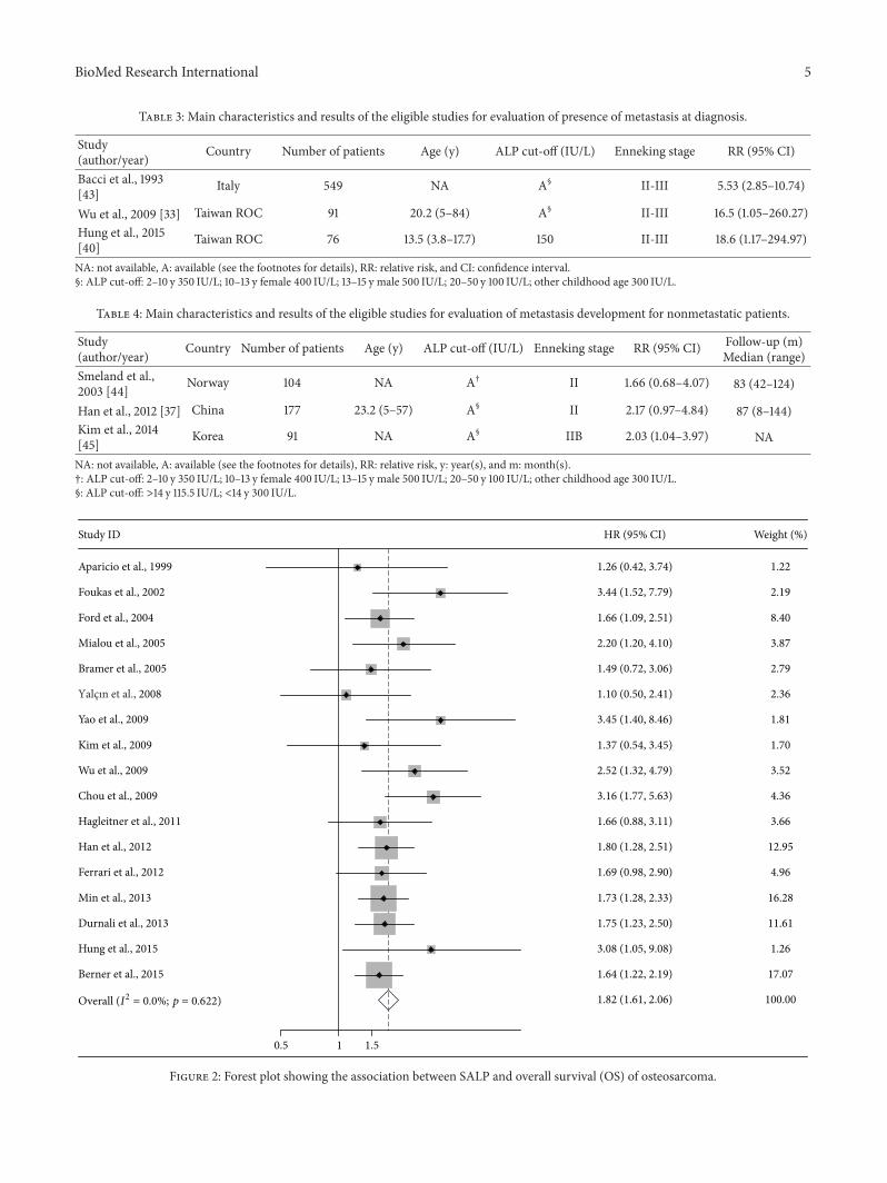
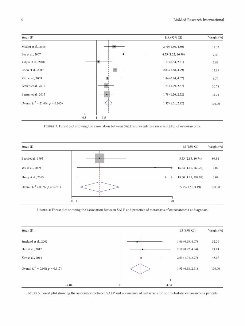
3.2. SALP Level and EFS or OS. 17 studies with a total of 2272osteosarcoma patients dealing with SALP level and OS weremeta-analyzed [25–41]. Because of heterogeneity (𝐼2 = 0%),a fixed effect model was selected. The pooled HR was 1.82(95% CI: 1.61–2.06; 𝑍 = 9.73; 𝑝 < 0.001), illustrating thatSALP level was significantly associated with the poor OS ofosteosarcoma patients (Figure 2). Seven studies including 752patients which reported the correlation between SALP leveland EFS were also meta-analyzed [29–32, 36, 41, 42]. Noheterogeneity was detected (𝐼2 = 21.6%), so a fixed effectmodel was adopted.The combinedHRwas 1.97 (95%CI: 1.61–2.42; 𝑍 = 6.50; 𝑝 < 0.001), demonstrating that SALP level ofosteosarcoma patients was significantly associated with poorEFS (Figure 3).
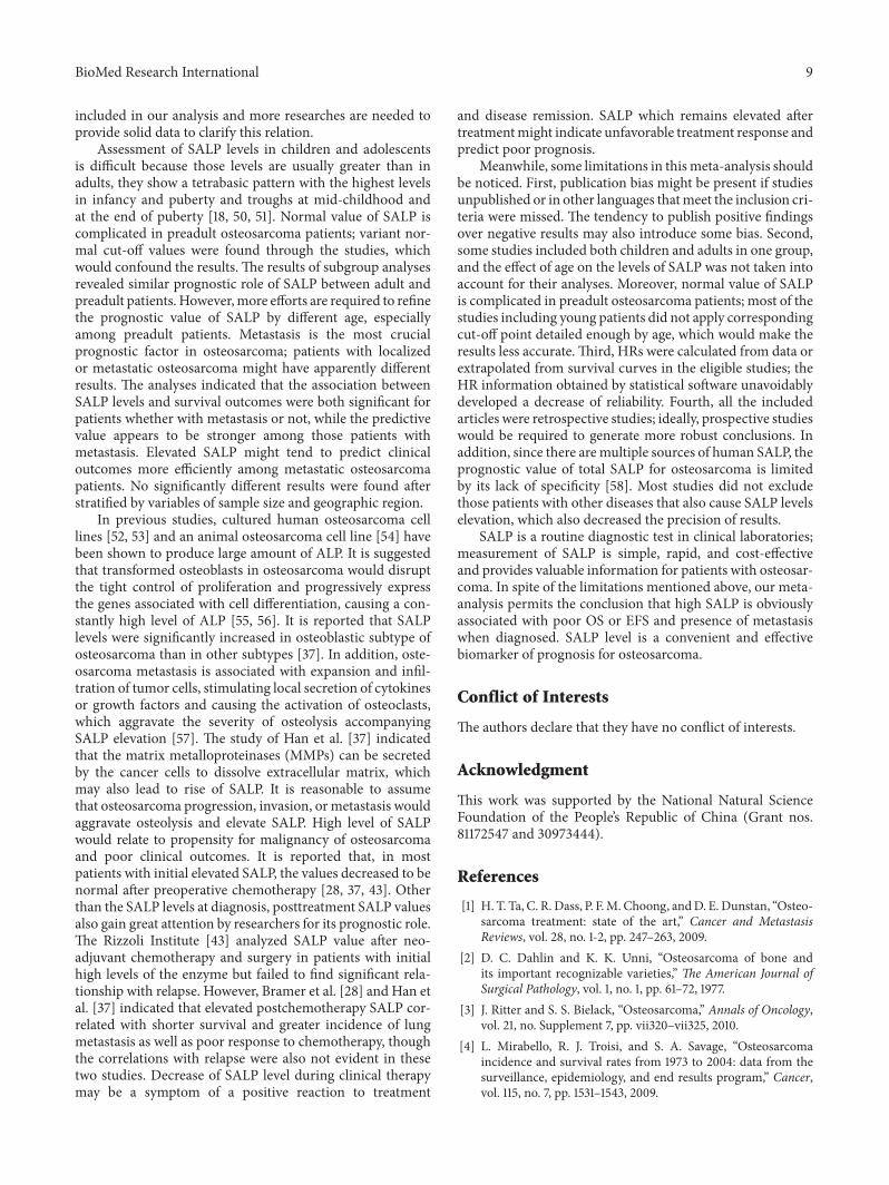
3.3. SALP Level and Metastasis. Three studies with 816patients investigated the relationship between SALP level andpresence of metastasis at diagnosis [33, 40, 43]. A fixed effectmodel was employed for analysis since no heterogeneity wasdetected (𝐼2 = 0.0%). The combined RR was 5.55 (95%

BioMed Research International 3
Table 1: Main characteristics and results of the eligible studies for evaluation of overall survival.
Study(author/year) Country Number of
patientsAge (y)
Median (range)ALP cut-off
(IU/L)Ennekingstage HR (95% CI)
Follow-up (m)Median(range)
Aparicio et al.,1999 [25] Spain 33 17 (12–42) 115 II 1.26
(0.42–3.74) 96 (60–156)
Foukas et al.,2002 [26] UK 45 18 (6–48) NA IIB 3.44
(1.52–7.79) 68 (28–88)
Ford et al., 2004[27] UK 350 NS (<40) NA II 1.66
(1.09–2.51) NA
Bramer et al.,2005 [28] UK 89 NA NA II 1.49
(0.72–3.06) NA
Mialou et al.,2005 [29] France 60 13.5 (2–19) 500 IIIB 2.2 (1.2–4.1) NA
Yalcın et al.,2008 [30] Turkey 55 13 (7–17) NA II-III 1.1 (0.5–2.41) NA
Chou et al.,2009 [31] USA 91 NA NA III 3.16
(1.77–5.36) 89 (1–141)
Kim et al., 2009[32] Korea 67 15.7 (3.8–644) NA II 1.37
(0.54–3.45) 59.9
Wu et al., 2009[33] Taiwan ROC 91 20.2 (5–84) A† II-III 2.52
(1.32–4.75) 58.2 (2–233)
Yao et al., 2009[34] China 57 16 (6–70) 136 II-III 3.45
(1.4–8.46) 32.5 (10–52)
Hagleitner et al.,2011 [35] Netherlands 94 17.8 (4.5–39.5) NA II-III 1.66
(0.88–3.11) 67.2 (28.8–360)
Ferrari et al.,2012 [36] Italy 209 14 (4–39) A† II 1.69
(0.98–2.9) 76 (31–115)
Han et al., 2012[37] China 177 23.2 (5–57) A§ II 1.80
(1.28–2.51) 87 (8–144)
Durnali et al.,2013 [38] Turkey 211 20 (13–74) A$ II-III 1.75 (1.23–2.5) 30.5 (0.5–213)
Min et al., 2013[39] China 333 19 (5–78) NA II-III 1.73
(1.28–2.33) NA (1–100)
Hung et al., 2015[40] Taiwan ROC 69 13.5 (3.8–17.7) 150 II-III 3.08
(1.05–9.08) 51.6 (18–111.6)
Berner et al.,2015 [41] Norway 301 NA AΔ II-III 1.64
(1.22–2.19) NA
NA: not available, A: available (see the footnotes for details), HR: hazard ratio, CI: confidence interval, y: year(s), and m: month(s).$: 2 times of upper limit normal level.†: ALP cut-off: 2–10 y 100–350 IU/L; 10–13 y female 110–400 IU/L; 13–15 y male 125–500 IU/L; 20–50 y 25–100 IU/L; other childhood age 73–300 IU/L.§: ALP cut-off: >18 y 150 IU/L; <18 y 110 IU/L.Δ: ALP cut-off: 0–17 y 400 IU/L; >17 y 105 IU/L.
CI: 1.61–9.49; 𝑍 = 2.76; 𝑝 = 0.006), indicating significantrelationship between elevated SALP level and metastaticdisease of osteosarcoma patients (Figure 4). Moreover, other3 studies, including 372 nonmetastatic osteosarcoma patients,observed the linkage of SALP level and metastasis develop-ment [37, 44, 45]. Because of heterogeneity (𝐼2 = 0%), a fixedeffect model was used in this analysis. However, the resultshowed no statistically significant correlation between highSALP and metastasis development, with RR being 1.95 (95%CI: 0.98–2.91; 𝑍 = 3.96; 𝑝 < 0.001) (Figure 5).
3.4. Subgroup Analyses. Because of the limited articles aboutmetastasis, stratifying analysis was only conducted on thecorrelation between SALP and OS or EFS. Main results of
subgroup analysis for OS and EFS were listed in Tables 5 and6. After stratified by age range, the pooled HRs of preadultgroup (patients’ age less than 19 years old) of OS and EFSwere 1.87 (95% CI: 1.20–2.91; 𝑍 = 2.78; 𝑝 = 0.242) and 2.07(95% CI: 1.51–2.83; 𝑍 = 4.56; 𝑝 = 0.341), respectively, similarto the studies comprising both preadult and adult patients,of which the HRs of OS and EFS were 1.82 (95% CI: 1.60–2.06; 𝑍 = 9.33; 𝑝 = 0.625) and 1.90 (95% CI: 1.45–2.48;𝑍 = 4.66; 𝑝 = 0.149), respectively. When stratified by cancerstage, the association between SALP levels and prognosisamong osteosarcoma patients seemed to be strengthened inthe subgroup ofmetastatic patients (Enneking stage III), withHRs of OS and EFS being 2.67 (95% CI: 1.75–4.06; 𝑍 =4.56; 𝑝 = 0.400) and 2.77 (95% CI: 1.88–4.09; 𝑍 = 5.13;

4 BioMed Research International
Table 2: Main characteristics and results of the eligible studies for evaluation of event-free survival.
Study(author/year) Country Number of
patients Age (y) ALP cut-off(IU/L)
Ennekingstage HR (95% CI) Follow-up (m)
Median (range)Mialou et al., 2005[29] France 48 13.5 (2–19) 500 IIIB 2.7 (1.5–4.8) NA
Lee et al., 2007 [42] Korea 45 <15 A† II 4.55 (1.22–16.99) 54 (6–153)Yalcın et al., 2008[30] Turkey 55 13 (7–17) NA II-III 1.11 (0.54–2.31) NA
Kim et al., 2009[32] Korea 67 15.7
(3.8–64.4) NA II 1.84 (0.84–4.07) 59.9
Chou et al., 2009[31] USA 91 NA NA III 2.83 (1.68–4.79) 89 (1–141)
Ferrari et al., 2012[36] Italy 209 14 (4–39) A§ II 1.71 (1.09–2.67) 76 (31–115)
Berner et al., 2015[41] Norway 237 NA AΔ II-III 1.78 (1.26–2.52) NA
NA: not available, A: available (see the footnotes for details), HR: hazard ratio, CI: confidence interval, y: year(s), and m: month(s).†: ALP cut-off: 2–10 y 420 IU/L; 10-11 y 560 IU/L; 12–15 y male 495 IU/L; 12-13 y female 420 IU/L; 14-15 y female 230 IU/L.§: ALP cut-off: 2–10 y 350 IU/L; 10–13 y female 400 IU/L; 13–15 y male 500 IU/L; 20–50 y 100 IU/L; other childhood age 300 IU/L.Δ: ALP cut-off: 0–17 y 400 IU/L; >17 y 105 IU/L.
Records identified through database searching
Additional records identified through other sources
Records after duplicates removed
Records screened Records excluded
Full-text articles assessed for eligibility
Without sufficient information for
Possibly overlapping
Studies included in qualitative synthesis
Studies included in quantitative synthesis
(meta-analysis)
(n = 2186) (n = 0)
(n = 2186)
(n = 2186) (n = 2123)
(n = 63)
extraction (n = 28)On bone-specific ALP (n = 7)
data set (n = 7)
(n = 21)
(n = 21)
Figure 1: Flow diagram of the study selection process.
𝑝 = 0.906), respectively, while in the subgroup of localizedosteosarcoma patients (Enneking stage II), the HRs of OSand EFS were 1.75 (95% CI: 1.42–2.15; 𝑍 = 5.29; 𝑝 =0.740) and 1.88 (95% CI: 1.29–2.73; 𝑍 = 3.31; 𝑝 = 0.386),respectively. Stratified analysis according to sample size was
also conducted. Whether patient number is greater than100 or not, similar results were found about the associationbetween SALP level and poor OS or EFS, while apparent lessheterogeneity was obtained in studies with larger sample size(𝑝 = 0.999; 𝐼2 = 0.0%). When stratifying by geographic
BioMed Research International 5
Table 3: Main characteristics and results of the eligible studies for evaluation of presence of metastasis at diagnosis.
Study(author/year) Country Number of patients Age (y) ALP cut-off (IU/L) Enneking stage RR (95% CI)
Bacci et al., 1993[43] Italy 549 NA A§ II-III 5.53 (2.85–10.74)
Wu et al., 2009 [33] Taiwan ROC 91 20.2 (5–84) A§ II-III 16.5 (1.05–260.27)Hung et al., 2015[40] Taiwan ROC 76 13.5 (3.8–17.7) 150 II-III 18.6 (1.17–294.97)
NA: not available, A: available (see the footnotes for details), RR: relative risk, and CI: confidence interval.§: ALP cut-off: 2–10 y 350 IU/L; 10–13 y female 400 IU/L; 13–15 y male 500 IU/L; 20–50 y 100 IU/L; other childhood age 300 IU/L.
Table 4: Main characteristics and results of the eligible studies for evaluation of metastasis development for nonmetastatic patients.
Study(author/year) Country Number of patients Age (y) ALP cut-off (IU/L) Enneking stage RR (95% CI) Follow-up (m)
Median (range)Smeland et al.,2003 [44] Norway 104 NA A† II 1.66 (0.68–4.07) 83 (42–124)
Han et al., 2012 [37] China 177 23.2 (5–57) A§ II 2.17 (0.97–4.84) 87 (8–144)Kim et al., 2014[45] Korea 91 NA A§ IIB 2.03 (1.04–3.97) NA
NA: not available, A: available (see the footnotes for details), RR: relative risk, y: year(s), and m: month(s).†: ALP cut-off: 2–10 y 350 IU/L; 10–13 y female 400 IU/L; 13–15 y male 500 IU/L; 20–50 y 100 IU/L; other childhood age 300 IU/L.§: ALP cut-off: >14 y 115.5 IU/L; <14 y 300 IU/L.
Hung et al., 2015
Durnali et al., 2013
Hagleitner et al., 2011
Yalçın et al., 2008
Yao et al., 2009
Ferrari et al., 2012
Han et al., 2012
Berner et al., 2015
Wu et al., 2009
Bramer et al., 2005
Kim et al., 2009
Aparicio et al., 1999
Ford et al., 2004
Foukas et al., 2002
Chou et al., 2009
Min et al., 2013
Mialou et al., 2005
Study ID HR (95% CI)
1.82 (1.61, 2.06)
3.08 (1.05, 9.08)
1.75 (1.23, 2.50)
1.66 (0.88, 3.11)
1.10 (0.50, 2.41)
3.45 (1.40, 8.46)
1.69 (0.98, 2.90)
1.80 (1.28, 2.51)
1.64 (1.22, 2.19)
2.52 (1.32, 4.79)
1.49 (0.72, 3.06)
1.37 (0.54, 3.45)
1.26 (0.42, 3.74)
1.66 (1.09, 2.51)
3.44 (1.52, 7.79)
3.16 (1.77, 5.63)
1.73 (1.28, 2.33)
2.20 (1.20, 4.10)
100.00
1.26
11.61
3.66
Weight (%)
2.36
1.81
4.96
12.95
17.07
3.52
2.79
1.70
1.22
8.40
2.19
4.36
16.28
3.87
1 1.50.5
Overall (I2 = 0.0%; p = 0.622)
Figure 2: Forest plot showing the association between SALP and overall survival (OS) of osteosarcoma.
6 BioMed Research International
Kim et al., 2009
Berner et al., 2015
Ferrari et al., 2012
Lee et al., 2007
Mialou et al., 2005
Chou et al., 2009
Yalçın et al., 2008
Study ID
1.97 (1.61, 2.42)
1.84 (0.84, 4.07)
1.78 (1.26, 2.52)
1.71 (1.09, 2.67)
HR (95% CI)
4.55 (1.22, 16.99)
2.70 (1.50, 4.80)
2.83 (1.68, 4.79)
1.11 (0.54, 2.31)
100.00
6.70
34.71
20.78
Weight (%)
2.40
12.33
15.19
7.89
0.5 1 1.5
Overall (I2 = 21.6%; p = 0.265)
Figure 3: Forest plot showing the association between SALP and event-free survival (EFS) of osteosarcoma.
Wu et al., 2009
Study ID
Hung et al., 2015
Bacci et al., 1993
5.55 (1.61, 9.49)
ES (95% CI)
16.54 (1.05, 260.27)
18.60 (1.17, 294.97)
5.53 (2.85, 10.74)
100.00
Weight (%)
0.09
0.07
99.84
0 1 20
Overall (I2 = 0.0%; p = 0.971)
Figure 4: Forest plot showing the association between SALP and presence of metastasis of osteosarcoma at diagnosis.
Study ID
Kim et al., 2014
Han et al., 2012
Smeland et al., 2003
1.95 (0.98, 2.91)
2.03 (1.04, 3.97)
ES (95% CI)
2.17 (0.97, 4.84)
1.66 (0.68, 4.07)
100.00
43.07
Weight (%)
24.74
32.20
−4.84 0 4.84
Overall (I2 = 0.0%; p = 0.917)
Figure 5: Forest plot showing the association between SALP and occurrence of metastasis for nonmetastatic osteosarcoma patients.

BioMed Research International 7
Table 5: A summary of HRs for the overall and subgroup analyses of SALP and OS of osteosarcoma patients.
Number of studies Patients number HR (95% CI) HeterogeneityChi-squared 𝐼
2𝑝 value
Overall 17 2272 1.82 (1.61–2.06) 13.68 0% 0.622Age
Preadult and adult 14 2088 1.82 (1.60–2.06) 10.83 0% 0.625Preadult only 3 184 1.87 (1.20–2.91) 2.84 2.96% 0.242
Enneking stageII 7 910 1.75 (1.42–2.15) 3.53 0% 0.740II-III 8 1211 1.77 (1.61–2.06) 6.02 0% 0.538III 2 151 2.67 (1.75–4.06) 0.71 0% 0.400
Sample size<100 11 751 2.13 (1.70–2.66) 10.94 8.6% 0.362>100 6 1521 1.71 (1.48–1.98) 0.20 0% 0.999
Geographic regionAsia 6 734 1.89 (1.55–2.31) 4.16 0% 0.527Non-Asia 11 1538 1.78 (1.53–2.07) 9.29 0% 0.505
HR: hazard ratio, OS: overall survival, and CI: confidence interval.
Table 6: A summary of HRs for the overall and subgroup analyses of SALP and EFS of osteosarcoma patients.
Number of studies Patients number HR (95% CI) HeterogeneityChi-squared 𝐼
2𝑝 value
Overall 7 752 1.97 (1.61–2.42) 7.65 21.6% 0.265Age
Preadult and adult 4 385 1.90 (1.45–2.48) 5.33 43.7% 0.149Preadult only 3 367 2.07 (1.51–2.83) 2.15 7.1% 0.341
Enneking stageII 3 321 1.88 (1.29–2.73) 1.91 0% 0.386II-III 2 292 1.63 (1.19–2.23) 1.32 24.3% 0.250III 2 139 2.77 (1.88–4.09) 0.01 0% 0.906
Sample size<100 5 306 2.28 (1.68–3.09) 6.09 34.3% 0.193>100 2 446 1.75 (1.33–2.31) 0.02 0% 0.89
Geographic regionAsia 2 112 2.34 (1.19–4.60) 1.34 25.2% 0.248Non-Asia 5 640 1.94 (1.56–2.40) 6.05 33.8% 0.196
HR: Hazard ratio, EFS: event-free survival, and CI: confidence interval.
region, the HRs of high SALP for OS and EFS were not sig-nificantly different between subgroups of Asia or non-Asia.
3.5. PublicationBias and SensitivityAnalysis. Publication biasof the included studieswas assessed by funnel plots andBegg’stest. As shown in Figure 6, the funnel plots were almost sym-metric in each analysis. Meanwhile, one study was omitted ata time in the sensitivity analysis to measure its effect on thepooledHR for theOSor EFS.No individual study dominantlyinfluenced overallHR, as presented in Figure 7, indicating therobustness of the results in this meta-analysis.
4. Conclusion
In the early studies, elevated SALP levels had been reported in40% to 80% of patients with osteosarcoma [46–49]. In accor-dance with that ratio, of these selected studies which have
sample size larger than 100 [27, 36–39, 41, 43, 44], elevatedSALP levels were found in 40.2% to 83.7% of osteosarcomapatients. The relationship of serum total ALP activity withclinical outcomes of osteosarcoma has been recognized forover 50 years [17]; however, this remains controversial. Thus,to derive amore precise estimation of the correlation betweenSALP levels and survival rates or metastasis in patients withosteosarcoma, we carried out this meta-analysis.
The present meta-analysis suggested that osteosarcomapatients with high SALP levels have significantly poorer OSor EFS when compared with those with normal levels. Theresults also showed that patients with high SALP significantlycorrelated with greater ratio of presence of metastasis atdiagnosis, indicating that osteosarcomametastases obviouslyrelate to higher SALP levels. However, it failed to obtainsignificant correlation between SALP level and metastasisdevelopment through nonmetastatic osteosarcoma patients,

8 BioMed Research International
Begg’s funnel plot with pseudo 95% confidence limits
0.2 0.4 0.60s.e. of ln (hr)
−1
0
1
2ln
(hr)
(a)
Begg’s funnel plot with pseudo 95% confidence limits
0.2 0.4 0.6 0.80s.e. of ln (hr)
−1
0
1
2
ln (h
r)
(b)
Begg’s funnel plot with pseudo 95% confidence limits
0.5 1 1.50s.e. of ln (hr)
−2
0
2
4
6
ln (h
r)
(c)
Begg’s funnel plot with pseudo 95% confidence limits
−0.5
0
0.5
1
1.5
ln (h
r)
0.2 0.4 0.60s.e. of ln (hr)
(d)
Figure 6: Funnel plots assessing possible publication bias for prognosis ((a) OS; (b) EFS; (c) presence ofmetastasis at diagnosis; (d)metastasisdevelopment for nonmetastatic patients).
0.45 0.600.48 0.72 0.75
Aparicio et al., 1999Foukas et al., 2002
Ford et al., 2004Mialou et al., 2005Bramer et al., 2005Yalçın et al., 2008
Yao et al., 2009Kim et al., 2009Wu et al., 2009
Chou et al., 2009Hagleitner at al., 2011
Han et al., 2012Ferrari et al., 2012
Min et al., 2013Durnali et al., 2013
Hung et al., 2015Berner et al., 2015
study ommitted Meta-analysis fixed effects estimates (linear form),
Lower CI limit Estimate Upper CI limit
(a)
0.39 0.680.47 0.88 0.98
Mialou et al., 2005
Lee et al., 2007
Yalçın et al., 2008
Chou et al., 2009
Kim et al., 2009
Ferrari et al., 2012
Berner et al., 2015
study ommitted Meta-analysis fixed effects estimates (linear form),
Lower CI limit Estimate Upper CI limit
(b)
Figure 7: Sensitivity analysis for prognosis of survival rates ((a) OS; (b) EFS).
with the combinedRR being 1.95 (95%CI: 0.98–2.91). Amongthe included three studies, it is worthwhile to notice that Kimet al. [45] developed a high-performance nomogram withseveral predictors to predict the probability of metastasis,
including the factor of SALP level. Though the meta-analysisshowed no statistically significant result, some relevancemight exist between SALP level and metastasis developmentin localized osteosarcoma patients. Merely three studies were
BioMed Research International 9
included in our analysis and more researches are needed toprovide solid data to clarify this relation.
Assessment of SALP levels in children and adolescentsis difficult because those levels are usually greater than inadults, they show a tetrabasic pattern with the highest levelsin infancy and puberty and troughs at mid-childhood andat the end of puberty [18, 50, 51]. Normal value of SALP iscomplicated in preadult osteosarcoma patients; variant nor-mal cut-off values were found through the studies, whichwould confound the results. The results of subgroup analysesrevealed similar prognostic role of SALP between adult andpreadult patients. However,more efforts are required to refinethe prognostic value of SALP by different age, especiallyamong preadult patients. Metastasis is the most crucialprognostic factor in osteosarcoma; patients with localizedor metastatic osteosarcoma might have apparently differentresults. The analyses indicated that the association betweenSALP levels and survival outcomes were both significant forpatients whether with metastasis or not, while the predictivevalue appears to be stronger among those patients withmetastasis. Elevated SALP might tend to predict clinicaloutcomes more efficiently among metastatic osteosarcomapatients. No significantly different results were found afterstratified by variables of sample size and geographic region.
In previous studies, cultured human osteosarcoma celllines [52, 53] and an animal osteosarcoma cell line [54] havebeen shown to produce large amount of ALP. It is suggestedthat transformed osteoblasts in osteosarcoma would disruptthe tight control of proliferation and progressively expressthe genes associated with cell differentiation, causing a con-stantly high level of ALP [55, 56]. It is reported that SALPlevels were significantly increased in osteoblastic subtype ofosteosarcoma than in other subtypes [37]. In addition, oste-osarcoma metastasis is associated with expansion and infil-tration of tumor cells, stimulating local secretion of cytokinesor growth factors and causing the activation of osteoclasts,which aggravate the severity of osteolysis accompanyingSALP elevation [57]. The study of Han et al. [37] indicatedthat the matrix metalloproteinases (MMPs) can be secretedby the cancer cells to dissolve extracellular matrix, whichmay also lead to rise of SALP. It is reasonable to assumethat osteosarcoma progression, invasion, or metastasis wouldaggravate osteolysis and elevate SALP. High level of SALPwould relate to propensity for malignancy of osteosarcomaand poor clinical outcomes. It is reported that, in mostpatients with initial elevated SALP, the values decreased to benormal after preoperative chemotherapy [28, 37, 43]. Otherthan the SALP levels at diagnosis, posttreatment SALP valuesalso gain great attention by researchers for its prognostic role.The Rizzoli Institute [43] analyzed SALP value after neo-adjuvant chemotherapy and surgery in patients with initialhigh levels of the enzyme but failed to find significant rela-tionship with relapse. However, Bramer et al. [28] and Han etal. [37] indicated that elevated postchemotherapy SALP cor-related with shorter survival and greater incidence of lungmetastasis as well as poor response to chemotherapy, thoughthe correlations with relapse were also not evident in thesetwo studies. Decrease of SALP level during clinical therapymay be a symptom of a positive reaction to treatment
and disease remission. SALP which remains elevated aftertreatmentmight indicate unfavorable treatment response andpredict poor prognosis.
Meanwhile, some limitations in thismeta-analysis shouldbe noticed. First, publication bias might be present if studiesunpublished or in other languages thatmeet the inclusion cri-teria were missed. The tendency to publish positive findingsover negative results may also introduce some bias. Second,some studies included both children and adults in one group,and the effect of age on the levels of SALP was not taken intoaccount for their analyses. Moreover, normal value of SALPis complicated in preadult osteosarcoma patients; most of thestudies including young patients did not apply correspondingcut-off point detailed enough by age, which would make theresults less accurate. Third, HRs were calculated from data orextrapolated from survival curves in the eligible studies; theHR information obtained by statistical software unavoidablydeveloped a decrease of reliability. Fourth, all the includedarticles were retrospective studies; ideally, prospective studieswould be required to generate more robust conclusions. Inaddition, since there aremultiple sources of human SALP, theprognostic value of total SALP for osteosarcoma is limitedby its lack of specificity [58]. Most studies did not excludethose patients with other diseases that also cause SALP levelselevation, which also decreased the precision of results.
SALP is a routine diagnostic test in clinical laboratories;measurement of SALP is simple, rapid, and cost-effectiveand provides valuable information for patients with osteosar-coma. In spite of the limitations mentioned above, our meta-analysis permits the conclusion that high SALP is obviouslyassociated with poor OS or EFS and presence of metastasiswhen diagnosed. SALP level is a convenient and effectivebiomarker of prognosis for osteosarcoma.
Conflict of Interests
The authors declare that they have no conflict of interests.
Acknowledgment
This work was supported by the National Natural ScienceFoundation of the People’s Republic of China (Grant nos.81172547 and 30973444).
References
[1] H. T. Ta, C. R. Dass, P. F.M. Choong, andD. E.Dunstan, “Osteo-sarcoma treatment: state of the art,” Cancer and MetastasisReviews, vol. 28, no. 1-2, pp. 247–263, 2009.
[2] D. C. Dahlin and K. K. Unni, “Osteosarcoma of bone andits important recognizable varieties,” The American Journal ofSurgical Pathology, vol. 1, no. 1, pp. 61–72, 1977.
[3] J. Ritter and S. S. Bielack, “Osteosarcoma,” Annals of Oncology,vol. 21, no. Supplement 7, pp. vii320–vii325, 2010.
[4] L. Mirabello, R. J. Troisi, and S. A. Savage, “Osteosarcomaincidence and survival rates from 1973 to 2004: data from thesurveillance, epidemiology, and end results program,” Cancer,vol. 115, no. 7, pp. 1531–1543, 2009.

10 BioMed Research International
[5] M. Hameed and H. Dorfman, “Primary malignant bone tu-mors—recent developments,” Seminars in Diagnostic Pathology,vol. 28, no. 1, pp. 86–101, 2011.
[6] L. Kager, A. Zoubek, U. Potschger et al., “Primary metastaticosteosarcoma: presentation and outcome of patients treated onneoadjuvant Cooperative Osteosarcoma Study Group proto-cols,” Journal of Clinical Oncology, vol. 21, no. 10, pp. 2011–2018,2003.
[7] S. S. Bielack, B. Kempf-Bielack, G. Delling et al., “Prognosticfactors in high-grade osteosarcoma of the extremities or trunk:an analysis of 1,702 patients treated on neoadjuvant cooperativeosteosarcoma study group protocols,” Journal of Clinical Oncol-ogy, vol. 20, no. 3, pp. 776–790, 2002.
[8] A. J. Chou, D. S. Geller, and R. Gorlick, “Therapy for osteosar-coma: where do we go from here?” Pediatric Drugs, vol. 10, no.5, pp. 315–327, 2008.
[9] V. H. C. Bramwell, “Osteosarcomas and other cancers of bone,”Current Opinion in Oncology, vol. 12, no. 4, pp. 330–336, 2000.
[10] D. S. Geller and R. Gorlick, “Osteosarcoma: a review of diagno-sis, management, and treatment strategies,”Clinical Advances inHematology & Oncology, vol. 8, no. 10, pp. 705–718, 2010.
[11] J. C. Wittig, J. Bickels, D. Priebat et al., “Osteosarcoma: a mul-tidisciplinary approach to diagnosis and treatment,” AmericanFamily Physician, vol. 65, no. 6, pp. 1123–1136, 2002.
[12] J. Gill, M. K. Ahluwalia, D. Geller, and R. Gorlick, “New tar-gets and approaches in osteosarcoma,” Pharmacology & Thera-peutics, vol. 137, no. 1, pp. 89–99, 2013.
[13] D. C. Allison, S. C. Carney, E. R. Ahlmann et al., “A meta-analysis of osteosarcoma outcomes in the modern medical era,”Sarcoma, vol. 2012, Article ID 704872, 10 pages, 2012.
[14] M.M.Kaplan, “Alkaline phosphatase,”TheNewEngland Journalof Medicine, vol. 286, no. 4, pp. 200–202, 1972.
[15] D. W. Moss, “Perspectives in alkaline phosphatase research,”Clinical Chemistry, vol. 38, no. 12, pp. 2486–2492, 1992.
[16] S. R. Cho, Y. A. Lim, and W. G. Lee, “Unusually high alkalinephosphatase due to intestinal isoenzyme in a healthy adult,”Clinical Chemistry and Laboratory Medicine, vol. 43, no. 11, pp.1274–1275, 2005.
[17] R. J. McKenna, C. P. Schwinn, K. Y. Soong, and N. L. Higin-botham, “Osteogenic sarcoma arising in Paget’s disease,” Can-cer, vol. 17, pp. 42–66, 1964.
[18] M. Rauchenzauner, A. Schmid, P. Heinz-Erian et al., “Sex-and age-specific reference curves for serum markers of boneturnover in healthy children from 2 months to 18 years,” TheJournal of Clinical Endocrinology and Metabolism, vol. 92, no. 2,pp. 443–449, 2007.
[19] A. Stang, “Critical evaluation of the Newcastle-Ottawa scale forthe assessment of the quality of nonrandomized studies inmeta-analyses,” European Journal of Epidemiology, vol. 25, no. 9, pp.603–605, 2010.
[20] M. K. B. Parmar, V. Torri, and L. Stewart, “Extracting summarystatistics to perform meta-analyses of the published literaturefor survival endpoints,” Statistics in Medicine, vol. 17, no. 24, pp.2815–2834, 1998.
[21] R. DerSimonian, “Meta-analysis in the design and monitoringof clinical trials,” Statistics in Medicine, vol. 15, no. 12, pp. 1237–1248, 1996.
[22] J. P. T. Higgins, S. G. Thompson, J. J. Deeks, and D. G. Altman,“Measuring inconsistency in meta-analyses,” British MedicalJournal, vol. 327, no. 7414, pp. 557–560, 2003.
[23] C. B. Begg and M. Mazumdar, “Operating characteristics of arank correlation test for publication bias,” Biometrics, vol. 50,no. 4, pp. 1088–1101, 1994.
[24] L. Bax, N. Ikeda, N. Fukui, Y. Yaju, H. Tsuruta, and K. G. M.Moons, “More than numbers: the power of graphs in meta-analysis,” American Journal of Epidemiology, vol. 169, no. 2, pp.249–255, 2009.
[25] J. Aparicio, A. Segura, J. Montalar et al., “Long-term resultsafter combinedmodality treatment for non-metastatic osteosar-coma,”Medical Oncology, vol. 16, no. 4, pp. 255–260, 1999.
[26] A. F. Foukas, N. S. Deshmukh, R. J. Grimer, D. C. Mangham, E.G. Mangos, and S. Taylor, “Stage-IIB osteosarcomas around theknee. A study of MMP-9 in surviving tumour cells,”The Journalof Bone and Joint Surgery—British Volume, vol. 84, no. 5, pp.706–711, 2002.
[27] S. Ford, A. Saithna, R. J. Grimer, and P. Picci, “Comparisonof the outcome of conventional osteosarcoma at two specialistinternational orthopaedic oncology centres,” Sarcoma, vol. 8,no. 1, pp. 13–18, 2004.
[28] J. A. M. Bramer, A. A. Abudu, R. M. Tillman, S. R. Carter,V. P. Sumathi, and R. J. Grimer, “Pre- and post-chemotherapyalkaline phosphatase levels as prognostic indicators in adultswith localised osteosarcoma,” European Journal of Cancer, vol.41, no. 18, pp. 2846–2852, 2005.
[29] V. Mialou, T. Philip, C. Kalifa et al., “Metastatic osteosarcomaat diagnosis: prognostic factors and long-term outcome—theFrench pediatric experience,” Cancer, vol. 104, no. 5, pp. 1100–1109, 2005.
[30] B. Yalcın, G. Gedikoglu, T. Kutluk, A. Varan, C. Akyuz,and M. Buyukpamukcu, “C-erbB-2 expression and prognosticsignificance in osteosarcoma,” Pediatric Blood—Cancer, vol. 51,no. 2, pp. 222–227, 2008.
[31] A. J. Chou, E. S. Kleinerman, M. D. Krailo et al., “Addition ofmuramyl tripeptide to chemotherapy for patients with newlydiagnosed metastatic osteosarcoma: a report from the Chil-dren’s Oncology Group,” Cancer, vol. 115, no. 22, pp. 5339–5348,2009.
[32] C. Kim, E. Shin, S. Hong et al., “Clinical value of ezrin expres-sion in primary osteosarcoma,”Cancer Research and Treatment,vol. 41, no. 3, pp. 138–144, 2009.
[33] P. K. Wu, W. M. Chen, C. F. Chen, O. K. Lee, C. K. Haung,and T. H. Chen, “Primary osteogenic sarcoma with pulmonarymetastasis: clinical results and prognostic factors in 91 patients,”Japanese Journal of Clinical Oncology, vol. 39, no. 8, pp. 514–522,2009.
[34] Y. Yao, Y. Dong, F. Lin et al., “The expression of CRM1 isassociated with prognosis in human osteosarcoma,” OncologyReports, vol. 21, no. 1, pp. 229–235, 2009.
[35] M. M. Hagleitner, P. M. Hoogerbrugge, W. T. A. van der Graaf,U. Flucke, H. W. B. Schreuder, and D. M. W. M. te Loo, “Age asprognostic factor in patients with osteosarcoma,” Bone, vol. 49,no. 6, pp. 1173–1177, 2011.
[36] S. Ferrari, P. Ruggieri, G. Cefalo et al., “Neoadjuvant chemother-apy with methotrexate, cisplatin, and doxorubicin with or with-out ifosfamide in nonmetastatic osteosarcoma of the extremity:an Italian sarcoma group trial ISG/OS-1,” Journal of ClinicalOncology, vol. 30, no. 17, pp. 2112–2118, 2012.
[37] J. Han, B. Yong, C. Luo, P. Tan, T. Peng, and J. Shen, “High serumalkaline phosphatase cooperating with MMP-9 predicts metas-tasis and poor prognosis in patients with primary osteosarcomain Southern China,”World Journal of Surgical Oncology, vol. 10,article 37, 2012.

BioMed Research International 11
[38] A. Durnali, N. Alkis, S. Cangur et al., “Prognostic factors forteenage and adult patients with high-grade osteosarcoma: ananalysis of 240 patients,”Medical Oncology, vol. 30, no. 3, article624, 2013.
[39] D. Min, F. Lin, Z. Shen et al., “Analysis of prognostic factorsin 333 Chinese patients with high-grade osteosarcoma treatedby multidisciplinary combined therapy,” Asia-Pacific Journal ofClinical Oncology, vol. 9, no. 1, pp. 71–79, 2013.
[40] G. Y. Hung, H. J. Yen, C. C. Yen et al., “Experience of pediatricosteosarcoma of the extremity at a single institution in Taiwan:prognostic factors and impact on survival,” Annals of SurgicalOncology, vol. 22, no. 4, pp. 1080–1087, 2015.
[41] K. Berner, K. S. Hall, O. R. Monge, H. Weedon-Fekjær, O.Zaikova, and Ø. S. Bruland, “Prognostic factors and treatmentresults of high-grade osteosarcoma in norway: a scope beyondthe ‘classical’ patient,” Sarcoma, vol. 2015, Article ID 516843, 14pages, 2015.
[42] J. A. Lee, D. H. Kim, J. S. Lim et al., “The survival of osteosar-coma patients 10 years old or younger is not worse than thesurvival of older patients: a retrospective analysis,” CancerResearch and Treatment, vol. 39, no. 4, pp. 160–164, 2007.
[43] G. Bacci, P. Picci, S. Ferrari et al., “Prognostic significanceof serum alkaline phosphatase measurements in patients withosteosarcoma treated with adjuvant or neoadjuvant chemother-apy,” Cancer, vol. 71, no. 4, pp. 1224–1230, 1993.
[44] S. Smeland, C. Muller, T. A. Alvegard et al., “Scandinavian Sar-coma Group Osteosarcoma Study SSG VIII: prognostic factorsfor outcome and the role of replacement salvage chemotherapyfor poor histological responders,” European Journal of Cancer,vol. 39, no. 4, pp. 488–494, 2003.
[45] S. H. Kim, K.-H. Shin, H. Y. Kim et al., “Postoperative nomo-gram to predict the probability of metastasis in enneking stageIIB extremity osteosarcoma,” BMC Cancer, vol. 14, article 666,2014.
[46] F. R. Eilber and E. Caulkins, “Bone tumors—clinical andradiologic investigation,” in Current Concepts of Diagnosis andTreatment of Bone and Soft Tissue Tumors, H. Uhthoff, Ed., pp.47–54, Springer, Berlin, Germany, 1984.
[47] M. D. Lockshin and I. T. Higgins, “Prognosis in osteogenicsarcoma,” Clinical Orthopaedics and Related Research, vol. 58,pp. 85–103, 1968.
[48] P. E. Scranton Jr., F. A. DeCicco, R. S. Totten, and E. J.Yunis, “Prognostic factors in osteosarcoma. A review of 20year’s experience at the University of Pittsburgh Health CenterHospitals,” Cancer, vol. 36, no. 6, pp. 2179–2191, 1975.
[49] R. J. Mckenna, C. P. Schwinn, K. Y. Soong, and N. L. Hig-inbotham, “Sarcomata of the osteogenic series (osteosarcoma,fibrosarcoma, chondrosarcoma, parosteal osteogenic sarcoma,and sarcomata arising in abnormal bone). An analysis of 552cases,”The Journal of Bone & Joint Surgery—American Volume,vol. 48, no. 1, pp. 1–26, 1966.
[50] S. Turan, B. Topcu, I. Gokce et al., “Serum alkaline phosphataselevels in healthy children and evaluation of alkaline phosphatasez-scores in different types of rickets,” Journal of Clinical Researchin Pediatric Endocrinology, vol. 3, no. 1, pp. 7–11, 2011.
[51] P. Szulc, E. Seeman, and P. D. Delmas, “Biochemical measure-ments of bone turnover in children and adolescents,” Oste-oporosis International, vol. 11, no. 4, pp. 281–294, 2000.
[52] J. R. Farley, E. Kyeyune-Nyombi, N. M. Tarbaux, S. L. Hall,and D. D. Strong, “Alkaline phosphatase activity from humanosteosarcoma cell line SaOS-2: an isoenzyme standard for
quantifying skeletal alkaline phosphatase activity in serum,”Clinical Chemistry, vol. 35, no. 2, pp. 223–229, 1989.
[53] C. Pautke, M. Schieker, T. Tischer et al., “Characterization ofosteosarcoma cell lines MG-63, Saos-2 and U-2 OS in compar-ison to human osteoblasts,” Anticancer Research, vol. 24, no. 6,pp. 3743–3748, 2004.
[54] N. N. Ali, M. A. Harrison, J. Rowe, and N. M. Teich, “Spectrumof osteoblastic differentiation in new cell lines derived fromspontaneous murine osteosarcomas,” Bone, vol. 14, no. 6, pp.847–858, 1993.
[55] G. S. Stein, J. B. Lian, and T. A. Owen, “Relationship of cellgrowth to the regulation of tissue-specific gene expression dur-ing osteoblast differentiation,”The FASEB Journal, vol. 4, no. 13,pp. 3111–3123, 1990.
[56] S. Limmahakhun, P. Pothacharoen, N. Theera-Umpon et al.,“Relationships between serum biomarker levels and clinicalpresentation of human osteosarcomas,” Asian Pacific Journal ofCancer Prevention, vol. 12, no. 7, pp. 1717–1722, 2011.
[57] J. Ambroszkiewicz, J. Gajewska, T. Klepacka, M. Chełchowska,T. Laskowska-Klita, and W. Wozniak, “Clinical utility of bio-chemical bone turnover markers in children and adolescentswith osteosarcoma,” Advances in Medical Sciences, vol. 55, no.2, pp. 266–272, 2010.
[58] P. P. L. Liu, K. S. Leung, S. M. Kumta, K. M. Lee, and K. P.Fung, “Bone-specific alkaline phosphatase in plasma as tumourmarker for osteosarcoma,”Oncology, vol. 53, no. 4, pp. 275–280,1996.

Submit your manuscripts athttp://www.hindawi.com
Stem CellsInternational
Hindawi Publishing Corporationhttp://www.hindawi.com Volume 2014
Hindawi Publishing Corporationhttp://www.hindawi.com Volume 2014
MEDIATORSINFLAMMATION
of
Hindawi Publishing Corporationhttp://www.hindawi.com Volume 2014
Behavioural Neurology
EndocrinologyInternational Journal of
Hindawi Publishing Corporationhttp://www.hindawi.com Volume 2014
Hindawi Publishing Corporationhttp://www.hindawi.com Volume 2014
Disease Markers
Hindawi Publishing Corporationhttp://www.hindawi.com Volume 2014
BioMed Research International
OncologyJournal of
Hindawi Publishing Corporationhttp://www.hindawi.com Volume 2014
Hindawi Publishing Corporationhttp://www.hindawi.com Volume 2014
Oxidative Medicine and Cellular Longevity
Hindawi Publishing Corporationhttp://www.hindawi.com Volume 2014
PPAR Research
The Scientific World JournalHindawi Publishing Corporation http://www.hindawi.com Volume 2014
Immunology ResearchHindawi Publishing Corporationhttp://www.hindawi.com Volume 2014
Journal of
ObesityJournal of
Hindawi Publishing Corporationhttp://www.hindawi.com Volume 2014
Hindawi Publishing Corporationhttp://www.hindawi.com Volume 2014
Computational and Mathematical Methods in Medicine
OphthalmologyJournal of
Hindawi Publishing Corporationhttp://www.hindawi.com Volume 2014
Diabetes ResearchJournal of
Hindawi Publishing Corporationhttp://www.hindawi.com Volume 2014
Hindawi Publishing Corporationhttp://www.hindawi.com Volume 2014
Research and TreatmentAIDS
Hindawi Publishing Corporationhttp://www.hindawi.com Volume 2014
Gastroenterology Research and Practice
Hindawi Publishing Corporationhttp://www.hindawi.com Volume 2014
Parkinson’s Disease
Evidence-Based Complementary and Alternative Medicine
Volume 2014Hindawi Publishing Corporationhttp://www.hindawi.com


![Research Article Prognostic Significance of Serum Free Light … · 2019. 7. 31. · the progression of MGUS [ ], solitary plasmacytoma [ ], and smoldering myeloma [ ]intomultiplemyeloma.](https://static.fdocuments.us/doc/165x107/60b139df8dfefb1baa01f551/research-article-prognostic-significance-of-serum-free-light-2019-7-31-the.jpg)