Review article: NONALCOHOLIC FATTY LIVER …EXCLI Journal 2014;13:461-490 – ISSN 1611-2156...
30
EXCLI Journal 2014;13:461-490 – ISSN 1611-2156 Received: March 12, 2014, accepted: March 31, 2014, published: May 07, 2014 461 Review article: NONALCOHOLIC FATTY LIVER DISEASE, DIET AND GUT MICROBIOTA Carmine Finelli 1 and Giovanni Tarantino 2,3 * 1 Center of Obesity and Eating Disorders, Stella Maris Mediterraneum Foundation, C/da S. Lucia, Chiaromonte, 80035 Potenza, Italy 2 Department of Clinical Medicine and Surgery, Federico II University Medical School of Naples, Via Sergio Pansini, 5, 80131Naples, Italy 3 National Cancer Institute "Foundation G. Pascale" -IRCS- 83013 Mercogliano (Av), Italy * Corresponding author: G. Tarantino, [email protected] ABSTRACT Non-alcoholic fatty liver disease (NAFLD) is a severe liver disease that is increasing in preva- lence with the worldwide epidemic of obesity and its related insulin-resistance state. Evidence for the role of the gut microbiota in energy storage and the subsequent development of obesity and some of its related diseases is now well established. More recently, a new role of gut mi- crobiota has emerged in NAFLD. The gut microbiota is involved in gut permeability, low- grade inflammation and immune balance, it modulates dietary choline metabolism, regulates bile acid metabolism and produces endogenous ethanol. All of these factors are molecular mechanisms by which the microbiota can induce NAFLD or its progression toward overt non- alcoholic steatohepatitis. Modification of the gut microbiota composition and/or its biochemi- cal capacity by specific dietary or pharmacological interventions may advantageously affect host metabolism. Large-scale intervention trials, investigating the potential benefit of prebiot- ics and probiotics in improving cardiometabolic health in high-risk populations, are fervently awaited. Keywords: Gut microbiome, NAFLD, interventions INTRODUCTION The rising incidence of obesity in to- day’s environment is associated with many obesity-related health complications, in- cluding cardiovascular disease, type 2 dia- betes (T2D), hyperlipidemia, hypertension, and nonalcoholic fatty liver disease (NAFLD) (Tarantino et al., 2007; 2012; 2013; Finelli and Tarantino, 2013a). This constellation is also recognized as the met- abolic syndrome and is characterized by underlying insulin resistance (IR). NAFLD or generally speaking hepatic steatosis (HS) is defined as the accumulation of lipid, pri- marily in the form of triacylglycerols in in- dividuals who do not consume significant amounts of alcohol (< 20 g ethanol/d) and in whom other known causes of steatosis, such as certain drugs and toxins, have been excluded (Vuppalanchi and Chalasani, 2009). The spectrum of NAFLD includes simple fatty liver, non alcoholic steatohepa- titis (NASH) - characterized by inflamma- tion, apoptosis, ballooning degeneration, Mallory hyaline, fibrosis-, cirrhosis post NASH, hepatocellular carcinoma and ad- vanced liver disease, which leads to liver- related death (Vuppalanchi and Chalasani, 2009; Sorrentino et al., 2004; Tarantino and Finelli, 2013; Tarantino, 2007; Tarantino et al., 2011a; Finelli and Tarantino, 2013c).
Transcript of Review article: NONALCOHOLIC FATTY LIVER …EXCLI Journal 2014;13:461-490 – ISSN 1611-2156...

EXCLI Journal 2014;13:461-490 – ISSN 1611-2156 Received: March 12, 2014, accepted: March 31, 2014, published: May 07, 2014
461
Review article:
NONALCOHOLIC FATTY LIVER DISEASE, DIET AND GUT MICROBIOTA
Carmine Finelli1 and Giovanni Tarantino2,3* 1 Center of Obesity and Eating Disorders, Stella Maris Mediterraneum Foundation,
C/da S. Lucia, Chiaromonte, 80035 Potenza, Italy 2 Department of Clinical Medicine and Surgery, Federico II University Medical School
of Naples, Via Sergio Pansini, 5, 80131Naples, Italy 3 National Cancer Institute "Foundation G. Pascale" -IRCS- 83013 Mercogliano (Av), Italy * Corresponding author: G. Tarantino, [email protected]
ABSTRACT
Non-alcoholic fatty liver disease (NAFLD) is a severe liver disease that is increasing in preva-lence with the worldwide epidemic of obesity and its related insulin-resistance state. Evidence for the role of the gut microbiota in energy storage and the subsequent development of obesity and some of its related diseases is now well established. More recently, a new role of gut mi-crobiota has emerged in NAFLD. The gut microbiota is involved in gut permeability, low-grade inflammation and immune balance, it modulates dietary choline metabolism, regulates bile acid metabolism and produces endogenous ethanol. All of these factors are molecular mechanisms by which the microbiota can induce NAFLD or its progression toward overt non-alcoholic steatohepatitis. Modification of the gut microbiota composition and/or its biochemi-cal capacity by specific dietary or pharmacological interventions may advantageously affect host metabolism. Large-scale intervention trials, investigating the potential benefit of prebiot-ics and probiotics in improving cardiometabolic health in high-risk populations, are fervently awaited. Keywords: Gut microbiome, NAFLD, interventions
INTRODUCTION
The rising incidence of obesity in to-day’s environment is associated with many obesity-related health complications, in-cluding cardiovascular disease, type 2 dia-betes (T2D), hyperlipidemia, hypertension, and nonalcoholic fatty liver disease (NAFLD) (Tarantino et al., 2007; 2012; 2013; Finelli and Tarantino, 2013a). This constellation is also recognized as the met-abolic syndrome and is characterized by underlying insulin resistance (IR). NAFLD or generally speaking hepatic steatosis (HS) is defined as the accumulation of lipid, pri-marily in the form of triacylglycerols in in-
dividuals who do not consume significant amounts of alcohol (< 20 g ethanol/d) and in whom other known causes of steatosis, such as certain drugs and toxins, have been excluded (Vuppalanchi and Chalasani, 2009). The spectrum of NAFLD includes simple fatty liver, non alcoholic steatohepa-titis (NASH) - characterized by inflamma-tion, apoptosis, ballooning degeneration, Mallory hyaline, fibrosis-, cirrhosis post NASH, hepatocellular carcinoma and ad-vanced liver disease, which leads to liver-related death (Vuppalanchi and Chalasani, 2009; Sorrentino et al., 2004; Tarantino and Finelli, 2013; Tarantino, 2007; Tarantino et al., 2011a; Finelli and Tarantino, 2013c).

EXCLI Journal 2014;13:461-490 – ISSN 1611-2156 Received: March 12, 2014, accepted: March 31, 2014, published: May 07, 2014
462
Given the close relations between obesi-ty, the metabolic syndrome, and the devel-opment of NAFLD, it is not surprising that many NAFLD patients have multiple com-ponents of the metabolic syndrome, wheth-er or not they are overweight or obese. IR is present in and is a significant predictor of NAFLD and NASH in most patients (Clark, 2006), even the ~ 10–15 % of patients who are not overweight (Chiang et al., 2011; Hamaguchi et al., 2012). NAFLD is a mul-tifactorial disease that involves a complex interaction of genetics, diet, and lifestyle, all of which combine to form the NAFLD phenotype. A cornerstone of the manage-ment strategy in such patients with fatty liver is the use of diet to decrease body weight, and improve glycemic control, dyslipidemia and cardiovascular risks as well (Finelli and Tarantino, 2012b).
Gut microbiota are thought to play a role in the pathogenesis of NASH for sev-eral reasons. First, gut microbiota are known to have a large effect on the diges-tion and absorption of nutrients (van der Hoeven-Hangoor et al., 2013). Microbiota transplantation experiments in mice sug-gested that certain microbiota is capable of inducing obesity independent of other envi-ronmental factors (Kallus and Brandt, 2012). Second, gut microbiota participate in the development and homeostasis of the overall immunity of the host (Gigante et al., 2011). Therefore, certain microbiota may influence the development of liver inflam-mation. The links between gut microbiota and the host immune system include TLRs and short-chain fatty acids (Vinolo et al., 2011). In fact, the innate immune system might influence the metabolic syndrome and obesity, as mice deficient in Toll-like receptor 5 develop hyperphagia, become obese and insulin resistant (Tilg, 2010). Third, gut microbiota may influence the production of gut hormones, such as gluca-gon-like peptide 1, and, subsequently, have an effect on the overall metabolism of the host (Flint, 2011).
The liver appears as the first point of contact for (and produces the initial immu-nological response to) bacteria and micro-bial components, as well as other endoge-nous and exogenous toxins present in the portal blood. Given the capacity of the liver to regulate metabolism in a form that can affect the entire organism, to distribute nu-merous substances to the gut through bile and the entero hepatic circulation, and to regulate numerous hormonal and immuno-logical responses, the potential for the liver to influence gut function can be quickly ap-preciated. Interactions between the gut, the diet and the liver are, naturally, bidirection-al; hormones, inflammatory mediators and the products of digestion and absorption all unequivocally influence liver function. The focus of this review is on gut–liver-diet in-teractions that contribute to the pathogene-sis of a common liver disorder, NAFLD.
Interactions between the intestinal micro-biota and liver
The interactions of the gut microbiota and the liver have only recently been inves-tigated in detail. Receiving approximately 70 % of its blood supply from the intestinal venous outflow, the liver represents the first line of defense against gut-derived antigens, food antigens, toxins, microbial-derived products, and microorganisms (Henao-Mejia et al., 2013). The liver, therefore, is equipped with a broad array of immune cells (i.e., macrophages, lymphocytes, natu-ral killer cells, and dendritic cells) to ac-complish this function (Henao-Mejia et al., 2013). Small intestinal bacterial overgrowth (SIBO) is common in patients with cirrho-sis (Wiest et al., 2014; Savarino et al., 2011; Bellot et al., 2013; Gupta et al., 2010) and its prevalence correlates directly with the severity of liver disease (Pande et al., 2009). A physiological basis for SIBO in liver disease was added by reports of im-paired small bowel motility and prolonged orocecal transit time in patients with cirrho-sis (Seo and Shah, 2012; Gunnarsdottir et al., 2003). Some studies reinforced the con-
EXCLI Journal 2014;13:461-490 – ISSN 1611-2156 Received: March 12, 2014, accepted: March 31, 2014, published: May 07, 2014
463
cept that disturbed motility predisposes to SIBO, and suggested a link between altera-tions in intestinal motility and the develop-ment of both hepatic encephalopathy and spontaneous bacterial peritonitis (Romeiro et al., 2013; Ancel et al., 2006; Bouin et al., 2004; Garcovich et al., 2012). Bacterial translocation (passage of viable bacteria re-sident in the gastrointestinal tract to gener-ally sterile tissues) in the context of SIBO is facilitated by augmented intestinal permea-bility, another feature of liver cirrhosis (Hada et al., 2010; Cariello et al., 2010). Indeed, increased intestinal permeability is associated with an increased risk of sponta-neous bacterial peritonitis (Benjamin et al., 2013; Assimakopoulos et al., 2012).
Having identified bacterial products as hepatotoxins (Lamontagne et al., 2013; Kisch et al., 2006) and having figured out that multiple metabolic activities of the gut microbiota could affect the liver function, the potential for the gut microbiota to indu-ce or maintain various liver diseases, main-ly at the light of its immunological interac-tions with the host has been clearly reck-oned.
Nafld, intestinal microbiota and metabolic changes
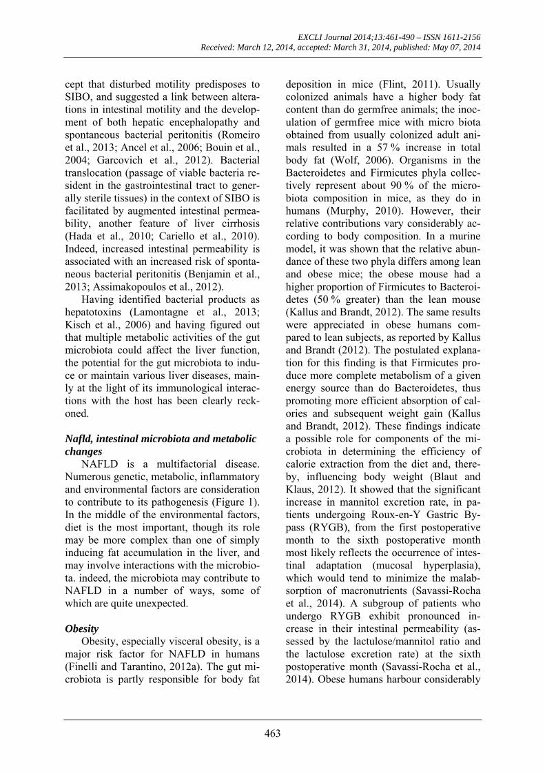
NAFLD is a multifactorial disease. Numerous genetic, metabolic, inflammatory and environmental factors are consideration to contribute to its pathogenesis (Figure 1). In the middle of the environmental factors, diet is the most important, though its role may be more complex than one of simply inducing fat accumulation in the liver, and may involve interactions with the microbio-ta. indeed, the microbiota may contribute to NAFLD in a number of ways, some of which are quite unexpected.
Obesity
Obesity, especially visceral obesity, is a major risk factor for NAFLD in humans (Finelli and Tarantino, 2012a). The gut mi-crobiota is partly responsible for body fat
deposition in mice (Flint, 2011). Usually colonized animals have a higher body fat content than do germfree animals; the inoc-ulation of germfree mice with micro biota obtained from usually colonized adult ani-mals resulted in a 57 % increase in total body fat (Wolf, 2006). Organisms in the Bacteroidetes and Firmicutes phyla collec-tively represent about 90 % of the micro-biota composition in mice, as they do in humans (Murphy, 2010). However, their relative contributions vary considerably ac-cording to body composition. In a murine model, it was shown that the relative abun-dance of these two phyla differs among lean and obese mice; the obese mouse had a higher proportion of Firmicutes to Bacteroi-detes (50 % greater) than the lean mouse (Kallus and Brandt, 2012). The same results were appreciated in obese humans com-pared to lean subjects, as reported by Kallus and Brandt (2012). The postulated explana-tion for this finding is that Firmicutes pro-duce more complete metabolism of a given energy source than do Bacteroidetes, thus promoting more efficient absorption of cal-ories and subsequent weight gain (Kallus and Brandt, 2012). These findings indicate a possible role for components of the mi-crobiota in determining the efficiency of calorie extraction from the diet and, there-by, influencing body weight (Blaut and Klaus, 2012). It showed that the significant increase in mannitol excretion rate, in pa-tients undergoing Roux-en-Y Gastric By-pass (RYGB), from the first postoperative month to the sixth postoperative month most likely reflects the occurrence of intes-tinal adaptation (mucosal hyperplasia), which would tend to minimize the malab-sorption of macronutrients (Savassi-Rocha et al., 2014). A subgroup of patients who undergo RYGB exhibit pronounced in-crease in their intestinal permeability (as-sessed by the lactulose/mannitol ratio and the lactulose excretion rate) at the sixth postoperative month (Savassi-Rocha et al., 2014). Obese humans harbour considerably

EXCLI Journal 2014;13:461-490 – ISSN 1611-2156 Received: March 12, 2014, accepted: March 31, 2014, published: May 07, 2014
464
Diabetes Mellitus
• Insulitis
• LPS-CD14
Gut microbiota
Body weight Choline metabolism
Insulin resistance
Fatty liver
• Cleavage of dietary polysaccharides
• Synthesis of acetyl coenzyme A carboxylase and fatty acid synthetase
• Angiopoietin-related protein 4
• Bile acid signaling
• Choline bioavailability polysaccharides
• Phosphatidylcholine synthesis
Free fatty acid Hepatic VLDL secretion
Figure 1: Effects of the gut microbita on pathogenesis of NAFLD (Abbreviations: LPS – lipopolysaccharide; VLDL – very-low-density lipoprotein)
fewer Bacteroidetes and more Firmicutes than lean controls (Krznarić et al., 2012). Changes in the gut microbiota have also been documented among obese individuals after gastric bypass surgery (Osto et al., 2013). Osto et al. (2013) showed that RYGB surgery might differently modify the gut microbiota composition in the three dis-tinct anatomical sections of the small intes-tine compared to sham surgery. RYGB in-duced changes in the microbiota of the ali-mentary limb and the common channel re-sembling those seen after prebiotic treat-ment or weight loss by dieting, as reported by Osto et al. (2013). These changes may be associated with altered production of in-testinal hormones known to control energy balance (Osto et al., 2013). Postsurgical modulation of gut microbiota may signifi-
cantly contribute to the beneficial metabolic effects of RYGB surgery (Osto et al., 2013), not excluding those on NAFLD.
Santacruz et al. (2009) indicated that calorie restriction and physical activity have an impact on gut microbiota composition related to body weight loss, which also seem to be influenced by the individual's microbiota. Limited evidence suggests a role for increased microbial extraction of calories among obese humans. DiBaise et al. (2008), in their review, suggests that the gut microbiota affects nutrient acquisition and energy regulation and its composition has also been shown to differ in lean vs obese animals and humans. Some evidence suggests that the metabolic activities of the gut microbiota facilitate the extraction of calories from ingested dietary substances

EXCLI Journal 2014;13:461-490 – ISSN 1611-2156 Received: March 12, 2014, accepted: March 31, 2014, published: May 07, 2014
465
and help to store these calories in host adi-pose tissue for later use (DiBaise et al., 2008). Furthermore, the gut bacterial flora of obese mice and humans include fewer Bacteroidetes and correspondingly more Firmicutes than that of their lean counter-parts, suggesting that differences in caloric extraction of ingested food substances may be due to the composition of the gut micro-biota (DiBaise et al., 2008). Bacterial lipo-polysaccharide derived from the intestinal microbiota may act as a triggering factor linking inflammation to high-fat diet-induced metabolic syndrome (DiBaise et al., 2008). DiBaise et al. (2008) concluded that existing evidence warrants further in-vestigation of the microbial ecology of the human gut and points to modification of the gut microbiota as one means to treat people who are over-weight or obese.
Several studies reveal how the microbi-ota might influence body weight and com-position. Gut microbiota could precisely af-fect the proportion of calories obtained from the intestinal contents (caloric sal-vage). For example, Bacteroides thetai-otamicron is able to break most glycosidic linkages of numerous constituents of our diet. This Gram-negative anaerobic bacte-rium can degrade indigestible poly saccha-rides from plant, representing a quota part of calorie needs, nearly 10–15 % (Zocco et al., 2007). Furthermore, microbes are pre-dominantly found in surface-attached and spatially structured polymicrobial commu-nities (Estrela and Brown, 2013). Within these communities, microbial cells excrete a wide range of metabolites, setting the stage for interspecific metabolic interac-tions (Estrela and Brown, 2013). The links, however, between metabolic and ecological interactions (functional relationships), and species spatial organization (structural rela-tionships) are still poorly understood, as re-ported by Estrela et al. (2013). It showed that strong metabolic interdependence drives the emergence of mutualism, robust interspecific mixing, and increased com-munity productivity. These emergent com-
munity properties are driven by demograph-ic feedbacks, such that aid from neighbor-ing cells directly enhances focal cell growth, which in turn feeds back to neigh-bour fecundity. In contrast, weak metabolic interdependence drives conflict (exploita-tion or competition), and in turn greater in-terspecific segregation. Together, these re-sults support the idea that species structural and functional relationships represent the net balance of metabolic interdependencies (Estrela and Brown, 2013).
The caloric restore is furnished by the presence of genes encoding enzymes that split vegetable and dietary polysaccharides in the microbiota of obese mouse. Admin-istration of a conventional microbiota to germ free mice induced a rapid increase in body fat associated with increased hepatic triglyceride production related to increased activity of two crucial enzymes involved in de novo fatty acid synthesis—acetyl coen-zyme a carboxylase and fatty acid synthe-tase (Wolf, 2006). The microbiota inhibits angiopoietin related protein 4, which sup-presses lipoprotein lipase, a key regulator of fatty acid release from triglyceride rich chy-lomicrons. Inhibition of angiopoietin relat-ed protein 4 causes hyper-expression of lip-oprotein lipase, giving place to an augment-ed uptake of fatty acids and storage of tri-glycerides in adipocytes (Wolf, 2006; Fleissner et al., 2010) and liver (Fleissner et al., 2010).
Gut microbes have a pivotal role in the intra luminal metabolism of bile acids (Ruiz et al., 2013). Given that bile acids are fun-damental for the absorption and emulsifica-tion of dietary fats and lipid soluble vita-mins in the small intestine, disorganized bile acid physiology could result in an al-tered energy balance. The role of bile acids in maintaining the intestinal barrier function and the luminal environment must too be called to mind, including their capacity to prevent SIBO and bacterial translocation (Hagey and Krasowski, 2013; Karatepete al., 2010). Additionally, bile acids are in-volved in energy and lipid metabolism, be-

EXCLI Journal 2014;13:461-490 – ISSN 1611-2156 Received: March 12, 2014, accepted: March 31, 2014, published: May 07, 2014
466
ing capable of lowering triglyceride levels, for example (Trauner et al., 2010). There-fore, the microbiota could, because to ef-fects on bile acid metabolism in the gut lu-men, influence signalling pathways in-volved in energy and lipid metabolism. The consequences of this involvement include the regulation of secondary bile acid me-tabolism, the inhibition of bile acid synthe-sis, modifications to lipid peroxidation and the storage of fatty acids in the liver (Sayin et al. 2013).
Insulin resistance
Insulin resistance is crucial in the path-ogenesis of the metabolic syndrome, of which NAFLD is considered as the hepatic component. Insulin resistance appears to have a crucial role in the pathogenesis of NAFLD and NASH (Finelli and Tarantino, 2013a). Besides the suggested role of insu-lin resistance in the development of steato-sis, hepatic insulin resistance could promote hepatocyte injury and inflammation (Tar-antino et al., 2009). Gut flora and gut de-rived endotoxemia are considered main fac-tors in developing insulin resistance. The key link is represented by the lipopolysac-charide–toll like receptor 4 (TLR4)–monocyte differentiation antigen CD14 sys-tem (Penas-Steinhardt et al., 2012; Ma et al., 2013; Belforte et al., 2013; Krautbauer et al., 2014). Even if this evidence has been largely gleaned from animal models, one study documented elevated plasma levels of lipopolysaccharide among patients with obesity and type 2 diabetes mellitus (Ha-wkesworth et al., 2013). Confirmation of these findings and elucidation of the role of the microbiota, gut damage and the path-ways for translocation of bacterial debris could open new avenues for prevention and treatment of type 2 diabetes, as reported by Hawkesworth et al. (2013). Suppression or modification of SIBO, by leading to re-duced proinflammatory cytokine produc-tion, results in a fall in fasting insulin con-centrations and decreased insulin resistance (Penas-Steinhardt et al., 2012; Rodríguez-
Hernández et al., 2013). Moreover, Car-valho and Saad (2013) suggested that sev-eral strategies focusing on modulation of the gut microbiota (antibiotics, probiotics, and prebiotics) are being experimentally employed in metabolic derangement in or-der to reduce intestinal permeability, in-crease the production of short chain fatty acids and anorectic gut hormones, and pro-mote insulin sensitivity to counteract the in-flammatory status and insulin resistance found in obese individuals. In another study, it hypothesized that ampicillin im-prove glucose tolerance in mice only if treatment is initiated prior to weaning and that it disappears when treatment is termi-nated (Rune et al., 2013). The results sup-ported the hypothesis that a "window" ex-ists early in life in which an alteration of the gut microbiota affects glucose tolerance as well as development of gut immunity and that this window may disappear after wean-ing (Rune et al., 2013).
The gut microbiota has also been impli-cated, though in a very different manner, in the pathogenesis of type 1 diabetes mellitus and is an exceedingly complex microenvi-ronment that is intimately linked with the immune system, including the regulation of immune responses (Atkinson and Cher-vonsky, 2012). Murri et al. (2013) hypothe-sized that type 1 diabetes in humans could also be linked to a specific gut microbiota Their aim was to quantify and evaluate the difference in the composition of gut micro-biota between children with type 1 diabetes and healthy children and to determine the possible relationship of the gut microbiota of children with type 1 diabetes with the glycemic level This is the first study show-ing that type 1 diabetes is associated with compositional changes in gut microbiota The significant differences in the number of Bifidobacterium, Lactobacillus and Clos-tridium and in the Firmicutes to Bacteroide-tes ratio observed between the two groups could be related to the glycemic level in the group with diabetes (Murri et al., 2013). Moreover, the quantity of bacteria essential
EXCLI Journal 2014;13:461-490 – ISSN 1611-2156 Received: March 12, 2014, accepted: March 31, 2014, published: May 07, 2014
467
to maintain gut integrity was significantly lower in the children with diabetes than the healthy children. Therefore, Murri et al. (2013) suggested that these findings could be useful for developing strategies to con-trol the development of type 1 diabetes by modifying the gut microbiota. There is in-creasing evidence that environmental fac-tors acting at the intestinal level, with a special regard to the diverse bacterial spe-cies that constitute the microbiota, influ-ence the course of autoimmune diseases in tissues outside the intestine both in humans and in preclinical models (Sorini and Fal-cone, 2013). These observations suggest factors in the modern environment promote pancreatic islet autoimmunity and destruc-tion of insulin-producing beta cells (Penno et al., 2013). The Environmental Determi-nants of Islet Autoimmunity (ENDIA) Study is investigating candidate environ-mental exposures and gene-environment in-teractions that may contribute to the devel-opment of islet autoimmunity and type 1 diabetes (Penno et al., 2013). ENDIA eval-uated the microbiome, nutrition, body-weight/composition, metabolome-lipidome, insulin resistance, innate and adaptive im-mune function and viral infections (Penno et al., 2013). Therefore, Penno et al. (2013) suggested that defining gene-environment interactions that initiate and/or promote de-struction of the insulin-producing beta cells in early life will inform approaches to pri-mary prevention of type 1 diabetes.
Altered choline metabolism
Diets deficient in both methionine and choline have been consistently associated with the development and progression of hepatic steatosis, and have been indicated that synergistic effects of protein restriction and choline deficiency influence integrated metabolism and hepatic pathology in mice when nutritional fat content is very high, and support the consideration of dietary choline content in ketogenic diet studies in rodents to limit hepatic mitochondrial dys-function and fat accumulation (Schugar et
al., 2013). Decreased choline intake is sig-nificantly associated with increased fibrosis in postmenopausal women with NAFLD (Guerrerio et al., 2012). Wattacheril et al. (2013) suggest that phospholipid zonation may be associated with the presence of an intrahepatic proinflammatory phenotype and thus have broad implications in the eti-opathogenesis of.
Enzymes produced by the gut microbio-ta catalyze the first step in the conversion of dietary choline to dimethylamine and trime-thylamine (Craciun and Balskus, 2012). These metabolites (Rezzi et al., 2007) are absorbed through the microvilli and reach the liver via the portal vein (Tang et al., 2013) where trimethylamine is largely cleared by hepatic first-pass metabolism be-fore it enters the systemic circulation. Germfree mice do not excrete trimethyla-mine, supporting an essential role for the gut microbiota in the conversion of choline to this compound (Bain et al., 2005). Létof-fé et al. (2014) showed that exposure to trimethylamine increases the pH of the growth medium of exposed bacteria, result-ing in modifications in antibiotic uptake and transient alteration of antibiotic re-sistance. This study therefore presents a new mechanism by which volatile com-pounds, during food transformation and fermentation, can affect community behav-ior and structure in physically separated bacteria, and it illustrates how airborne chemical interactions between bacteria con-tribute to the development of bacterial communities (Létoffé et al., 2014).
Diet and mutations of the gut microbiota and host metabolism
Modification of gut microbiota and/or its biochemical ability by specific dietary or pharmacological interventions, may con-veniently affect host metabolism. However, in humans to date it is unclear, whether the diet-induced effects depend on pre-existent gut microbial composition, in interaction with the host phenotype, whether oral co-administration of specific bacterial species

EXCLI Journal 2014;13:461-490 – ISSN 1611-2156 Received: March 12, 2014, accepted: March 31, 2014, published: May 07, 2014
468
together with the dietary substrate is re-quired, and which mechanisms are in-volved. Crucially, it is unknown whether the observations made in rodents can be ex-trapolated to humans and ultimately har-nessed for clinical purposes. In association with the tools to modulate the gut microbio-ta, prebiotics (i.e. ‘non-digestible food in-gredients that beneficially affect the host by selectively stimulating the growth and/or activity of one or a limited number of the bacteria in the guts’) (Gibson et al., 2004), and probiotics (i.e. ‘live microorganisms which, when given orally in quantities ade-quate to allow colonization of the colon, confer a health benefit to the host’) (Bertaz-zoni et al., 2013), are the most important. Supplementation with inulin-type fructooli-gosaccharides (FOS), stimulated growth of Bifidobacterium spp. and in some cases Lactobacillus spp. in humans (Dewulf et al., 2013; Bedani et al., 2013; O'Connell Motherway et al., 2013). These groups of bacteria, often administrated as probiotics, were associated with reduction of intestinal endotoxin levels and improvement of mu-cosal barrier function (Pinzone et al., 2012; Rao and Samak, 2013; Fouhy et al., 2013). The Bifidobacteria count at baseline is strictly related to the increased count after treatment, clearly showing that pre-existent composition of the gut microbiota is central to the response of the intervention (Dewulf et al., 2013). Van Bearlen et al. (2009) showed that expression profiles of human mucosa displayed striking differences in modulation of NF-kappaB-dependent path-ways, notably after consumption of living Lactobacillus plantarum bacteria in differ-ent growth phases. In a randomized, dou-ble-blind, placebo-controlled trial, inde-pendently of other lifestyle changes, oli-gofructose supplementation has the poten-tial to promote weight loss and improve glucose regulation in overweight adults (Parnell and Reimer, 2009). Also, Pedersen et al. showed that oligofructose dose-dependently increased peptide YY, de-creased pancreatic polypeptide and tended
to decrease ghrelin, but did not significantly affect appetite profile, energy intake, glu-cose, insulin, or glucagon-like peptide 1 concentrations during appetite study ses-sions (Pedersen et al., 2013). Pedersen et al. concluded that oligo-fructose supplementa-tion at ≥ 35 g/day increased peptide YY and suppressed pancreatic polypeptide and hun-ger; however, energy intake did not change significantly (Pedersen et al., 2013). It has demonstrated that a single gene (encoding linoleic acid isomerase) expressed in an in-testinal microbe can influence the fatty acid composition of host fat (Rosberg-Cody et al., 2011). Fava et al. (2013) suggested a new evidence from a large-scale dietary in-tervention study that high carbohydrate di-ets, irrespective of glycemic index, can modulate human faecal saccharolytic bacte-ria, including bacteroides and bifidobacteria Conversely, high fat diets reduced bacterial numbers, and in the high saturated fat diet, increased excretion of short-chain fatty ac-ids (SCFAs), which may suggest a compen-satory mechanism to eliminate excess die-tary energy (Fava et al., 2013). In contrast, supplementation of non-fermentable carbo-hydrates such as FOS, which lead to an in-crease in SFCA formation, had beneficial effects on the host metabolic phenotype, in-cluding increased satiety, body weight and -fat loss and improvement in insulin sensi-tivity and glucose tolerance, with several mechanisms involved (Fouhyet al., 2013; Pourghassem Gargari et al., 2013; Schroed-er et al., 2013; Whelan, 2013). Of note, bu-tyrate shows an obvious function of anti-obesity, and can alleviate the metabolic stress, maintain the β-cell function and pro-tect them from inflammatory response in pregnant obese mouse without obvious fe-tus toxicity (Li et al., 2013). Evidence sup-porting that dietary inulin alone was effec-tive to prevent the development of hepatic steatosis, ameliorate nutritional effects, and alleviate the hepatic change in the expres-sion of hepatic cytochrome P450 (CYP) mRNA, while co-treatment with statin did not have additive or synergistic effects and

EXCLI Journal 2014;13:461-490 – ISSN 1611-2156 Received: March 12, 2014, accepted: March 31, 2014, published: May 07, 2014
469
statin may cause adverse effects in rats fed the high-fat and high-sucrose diet (Sugatani et al., 2012). Reimer et al. (2012) reported that novel polysaccharide (NPS) PolyGly-copleX (PGX) and Sitagliptin improve sev-eral metabolic outcomes in Zucher diabetic fatty rats, but combined, their ability to markedly reduce glycemia suggests they may be a promising dietary/pharmacologi-cal co-therapy for type 2 diabetes manage-ment. Probably, the SCFA-induced physio-logical effects on colonic functions might be attributable to the activation of SCFA receptors on epithelial cells in the colon. (Tazoe et al., 2008). However, highly vis-cous, non-fermentable fibers may limit weight gain and reduce adiposity and non-fermentable fibers, regardless of viscosity, may promote meal termination (Schroeder et al., 2013). Another, fermentable indigest-ible carbohydrate increases the number of free fatty acid receptor 2 -positive L-cells in the proximal colon (Schroeder et al., 2013). Free fatty acid receptor 2 activation by SCFAs might be an important trigger for produce and release GLP-1 by enteroendo-crine L-cells in the lower intestine (Schroeder et al., 2013). Also, FOS in mice increased the number of intestinal bifidobacteria and reduced the impact of high-fat diet-induced endotoxaemia and in-flammation (Pourghassem Gargari et al., 2013; Schroeder et al., 2013). Several stud-ies in humans already support interest in FOS in the control of satiety, triglyceridem-ia, or steatohepatitis (Delzenne et la., 2007). Moran-Ramos et al. (2012) suggested that the potential for diet interventions as a promising strategy for modulating gut hor-mone responses to food ingestion and, ulti-mately, preventing or treating metabolic diseases is being emphasized considering that these diseases are currently a public health burden. The link with gut peptides production in humans remains to be proven.
Therefore, we hypothesize that the ef-fects of dietary factors on gut microbiota and host metabolism, particularly in hu-mans, are as yet widely unknown. These
may depend on both the dietary interven-tion and the pre-existent gut microbial composition, in relation to the host pheno-type.
GUT MICROFLORA'S COMPOSI-
TION AND NON-DIETARY FACTORS
Gut microflora composition in an indi-vidual’s colon likely is influenced by a combination of dietary habits and other host- and non-host-associated factors. For example, the exposure of individuals to mi-crobes capable of establishing residence in the gut might depend on geographic loca-tion, with large differences expected be-tween individuals living in areas with dif-ferent levels of drinking water purity and food quality; different levels of hygiene; or with different climates. Other factors that may contribute to the progression of the in-testinal microflora include initial coloniza-tion after birth, driven by the presence of selective nutrients in the mother’s milk; host genetic factors that influence the secre-tion of substances that facilitate selection for specific bacteria; immune control that favors growth of some groups of bacteria; and random chance that results in a coloni-zation cascade. Another factor that can alter an established microflora is antibiotic treatment. Antibiotics interfere with the ex-isting microflora by selecting in contrast to vulnerable bacteria and, even after treat-ment, the re-establishment of the full com-plexity of the microflora might result in a changed composition. Buccigrossi et al. (2013) sustained that a relationship exists between eubiosis and functions and con-versely between dysbiosis and dysfunctions or even diseases. Abnormalities in micro-flora composition may trigger or contribute to specific diseases. This raises the hypoth-esis to target microflora in order to restore eubiosis through the use of antibiotics, pro-biotics or nutrients (Buccigrossi et al., 2013).
Differences between physical activity levels might too change gut microflora composition. Even if moderate exercise has

EXCLI Journal 2014;13:461-490 – ISSN 1611-2156 Received: March 12, 2014, accepted: March 31, 2014, published: May 07, 2014
470
not been shown to reduce transit time through the intestinal tract, elevated activity levels might change other aspects of intesti-nal physiology and, in this manner, the con-ditions for microbial growth (Kim, 2012; Cho et al., 2013). Valdés-Ramos et al. (2010) suggest that high-fat diets combined with exercise are able to induce an increase in CD3+ lymphocytes due to increased CD8+ cells and a decrease in B-cells and the authors concluded that explanations and consequences of the effects of diet and ex-ercise on the gut mucosal immunity are still being explored (Valdés-Ramos et al., 2010). Another, it observed substantial tax-onomic changes in the microbiome, chang-es in copies of key genes involved in the metabolism of carbohydrates to short-chain fatty acids, increases in colonic short-chain fatty acid levels, and alterations in the regu-lation of hepatic metabolism of lipids and cholesterol (Cho et al., 2012). Therefore, Cho et al. (2012) demonstrated the altera-tion of early-life murine metabolic homeo-stasis through antibiotic manipulation. For these findings, we hypothesized that as-yet-undiscovered factors, or random chance, too is partly responsible for the establish-ment and maintenance of the intestinal mi-crobiota.
In a human dietary intervention study reporting beneficial effects of green and black tea drinking on serum lipids (Hartley et al., 2013), Henning et al. (2013) observed that the consumption of both, green tea and black tea, was associated with a significant increase in urinary and serum phenolic ac-ids. Tea polyphenols are metabolized by the colonic microflora yielding phenolic me-tabolites, which may contribute to the health benefits of tea (Henning et al., 2013). We hypothesize that, at least for short study intervals, the constituted microflora had an overwhelming effect on the flora’s final composition, thus indicating the need for a longer follow-up in dietary studies.
Due to substrate availability, water con-tent, and other physiologic conditions, the highest microbial activity is found in the
proximal colon (Tannock, 2002; Macfar-lane and Macfarlane, 2012). In that regard, although the microflora’s composition ap-pears to be affected by factors that are pri-marily associated with diet (i.e., changes in substrate availability, pH, and reduction po-tential), it might also be influenced by ge-netic and other as-yet-undiscovered factors.
SMALL INTESTINAL BACTERIAL
OVERGROWTH (SIBO)
The progression of simple steatosis to steatohepatitis is essentially an inflamma-tory rather than a metabolic process; risk factors associated with this change include obesity and a high BMI (Tarantino et al., 2010; Greene et al., 2014; Alkhouri et al., 2014a). Several lines of evidence, detailed below, have suggested that SIBO might play an important part in progression of NAFLD to NASH. Intestinal failure and to-tal parenteral nutrition (TPN) are associated with NAFLD and progression to NASH (Corbin and Zeisel, 2012; Rollins et al., 2013). SIBO, related probably to intestinal hypomotility as well as other factors, such as suppressed secretion of gastric acid and intestinal enzymes and reduced bile flow, has been considered as a causative factor (Corbin and Zeisel, 2012; Rollins et al., 2013).
The use of TPN in the treatment of crit-ically ill patients has been the subject of debate because it has been associated with alterations in intestinal homeostasis (Hodin et al., 2012). Important factors in maintain-ing intestinal homeostasis are the intestinal microbiota and Paneth cells, which exist in a mutually amendable relationship. Hodin et al. (2012) showed a shift in intestinal mi-crobiota in TPN-fed rats that correlated with changes in Paneth cell lysozyme ex-pression. Further studies that include inter-ventions with microbiota or nutrients that modulate them may yield information on the involvement of the microbiota and Pan-eth cells in TPN-associated intestinal com-promise (Hodin et al., 2012). However, the contribution of the intestinal microbiome to

EXCLI Journal 2014;13:461-490 – ISSN 1611-2156 Received: March 12, 2014, accepted: March 31, 2014, published: May 07, 2014
471
liver disease goes beyond simple transloca-tion of bacterial products that promote he-patic injury and inflammation (Schnabl and Brenner, 2014). Microbial metabolites pro-duced in a dysbiotic intestinal environment and host factors are equally important in the pathogenesis of liver disease (Schnabl and Brenner, 2014). Therefore, we hypothesize that the combination of liver insult and dis-ruptions in intestinal homeostasis contribute to liver disease.
The increased abundance of alcohol-producing bacteria in NASH microbiomes, elevated blood-ethanol concentration in NASH patients, and the well-established role of alcohol metabolism in oxidative stress and, thus, liver inflammation suggest a role for alcohol-producing microbiota in the pathogenesis of NASH (Zhu et al., 2013). Zhu et al. (2013) postulated that the distinct composition of the gut microbiota among NASH, obese, and healthy controls could offer a target for intervention or a marker for disease. In addition, several ex-perimental studies and clinical trials re-vealed promising effects of probiotics in improving NAFLD; however given the lim-ited experience in this field, generalization of probiotics as treatment of NAFLD needs substantiation through more trials with a larger sample sizes and with longer-term follow up (Kelishadi et al., 2013).
Younossi et al. (2014) suggested that NASH associated with metabolic syndrome can progress advanced fibrosis and cirrho-sis. Weight loss and lifestyle modification have been shown to improve NASH. Other medications used for weight loss and meta-bolic syndrome have been evaluated, such as orlistat, metformin and thiazolidinedi-ones, as reported by Younossi et al. (Younossi et al., 2014). Alternative regi-mens using ursodeoxycholic acid, statins and probiotics as well as bariatric surgery have been evaluated, but have not been rec-ommended as first-line treatment for NASH (Younossi et al., 2014). Vitamin E for NASH patients without diabetes seems to be promising (Younossi et al., 2014). The
lack of effective treatment for NASH sug-gests the heterogeneity of patients present-ing with the NASH phenotype (Younossi et al., 2014). The best treatment strategy for these patients may be to identify their path-ogenic target and develop personalised treatment protocols (Younossi et al., 2014). Shanab et al. showed that NASH patients have a higher prevalence of SIBO which is associated with enhanced expression of TLR-4 and release of IL-8 (Shanab et al., 2011). SIBO may have an important role in NASH through interactions with TLR-4 and induction of the pro-inflammatory cytokine, IL-8 (Shanab et al., 2011). It showed that probiotic combination with metformin im-proves liver aminotransferases better than metformin alone in patients with NASH (Shavakhi et al., 2013).
Nevertheless, NASH recurs immediate-ly after liver transplantation unless the jeju-noileal bypass is removed (Charlton, 2013). However, Wu et al. (2008) suggested that SIBO may decrease small intestinal move-ment in NASH rats. Another, SIBO may be an important pathogenesis of NASH and treatment with cidomycin by mouth can al-leviate the severity of NASH (Wu et al., 2008). In addition, gut flora and bacterial translocation play important roles in the pathogenesis of chronic liver disease, in-cluding cirrhosis and its complications (Ilan, 2012). Intestinal bacterial overgrowth and increased bacterial translocation of gut flora from the intestinal lumen predispose patients to bacterial infections, major com-plications and also play a role in the patho-genesis of chronic liver disorders (Ilan, 2012). A better understanding of the cell-specific recognition and intracellular signal-ing events involved in sensing gut-derived microbes will help in the development of means to achieve an optimal balance in the gut-liver axis and ameliorate liver diseases (Ilan, 2012). These may suggest new targets for potential therapeutic interventions for the treatment of NASH (Ilan, 2012). Both obesity and diabetes, key factors in NASH progression, are also associated with intes-

EXCLI Journal 2014;13:461-490 – ISSN 1611-2156 Received: March 12, 2014, accepted: March 31, 2014, published: May 07, 2014
472
tinal dysmotility (Stenkamp-Strahm et al., 2013), which could potentially lead to SIBO (Bures et al., 2010; Jacobs et al., 2013; Mushref and Srinivasan, 2013). Ghrelin, a gastric hormone that regulates food intake, also exerts prokinetic effects (Strasser, 2012; Queipo-Ortuño et al., 2013). Patients with NASH show low ghrelin levels (Gonciarz et al., 2013; Ma-chado et al., 2012), which could lead to re-duced gut motility and encourage retro-grade colonization of the small intestine by colonic bacteria and, probably, the progres-sion of SIBO.
Rana et al. (2014) showed that increase in cytokines and decrease in anti-oxidants in ulcerative colitis patients would have re-sulted in oxidative stress causing delayed gastrointestinal motility leading to SIBO.
Miele et al. (2009) suggested that NAFLD in humans is associated with in-creased gut permeability and that this ab-normality is related to the increased preva-lence of SIBO in these patients. The in-creased permeability appears to be caused by disruption of intercellular tight junctions in the intestine, and it may play an im-portant role in the pathogenesis of hepatic fat deposition (Miele et al., 2009). Disrup-tion of tight junctions between intestinal ep-ithelial cells by bacterial toxins or other in-flammatory mediators leads to translocation of intraluminal contents (and, notably, bac-terial endotoxins) into the systemic circula-tion.
Sachdev and Pimentel (2013) suggested that quantitative culture of small bowel contents and a variety of indirect tests have been used over the years in an attempt to facilitate the diagnosis of SIBO. The indi-rect tests include breath tests and biochemi-cal tests based on bacterial metabolism of a variety of substrates. Infact, Rana and Bhardwaj (2008) suggested that SIBO can be diagnosed by: 1) culture of jejunum aspi-rate for bacterial counts, 2) 14C-D-xylose breath testing, 3) non-invasive hydrogen breath testing using glucose or lactulose or 4) 14C-glycocholic acid breath testing. Ac-
tually, there is no single valid test for SIBO, and the accuracy of all current tests remains limited due to the failure of culture to be a gold standard and the lack of standardiza-tion of the normal bowel flora in the small intestine (Sachdev and Pimentel, 2013). In-terestingly, in morbidly obese patients, bac-terial overgrowth prevalence is higher than in healthy subjects and is associated with severe hepatic steatosis (Sabaté et al., 2008). Therefore, the ideal approach to treat SIBO is to treat the underlying disease, eradicate overgrowth, and address nutri-tional deficiencies that may be associated with the development of SIBO (Sachdev and Pimentel, 2013).
THE GUT MICROBIOTA AND
HEPATOTOXIC EFFECTS
The gut microflora has been identified to have possible hepatotoxic effects for numerous years. Indeed, the intestinal mi-crobiota produces a number of probably hepatotoxic compounds, such as ammonia, ethanol, acetaldehyde, phenols and benzo-diazepines, which must be consequently metabolized in the liver. Bacterial endotox-ins reaching the liver through the portal cir-culation activate the hepatic Kupffer cells and stimulate their production of nitric ox-ide and cytokines. Altered intestinal perme-ability might ease the delivery of these hepatotoxic factors to the liver. Bacterial endotoxin, such as lipopolysaccharide (LPS), plays an important role in the patho-genesis of NAFLD (Fukunishi et al., 2014). In fact, Fukunishi et al. (2014) suggest that LPS may accelerate the progression of he-patic steatosis. In association with the nu-merous bacterial products, lipopolysaccha-ride and ethanol appear to be the most im-portant factors in NAFLD pathogenesis.
Lipopolysaccharide
Lipopolysaccharide (LPS), the active component of endotoxin, binds to lipopoly-saccharide binding protein (LBP), CD14, TLR4 and lymphocyte antigen 96, among other receptors. Roh and Seki (2013) sug-

EXCLI Journal 2014;13:461-490 – ISSN 1611-2156 Received: March 12, 2014, accepted: March 31, 2014, published: May 07, 2014
473
gested that gut microflora-derived bacterial products (i.e. LPS) and endogenous sub-stances (i.e. high-mobility group protein B1 [HMGB1], free fatty acids) released from damaged cells activate hepatic TLRs that contribute to the development of alcoholic and NASH and liver fibrosis. The crucial role of TLR4, a receptor for LPS, has been implicated in the development of alcoholic steatohepatitis, NASH, liver fibrosis, and hepatocellular carcinoma (Roh and Seki, 2013).
In fact, LPS binds to LBP and the LBP–LPS complex binds to CD14 on Kupffer cells. Then TLR associates with CD14 on the cell surface, triggering an essential in-tracellular inflammatory cascade, including stress-activated and mitogen-activated pro-tein kinases, c-Jun N-terminal kinase (JNK), p38 and the nuclear factor κB (NFκB) pathway. Activation of Inhibitor of NFκB kinase β subunit kinase (IKK) leads to the phosphorylation and complete degra-dation of IKK-β, an NFκB inhibitor. NFκB translocates to the nucleus, where it binds to the promoter region of a number of target genes involved in the inflammatory path-way, such as TNF and IL-1β.
Metabolic effects
Endogenous lipopolysaccharide is a complex of polysaccharide components and lipids. The lipid moiety, termed lipid a, is thought to be relevant to the induction of metabolic effects. In mice, lipopolysaccha-ride infusion resulted in increased fasting levels of glucose and insulin, as well as weight gain; the effects of this treatment on total body fat, steatosis and adipose tissue were similar to those induced by a high-fat diet. In parallel with these changes, the numbers of macrophages in adipose tissue and levels of inflammatory markers and he-patic triglycerides increased. In addition, insulin sensitivity in the liver (but not in other body tissues) was modified in lipo-polysaccharide infused mice. Visceral and subcutaneous fat deposition was likewise increased in both the high-fat diet and lipo-
polysaccharide infused groups of animals (Krautbauer et al., 2014).
Moreover, fat ingestion elevates the ef-fectiveness of translocation of intestinal bacterial LPS (Lee, 2013). A high-fat diet produces a considerable quantity of lipopro-tein containing chylomicrons, which pro-mote LPS translocation to extraintestinal tissues (Demignot et al., 2014). For indi-viduals on a high-fat diet, therefore, a pri-mary factor in the induction of metabolic diseases could be activation of an inflam-matory cascade, induced by lipopolysaccha-ride binding to the complex of lymphocyte antigen 96, CD14 and TRL4 on the surface of immune cells (Racioppi et al., 2012). Another, Racioppi et al. (2012) sustained that calcium/calmodulin-dependent kinase kinase 2 (CaMKK2) plays a key role in regulating food intake and energy expendi-ture at least in part by its actions in hypo-thalamic neurons.
Lipopolysaccharide can stimulate mon-ocytes and macrophages to produce the pro-inflammatory cytokines TNF, IL1 and IL6 (Li et al., 2014). Accordingly, several stud-ies have reported high levels of proinflam-matory cytokines, notably TNF, in obese individuals (Miele et al., 2009; Zhong et al., 2013; Gonzalez-Quintela et al., 2013; Zunino et al., 2013). TNF can induce insu-lin resistance by dual effects on insulin sen-sitive tissues, and this cytokine rapidly abolishes insulin receptor signalling in adi-pocytes, hepatocytes and skeletal muscle cells in tissue culture (Lorenzo et al., 2008; Di Renzo et al., 2013; Carstensen et al., 2014). Furthermore, TNF-α in male Wistar rats models showed improved glucose and insulin homeostasis (Ahmed et al., 2014). CD14, which acts as a lipopolysaccharide co-receptor along with lymphocyte antigen 96 and TLR4, might be the main molecule mediating insulin resistance and, hence, the occurrence of obesity and diabetes. Obese rodents lacking CD14 were protected from obesity, diabetes, the development of stea-tosis and visceral fat mass accumulation af-

EXCLI Journal 2014;13:461-490 – ISSN 1611-2156 Received: March 12, 2014, accepted: March 31, 2014, published: May 07, 2014
474
ter lipopolysaccharide administration (Krautbauer et al., 2014).
Proinflammatory effects
In a murine model with NAFLD, hepat-ic fat accumulation induces the liver to fur-ther grave injury by hepatotoxins and/or in-fectious agents, leading to NASH and the eventual progression of cirrhosis (Vansaun et al., 2013). Shen et al. (2005) reported that addition of leptin to normal rats in-creased LPS-induced hepatic TNF-alpha production in vivo and leptin receptor-deficient Zucker rats showed reduced he-patic TNF-alpha production on addition of LPS in vivo. These findings indicate that P38 and JNK pathways are involved in the signal transduction of leptin enhancement of LPS-induced TNF-alpha production (Shen et al., 2005). Furthermore, Imajo et al. demonstrated that up-regulation of CD14 by leptin-mediated signaling is criti-cal to hyperreactivity against endotoxin during NASH progression (Imajo et al., 2012). Up-regulation of CD14 in Kupffer cells and hyperreactivity against low-dose LPS were observed in high-fat diet (HFD)-induced steatosis mice, but not chow-fed-control mice (Imajo et al., 2012). Hyperre-sponsivity against low-dose LPS led to ac-celerated NASH progression, including liv-er inflammation and fibrosis. Administering leptin in chow-fed mice caused increased hepatic expression of CD14 via STAT3 signaling, resulting in hyperreactivity against low-dose LPS without steatosis. In contrast, a marked decrease in hepatic CD14 expression was observed in leptin-deficient ob/ob mice, despite severe steato-sis (Imajo et al., 2012).
Lipopolysaccharide induced production of cytokines is initiated by binding of lipo-polysaccharide to LBP, followed by the at-tachment of this complex to CD14 on Kup-ffer cells. TLR4 associates with CD14 on the cell surface to initiate lipopolysaccha-ride induced signal transduction - notably, activation of nuclear factor Κb (NFκB) and the subsequent production of proinflamma-
tory cytokines, such as TNF and cyclooxy-genase 2 (Imajo et al., 2012; Ling et al., 2014). Activation of TLR4 by lipopolysac-charide triggers an essential intracellular in-flammatory cascade, including stress-activated and mitogen-activated protein ki-nases, c-Jun-N-terminal kinase, p38 and the nFκB pathway. Activation of inhibitor of NFκB kinase subunit β (IKKβ) kinase leads to the phosphorylation and complete degra-dation of IKKβ, an NFκB inhibitor. Re-moval of IKKβ allows NFκB to translocate to the nucleus, where it binds to the pro-moter region of a number of target genes involved in the inflammatory pathway, such as TNF and IL-1β (Huang and Hung, 2013). Thus, NFκB might be a key factor in the induction of pro-inflammatory cytokines.
Evidence, mostly from animal models, shows that this pathway is activated in the presence of NASH. Ruiz et al. (2007) showed that NAFLD patients have elevated plasma levels of LPS-binding and they are further increased in patients with NASH. This increase is related to a rise in TNF-alpha gene expression in the hepatic tissue which supports a role for endotoxemia in the development of steatohepatitis in obese patients, as reported by Ruiz et al. (2007).
Stanković et al. (2014) suggested that methionine-choline deficient (MCD) diet duration necessary for development of NAFLD and the dynamic of lipid profile and fatty acids are not completely estab-lished. Therefore, in their study examined dynamics and association between liver free fatty acids, serum lipid profile and liver morphological changes on MCD diet-induced NAFLD in mice. Stanković et al. (2014) concluded that supplementation with n-3 polyunsaturated acid, especially in the initial stage of fatty liver disease, may po-tentially have preventive effects and allevi-ate development of NAFLD/NASH and may also potentially reduce cardiovascular risk by moderating dyslipidemia (Stanković et al., 2014).
Some studies have suggested that bacte-rial overgrowth and endotoxemia along

EXCLI Journal 2014;13:461-490 – ISSN 1611-2156 Received: March 12, 2014, accepted: March 31, 2014, published: May 07, 2014
475
with its receptor, TLR-4, play a role in the pathogenesis of NAFLD. Kiziltas et al. (2014) reported that as the first-time-in-humans controlled study related to investi-gation of TLR4 gene polymorphism in NAFLD, their findings contribute to the available data that TLR-4 signaling is piv-otal for the pathogenesis of NASH and in-dicate that the TLR4 codon 299 heterozy-gous gene mutation (Asp299Gly) in hu-mans may have a preventive role against the genesis of NAFLD.
Inflammatory cytokines, such as TNF-α and IFN-γ, induce, as reported by Kawara-tani et al. (2013), liver injury in the rat model of NASH. Another, hepatoprotective cytokines, such as IL-6, and anti-inflam-matory cytokines, such as IL-10, are also associated with NASH (Kawaratani et al., 2013). Besides, IL-6 improves NASH via activation of the signal transducer and acti-vator of transcription 3 (STAT3) and the subsequent induction of a variety of hepa-toprotective genes in hepatocytes (Kawara-tani et al., 2013). IL-10 inhibits alcoholic liver inflammation via activation of STAT3 in Kupffer cells and the subsequent inhibi-tion of liver inflammation (Kawaratani et al., 2013). Alcohol consumption promotes liver inflammation by increasing transloca-tion of gut-derived endotoxins to the portal circulation and activating Kupffer cells through the LPS/TLR 4 pathways. Another, oxidative stress and microflora products are also associated with NASH (Kawaratani et al., 2013). Therefore, interactions between pro- and anti-inflammatory cytokines and other cytokines and chemokines are likely to play important roles in the development of NASH (Kawaratani et al., 2013).
Hepatic stellate cells (HSCs) could play a main role in generating the liver inflam-matory cascade associated with endotoxe-mia (Harvey et al., 2013; Stewart et al., 2014).
HSCs are the major cell type involved in liver fibrosis. Lipopolysaccharide (LPS)-mediated signaling through TLR4 in HSCs has been identified as a key event in liver
fibrosis, and as the molecular link between inflammation and liver fibrosis (Zhao et al., 2014). Therefore, Zhao et al. investigated the effects of caffeic acid phenethyl ester (CAPE), one of the main medicinal compo-nents of propolis, on the pro-inflammatory and fibrogenic phenotypes of LPS-stimu-lated HSCs (Zhao et al., 2014). HSCs from rats were isolated and cultured in Dulbec-co's modified Eagle's medium (DMEM) (Zhao et al., 2014). Following treatment with LPS, HSCs showed a strong pro-in-flammatory phenotype with an up regula-tion of pro-inflammatory mediators, and a fibrogenic phenotype with enhanced colla-gen synthesis, mediated by transforming growth factor-β1 (TGF-β1) (Zhao et al., 2014). CAPE significantly and dose-dependently reduced LPS-induced nitrite production, as well as the transcription and protein synthesis of monocyte chemoat-tractant protein-1 (MCP-1), interleukin-6 (IL-6) and inducible nitric oxide synthase (iNOS), as determined by quantitative re-verse transcription-polymerase chain reac-tion (qRT-PCR), western blotting and en-zyme-linked immunosorbent assays (ELI-SA) (Zhao et al., 2014). CAPE further re-duced the TGF-β1-induced transcription and translation (protein synthesis) of the gene coding for collagen type I α1 (col1A1), in LPS-stimulated HSCs (Zhao et al., 2014). Following LPS stimulation, the phosphorylation of the nuclear factor-κB (NF-κB) inhibitor IκBα and consequently, the nuclear translocation of NF-κB, were markedly increased in the HSCs, and these changes were reversed by pre-treatment with CAPE (Zhao et al., 2014). Zhao et al. (2014) concluded that CAPE attenuates the pro-inflammatory phenotype of LPS-stimulated HSCs, as well as the LPS-induced sensitization of HSCs to fibrogenic cytokines by inhibiting NF-κB signaling. These results provide new insight into the treatment of hepatic fibrosis through regula-tion of the TLR4 signaling pathway (Zhao et al., 2014). Thus, HSCs play an important role both in endotoxin-induced acute

EXCLI Journal 2014;13:461-490 – ISSN 1611-2156 Received: March 12, 2014, accepted: March 31, 2014, published: May 07, 2014
476
hepatocyte injury, with TNF-α and endo-thelin-1 as important mediators of these ef-fects (Stewart et al., 2014).
Ethanol
Acetaldehyde and acetate are two major metabolites of ethanol. Ethanol can increase production of acetate via inhibition of the tricarboxylic acid cycle. In turn, acetate is a substrate for fatty acid synthesis (Sato et al., 2014). Acetaldehyde and its metabolites might lead to the formation of reactive oxy-gen species, which increase oxidative stress and, ultimately, induce liver injury (Tar-antino et al., 2014).
Ye et al. (2013) investigated the role of Cytochrome P4502E1 in sensitizing Kup-ffer cells to LPS-mediated inflammation af-ter ethanol induction. As reported by Ye et al. (2013), in cultured Kupffer cell, using chlormethiazole as inhibitor, ethanol-indu-ced CYP2E1 overexpression was proved to contribute to the sensitization of Kupffer cells to LPS stimuli, with amplification of ROS production and activation of NF-κB, resulting in increased TNF-α production.
Alkhouri et al. (2014b) showed that ex-haled breath analysis is a promising non in-vasive method to detect fatty liver in chil-dren. Therefore, isoprene, acetone, trime-thylamine, acetaldehyde, and pentane are novel biomarkers that may help to gain in-sight into pathophysiological processes leading to the development of NAFLD (Alkhouri et al., 2014b). Treating these an-imals with probiotics to modify the gut mi-crobiota improved NAFLD histology and decreased serum levels of liver enzymes (Penas-Steinhardt et al., 2012). Zhu et al. (2013) showed that the increased abun-dance of alcohol-producing bacteria in NASH microbiomes, elevated blood-etha-nol concentration in NASH patients, and the well-established role of alcohol metabo-lism in oxidative stress and, consequently, liver inflammation suggest a role for alco-hol-producing microbiota in the pathogene-sis of NASH. Ethanol is partly responsible for the physiological and morphological
modifications in the intestinal barrier asso-ciated with SIBO, and thus enhances the passage of endotoxins from the gut lumen into the portal blood (Cariello et al., 2010). Nair et al. (2001) suggested that higher breath ethanol concentrations are observed in obese subjects than in leaner ones (Nair et al., 2001). It is possible that intestinally derived ethanol may contribute to the path-ogenesis of NASH.
PROBIOTICS AND PREBIOTICS
In vitro studies, as reported by Druart et al. (2014), have suggested that isolated gut bacteria are able to metabolize PUFA into CLA (conjugated linoleic acids) and CLnA (conjugated linolenic acids). However, the bioavailability of fatty acid metabolites produced in vivo by the gut microbes re-mains to be studied. Druart et al. (2014) concluded that the accumulation of the main metabolites (CLA cis-9,trans-11-18:2 and CLnA cis-9, trans-11, cis-15-18:3) in the caecal tissue was not associated with their increase in the plasma, therefore sug-gesting that, if endogenously produced CLA and CLnA have any biological role in host metabolism regulation, their effect would be confined at the intestinal level, where the microbiota is abundant (Druart et al., 2014). The effects of administering prebiotics have illustrated the ability of the gut microbiota to affect host metabolism by both reducing energy intake and protecting the host from weight gain; the latter effect might be mediated by altered release of gut peptides involved in appetite and weight control (Pyra et al., 2012; Koleva et al., 2012; Everard et al., 2013; Bomhof et al., 2014; Dewulf et al., 2013; Closa-Monasterolo et al., 2013).
Rauch and Lynch (2012) suggested that modulating microbial exposure through probiotic supplementation represents a long-held strategy towards ameliorating disease via intestinal microbial community restructuring. Therefore, this field has expe-rienced somewhat of resurgence over the past few years, primarily due to the expo-

EXCLI Journal 2014;13:461-490 – ISSN 1611-2156 Received: March 12, 2014, accepted: March 31, 2014, published: May 07, 2014
477
nential increase in human microbiome stud-ies and a growing appreciation of our de-pendence on resident microbiota to modu-late human health (Rauch and Lynch, 2012). Wang et al. (2013b) reported that the therapeutic effects of probiotic treatment in alcoholic liver disease have been studied in both patients and experimental animal models. Although the precise mechanisms of the pathogenesis of alcoholic liver dis-ease are not fully understood, gut-derived endotoxin has been postulated to play a crucial role in hepatic inflammation (Wang et al., 2013b). Previous studies have de-monstrated that probiotic therapy reduces circulating endotoxin derived from intesti-nal gram-negative bacteria in alcoholic liver disease. Wang et al. (2013b) concluded that probiotic Lactobacillus rhamnosus GG (LGG) treatment reduced alcohol-induced hepatic inflammation by attenuation of TNFα production via inhibition of TLR4- and TLR5-mediated endotoxin activation (Wang et al., 2013b). Another, early low volume oral synbiotic/prebiotic supple-mented enteral stimulation of the gut seems to be a potentially valuable complement to the routine treatment protocol of severe acute pancreatitis, as reported by Plaudis et al. (Plaudis et al., 2012). Therefore, the eth-anol-induced pathogenic changes in the mi-crobiome and the liver were prevented by LGG supplementation (Bull-Otterson et al., 2013). Overall, significant alterations in the gut microbiome over time occur in response to chronic alcohol exposure and correspond to increases in intestinal barrier dysfunction and development of alcoholic liver disease (Bull-Otterson et al., 2013). Furthermore, the altered bacterial communities of the gut may serve as significant therapeutic target for the prevention/treatment of chronic al-cohol intake induced intestinal barrier dys-function and liver disease (Bull-Otterson et al., 2013). Dewulf et al showed that inulin-type fructans, which promote gut fermenta-tion, paradoxically counteract GPR43 (a G protein-coupled receptor, potential link be-tween gut fermentation processes and white
adipose tissue development) overexpression induced in the adipose tissue by an high-fat diet, a phenomenon that correlates with a beneficial effect on adiposity and with po-tential decrease in PPARγ-activated pro-cesses (Dewulf et al., 2011).
Probiotics alter the intestinal microbiota with non-urease-producing organisms that reduce production of ammonia (Lunia et al., 2013). In a prospective, randomized con-trolled trial conducted by Lunia et al., pro-biotics were found to be effective in pre-venting hepatic encephalopathy in patients with cirrhosis (Lunia et al., 2013).
Lactulose promotes equol production and changes the microbial community dur-ing in vitro fermentation of daidzein by fe-cal inocula of sows (Zheng et al., 2014). Equol has higher biological effects than other isoflavones. However, only about 30-50 % of humans possess a microbiota capa-ble of producing equol from dietary daidze-in. In recent years, interest has grown in di-etary applications to improve equol produc-tion in human and other animals. Zheng et al. (2014) showed that lactulose was used as a potential equol-promoting prebiotic in vitro. The effect of lactulose on transfor-mation of daidzein into equol by sows' fecal microbiota was investigated (Zheng et al., 2014). Results showed that lactulose treat-ment improved bacteria growth parameters, changing the kinetics of fermentation in vitro. Lactulose significantly increased total gas production (Zheng et al., 2014). Fur-thermore, lactulose altered the microflora composition, increased equol production associated with a reduction in the popula-tion of methanogen and increased the sul-fate-reducing bacteria population during 24 h of incubation. Zheng et al. (2014) report-ed for the first time that in a certain condi-tion (sealing or high pressure), via a dihy-drodaidzein pathway equol might be able to reform to daidzein by further metabolism using lactulose as a substrate. Zheng et al. (2014), in this study, proposed that "hydro-gen-producing prebiotic" might be a novel way to promote equol production in vivo or

EXCLI Journal 2014;13:461-490 – ISSN 1611-2156 Received: March 12, 2014, accepted: March 31, 2014, published: May 07, 2014
478
in vitro (Zheng et al., 2014). Finally, exper-imental models, as reported by Imajo et al. (2014), have highlighted several mecha-nisms connecting microbiota to the devel-opment of liver dysfunction in NASH such as increased energy harvesting from the di-et, small intestine bacterial overgrowth, modulation of the intestinal barrier by glu-cagon-like peptide-2 secretions, activation of innate immunity through the lipopoly-saccharide-CD14 axis caused by obesity-induced leptin, periodontitis, and sterile in-flammation. The manipulation of microbio-ta through probiotics, prebiotics, antibiotics, and periodontitis treatment yields encourag-ing results for the treatment of obesity, dia-betes, and NASH, but data in humans is scarce (Imajo et al., 2014).
Metabolic effects
Tomaro-Duchesneau et al. (2014) indi-cated that administration of the ferulic acid (a phenolic acid found in foods normally consumed by humans that has demonstrated antioxidant activity, cholesterol-lowering capabilities, and anti-tumorigenic proper-ties) producing L. fermentum NCIMB 5221 has the potential to reduce insulin re-sistance, hyperinsulinemia, hypercholester-olemia, and other markers involved in the pathogenesis of metabolic syndrome. Cer-tain probiotics, including Lactobacillus and Bifidobacterium spp., have the capacity to synthesize bile salt hydrolase (Ruiz et al., 2013), a key enzyme in the deconjugation of bile acids. Deconjugated bile acids are less effective in micelle formation and the emulsification of ingested lipids than con-jugated bile acids and, therefore, reduce the efficiency of fat absorption (Cirin et al., 2011; Yokota et al., 2012; Hagey and Kra-sowski, 2013; Cherrington et al., 2013; Chen et al., 2013). Through cholesterol-lowering effects, Lactobacillus and Bifido-bacterium spp. can ameliorate dyslipidemia (Banjoko et al., 2012; Wang et al., 2013a). In obese and/or dyslipidemic patients, ad-ministration of the LAB probiotic mixture ameliorated the levels of total cholesterol
and LDL-cholesterol (Jones et al., 2013; Rai et al., 2013; Tuohy et al., 2014). The ef-fects Lactobacillus reuteri GMNL-263 (Lr263), a new probiotic strain developed by Hsieh’s laboratory, on insulin resistance and the development of hepatic steatosis in high-fructose fed rats were explored (Hsieh et al., 2013). The levels of serum glucose, insulin, leptin, C-peptide, glycated hemo-globin, GLP-1, liver injury markers, lipid profile in serum and liver were significantly increased in high-fructose-fed rats (Hsieh et al., 2013). However, after Lr263 admin-istration, the elevation of these parameters was significantly suppressed (Hsieh et al., 2013). Therefore, the Hsieh’s study provid-ed evidences clarifying the effectiveness of Lr263 on reducing insulin resistance as well as hepatic steatosis formation in high-fructose-fed rats and suggested that Lr263 may be a promising therapeutic agent in treating type 2 diabetes (Hsieh et al., 2013). Experimental evidence revealed that obesi-ty-associated NAFLD is linked to changes in intestinal permeability and translocation of bacterial products to the liver (Ritze et al., 2014). Actually, no reliable therapy is available except for weight reduction. Ritze et al. (2014) examined the possible effect of the probiotic bacterial strain Lactobacillus rhamnosus GG (LGG) as protective agent against experimental NAFLD in a mouse model. LGG increased beneficial bacteria in the distal small intestine. Moreover, LGG reduced duodenal IκB protein levels and restored the duodenal tight junction protein concentration (Ritze et al., 2014). Ritze et al. showed for the first time that LGG protects mice from NAFLD induced by a high-fructose diet. The underlying me-chanisms of protection likely involve an in-crease of beneficial bacteria, restoration of gut barrier function and subsequent attenua-tion of liver inflammation and steatosis (Ritze et al., 2014). Rosberg-Cody et al. (2011) demonstrated that a single gene (en-coding linoleic acid isomerase) expressed in an intestinal microbe can influence the fatty acid composition of host fat.

EXCLI Journal 2014;13:461-490 – ISSN 1611-2156 Received: March 12, 2014, accepted: March 31, 2014, published: May 07, 2014
479
Anti-inflammatory effects Probiotics have several anti-inflam-
matory effects (Table 1) that could contrib-ute to clinical benefit in NAFLD (Ritze et al., 2014): competition with and displace-ment of pathogenic strains in SIBO, par-ticularly those with limited adherence abil-ity in vitro (Abedi et al., 2013); alteration of inflammatory pathways produced by intes-tinal bacterial overgrowth via alteration of cytokine signalling (Audy et al., 2012); amelioration of intestinal barrier function through modulation of cytoskeletal and tight-junction proteins (Miyauchi et al., 2013; Noda et al., 2013); enhancement of the integrity of the intestinal epithelium by providing essential nutrients, especially in the form of medium-chain fatty acids that inhibit apoptosis (Wen et al., 2012); direct inhibition of the production of pro-inflam-matory mediators, such as TNF and induc-tion of anti-inflammatory responses in in-testinal-epithelial-cell–leukocyte co-cultu-res (Trapecar et al., 2014); and stimulation of IgA release (Ashraf and Shah, 2014).
In conventional culture there was no Escherichia Coli bacterial translocation in control animals (Eizaguirre et al., 2011). Polymerase chain reaction detected Esche-richia Coli bacterial translocation showing higher sensitivity (Eizaguirre et al., 2011). Administration of Lactobacillus johnsonii La1, without addition to antioxidants, not reduced bacterial translocation and not at-tenuated endotoxemia in a rat model of cir-rhosis (Soriano et al., 2012). Furthermore, mouse models of acute hepatitis have too showed reductions in the incidence of bac-terial translocation and hepatic injury after the administration of several strains of Lac-tobacillus and Bifidobacterium (Osman et al., 2007; Ahrne and Hagslat, 2011).
FUTURE DIRECTIONS
The importance of gut–liver interactions is also accentuated by the role of the intes-tinal microbiota in NAFLD. The gut micro-biota and SIBO, in particular, are now con-sidered to be crucial factors in the patho-
Table 1: Probiotics and clinical benefit in NAFLD
Metabolic effects
• Reduction availability of calories from indigestible carbohydrates
• Enhancement insulin sensitivity
• Modulation intraluminal bile salt me-tabolism
• Lower cholesterol
• Production conjugated linoleic acid
• Reduction hepatic fatty acid oxidation
Anti-inflammatory effects
• Competition with and displacement pathogenic strains in small intestinal bacterial overgrowth
• Antibacterial effects mediated by bacteriocins
• Modulation inflammatory pathways induced by bacteria involved in intes-tinal bacterial overgrowth
• Amelioration intestinal barrier func-tion
• Enhancement integrity of the intesti-nal epithelium
• Direct inhibition of the production of pro-inflammatory mediators and in-duction anti-inflammatory responses
• Stimulation release of immunoglobulin A
genesis of NAFLD. Actually, evidence has been widely derived from a variety of ani-mal models; the definition and diagnosis of SIBO in man continues to present a sub-stantial challenge.
Several bacterial components and prod-ucts have been implicated in the pathogene-sis of NAFLD and NASH; in animal mod-els the role of lipopolysaccharide, through its capacity to regulate metabolic processes and activate proinflammatory cytokine pro-duction, has been particularly prominent.
Since it is clear, from everything that has been described above, the possible im-portant role of gut derived microbial factors in the development and/or progression of NAFLD, a logical proposition is that modi-
EXCLI Journal 2014;13:461-490 – ISSN 1611-2156 Received: March 12, 2014, accepted: March 31, 2014, published: May 07, 2014
480
fying the microbiota might have a benefi-cial effect on this pathological condition.
Complications of liver disease could probably be reduced by altering the micro-biota either qualitatively or quantitatively. For example, alteration of the gut microbio-ta by prebiotics or probiotics might be an important therapeutic strategy in the treat-ment of NAFLD.
To understand the impact of gut mi-crobes on human health and well-being it is crucial to assess their genetic potential (Qin et al., 2010). The gene set, approximately 150 times larger than the human gene com-plement, contains an overwhelming majori-ty of the prevalent (more frequent) microbi-al genes of the cohort and probably includes a large proportion of the prevalent human intestinal microbial genes (Qin et al., 2010).
Several issues remain to be determined: the exact prevalence of SIBO (defined us-ing modern molecular techniques) in NAFLD and NASH, whether the transloca-tion of bacterial products, such as LPs, across the gut wall is significant to these disorders in man and whether tailored in-terventions (with probiotics, prebiotics, an-tibiotics, or some combinations thereof) will exert meaningful benefits.
Alcohol consumption increases the SIBO and intestinal permeability of endo-toxin (Abhilash et al., 2014). The endotoxin mediated inflammatory signaling plays a major role in alcoholic liver fibrosis (Abhi-lash et al., 2014). The possible mechanism may be the inhibitory effect of acid ascorbic on SIBO, intestinal barrier defect and IKKβ, which decreased the activation of NF-κB and synthesis of cytokines, as re-ported by Abhilash (Abhilash et al., 2014). SIBO is also responsible for endotoxemia, systemic inflammation, and its consequenc-es including obesity and NAFLD (Duseja and Chawla, 2014). Relationship between gut microbiota and NAFLD is also depend-ent on altered choline and bile acid metabo-lism and endogenous alcohol production by gut bacteria (Duseja and Chawla, 2014). Further evidence linking gut microbiota
with obesity and NAFLD comes from stud-ies showing usefulness of probiotics in an-imals and patients with NAFLD, as sug-gested by Duseja et al. (Duseja and Chawla, 2014).
Diet and nutritional status are among the most important, modifiable determi-nants of human health. The nutritional val-ue of food is determined partially by a per-son’s gut microbial community (microbio-ta) and its component genes (microbiome). Separating the interactions between diet, the structure and operations of the gut mi-crobiota, and nutrient and energy harvest is confounded by changes in human environ-mental exposures, microbial ecology and genotype.
The human gut microbiota and microbi-al influences on lipid and glucose metabo-lism, satiety, and chronic low-grade in-flammation are known to be involved in metabolic syndrome (Remely et al., 2014). Fermentation end products, especially short chain fatty acids, are believed to engage the epigenetic regulation of inflammatory reac-tions via free fatty acid receptor and other short chain fatty acid receptors. Remely et al. (2014) suggested that a different compo-sition of gut microbiota in obesity and type 2 diabetes affect the epigenetic regulation of genes. Interactions between the microbi-ota and epigenetic regulation may involve not only short chain fatty acids binding to free fatty acid receptor. Therefore dietary interventions influencing microbial compo-sition may be considered as an option in the engagement against metabolic syndrome (Remely et al., 2014).
We hypothesized that each step in the process to ultimately bring gut microbiota manipulation to the clinic, in particular to harness its potential for the prevention and treatment of dysmetabolic disorders, needs to be a small, careful and well-controlled one, to be taken in the setting of expert multidisciplinary collaborations (Karlsson et al., 2013). The latter can only be an-swered by appropriately powered, random-ized, controlled clinical trials. Further stud-

EXCLI Journal 2014;13:461-490 – ISSN 1611-2156 Received: March 12, 2014, accepted: March 31, 2014, published: May 07, 2014
481
ies are required to investigate the human clinical potential of the probiotic formula-tion in affecting the markers and pathogen-esis of metabolic syndrome (Tomaro-Du-chesneau et al., 2014), or associated mor-bidities, according to recent findings show-ing that modulation of gut microbiota by probiotics has beneficial effects on brain activity in stress conditions, displays an-xiolytic-like activity and reduces apoptosis in the lymbic system in animal models of depression (Ait-Belgnaoui et al., 2013). This long-distance effect of probiotics opens up a new field of research mainly at the light of the possible impact on the un-balance of apoptosis-antiapoptosis process , key mechanism inducing NAFLD/NASH (Tarantino et al., 2011).
The main approach to obesity is to un-ravel the mechanisms involved in nutrient absorption and then the role of gut flora. In conditions of over-nutrition, cells must cope with a multitude of extracellular sig-nals generated by changes in nutrient load, hormonal milieu, adverse cytokine/adipo-kine profile, and apoptosis/anti-apoptosis processes. To date studies have demonstrate that among all nutrients, lipids and carbo-hydrates play a major regulatory role in the gene transcription of glycolytic and lipo-genic enzymes (epigenetics), insulin, and adipokines. These nutrients mainly exert their effects through the gene expression of sterol responsive binding protein 1 and 2 (SREBP) and the mammalian target of ra-pamycin (mTOR) (Tarantino and Capone, 2013).
REFERENCES
Abedi D, Feizizadeh S, Akbari V, Jafarian-Dehkordi A. In vitro anti-bacterial and anti-adherence effects of Lactobacillus delbrueckii subsp bulgaricus on Escherichia coli. Res Pharm Sci 2013;8:260-8.
Abhilash PA, Harikrishnan R, Indira M. Ascorbic acid suppresses endotoxemia and NF-κB signaling cascade in alcoholic liver fibrosis in guinea pigs: A mechanistic approach. Toxicol Appl Pharmacol 2014;274:215-24.
Ahmed M, El-Bakly WM, Zaki AM, Abd Alzez LF, El Serafi O. Bupropion effects on high-fat diet-induced steatohepatitis and endothelial dysfunction in rats: role of tumour necrosis factor-alpha. J Pharm Pharmacol 2014. doi: 10.1111/jphp.12213. [Epub ahead of print].
Ahrne S, Hagslatt ML. Effect of lactobacilli on pa-racellular permeability in the gut. Nutrients 2011;3: 104-17.
Ait-Belgnaoui A, Colom A, Braniste V, Ramalho L, Marrot A, Cartier C et al. Probiotic gut effect pre-vents the chronic psychological stress-induced brain activity abnormality in mice. Neurogastroenterol Motil 2013. doi: 10.1111/nmo.12295. [Epub ahead of print]
Ancel D, Barraud H, Peyrin-Biroulet L, Bronowicki JP. Intestinal permeability and cirrhosis. Gastroen-terol Clin Biol 2006;30:460-8.
Alkhouri N, Berk M, Yerian L, Lopez R, Chung YM, Zhang R et al. OxNASH Score Correlates with Histologic Features and Severity of Nonalcoholic Fatty Liver Disease. Dig Dis Sci 2014a [Epub ahead of print].
Alkhouri N, Cikach F, Eng K, Moses J, Patel N, Yan C et al. Analysis of breath volatile organic com-pounds as a noninvasive tool to diagnose nonalco-holic fatty liver disease in children. Eur J Gastroen-terol Hepatol 2014b;26:82-7.
Ashraf R, Shah NP. Immune system stimulation by probiotic microorganisms. Crit Rev Food Sci Nutr 2014;54:938-56.
Assimakopoulos SF, Tsamandas AC, Tsiaoussis GI, Karatza E, Triantos C, Vagianos CE et al. Altered intestinal tight junctions' expression in patients with liver cirrhosis: a pathogenetic mechanism of intesti-nal hyperpermeability. Eur J Clin Invest 2012;42: 439-46.
Atkinson MA, Chervonsky A. Does the gut microbi-ota have a role in type 1 diabetes? Early evidence from humans and animal models of the disease. Dia-betologia 2012;55:2868-77.
Audy J, Mathieu O, Belvis J, Tompkins TA. Trans-criptomic response of immune signalling pathways in intestinal epithelial cells exposed to lipopoly-saccharides, Gram-negative bacteria or potentially probiotic microbes. Benef Microbes 2012;3:273-86.
Bain MA, Fornasini G, Evans AM. Trimethylamine: metabolic, pharmacokinetic and safety aspects. Curr Drug Metab 2005;6:227-40.

EXCLI Journal 2014;13:461-490 – ISSN 1611-2156 Received: March 12, 2014, accepted: March 31, 2014, published: May 07, 2014
482
Banjoko IO, Adeyanju MM, Ademuyiwa O, Ade-bawo OO, Olalere RA, Kolawole MO et al. Hypoli-pidemic effects of lactic acid bacteria fermented ce-real in rats. Lipids Health Dis 2012;11:170.
Bedani R, Rossi EA, Isay Saad SM. Impact of inulin and okara on Lactobacillus acidophilus La-5 and Bifidobacterium animalis Bb-12 viability in a fer-mented soy product and probiotic survival under in vitro simulated gastrointestinal conditions. Food Mi-crobiol 2013;34:382-9.
Bellot P, Francés R, Such J. Pathological bacterial translocation in cirrhosis: pathophysiology, diagno-sis and clinical implications. Liver Int 2013;33:31-9.
Belforte FS, Coluccio Leskow F, Poskus E, Penas Steinhardt A. Toll-like receptor 4 D299G polymor-phism in metabolic disorders: a meta-analysis. Mol Biol Rep 2013;40:3015-20.
Benjamin J, Singla V, Arora I, Sood S, Joshi YK. In-testinal permeability and complications in liver cir-rhosis: A prospective cohort study. Hepatol Res 2013;43:200-7.
Bertazzoni E, Donelli G, Midtvedt T, Nicoli J, Sanz Y. Probiotics and clinical effects: is the number what counts? J Chemother 2013;25:193-212.
Blaut M, Klaus S. Intestinal microbiota and obesity. Handb Exp Pharmacol 2012;(209): 251-73. doi: 10.1007/978-3-642-24716-3_11
Bouin M, Vincent C, Bouhier K, Debruyne D, Fatome A, Piquet MA et al. Increased oro-cecal transit time in grade I or II hepatic encephalopathy. Gastroenterol Clin Biol 2004;28:1240-4.
Bomhof MR, Saha DC, Reid DT, Paul HA, Reimer RA. Combined effects of oligofructose and Bifido-bacterium animalis on gut microbiota and glycemia in obese rats. Obesity (Silver Spring) 2014;22:763-71.
Buccigrossi V, Nicastro E, Guarino A. Functions of intestinal microflora in children. Curr Opin Gastro-enterol 2013;29:31-8.
Bull-Otterson L, Feng W, Kirpich I, Wang Y, Qin X, Liu Y et al. Metagenomic analyses of alcohol in-duced pathogenic alterations in the intestinal micro-biome and the effect of Lactobacillus rhamnosus GG treatment. PLoS One 2013;8:e53028.
Bures J, Cyrany J, Kohoutova D, Förstl M, Rejchrt S, Kvetina J et al. Small intestinal bacterial over-growth syndrome. World J Gastroenterol 2010;16: 2978-90.
Cariello R, Federico A, Sapone A, Tuccillo C, Scialdone VR, Tiso A et al. Intestinal permeability in patients with chronic liver diseases: Its relation-ship with the aetiology and the entity of liver dama-ge. Dig Liver Dis 2010;42:200-4.
Carstensen M, Wiza C, Röhrig K, Fahlbusch P, Ro-den M, Herder C et al. Effect of sfrp5 on cytokine release and insulin action in primary human adipo-cytes and skeletal muscle cells. PLoS One 2014;9: e85906.
Carvalho BM, Saad MJ. Influence of gut microbiota on subclinical inflammation and insulin resistance. Mediators Inflamm. 2013; 2013: 986734.
Charlton M. Evolving aspects of liver transplanta-tion for non-alcoholic steatohepatitis. Curr Opin Or-gan Transplant 2013;18:251-8.
Chiang DJ, Pritchard MT, Nagy LE. Obesity, diabe-tes mellitus, and liver fibrosis. Am J Physiol Ga-strointest Liver Physiol 2011;300:G697-702.
Chen TF, Yadav PK, Wu RJ, Yu WH, Liu CQ, Lin H et al. Comparative evaluation of intragastric bile acids and hepatobiliary scintigraphy in the diagnosis of duodenogastric reflux. World J Gastroenterol 2013;19:2187-96.
Cherrington NJ, Estrada TE, Frisk HA, Canet MJ, Hardwick RN, Dvorak B et al. The hepatic bile acid transporters Ntcp and Mrp2 are downregulated in experimental necrotizing enterocolitis. Am J Physiol Gastrointest Liver Physiol 2013;304:G48-56.
Cho I, Yamanishi S, Cox L, Methé BA, Zavadil J, Li K et al. Antibiotics in early life alter the murine co-lonic microbiome and adiposity. Nature 2012;488: 621-6.
Cho KO, Jo YJ, Song BK, Oh JW, Kim YS. Colon transit time according to physical activity and char-acteristics in South Korean adults. World J Gastro-enterol 2013;19:550-5.
Cirin DM, Poša MM, Krstonošić VS. Interactions between selected bile salts and Triton X-100 or so-dium lauryl ether sulfate. Chem Cent J 2011;5:89.
Clark JM. The epidemiology of nonalcoholic fatty liver disease in adults. J Clin Gastroenterol 2006;40 (suppl 1):S5–10.
Closa-Monasterolo R, Gispert-Llaurado M, Luque V, Ferre N, Rubio-Torrents C, Zaragoza-Jordana M et al. Safety and efficacy of inulin and oligofructose supplementation in infant formula: results from a randomized clinical trial. Clin Nutr 2013;32:918-27.

EXCLI Journal 2014;13:461-490 – ISSN 1611-2156 Received: March 12, 2014, accepted: March 31, 2014, published: May 07, 2014
483
Corbin KD, Zeisel SH. Choline metabolism provides novel insights into nonalcoholic fatty liver disease and its progression. Curr Opin Gastroenterol 2012; 28:159-65.
Craciun S, Balskus EP. Microbial conversion of cho-line to trimethylamine requires a glycyl radical en-zyme. Proc Natl Acad Sci U S A 2012;109:21307-12.
Delzenne NM, Cani PD, Neyrinck AM. Modulation of glucagon-like peptide 1 and energy metabolism by inulin and oligofructose: experimental data. J Nutr 2007;137:2547S-51S.
Demignot S, Beilstein F, Morel E. Triglyceride-rich lipoproteins and cytosolic lipid droplets in ente-rocytes: key players in intestinal physiology and me-tabolic disorders. Biochimie 2014;96:48-55.
Dewulf EM, Cani PD, Neyrinck AM, Possemiers S, Van Holle A, Muccioli GG et al. Inulin-type fruc-tans with prebiotic properties counteract GPR43 overexpression and PPARγ-related adipogenesis in the white adipose tissue of high-fat diet-fed mice. J Nutr Biochem 2011;22:712-22.
Dewulf EM, Cani PD, Claus SP, Fuentes S, Puylaert PG, Neyrinck AM et al. Insight into the prebiotic concept: lessons from an exploratory, double blind intervention study with inulin-type fructans in obese women. Gut 2013;62:1112-21.
DiBaise JK, Zhang H, Crowell MD, Krajmalnik-Brown R, Decker GA, Rittmann BE. Gut microbiota and its possible relationship with obesity. Mayo Clin Proc 2008;83:460-9.
Di Renzo L, Sarlo F, Petramala L, Iacopino L, Mon-teleone G, Colica C et al. Association between -308 G/A TNF-α polymorphism and appendicular skeletal muscle mass index as a marker of sarcopenia in normal weight obese syndrome. Dis Markers 2013; 35:615-23.
Druart C, Neyrinck AM, Vlaeminck B, Fievez V, Cani PD, Delzenne NM. Role of the Lower and Up-per Intestine in the Production and Absorption of Gut Microbiota-Derived PUFA Metabolites. PLoS One 2014;9:e87560.
Duseja A, Chawla YK. Obesity and NAFLD: The role of bacteria and microbiota. Clin Liver Dis 2014; 18:59-71.
Eizaguirre I, Aldazabal P, Urkia NG, Asensio A, Arenzxana JM. Escherichia coli translocation in ex-perimental short bowel syndrome: probiotic supple-mentation and detection by polymerase chain reac-tion. Pediatr Surg Int 2011;27:1301-5.
Estrela S, Brown SP. Metabolic and demographic feedbacks shape the emergent spatial structure and function of microbial communities. PLoS Comput Biol 2013;9:e1003398.
Everard A, Belzer C, Geurts L, Ouwerkerk JP, Druart C, Bindels LB et al. Cross-talk between Ak-kermansia muciniphila and intestinal epithelium controls diet-induced obesity. Proc Natl Acad Sci U S A 2013;110:9066-71.
Fava F, Gitau R, Griffin BA, Gibson GR, Tuohy KM, Lovegrove JA. The type and quantity of dietary fat and carbohydrate alter faecal microbiome and short-chain fatty acid excretion in a metabolic syn-drome 'at-risk' population. Int J Obes (Lond) 2013; 37:216-23.
Finelli C, Tarantino G. Is visceral fat reduction nec-essary to favour metabolic changes in the liver? J Gastrointestin Liver Dis 2012a;21:205-8.
Finelli C, Tarantino G. Is there any consensus as to what diet or lifestyle approach is the right one for NAFLD patients? J Gastrointest Liver Dis 2012b; 21:293-302.
Finelli C, Tarantino G. What is the role of adiponec-tin in obesity related non-alcoholic fatty liver dis-ease? World J Gastroenterol 2013a;19:802-12.
Finelli C, Tarantino G. "Obesity paradox" or "meta-bolically benign obesity"? Eat Weight Disord 2013b; 18:337-8.
Finelli C, Tarantino G. Should visceral fat, strictly linked to hepatic steatosis, be depleted to improve survival? Hepatol Int 2013c;7:413-28.
Fleissner CK, Huebel N, Abd El-Bary MM, Loh G, Klaus S, Blaut M. Absence of intestinal microbiota does not protect mice from diet-induced obesity. Br J Nutr 2010;104:919-29.
Flint HJ. Obesity and the gut microbiota. J Clin Gas-troenterol 2011;45(Suppl):S128-32.
Fouhy F, O'Connell Motherway M, Fitzgerald GF, Ross RP, Stanton C, Cotter PD. In silico assigned resistance genes confer bifidobacterium with partial resistance to aminoglycosides but not to β-lactams. PLoS One 2013;8:e82653.
Fukunishi S, Sujishi T, Takeshita A, Ohama H, Tsu-chimoto Y, Asai A et al. Lipopolysaccharides acce-lerate hepatic steatosis in the development of nonal-coholic fatty liver disease in Zucker rats. J Clin Bio-chem Nutr 2014;54:39-44.

EXCLI Journal 2014;13:461-490 – ISSN 1611-2156 Received: March 12, 2014, accepted: March 31, 2014, published: May 07, 2014
484
Garcovich M, Zocco MA, Roccarina D, Ponziani FR, Gasbarrini A. Prevention and treatment of hepa-tic encephalopathy:focusing on gut microbiota. World J Gastroenterol 2012;18:6693-700.
Gibson GR, Probert HM, Loo JV, Rastall RA, Roberfroid MB. Dietary modulation of the human colonic microbiota:updating the concept of prebiot-ics. Nutr Res Rev 2004;17:259-75.
Gigante G, Tortora A, Ianiro G, Ojetti V, Purchiaro-ni F, Campanale M et al. Role of gut microbiota in food tolerance and allergies. Dig Dis 2011;29:540-9.
Gonciarz M, Bielański W, Partyka R, Brzozowski T, Konturek PC, Eszyk J et al. Plasma insulin, leptin, adiponectin, resistin, ghrelin, and melatonin in non-alcoholic steatohepatitis patients treated with mela-tonin. J Pineal Res 2013;54:154-61.
Gonzalez-Quintela A, Alonso M, Campos J, Vizcai-no L, Loidi L, Gude F. Determinants of serum con-centrations of lipopolysaccharide-binding protein (LBP) in the adult population:the role of obesity. PLoS One 2013;8:e54600.
Greene MW, Burrington CM, Lynch DT, Davenport SK, Johnson AK, Horsman MJ et al. Lipid metabo-lism, oxidative stress and cell death are regulated by PKC delta in a dietary model of nonalcoholic steato-hepatitis. PLoS One 2014;9:e85848.
Gunnarsdottir SA, Sadik R, Shev S, Simrén M, Sjövall H, Stotzer PO et al. Small intestinal motility disturbances and bacterial overgrowth in patients with liver cirrhosis and portal hypertension. Am J Gastroenterol 2003;98:1362-70.
Gupta A, Dhiman RK, Kumari S, Rana S, Agarwal R, Duseja A et al. Role of small intestinal bacterial overgrowth and delayed gastrointestinal transit time in cirrhotic patients with minimal hepatic encepha-lopathy. J Hepatol 2010;53:849-55.
Guerrerio AL, Colvin RM, Schwartz AK, Molleston JP, Murray KF, Diehl A et al. Choline intake in a large cohort of patients with nonalcoholic fatty liver disease. Am J Clin Nutr 2012;95:892-900.
Hada M, Omura K, Hirano Y, Watanabe G. Changes in bowel mucosal permeability and wound healing after neoadjuvant chemotherapy. Oncol Lett 2010; 1:161-5.
Hagey LR, Krasowski MD. Microbial biotransfor-mations of bile acids as detected by electrospray mass spectrometry. Adv Nutr 2013;4:29-35.
Hamaguchi M, Kojima T, Ohbora A, Takeda N, Fu-kui M, Kato T. Protective effect of alcohol con-sumption for fatty liver but not metabolic syndrome. World J Gastroenterol 2012;18:156-67.
Hartley L, Flowers N, Holmes J, Clarke A, Stranges S, Hooper L et al. Green and black tea for the prima-ry prevention of cardiovascular disease. Cochrane Database Syst Rev 2013;6:CD009934.
Harvey SA, Dangi A, Tandon A, Gandhi CR. The transcriptomic response of rat hepatic stellate cells to endotoxin:implications for hepatic inflammation and immune regulation. PLoS One 2013;8:e82159.
Hawkesworth S, Moore SE, Fulford AJ, Barclay GR, Darboe AA, Mark H et al. Evidence for meta-bolic endotoxemia in obese and diabetic Gambian women. Nutr Diabetes 2013;3:e83.
Henao-Mejia J, Elinav E, Thaiss CA, Flavell RA. The intestinal microbiota in chronic liver disease. Adv Immunol 2013;117:73-97.
Henning SM, Wang P, Abgaryan N, Vicinanza R, de Oliveira DM, Zhang Y et al. Phenolic acid concen-trations in plasma and urine from men consuming green or black tea and potential chemopreventive properties for colon cancer. Mol Nutr Food Res 2013;57:483-93.
Hodin CM, Visschers RG, Rensen SS, Boonen B, Olde Damink SW, Lenaerts K et al. Total parenteral nutrition induces a shift in the firmicutes to bac-teroidetes ratio in association with paneth cell acti-vation in rats. J Nutr 2012;142:2141-7.
Hsieh FC, Lee CL, Chai CY, Chen WT, Lu YC, Wu CS. Oral administration of Lactobacillus reuteri GMNL-263 improves insulin resistance and amelio-rates hepatic steatosis in high fructose-fed rats. Nutr Metab (Lond) 2013;10:35.
Huang WC, Hung MC. Beyond NF-κB activation: nuclear functions of IκB kinase α. J Biomed Sci 2013;20:3.
Ilan Y. Leaky gut and the liver:a role for bacterial translocation in nonalcoholic steatohepatitis. World J Gastroenterol 2012;18:2609-18.
Imajo K, Fujita K, Yoneda M, Nozaki Y, Ogawa Y, Shinohara Y et al. Hyperresponsivity to low-dose endotoxin during progression to nonalcoholic stea-tohepatitis is regulated by leptin-mediated signaling. Cell Metab 2012;16:44-54.
Imajo K, Yoneda M, Ogawa Y, Wada K, Nakajima A. Microbiota and nonalcoholic steatohepatitis. Se-min Immunopathol 2014;36:115-32.

EXCLI Journal 2014;13:461-490 – ISSN 1611-2156 Received: March 12, 2014, accepted: March 31, 2014, published: May 07, 2014
485
Jacobs C, Coss Adame E, Attaluri A, Valestin J, Rao SS. Dysmotility and proton pump inhibitor use are independent risk factors for small intestinal bacterial and/or fungal overgrowth. Aliment Pharmacol Ther 2013;37:1103-11.
Jones ML, Tomaro-Duchesneau C, Martoni CJ, Pra-kash S. Cholesterol lowering with bile salt hydrola-se-active probiotic bacteria, mechanism of action, clinical evidence, and future direction for heart health applications. Expert Opin Biol Ther 2013;13: 631-42.
Kallus SJ, Brandt LJ. The intestinal microbiota and obesity. J Clin Gastroenterol 2012;46:16-24.
Karatepe O, Acet E, Battal M, Adas G, Kemik A, Altiok M et al. Effects of glutamine and curcumin on bacterial translocation in jaundiced rats. World J Gastroenterol 2010;16:4313-20.
Karlsson F, Tremaroli V, Nielsen J, Bäckhed F. As-sessing the human gut microbiota in metabolic di-seases. Diabetes 2013;62:3341-9.
Kawaratani H, Tsujimoto T, Douhara A, Takaya H, Moriya K, Namisaki T et al. The effect of inflamma-tory cytokines in alcoholic liver disease. Mediators Inflamm 2013;2013:495156.
Kelishadi R, Farajian S, Mirlohi M. Probiotics as a novel treatment for non-alcoholic Fatty liver dis-ease;a systematic review on the current evidences. Hepat Mon 2013;13:e7233.
Kim JH. The physical activity level in female affects colon transit time. J Neurogastroenterol Motil 2012; 18:4-5.
Kirsch R, Clarkson V, Verdonk RC, Marais AD, Shephard EG, Ryffel B et al. Rodent nutritional mo-del of steatohepatitis:effects of endotoxin (lipopoly-saccharide) and tumor necrosis factor alpha defi-ciency. J Gastroenterol Hepatol 2006;21:174-82.
Kiziltas S, Ata P, Colak Y, Mesçi B, Senates E, Enc F et al. TLR4 gene polymorphism in patients with nonalcoholic fatty liver disease in comparison to healthy controls. Metab Syndr Relat Disord 2014;12: 165-70.
Koleva PT, Valcheva RS, Sun X, Gänzle MG, Die-leman LA. Inulin and fructo-oligosaccharides have divergent effects on colitis and commensal micro-biota in HLA-B27 transgenic rats. Br J Nutr 2012; 108:1633-43.
Krautbauer S, Eisinger K, Neumeier M, Hader Y, Buettner R, Schmid PM et al. Free fatty acids, lipo-polysaccharide and IL-1α induce adipocyte manga-nese superoxide dismutase which is increased in vi-sceral adipose tissues of obese rodents. PLoS One 2014;9:e86866.
Krznarić Z, Vranešić Bender D, Kunović A, Kekez D, Stimac D. Gut microbiota and obesity. Dig Dis 2012;30:196-200.
Lamontagne A, Long RE, Comunale MA, Hafner J, Rodemich-Betesh L, Wang M et al. Altered functio-nality of anti-bacterial antibodies in patients with chronic hepatitis C virus infection. PLoS One 2013; 8:e64992.
Lee CY. The effect of high-fat diet-induced patho-physiological changes in the gut on obesity: what should be the ideal treatment? Clin Transl Gastroen-terol 2013;4:e39.
Létoffé S, Audrain B, Bernier SP, Delepierre M, Ghigo JM. Aerial exposure to the bacterial volatile compound trimethylamine modifies antibiotic re-sistance of physically separated bacteria by raising culture medium pH. MBio 2014;5:e00944-13.
Li HP, Chen X, Li MQ. Butyrate alleviates metabol-ic impairments and protects pancreatic β cell func-tion in pregnant mice with obesity. Int J Clin Exp Pathol 2013;6:1574-84.
Li W, Yang S, Kim SO, Reid G, Challis JR, Bocking AD. Lipopolysaccharide-induced profiles of cytoki-ne, chemokine, and growth factors produced by hu-man decidual cells are altered by Lactobacillus rhamnosus GR-1 Supernatant. Reprod Sci 2014 [Epub ahead of print].
Ling GS, Bennett J, Woollard KJ, Szajna M, Fossa-ti-Jimack L, Taylor PR et al. Integrin CD11b positi-vely regulates TLR4-induced signalling pathways in dendritic cells but not in macrophages. Nat Commun 2014;5:3039.
Lorenzo M, Fernández-Veledo S, Vila-Bedmar R, Garcia-Guerra L, De Alvaro C, Nieto-Vazquez I. In-sulin resistance induced by tumor necrosis factor-alpha in myocytes and brown adipocytes. J Anim Sci 2008;86(14 Suppl):E94-104.
Lunia MK, Sharma BC, Sharma P, Sachdeva S, Sri-vastava S. Probiotics prevent hepatic encephalopa-thy in patients with cirrhosis:a randomized control-led trial. Clin Gastroenterol Hepatol 2013 [Epub ahead of print].

EXCLI Journal 2014;13:461-490 – ISSN 1611-2156 Received: March 12, 2014, accepted: March 31, 2014, published: May 07, 2014
486
Ma YY, Li L, Yu CH, Shen Z, Chen LH, Li YM. Ef-fects of probiotics on nonalcoholic fatty liver dis-ease:a meta-analysis. World J Gastroenterol 2013; 19:6911-8.
Macfarlane GT, Macfarlane S. Bacteria, colonic fermentation, and gastrointestinal health. J AOAC Int 2012;95:50-60.
Machado MV, Coutinho J, Carepa F, Costa A, Proença H, Cortez-Pinto H. How adiponectin, leptin, and ghrelin orchestrate together and correlate with the severity of nonalcoholic fatty liver disease. Eur J Gastroenterol Hepatol 2012;24:1166-72.
Miele L, Valenza V, La Torre G, Montalto M, Cammarota G, Ricci R et al. Increased intestinal permeability and tight junction alterations in nonal-coholic fatty liver disease. Hepatology 2009;49: 1877-87.
Miyauchi E, Ogita T, Miyamoto J, Kawamoto S, Morita H, Ohno H et al. Bifidobacterium longum al-leviates dextran sulfate sodium-induced colitis by suppressing IL-17A response:involvement of inte-stinal epithelial costimulatory molecules. PLoS One 2013;8:e79735.
Moran-Ramos S, Tovar AR, Torres N. Diet: friend or foe of enteroendocrine cells - how it interacts with enteroendocrine cells. Adv Nutr 2012;3:8-20.
Murphy EF, Cotter PD, Healy S, Marques TM, O'Sullivan O, Fouhy F et al. Composition and ener-gy harvesting capacity of the gut microbiota: rela-tionship to diet, obesity and time in mouse models. Gut 2010;59:1635-42.
Murri M, Leiva I, Gomez-Zumaquero JM, Tinaho-nes FJ, Cardona F, Soriguer F et al. Gut microbiota in children with type 1 diabetes differs from that in healthy children:a case-control study. BMC Med 2013;11:46.
Mushref MA, Srinivasan S. Effect of high fat-diet and obesity on gastrointestinal motility. Ann Transl Med 2013;1:14.
Nair S, Cope K, Risby TH, Diehl AM. Obesity and female gender increase breath ethanol concentration: potential implications for the pathogenesis of nonal-coholic steatohepatitis. Am J Gastroenterol 2001;96:1200-4. Erratum in: Am J Gastroenterol 2001;96:2809.
Noda S, Tanabe S, Suzuki T. Naringenin enhances intestinal barrier function through the expression and cytoskeletal association of tight junction proteins in Caco-2 cells. Mol Nutr Food Res 2013;57:2019-28.
O'Connell Motherway M, Kinsella M, Fitzgerald GF, van Sinderen D. Transcriptional and functional characterization of genetic elements involved in galacto-oligosaccharide utilization by Bifidobacte-rium breve UCC2003. Microb Biotechnol 2013;6: 67-79.
Osman N, Adawi D, Ahrné S, Jeppsson B, Molin G. Endotoxin- and D-galactosamine-induced liver inju-ry improved by the administration of Lactobacillus, Bifidobacterium and blueberry. Dig Liver Dis 2007; 39:849-56.
Osto M, Abegg K, Bueter M, le Roux CW, Cani PD, Lutz TA. Roux-en-Y gastric bypass surgery in rats alters gut microbiota profile along the intestine. Physiol Behav 2013;119:92-6.
Pande C, Kumar A, Sarin SK. Small-intestinal bac-terial overgrowth in cirrhosis is related to the severi-ty of liver disease. Aliment Pharmacol Ther 2009; 29:1273-81.
Parnell JA, Reimer RA. Weight loss during oligo-fructose supplementation is associated with de-creased ghrelin and increased peptide YY in over-weight and obese adults. Am J Clin Nutr 2009;89: 1751-9.
Penas-Steinhardt A, Barcos LS, Belforte FS, de Se-reday M, Vilariño J, Gonzalez CD et al. Functional characterization of TLR4 +3725 G/C polymorphism and association with protection against overweight. PLoS One 2012;7:e50992.
Pedersen C, Lefevre S, Peters V, Patterson M, Ghatei MA, Morgan LM et al. Gut hormone release and appetite regulation in healthy non-obese partici-pants following oligofructose intake. A dose-escalation study. Appetite 2013;66:44-53.
Penno MA, Couper JJ, Craig ME, Colman PG, Rawlinson WD, Cotterill AM et al. Environmental determinants of islet autoimmunity (ENDIA):a pregnancy to early life cohort study in children at-risk of type 1 diabetes. BMC Pediatr 2013;13:124.
Pinzone MR, Celesia BM, Di Rosa M, Cacopardo B, Nunnari G. Microbial translocation in chronic liver diseases. Int J Microbiol 2012;2012:694629.
Plaudis H, Pupelis G, Zeiza K, Boka V. Early low volume oral synbiotic/prebiotic supplemented ente-ral stimulation of the gut in patients with severe acu-te pancreatitis:a prospective feasibility study. Acta Chir Belg 2012;112:131-8.
EXCLI Journal 2014;13:461-490 – ISSN 1611-2156 Received: March 12, 2014, accepted: March 31, 2014, published: May 07, 2014
487
Pourghassem Gargari B, Dehghan P, Aliasgharzadeh A, Asghari Jafar-Abadi M. Effects of high perfor-mance inulin supplementation on glycemic control and antioxidant status in women with type 2 diabe-tes. Diabetes Metab J 2013;37:140-8.
Pyra KA, Saha DC, Reimer RA. Prebiotic fiber in-creases hepatic acetyl CoA carboxylase phosphory-lation and suppresses glucose-dependent insulino-tropic polypeptide secretion more effectively when used with metformin in obese rats. J Nutr 2012;142: 213-20.
Queipo-Ortuño MI, Seoane LM, Murri M, Pardo M, Gomez-Zumaquero JM, Cardona F et al. Gut micro-biota composition in male rat models under different nutritional status and physical activity and its asso-ciation with serum leptin and ghrelin levels. PLoS One 2013;8:e65465.
Qin J, Li R, Raes J, Arumugam M, Burgdorf KS, Manichanh C et al. A human gut microbial gene ca-talogue established by metagenomic sequencing. Nature 2010;464(7285):59-65.
Racioppi L, Noeldner PK, Lin F, Arvai S, Means AR. Calcium/calmodulin-dependent protein kinase kinase 2 regulates macrophage-mediated inflam-matory responses. J Biol Chem 2012;287:11579-91.
Rana SV, Bhardwaj SB. Small intestinal bacterial overgrowth. Scand J Gastroenterol 2008;43:1030-7.
Rana SV, Sharma S, Kaur J, Prasad KK, Sinha SK, Kochhar R et al. Relationship of cytokines, oxidative stress and GI motility with bacterial overgrowth in ulcerative colitis patients. J Crohns Colitis 2014 [Epub ahead of print].
Rai AK, Debetto P, Sala FD. Molecular regulation of cholesterol metabolism:HDL-based intervention through drugs and diet. Indian J Exp Biol 2013;51: 885-94.
Rao RK, Samak G. Protection and restitution of gut barrier by probiotics:nutritional and clinical impli-cations. Curr Nutr Food Sci 2013;9:99-107.
Rauch M, Lynch SV. The potential for probiotic manipulation of the gastrointestinal microbiome. Curr Opin Biotechnol 2012;23:192-201.
Reimer RA, Grover GJ, Koetzner L, Gahler RJ, Juneja P, Lyon MR et al. Sitagliptin reduces hyper-glycemia and increases satiety hormone secretion more effectively when used with a novel polysac-charide in obese Zucker rats. J Nutr 2012; 142:1812-20.
Remely M, Aumueller E, Merold C, Dworzak S, Hippe B, Zanner J et al. Effects of short chain fatty acid producing bacteria on epigenetic regulation of FFAR3 in type 2 diabetes and obesity. Gene 2014; 537:85-92.
Rezzi S, Ramadan Z, Fay LB, Kochhar S. Nutrit-ional metabonomics:applications and perspectives. J Proteome Res 2007;6:513-25.
Ritze Y, Bárdos G, Claus A, Ehrmann V, Bergheim I, Schwiertz A et al. Lactobacillus rhamnosus GG protects against non-alcoholic fatty liver disease in mice. PLoS One 2014;9:e80169.
Rodríguez-Hernández H, Simental-Mendía LE, Rodríguez-Ramírez G, Reyes-Romero MA. Obesity and inflammation: epidemiology, risk factors, and markers of inflammation. Int J Endocrinol 2013; 2013:678159.
Roh YS, Seki E. Toll-like receptors in alcoholic li-ver disease, non-alcoholic steatohepatitis and carci-nogenesis. J Gastroenterol Hepatol 2013;28 (Suppl 1):38-42.
Rollins MD, Ward RM, Jackson WD, Mulroy CW, Spencer CP, Ying J et al. Effect of decreased paren-teral soybean lipid emulsion on hepatic function in infants at risk for parenteral nutrition-associated li-ver disease:a pilot study. J Pediatr Surg 2013;48:1348-56.
Romeiro FG, da Silva Yamashiro F, Américo MF, Corá LA, Silva GF, Miranda JR et al. Erythromycin versus neomycin in the treatment of hepatic enceph-alopathy in cirrhosis:a randomized double-blind study. BMC Gastroenterol 2013;13:13.
Rosberg-Cody E, Stanton C, O'Mahony L, Wall R, Shanahan F, Quigley EM et al. Recombinant lacto-bacilli expressing linoleic acid isomerase can modu-late the fatty acid composition of host adipose tissue in mice. Microbiology 2011;157:609-15.
Ruiz L, Margolles A, Sánchez B. Bile resistance mechanisms in lactobacillus and bifidobacterium. Front Microbiol 2013;4:396.
Rune I, Hansen CH, Ellekilde M, Nielsen DS, Skov-gaard K, Rolin BC et al. Ampicillin-improved glu-cose tolerance in diet-induced obese C57BL/6NTac mice is age dependent. J Diabetes Res 2013;2013: 319321.

EXCLI Journal 2014;13:461-490 – ISSN 1611-2156 Received: March 12, 2014, accepted: March 31, 2014, published: May 07, 2014
488
Ruiz AG, Casafont F, Crespo J, Cayón A, Mayorga M, Estebanez A et al. Lipopolysaccharide-binding protein plasma levels and liver TNF-alpha gene ex-pression in obese patients:evidence for the potential role of endotoxin in the pathogenesis of non-alcoholic steatohepatitis. Obes Surg 2007;17: 1374-80.
Sabaté JM, Jouët P, Harnois F, Mechler C, Msika S, Grossin M et al. High prevalence of small intestinal bacterial overgrowth in patients with morbid obesi-ty:a contributor to severe hepatic steatosis. Obes Surg 2008;18:371-7.
Sachdev AH, Pimentel M. Gastrointestinal bacterial overgrowth:pathogenesis and clinical significance. Ther Adv Chronic Dis 2013;4:223-31.
Santacruz A, Marcos A, Wärnberg J, Martí A, Mar-tin-Matillas M, Campoy C et al. Interplay between weight loss and gut microbiota composition in overweight adolescents. Obesity (Silver Spring) 2009;17:1906-15.
Sato T, Morita A, Mori N, Miura S. The role of gly-cerol-3-phosphate dehydrogenase 1 in the pro-gression of fatty liver after acute ethanol administra-tion in mice. Biochem Biophys Res Commun 2014; 444:525-30.
Savarino E, Assandri L, Giannini EG, Savarino V. Small intestinal bacterial overgrowth and Helicobac-ter pylori: can they be cause of thrombocytopenia in patients with chronic liver disease? Am J Gastro-enterol 2011;106:1171-2.
Savassi-Rocha AL, Diniz MT, Vilela EG, Diniz Mde F, Sanches SR, da Cunha AS et al. Changes in intestinal permeability after roux-en-y gastric by-pass. Obes Surg 2014;24:184-90.
Sayin SI, Wahlström A, Felin J, Jäntti S, Marschall HU, Bamberg K et al. Gut microbiota regulates bile acid metabolism by reducing the levels of tauro-beta-muricholic acid, a naturally occurring FXR an-tagonist. Cell Metab 2013;17:225-35.
Schnabl B, Brenner DA. Interactions between the in-testinal microbiome and liver diseases. Gastroente-rology 2014;146:1513-24.
Schroeder N, Marquart LF, Gallaher DD. The role of viscosity and fermentability of dietary fibers on sa-tiety- and adiposity-related hormones in rats. Nutri-ents 2013;5:2093-113.
Schugar RC, Huang X, Moll AR, Brunt EM, Craw-ford PA. Role of choline deficiency in the fatty liver phenotype of mice fed a low protein, very low car-bohydrate ketogenic diet. PLoS One 2013;8:e74806.
Seo YS, Shah VH. The role of gut-liver axis in the pathogenesis of liver cirrhosis and portal hyperten-sion. Clin Mol Hepatol 2012;18:337-46.
Shanab AA, Scully P, Crosbie O, Buckley M, O'Mahony L, Shanahan F et al. Small intestinal bac-terial overgrowth in non-alcoholic steatohepati-tis:association with toll-like receptor 4 expression and plasma levels of interleukin 8. Dig Dis Sci 2011;56:1524-34.
Shavakhi A, Minakari M, Firouzian H, Assali R, Hekmatdoost A, Ferns G. Effect of a probiotic and metformin on liver aminotransferases in non-alcoholic steatohepatitis: a double blind randomized clinical trial. Int J Prev Med 2013;4:531-7.
Shen J, Sakaida I, Uchida K, Terai S, Okita K. Lep-tin enhances TNF-alpha production via p38 and JNK MAPK in LPS-stimulated Kupffer cells. Life Sci 2005;77:1502-15.
Soriano G, Sánchez E, Guarner C, Schiffrin EJ. Lac-tobacillus johnsonii La1 without antioxidants does not decrease bacterial translocation in rats with car-bon tetrachloride-induced cirrhosis. J Hepatol 2012; 57:1395-6.
Sorini C, Falcone M. Shaping the (auto)immune re-sponse in the gut:the role of intestinal immune regu-lation in the prevention of type 1 diabetes. Am J Clin Exp Immunol 2013;2:156-71.
Sorrentino P, Tarantino G, Conca P, Perrella A, Ter-racciano ML, Vecchione et al. Silent non-alcoholic fatty liver disease-a clinical-histological study. J Hepatol 2004;41:751-7.
Stanković MN, Mladenović DR, Duričić I, Sobajić SS, Timić J, Jorgačević B et al. Time-dependent changes and association among liver free fatty acids, serum lipid profile and histological features in mice model of nonalchoholic fatty liver disease. Arch Med Res 2014;45:116-24.
Stenkamp-Strahm CM, Kappmeyer AJ, Schmalz JT, Gericke M, Balemba O. High-fat diet ingestion cor-relates with neuropathy in the duodenum myenteric plexus of obese mice with symptoms of type 2 dia-betes. Cell Tissue Res 2013;354:381-94.
Stewart RK, Dangi A, Huang C, Murase N, Kimura S, Stolz DB et al. A novel mouse model of depletion of stellate cells clarifies their role in ischemia/reper-fusion- and endotoxin-induced acute liver injury. J Hepatol 2014;60:298-305.
Strasser F. Clinical application of ghrelin. Curr Pharm Des 2012;18:4800-12.

EXCLI Journal 2014;13:461-490 – ISSN 1611-2156 Received: March 12, 2014, accepted: March 31, 2014, published: May 07, 2014
489
Sugatani J, Sadamitsu S, Wada T, Yamazaki Y, Ikari A, Miwa M. Effects of dietary inulin, statin, and their co-treatment on hyperlipidemia, hepatic steato-sis and changes in drug-metabolizing enzymes in rats fed a high-fat and high-sucrose diet. Nutr Metab (Lond) 2012;9:23.
Tang WH, Wang Z, Levison BS, Koeth RA, Britt EB, Fu X et al. Intestinal microbial metabolism of phosphatidylcholine and cardiovascular risk. N Engl J Med 2013;368:1575-84.
Tannock GW. Analysis of the intestinal microflora using molecular methods. Eur J Clin Nutr 2002;56 (Suppl 4):S44-9.
Tarantino G. Utility of sonography in diagnosing hepatocellular carcinoma. AJR Am J Roentgenol 2007;188:W494;author reply W495.
Tarantino G, Capone D. Inhibition of the mTOR pathway:a possible protective role in coronary artery disease. Ann Med 2013;45:348-56..
Tarantino G, Finelli C. What about non-alcoholic fatty liver disease as a new criterion to define meta-bolic syndrome? World J Gastroenterol 2013;19: 3375-84.
Tarantino G, Saldalamacchia G, Conca P, Arena A. Non-alcoholic fatty liver disease:further expression of the metabolic syndrome. J Gastroenterol Hepatol 2007;22:293-303.
Tarantino G, Colicchio P, Conca P, Finelli C, Di Minno MN, Tarantino M et al. Young adult obese subjects with and without insulin resistance:what is the role of chronic inflammation and how to weigh it non-invasively? J Inflamm (Lond) 2009;6:6.
Tarantino G, Savastano S, Colao A. Hepatic steato-sis, low-grade chronic inflammation and hormo-ne/growth factor/adipokine imbalance. World J Ga-stroenterol 2010;16:4773-83.
Tarantino G, Colao A, Capone D, Conca P, Taranti-no M, Grimaldi E et al. Circulating levels of cyto-chrome C, gamma-glutamyl transferase, triglycer-ides and unconjugated bilirubin in overweight/obese patients with non-alcoholic fatty liver disease. J Biol Regul Homeost Agents 2011a;25:47-56.
Tarantino G, Scopacasa F, Colao A, Capone D, Ta-rantino M, Grimaldi E et al. Serum Bcl-2 concentra-tions in overweight-obese subjects with nonalcoholic fatty liver disease.World J Gastroenterol 2011b;17: 5280-8.
Tarantino G, Finelli C, Colao A, Capone D, Taranti-no M, Grimaldi E et al. Are hepatic steatosis and ca-rotid intima media thickness associated in obese pa-tients with normal or slightly elevated gamma-glutamyl-transferase? J Transl Med 2012;10:50.
Tarantino G, Capone D, Finelli C. Exposure to am-bient air particulate matter and non-alcoholic fatty liver disease. World J Gastroenterol 2013;19:3951-6.
Tarantino G, Citro V, Finelli C. Recreational drugs: a new health hazard for patients with concomitant chronic liver diseases. J Gastrointest Liver Dis 2014; 23:79-84.
Tazoe H, Otomo Y, Kaji I, Tanaka R, Karaki SI, Kuwahara A. Roles of short-chain fatty acids recep-tors, GPR41 and GPR43 on colonic functions. J Physiol Pharmacol 2008;59(Suppl 2):251-62.
Tilg H. Obesity, metabolic syndrome, and microbio-ta:multiple interactions. J Clin Gastroenterol 2010; 44(Suppl 1):S16-8.
Tomaro-Duchesneau C, Saha S, Malhotra M, Jones ML, Labbé A, Rodes L et al. Effect of orally admi-nistered L. fermentum NCIMB 5221 on markers of metabolic syndrome:an in vivo analysis using ZDF rats. Appl Microbiol Biotechnol 2014;98:115-26.
Trapecar M, Goropevsek A, Gorenjak M, Gradisnik L, Slak Rupnik M. A co-culture model of the deve-loping small intestine offers new insight in the early immunomodulation of enterocytes and macrophages by lactobacillus spp. through STAT1 and NF-kB p65 translocation. PLoS One 2014;9:e86297.
Trauner M, Claudel T, Fickert P, Moustafa T, Wag-ner M. Bile acids as regulators of hepatic lipid and glucose metabolism. Dig Dis 2010;28:220-4.
Tuohy KM, Fava F, Viola R. The way to a man's heart is through his gut microbiota' - dietary pro- and prebiotics for the management of cardiovascular risk. Proc Nutr Soc 2014:1-14.
Valdés-Ramos R, Martínez-Carrillo BE, Aranda-González II, Guadarrama AL, Pardo-Morales RV, Tlatempa P et al. Diet, exercise and gut mucosal immunity. Proc Nutr Soc 2010;69:644-50.
van Baarlen P, Troost FJ, van Hemert S, van der Meer C, de Vos WM, de Groot PJ et al. Differential NF-kappaB pathways induction by Lactobacillus plantarum in the duodenum of healthy humans cor-relating with immune tolerance. Proc Natl Acad Sci U S A 2009;106:2371-6.
EXCLI Journal 2014;13:461-490 – ISSN 1611-2156 Received: March 12, 2014, accepted: March 31, 2014, published: May 07, 2014
490
van der Hoeven-Hangoor E, van der Vossen JM, Schuren FH, Verstegen MW, de Oliveira JE, Mon-tijn RC et al. Ileal microbiota composition of broil-ers fed various commercial diet compositions. Poult Sci 2013;92:2713-23.
Vansaun MN, Mendonsa AM, Lee Gorden D. Hepa-tocellular proliferation correlates with inflammatory cell and cytokine changes in a murine model of no-nalchoholic fatty liver disease. PLoS One 2013;8: e73054.
Vinolo MA, Rodrigues HG, Nachbar RT, Curi R. Regulation of inflammation by short chain fatty ac-ids. Nutrients 2011;3:858-76.
Vuppalanchi R, Chalasani N. Nonalcoholic fatty liv-er disease and nonalcoholic steatohepatitis: Selected practical issues in their evaluation and management. Hepatology 2009;49:306-17.
Wang LX, Liu K, Gao DW, Hao JK. Protective ef-fects of two Lactobacillus plantarum strains in hy-perlipidemic mice. World J Gastroenterol 2013a; 19:3150-6.
Wang Y, Liu Y, Kirpich I, Ma Z, Wang C, Zhang M et al. Lactobacillus rhamnosus GG reduces hepatic TNFα production and inflammation in chronic alco-hol-induced liver injury. J Nutr Biochem 2013b;24: 1609-15.
Wattacheril J, Seeley EH, Angel P, Chen H, Bowen BP, Lanciault C et al. Differential intrahepatic phos-pholipid zonation in simple steatosis and nonalco-holic steatohepatitis. PLoS One 2013;8:e57165.
Wen SH, Li Y, Li C, Xia ZQ, Liu WF, Zhang XY et al. Ischemic postconditioning during reperfusion at-tenuates intestinal injury and mucosal cell apoptosis by inhibiting JAK/STAT signaling activation. Shock 2012;38:411-9.
Whelan K. Mechanisms and effectiveness of prebi-otics in modifying the gastrointestinal microbiota for the management of digestive disorders. Proc Nutr Soc 2013;72:288-98.
Wiest R, Lawson M, Geuking M. Pathological bac-terial translocation in liver cirrhosis. J Hepatol 2014; 60:197-209.
Wolf G. Gut microbiota: a factor in energy regula-tion. Nutr Rev 2006;64:47-50.
Wu WC, Zhao W, Li S. Small intestinal bacteria overgrowth decreases small intestinal motility in the NASH rats. World J Gastroenterol 2008;14:313-7.
Ye Q, Wang X, Wang Q, Xia M, Zhu Y, Lian F et al. Cytochrome P4502E1 inhibitor, chlormethiazole, decreases lipopolysaccharide-induced inflammation in rat Kupffer cells with ethanol treatment. Hepatol Res 2013;43:1115-23.
Yokota A, Fukiya S, Islam KB, Ooka T, Ogura Y, Hayashi T et al. Is bile acid a determinant of the gut microbiota on a high-fat diet? Gut Microbes 2012;3: 455-9.
Younossi ZM, Reyes MJ, Mishra A, Mehta R, Hen-ry L. Systematic review with meta-analysis: non-alcoholic steatohepatitis - a case for personalised treatment based on pathogenic targets. Aliment Pharmacol Ther 2014;39:3-14.
Zhao WX, Wang L, Yang JL, Li LZ, Xu WM, Li T. Caffeic acid phenethyl ester attenuates pro-inflammatory and fibrogenic phenotypes of LPS-stimulated hepatic stellate cells through the inhibi-tion of NF-κB signaling. Int J Mol Med 2014;33: 687-94.
Zheng W, Hou Y, Su Y, Yao W. Lactulose promotes equol production and changes the microbial com-munity during in vitro fermentation of daidzein by fecal inocula of sows. Anaerobe 2014;25:47-52.
Zhong J, Rao X, Deiuliis J, Braunstein Z, Narula V, Hazey J et al. A potential role for dendritic cell/ ma-crophage-expressing DPP4 in obesity-induced visce-ral inflammation. Diabetes 2013;62:149-57.
Zhu L, Baker SS, Gill C, Liu W, Alkhouri R, Baker RD et al. Characterization of gut microbiomes in nonalcoholic steatohepatitis (NASH) patients:a con-nection between endogenous alcohol and NASH. Hepatology 2013;57:601-9.
Zocco MA, Ainora ME, Gasbarrini G, Gasbarrini A. Bacteroides thetaiotaomicron in the gut:molecular aspects of their interaction. Dig Liver Dis 2007;39: 707-12.
Zunino SJ, Storms DH, Freytag TL, Mackey BE, Zhao L, Gouffon JS et al. Dietary strawberries in-crease the proliferative response of CD3/CD28-activated CD8⁺ T cells and the production of TNF-α in lipopolysaccharide-stimulated monocytes from obese human subjects. Br J Nutr 2013;110:2011-9.