Review Article Biology of Bone Tissue: Structure, Function...
18
Review Article Biology of Bone Tissue: Structure, Function, and Factors That Influence Bone Cells Rinaldo Florencio-Silva, 1 Gisela Rodrigues da Silva Sasso, 1 Estela Sasso-Cerri, 2 Manuel Jesus Simões, 1 and Paulo Sérgio Cerri 2 1 Department of Morphology and Genetics, Laboratory of Histology and Structural Biology, Federal University of S˜ ao Paulo, 04023-900 S˜ ao Paulo, SP, Brazil 2 Department of Morphology, Laboratory of Histology and Embryology, Dental School, Universidade Estadual Paulista (UNESP), 14801-903 Araraquara, SP, Brazil Correspondence should be addressed to Paulo S´ ergio Cerri; [email protected] Received 3 December 2014; Revised 30 April 2015; Accepted 4 May 2015 Academic Editor: Wanda Lattanzi Copyright © 2015 Rinaldo Florencio-Silva et al. is is an open access article distributed under the Creative Commons Attribution License, which permits unrestricted use, distribution, and reproduction in any medium, provided the original work is properly cited. Bone tissue is continuously remodeled through the concerted actions of bone cells, which include bone resorption by osteoclasts and bone formation by osteoblasts, whereas osteocytes act as mechanosensors and orchestrators of the bone remodeling process. is process is under the control of local (e.g., growth factors and cytokines) and systemic (e.g., calcitonin and estrogens) factors that all together contribute for bone homeostasis. An imbalance between bone resorption and formation can result in bone diseases including osteoporosis. Recently, it has been recognized that, during bone remodeling, there are an intricate communication among bone cells. For instance, the coupling from bone resorption to bone formation is achieved by interaction between osteoclasts and osteoblasts. Moreover, osteocytes produce factors that influence osteoblast and osteoclast activities, whereas osteocyte apoptosis is followed by osteoclastic bone resorption. e increasing knowledge about the structure and functions of bone cells contributed to a better understanding of bone biology. It has been suggested that there is a complex communication between bone cells and other organs, indicating the dynamic nature of bone tissue. In this review, we discuss the current data about the structure and functions of bone cells and the factors that influence bone remodeling. 1. Introduction Bone is a mineralized connective tissue that exhibits four types of cells: osteoblasts, bone lining cells, osteocytes, and osteoclasts [1, 2]. Bone exerts important functions in the body, such as locomotion, support and protection of soſt tissues, calcium and phosphate storage, and harboring of bone marrow [3, 4]. Despite its inert appearance, bone is a highly dynamic organ that is continuously resorbed by osteo- clasts and neoformed by osteoblasts. ere is evidence that osteocytes act as mechanosensors and orchestrators of this bone remodeling process [5–8]. e function of bone lining cells is not well clear, but these cells seem to play an important role in coupling bone resorption to bone formation [9]. Bone remodeling is a highly complex process by which old bone is replaced by new bone, in a cycle comprised of three phases: (1) initiation of bone resorption by osteoclasts, (2) the transition (or reversal period) from resorption to new bone formation, and (3) the bone formation by osteoblasts [10, 11]. is process occurs due to coordinated actions of osteoclasts, osteoblasts, osteocytes, and bone lining cells which together form the temporary anatomical structure called basic multicellular unit (BMU) [12–14]. Normal bone remodeling is necessary for fracture healing and skeleton adaptation to mechanical use, as well as for calcium homeostasis [15]. On the other hand, an imbalance of bone resorption and formation results in several bone diseases. For example, excessive resorption by osteoclasts without the corresponding amount of nerformed bone by osteoblasts contributes to bone loss and osteoporosis [16], whereas the contrary may result in osteopetrosis [17]. us, the equilibrium between bone formation and resorption is Hindawi Publishing Corporation BioMed Research International Volume 2015, Article ID 421746, 17 pages http://dx.doi.org/10.1155/2015/421746
Transcript of Review Article Biology of Bone Tissue: Structure, Function...
Review ArticleBiology of Bone Tissue: Structure, Function,and Factors That Influence Bone Cells
Rinaldo Florencio-Silva,1 Gisela Rodrigues da Silva Sasso,1 Estela Sasso-Cerri,2
Manuel Jesus Simões,1 and Paulo Sérgio Cerri2
1Department of Morphology and Genetics, Laboratory of Histology and Structural Biology, Federal University of Sao Paulo,04023-900 Sao Paulo, SP, Brazil2Department of Morphology, Laboratory of Histology and Embryology, Dental School, Universidade Estadual Paulista (UNESP),14801-903 Araraquara, SP, Brazil
Correspondence should be addressed to Paulo Sergio Cerri; [email protected]
Received 3 December 2014; Revised 30 April 2015; Accepted 4 May 2015
Academic Editor: Wanda Lattanzi
Copyright © 2015 Rinaldo Florencio-Silva et al.This is an open access article distributed under the Creative Commons AttributionLicense, which permits unrestricted use, distribution, and reproduction in any medium, provided the original work is properlycited.
Bone tissue is continuously remodeled through the concerted actions of bone cells, which include bone resorption by osteoclastsand bone formation by osteoblasts, whereas osteocytes act as mechanosensors and orchestrators of the bone remodeling process.This process is under the control of local (e.g., growth factors and cytokines) and systemic (e.g., calcitonin and estrogens) factorsthat all together contribute for bone homeostasis. An imbalance between bone resorption and formation can result in bone diseasesincluding osteoporosis. Recently, it has been recognized that, during bone remodeling, there are an intricate communication amongbone cells. For instance, the coupling from bone resorption to bone formation is achieved by interaction between osteoclasts andosteoblasts. Moreover, osteocytes produce factors that influence osteoblast and osteoclast activities, whereas osteocyte apoptosis isfollowed by osteoclastic bone resorption. The increasing knowledge about the structure and functions of bone cells contributed toa better understanding of bone biology. It has been suggested that there is a complex communication between bone cells and otherorgans, indicating the dynamic nature of bone tissue. In this review, we discuss the current data about the structure and functionsof bone cells and the factors that influence bone remodeling.
1. Introduction
Bone is a mineralized connective tissue that exhibits fourtypes of cells: osteoblasts, bone lining cells, osteocytes, andosteoclasts [1, 2]. Bone exerts important functions in thebody, such as locomotion, support and protection of softtissues, calcium and phosphate storage, and harboring ofbone marrow [3, 4]. Despite its inert appearance, bone is ahighly dynamic organ that is continuously resorbed by osteo-clasts and neoformed by osteoblasts. There is evidence thatosteocytes act as mechanosensors and orchestrators of thisbone remodeling process [5–8]. The function of bone liningcells is not well clear, but these cells seem to play an importantrole in coupling bone resorption to bone formation [9].
Bone remodeling is a highly complex process by whichold bone is replaced by new bone, in a cycle comprised of
three phases: (1) initiation of bone resorption by osteoclasts,(2) the transition (or reversal period) from resorption to newbone formation, and (3) the bone formation by osteoblasts[10, 11]. This process occurs due to coordinated actionsof osteoclasts, osteoblasts, osteocytes, and bone lining cellswhich together form the temporary anatomical structurecalled basic multicellular unit (BMU) [12–14].
Normal bone remodeling is necessary for fracture healingand skeleton adaptation to mechanical use, as well as forcalcium homeostasis [15]. On the other hand, an imbalanceof bone resorption and formation results in several bonediseases. For example, excessive resorption by osteoclastswithout the corresponding amount of nerformed bone byosteoblasts contributes to bone loss and osteoporosis [16],whereas the contrary may result in osteopetrosis [17]. Thus,the equilibrium between bone formation and resorption is
Hindawi Publishing CorporationBioMed Research InternationalVolume 2015, Article ID 421746, 17 pageshttp://dx.doi.org/10.1155/2015/421746
2 BioMed Research International
Oc
Ot
Ob
Ob
B
B
(a)
BV
BV
B
B
(b)
B
B
BOb
Ob
(c)
BB
B
(d)
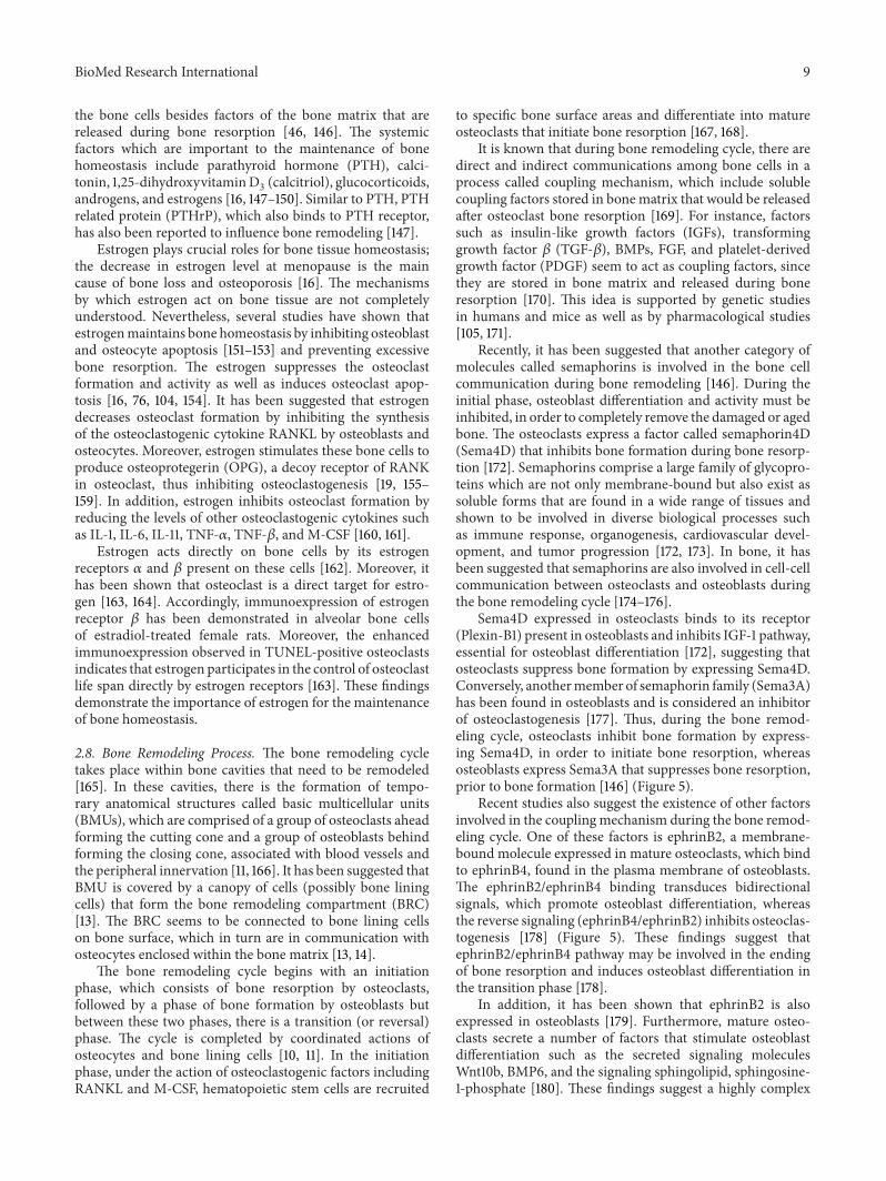
Figure 1: (a)–(d) Light micrographs of portions of alveolar bone of rats. (a) HE-stained section showing a portion of a bony trabecula (B).Polarized osteoblasts (Ob) and giant multinucleated osteoclasts (Oc) are observed in the bone surface; osteocyte (Ot) surrounding bonematrix is also observed. (b) Section subjected to immunohistochemistry for osteocalcin detection and counterstained with hematoxylin.Note osteocalcin-positive osteoblasts (arrows) on the surface of a bony trabecula (B). BV: blood vessel. (c) Undecalcified section subjected tothe Gomori method for the detection of alkaline phosphatase, evidencing a portion of bone matrix (B) positive to the alkaline phosphatase(in brown/black). Ob: osteoblasts. (d) Undecalcified section subjected to the von Kossa method for calcium detection (brown/dark color).von Kossa-positive bone matrix (B) is observed; some positive granules (arrow) can also be observed on the surface of the bone trabeculae.Scale bar: 15𝜇m.
necessary and depends on the action of several local andsystemic factors including hormones, cytokines, chemokines,and biomechanical stimulation [18–20].
Recent studies have shown that bone influences theactivity of other organs and the bone is also influenced byother organs and systems of the body [21], providing newinsights and evidencing the complexity and dynamic natureof bone tissue.
In this review we will address the current data about bonecells biology, bone matrix, and the factors that influence thebone remodeling process. Moreover, we will briefly discussthe role of estrogen on bone tissue under physiological andpathological conditions.
2. Bone Cells
2.1. Osteoblasts. Osteoblasts are cuboidal cells that are locatedalong the bone surface comprising 4–6% of the total residentbone cells and are largely known for their bone formingfunction [22].These cells showmorphological characteristicsof protein synthesizing cells, including abundant rough endo-plasmic reticulum and prominent Golgi apparatus, as well
as various secretory vesicles [22, 23]. As polarized cells, theosteoblasts secrete the osteoid toward the bone matrix [24](Figures 1(a), 1(b), and 2(a)).
Osteoblasts are derived from mesenchymal stem cells(MSC). The commitment of MSC towards the osteopro-genitor lineage requires the expression of specific genes,following timely programmed steps, including the synthesisof bone morphogenetic proteins (BMPs) and members ofthe Wingless (Wnt) pathways [25]. The expressions of Runt-related transcription factors 2,Distal-less homeobox 5 (Dlx5),and osterix (Osx) are crucial for osteoblast differentiation[22, 26]. Additionally, Runx2 is a master gene of osteoblastdifferentiation, as demonstrated by the fact that Runx2-nullmice are devoid of osteoblasts [26, 27]. Runx2 has demon-strated to upregulate osteoblast-related genes such as ColIA1,ALP, BSP, BGLAP, and OCN [28].
Once a pool of osteoblast progenitors expressing Runx2and ColIA1 has been established during osteoblast differenti-ation, there is a proliferation phase. In this phase, osteoblastprogenitors show alkaline phosphatase (ALP) activity, andare considered preosteoblasts [22]. The transition of pre-osteoblasts to mature osteoblasts is characterized by an

BioMed Research International 3
BOb
Otd
Otd
B
ObOb
B
(a)
B
N
N
BLC
OtdOtd
(b)
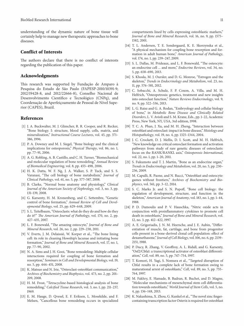
Figure 2: Electron micrographs of portions of alveolar bone of rats.(a) Oteoblasts exhibiting abundant rough endoplasmic reticulumare observed adjacent to the bone (B) surface. A layer of bundlesof collagen fibrils situated between osteoblasts (Ob) and calcifiedbone surface (B) constitutes the osteoid (Otd). Scale bar: 2.7 𝜇m. (b)Bone lining cells (BLC) exhibiting scarce cytoplasm are situated onthe osteoid surface (Otd). Bone lining cells (BLC) extend some thincytoplasmic projections (arrows) towards the osteoid (Otd). Scalebar: 2 𝜇m. N: nucleus.
increase in the expression of Osx and in the secretion of bonematrix proteins such as osteocalcin (OCN), bone sialopro-tein (BSP) I/II, and collagen type I. Moreover, the osteoblastsundergo morphological changes, becoming large and cu-boidal cells [26, 29–31].
There is evidence that other factors such as fibroblastgrowth factor (FGF), microRNAs, and connexin 43 playimportant roles in the osteoblast differentiation [32–35].FGF-2 knockoutmice showed a decreased bonemass coupledto increase of adipocytes in the bone marrow, indicatingthe participation of FGFs in the osteoblast differentiation[34]. It has also been demonstrated that FGF-18 upregulatesosteoblast differentiation in an autocrine mechanism [36].MicroRNAs are involved in the regulation of gene expressionin many cell types, including osteoblasts, in which somemic-roRNAs stimulate and others inhibit osteoblast differentia-tion [37, 38]. Connexin 43 is known to be the main con-nexin in bone [35]. The mutation in the gene encoding con-nexin 43 impairs osteoblast differentiation and causes skeletalmalformation in mouse [39].
The synthesis of bone matrix by osteoblasts occurs intwo main steps: deposition of organic matrix and its subse-quent mineralization (Figures 1(b)–1(d)). In the first step, theosteoblasts secrete collagen proteins, mainly type I collagen,noncollagen proteins (OCN, osteonectin, BSP II, and osteo-pontin), and proteoglycan including decorin and biglycan,which form the organic matrix. Thereafter, mineralizationof bone matrix takes place into two phases: the vesicular
and the fibrillar phases [40, 41]. The vesicular phase occurswhen portions with a variable diameter ranging from 30 to200 nm, called matrix vesicles, are released from the apicalmembrane domain of the osteoblasts into the newly formedbone matrix in which they bind to proteoglycans and otherorganic components. Because of its negative charge, thesulphated proteoglycans immobilize calcium ions that arestored within the matrix vesicles [41, 42]. When osteoblastssecrete enzymes that degrade the proteoglycans, the calciumions are released from the proteoglycans and cross thecalcium channels presented in thematrix vesicles membrane.These channels are formed by proteins called annexins [40].
On the other hand, phosphate-containing compounds aredegraded by the ALP secreted by osteoblasts, releasing phos-phate ions inside the matrix vesicles. Then, the phosphateand calcium ions inside the vesicles nucleate, forming thehydroxyapatite crystals [43]. The fibrillar phase occurs whenthe supersaturation of calcium and phosphate ions inside thematrix vesicles leads to the rupture of these structures andthe hydroxyapatite crystals spread to the surrounding matrix[44, 45].
Mature osteoblasts appear as a single layer of cuboidalcells containing abundant rough endoplasmic reticulum andlarge Golgi complex (Figures 2(a) and 3(a)). Some of theseosteoblasts show cytoplasmic processes towards the bonematrix and reach the osteocyte processes [46]. At this stage,the mature osteoblasts can undergo apoptosis or becomeosteocytes or bone lining cells [47, 48]. Interestingly, round/ovoid structures containing dense bodies and TUNEL-pos-itive structures have been observed inside osteoblast vac-uoles.These findings suggest that besides professional phago-cytes, osteoblasts are also able to engulf anddegrade apoptoticbodies during alveolar bone formation [49].
2.2. Bone Lining Cells. Bone lining cells are quiescent flat-shaped osteoblasts that cover the bone surfaces, where neitherbone resorption nor bone formation occurs [50]. Thesecells exhibit a thin and flat nuclear profile; its cytoplasmextends along the bone surface and displays few cytoplasmicorganelles such as profiles of rough endoplasmic reticulumand Golgi apparatus [50] (Figure 2(b)). Some of these cellsshow processes extending into canaliculi, and gap junctionsare also observed between adjacent bone lining cells andbetween these cells and osteocytes [50, 51].
The secretory activity of bone lining cells depends on thebone physiological status, whereby these cells can reacquiretheir secretory activity, enhancing their size and adoptinga cuboidal appearance [52]. Bone lining cells functions arenot completely understood, but it has been shown thatthese cells prevent the direct interaction between osteoclastsand bone matrix, when bone resorption should not occur,and also participate in osteoclast differentiation, producingosteoprotegerin (OPG) and the receptor activator of nuclearfactor kappa-B ligand (RANKL) [14, 53]. Moreover, the bonelining cells, together with other bone cells, are an importantcomponent of the BMU, an anatomical structure that ispresent during the bone remodeling cycle [9].

4 BioMed Research International
Ot
Ot
BV
BLC
Ob
BLC
B
BVOb
B
(a)
*
*
Ot
Ot
(b)
Ot
Ot
B
(c)
Ot N
La
Ca
B
(d)
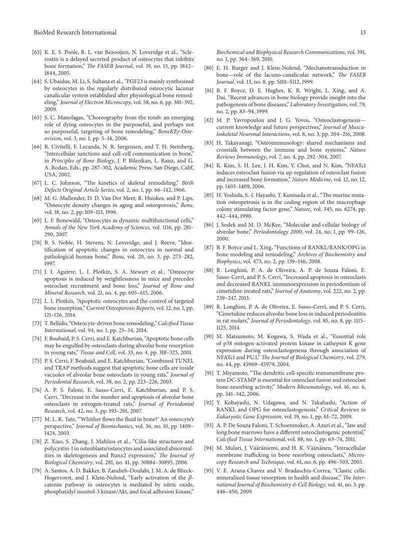
Figure 3: Light (a and b) and electron micrographs of portions of alveolar bone rats. (a) a semithin section stained with toluidine blueshowing a portion of a bony trabecula (B). Osteoblasts (Ob) and bone lining cells (BLC) are present on bone surface while osteocytes (Ot)are observed entrapped in the bone matrix. BV: blood vessels. Scale bar: 15𝜇m. (b) Section subjected to the silver impregnation method.Note the cytoplasmic processes (arrows) of the osteocytes (Ot) connecting them with each other. Scale bar: 15𝜇m. (c) Scanning electronmicrograph showing two osteocytes (Ot) surrounded by bonematrix (B). Note that the cytoplasmic processes (arrows) are observed betweenthe osteocytes (Ot) forming an interconnected network. Scale bar: 2𝜇m. (d) Transmission electron micrograph showing a typical osteocyte(Ot) inside a lacuna (La) in the bonematrix (B), with its cytoplasmic processes (arrows) inside the canaliculi (Ca). Scale bar: 2 𝜇m.N: nucleus.
2.3. Osteocytes. Osteocytes, which comprise 90–95% of thetotal bone cells, are the most abundant and long-lived cells,with a lifespan of up to 25 years [54]. Different from oste-oblasts and osteoclasts, which have been defined by theirrespective functions during bone formation and bone resorp-tion, osteocytes were earlier defined by their morphologyand location. For decades, due to difficulties in isolatingosteocytes from bone matrix led to the erroneous notion thatthese cells would be passive cells, and their functions weremisinterpreted [55]. The development of new technologiessuch as the identification of osteocyte-specific markers, newanimal models, development of techniques for bone cellisolation and culture, and the establishment of phenotypicallystable cell lines led to the improvement of the understandingof osteocyte biology. In fact, it has been recognized that thesecells play numerous important functions in bone [8].
The osteocytes are located within lacunae surroundedby mineralized bone matrix, wherein they show a dendriticmorphology [15, 55, 56] (Figures 3(a)–3(d)).Themorphologyof embedded osteocytes differs depending on the bone type.For instance, osteocytes from trabecular bone are more
rounded than osteocytes from cortical bone, which displayan elongated morphology [57].
Osteocytes are derived from MSCs lineage through oste-oblast differentiation. In this process, four recognizable stageshave been proposed: osteoid-osteocyte, preosteocyte, youngosteocyte, and mature osteocyte [54]. At the end of abone formation cycle, a subpopulation of osteoblasts becomesosteocytes incorporated into the bone matrix. This process isaccompanied by conspicuous morphological and ultrastruc-tural changes, including the reduction of the round osteoblastsize. The number of organelles such as rough endoplasmicreticulum andGolgi apparatus decreases, and the nucleus-to-cytoplasm ratio increases, which correspond to a decrease inthe protein synthesis and secretion [58].
During osteoblast/osteocyte transition, cytoplasmic pro-cess starts to emerge before the osteocytes have been encasedinto the bone matrix [22]. The mechanisms involved inthe development of osteocyte cytoplasmic processes are notwell understood. However, the protein E11/gp38, also calledpodoplanin may have an important role. E11/gp38 is highlyexpressed in embedding or recently embedded osteocytes,

BioMed Research International 5
similarly to other cell types with dendritic morphology suchas podocytes, type II lung alveolar cells, and cells of thechoroid plexus [59]. It has been suggested that E11/gp38 usesenergy from GTPase activity to interact with cytoskeletalcomponents andmolecules involved in cell motility, wherebyregulate actin cytoskeleton dynamics [60, 61]. Accordingly,inhibition of E11/gp38 expression in osteocyte-like MLO-Y4cells has been shown to block dendrite elongation, suggestingthat E11/gp38 is implicated in dendrite formation in osteo-cytes [59].
Once the stage of mature osteocyte totally entrappedwithin mineralized bone matrix is accomplished, several ofthe previously expressed osteoblast markers such as OCN,BSPII, collagen type I, and ALP are downregulated. On theother hand, osteocyte markers including dentine matrix pro-tein 1 (DMP1) and sclerostin are highly expressed [8, 62–64].
Whereas the osteocyte cell body is located inside thelacuna, its cytoplasmic processes (up to 50 per each cell)cross tiny tunnels that originate from the lacuna space calledcanaliculi, forming the osteocyte lacunocanalicular system[65] (Figures 3(b)–3(d)). These cytoplasmic processes areconnected to other neighboring osteocytes processes by gapjunctions, as well as to cytoplasmic processes of osteoblastsand bone lining cells on the bone surface, facilitating theintercellular transport of small signaling molecules suchas prostaglandins and nitric oxide among these cells [66].In addition, the osteocyte lacunocanalicular system is inclose proximity to the vascular supply, whereby oxygen andnutrients achieve osteocytes [15].
It has been estimated that osteocyte surface is 400-foldlarger than that of the all Haversian and Volkmann systemsand more than 100-fold larger than the trabecular bonesurface [67, 68].The cell-cell communication is also achievedby interstitial fluid that flows between the osteocytes pro-cesses and canaliculi [68]. By the lacunocanalicular system(Figure 3(b)), the osteocytes act as mechanosensors as theirinterconnected network has the capacity to detectmechanicalpressures and loads, thereby helping the adaptation of boneto daily mechanical forces [55]. By this way, the osteocytesseem to act as orchestrators of bone remodeling, throughregulation of osteoblast and osteoclast activities [15, 69].Moreover, osteocyte apoptosis has been recognized as achemotactic signal to osteoclastic bone resorption [70–73].In agreement, it has been shown that during bone resorption,apoptotic osteocytes are engulfed by osteoclasts [74–76].
The mechanosensitive function of osteocytes is accom-plished due to the strategic location of these cells withinbone matrix. Thus, the shape and spatial arrangement of theosteocytes are in agreement with their sensing and signaltransport functions, promoting the translation of mechanicalstimuli into biochemical signals, a phenomenon that is calledpiezoelectric effect [77].Themechanisms and components bywhich osteocytes convert mechanical stimuli to biochemicalsignals are not well known. However, two mechanisms havebeen proposed. One of them is that there is a protein complexformed by a cilium and its associated proteins PolyCystins 1and 2, which has been suggested to be crucial for osteocytemechanosensing and for osteoblast/osteocyte-mediated boneformation [78]. The second mechanism involves osteocyte
cytoskeleton components, including focal adhesion proteincomplex and its multiple actin-associated proteins such aspaxillin, vinculin, talin, and zyxin [79]. Upon mechanicalstimulation, osteocytes produce several secondary messen-gers, for example, ATP, nitric oxide (NO), Ca2+, and pros-taglandins (PGE
2and PGI
2,) which influence bone physiol-
ogy [8, 80]. Independently of the mechanism involved, it isimportant to mention that the mechanosensitive function ofosteocytes is possible due to the intricate canalicular network,which allows the communication among bone cells.
2.4. Osteoclasts. Osteoclasts are terminally differentiatedmultinucleated cells (Figures 4(a)–4(d)), which originatefrom mononuclear cells of the hematopoietic stem celllineage, under the influence of several factors. Among thesefactors the macrophage colony-stimulating factor (M-CSF),secreted by osteoprogenitor mesenchymal cells and oste-oblasts [81], and RANK ligand, secreted by osteoblasts,osteocytes, and stromal cells, are included [20]. Together,these factors promote the activation of transcription factors[81, 82] and gene expression in osteoclasts [83, 84].
M-CSF binds to its receptor (cFMS) present in osteo-clast precursors, which stimulates their proliferation andinhibits their apoptosis [82, 85]. RANKL is a crucial fac-tor for osteoclastogenesis and is expressed by osteoblasts,osteocytes, and stromal cells. When it binds to its recep-tor RANK in osteoclast precursors, osteoclast formation isinduced [86]. On the other hand, another factor called oste-oprotegerin (OPG), which is produced by a wide rangeof cells including osteoblasts, stromal cells, and gingivaland periodontal fibroblasts [87–89], binds to RANKL, pre-venting the RANK/RANKL interaction and, consequently,inhibiting the osteoclastogenesis [87] (Figure 5). Thus, theRANKL/RANK/OPG system is a key mediator of osteoclas-togenesis [19, 86, 89].
The RANKL/RANK interaction also promotes theexpression of other osteoclastogenic factors such as NFATc1and DC-STAMP. By interacting with the transcriptionfactors PU.1, cFos, and MITF, NFATc1 regulates osteoclast-specific genes including TRAP and cathepsin K, which arecrucial for osteoclast activity [90]. Under the influence ofthe RANKL/RANK interaction, NFATc1 also induces theexpression of DC-STAMP, which is crucial for the fusion ofosteoclast precursors [91, 92].
Despite these osteoclastogenic factors having been welldefined, it has recently been demonstrated that the osteo-clastogenic potential may differ depending on the bone siteconsidered. It has been reported that osteoclasts from longbone marrow are formed faster than in the jaw.This differentdynamic of osteoclastogenesis possibly could be, due to thecellular composition of the bone-site specific marrow [93].
During bone remodeling osteoclasts polarize; then, fourtypes of osteoclast membrane domains can be observed: thesealing zone and ruffled border that are in contact with thebonematrix (Figures 4(b) and 4(d)), as well as the basolateraland functional secretory domains, which are not in contactwith the bone matrix [94, 95]. Polarization of osteoclastsduring bone resorption involves rearrangement of the actin
6 BioMed Research International
Oc
OtBV B
Oc
(a)
B
NN
Oc
VV
RBRB
V
(b)
B
Ot
Oc
Ap
Oc1
(c)
OcN
N
B
V
Ap
CZCZRB
(d)
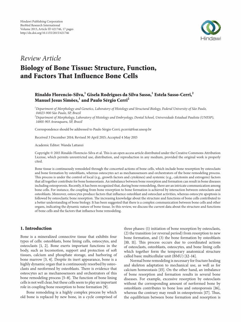
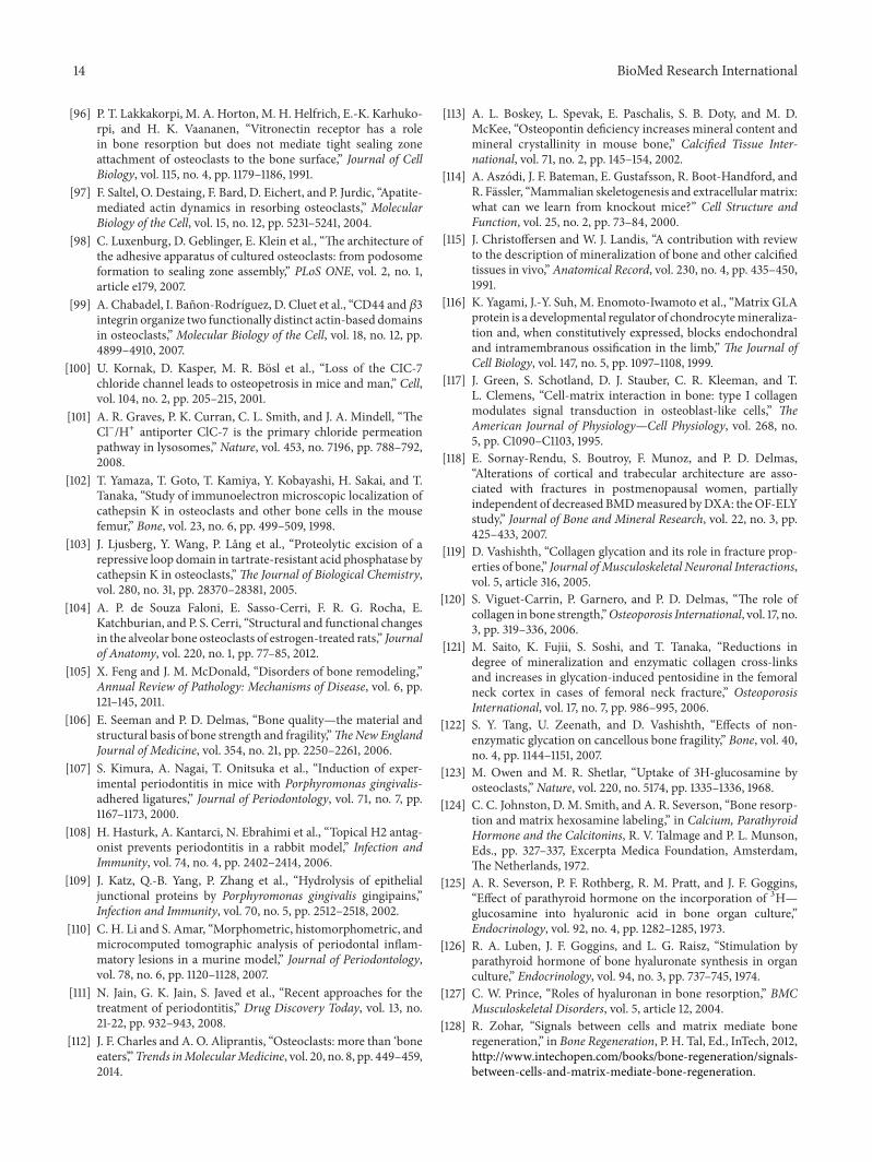
Figure 4: Light (a and c) and electron micrographs (b and d) of portions of alveolar bone of rats. In (a) tartrate-resistant acid phosphatase(TRAP) activity (in red color) is observed in the cytoplasm of osteoclasts (OC) adjacent to the alveolar bone (B) surface. Note that in theopposite side of the bony trabecula B is covered by large and polarized osteoblasts (Ob). Ot, osteocytes (Ot); BV: blood vessel. Bar: 40 𝜇m. (b)Multinucleated osteoclast (OC) shows evident ruffled border (RB) adjacent to the excavated bone surface (arrows). Several vacuoles (V) areobserved in the cytoplasm adjacent to ruffled border (RB). N: nucleus. Bar: 4𝜇m. (c) Portions of TRAP-positive osteoclasts (Oc and Oc
1) are
observed in a resorbing bone lacuna. A round cell (Ap) with condensed irregular blocks of chromatin, typical apoptotic cell, is observed insidea large vacuole of the Oc
1. B: bone matrix; Ot: osteocyte. Bar: 15𝜇m. (d) An osteoclast (Oc) showing ruffled border (RB) and clear zone (CZ)
is in close juxtaposition to the excavation of the bone surface (arrows), that is, Howship lacuna. Vacuoles (V) with varied size are present nextto the ruffled border (RB); one of them contains a round cell with masses of condensed chromatin (Ap), typical of cell undergoing apoptosis.B: bone matrix; N: nucleus. Bar: 3 𝜇m.
cytoskeleton, in which an F-actin ring that comprises a densecontinuous zone of highly dynamic podosome is formed andconsequently an area of membrane that develop into theruffled border is isolated. It is important tomention that thesedomains are only formed when osteoclasts are in contactwith extracellular mineralized matrix, in a process which𝛼v𝛽3-integrin, as well as the CD44, mediates the attachmentof the osteoclast podosomes to the bone surface [96–99].Ultrastructurally, the ruffled border is a membrane domainformed by microvilli, which is isolated from the surroundedtissue by the clear zone, also known as sealing zone. Theclear zone is an area devoid of organelles located in theperiphery of the osteoclast adjacent to the bone matrix [98].This sealing zone is formed by an actin ring and several otherproteins, including actin, talin, vinculin, paxillin, tensin,and actin-associated proteins such as 𝛼-actinin, fimbrin,gelsolin, and dynamin [95]. The 𝛼v𝛽3-integrin binds to non-collagenous bone matrix containing-RGD sequence such asbone sialoprotein, osteopontin, and vitronectin, establishinga peripheric sealing that delimits the central region, where theruffled border is located [98] (Figures 4(b)–4(d)).
The maintenance of the ruffled border is also essen-tial for osteoclast activity; this structure is formed due tointense trafficking of lysosomal and endosomal components.In the ruffled border, there is a vacuolar-type H+-ATPase(V-ATPase), which helps to acidify the resorption lacunaand hence to enable dissolution of hydroxyapatite crystals[20, 100, 101]. In this region, protons and enzymes, suchas tartrate-resistant acid phosphatase (TRAP), cathepsin K,and matrix metalloproteinase-9 (MMP-9) are transportedinto a compartment called Howship lacuna leading to bonedegradation [94, 101–104] (Figure 5). The products of thisdegradation are then endocytosed across the ruffled borderand transcytosed to the functional secretory domain at theplasma membrane [7, 95].
Abnormal increase in osteoclast formation and activityleads to some bone diseases such as osteoporosis, whereresorption exceeds formation causing decreased bone densityand increased bone fractures [105]. In some pathologic condi-tions including bone metastases and inflammatory arthritis,abnormal osteoclast activation results in periarticular ero-sions and painful osteolytic lesions, respectively [83, 105, 106].

BioMed Research International 7
CZ CZ
RB
Cp
TRAP
CAII
RANKL RANK
OPGSclerostin, DKK-1
PGE2, NO, IGF-1
RANK RANKL
OPG
Sema4DSema3A
Eph2 Eph4
Cx3
Oc
N
N
Ob
Ot
Col1OCNOSNOSPBSPBMP
BLC
Cx3
HL
Cx3
Ot
B
B
5
HCO3−
Cl−
Cl−
Cl−
CO2 + H2O
H+ H+
H+MMP-9
HCO3 + H
Figure 5: Schematic summary of bone tissue showing bone cells and the relationships among them and with bone matrix (B). Osteoclast(Oc) activation occurs after binding of RANKL to its receptor RANK, present in the membrane of osteoclast precursors. Then, osteoclastbecomes polarized through its cytoskeleton reorganization; the ruffled border (RB) and clear zone (CZ) are membrane specializationsobserved in the portion of the osteoclast juxtaposed to the bone resorption surface, Howship lacuna (HL). Dissolution of hydroxyapatiteoccurs in the bone surface adjacent to the ruffled border (RF) upon its acidification due to pumping of hydrogen ions (H+) to the HL. H+and ions bicarbonate (HCO3
−) originate from the cleavage of carbonic acid (H2CO3) under the action of carbonic anhydrase II (CAII).
After dissolution of mineral phase, osteoclast (Oc) releases cathepsin (Cp), matrix metalloproteinase-9 (MMP-9), and tartrate-resistantacid phosphatase (TRAP) that degrade the organic matrix. EphrinB2 (Eph2) present in osteoclast membrane binds to ephrinB4 (Eph4)in osteoblast (Ob) membrane, promoting its differentiation, whereas the reverse signaling (ephrinB4/ephrinB2) inhibits osteoclastogenesis.Sema4Dproduced by osteoclasts inhibits osteoblasts, while Sema3A secreted by osteoblasts inhibits osteoclasts. Osteoblasts (Ob) also producereceptor activator of nuclear factor KB (RANKL) and osteoprotegerin (OPG), which increase and decrease osteoclastogenesis, respectively.Osteoblasts (Ob) secrete collagenous (Col1) and noncollagenous proteins such as osteocalcin (OCN), osteopontin (OSP), osteonectin (OSN),bone sialoprotein (BSP), and bone morphogenetic proteins (BMP). Osteocytes (Ot) are located within lacunae surrounded by mineralizedbone matrix (B). Its cytoplasmic processes cross canaliculi to make connection with other neighboring osteocytes processes by gap junctions,mainly composed by connexin 43 (Cx3), as well as to cytoplasmic processes of osteoblasts (Ob) and bone lining cells (BLC) on bone surface.RANKL secreted by osteocytes stimulates osteoclastogenesis, while prostaglandin E
2(PGE2), nitric oxide (NO), and insulin-like growth
factor (IGF) stimulate osteoblast activity. Conversely, osteocytes produce OPG that inhibits osteoclastogenesis; moreover, osteocytes producesclerostin and dickkopf WNT signaling pathway inhibitor (DKK-1) that decrease osteoblast activity.
In periodontitis, a disease of the periodontium caused by bac-terial proliferation [107, 108] induces themigration of inflam-matory cells. These cells produce chemical mediators suchas IL-6 and RANKL that stimulate the migration of osteo-clasts [89, 109, 110]. As a result, an abnormal increased boneresorption occurs in the alveolar bone, contributing to theloss of the insertions of the teeth and to the progression ofperiodontitis [89, 111].
On the other hand, in osteopetrosis, which is a rare bonedisease, genetic mutations that affect formation and resorp-tion functions in osteoclasts lead to decreased bone resorp-tion, resulting in a disproportionate accumulation of bone
mass [17]. These diseases demonstrate the importance of thenormal bone remodeling process for themaintenance of bonehomeostasis.
Furthermore, there is evidence that osteoclasts displayseveral other functions. For example, it has been shown thatosteoclasts produce factors called clastokines that controlosteoblast during the bone remodeling cycle, which will bediscussed below.Other recent evidence is that osteoclastsmayalso directly regulate the hematopoietic stem cell niche [112].These findings indicate that osteoclasts are not only boneresorbing cells, but also a source of cytokines that influencethe activity of other cells.

8 BioMed Research International
2.5. Extracellular Bone Matrix. Bone is composed by inor-ganic salts and organic matrix [113]. The organic matrix con-tains collagenous proteins (90%), predominantly type I colla-gen, andnoncollagenous proteins including osteocalcin, oste-onectin, osteopontin, fibronectin and bone sialoprotein II,bone morphogenetic proteins (BMPs), and growth factors[114]. There are also small leucine-rich proteoglycans includ-ing decorin, biglycan, lumican, osteoaderin, and seric pro-teins [114–116].
The inorganic material of bone consists predominantlyof phosphate and calcium ions; however, significant amountsof bicarbonate, sodium, potassium, citrate, magnesium, car-bonate, fluorite, zinc, barium, and strontium are also present[1, 2]. Calcium and phosphate ions nucleate to form thehydroxyapatite crystals, which are represented by the chem-ical formula Ca
10(PO4)6(OH)2. Together with collagen, the
noncollagenous matrix proteins form a scaffold for hydrox-yapatite deposition and such association is responsible for thetypical stiffness and resistance of bone tissue [4].
Bone matrix constitutes a complex and organized frame-work that provides mechanical support and exerts essentialrole in the bone homeostasis. The bone matrix can releaseseveral molecules that interfere in the bone cells activityand, consequently, has a participation in the bone remod-eling [117]. Once loss of bone mass alone is insufficient tocause bone fractures [118], it is suggested that other factors,including changes in the bone matrix proteins and theirmodifications, are of crucial importance to the understandingand prediction of bone fractures [119]. In fact, it is known thatcollagen plays a critical role in the structure and function ofbone tissue [120].
Accordingly, it has been demonstrated that there is avariation in the concentration of bone matrix proteins withage, nutrition, disease, and antiosteoporotic treatments [119,121, 122] which may contribute to postyield deformation andfracture of bone [119]. For instance, in vivo and in vitrostudies have reported that the increase in hyaluronic acidsynthesis after parathyroid hormone (PTH) treatment wasrelated to a subsequent bone resorption [123–127] suggestinga possible relationship between hyaluronic acid synthesis andthe increase in osteoclast activity.
2.6. Interactions between Bone Cells and Bone Matrix. Aspreviously discussed, bone matrix does not only providessupport for bone cells, but also has a key role in regu-lating the activity of bone cells through several adhesionmolecules [117, 128]. Integrins are themost common adhesionmolecules involved in the interaction between bone cells andbone matrix [129]. Osteoblasts make interactions with bonematrix by integrins, which recognize and bind to RGD andother sequences present in bone matrix proteins includingosteopontin, fibronectin, collagen, osteopontin, and bonesialoprotein [130, 131]. The most common integrins presentin osteoblasts are 𝛼1𝛽1, 𝛼2𝛽1, and 𝛼5𝛽1 [132]. These proteinsalso play an important role in osteoblast organization on thebone surface during osteoid synthesis [129].
On the other hand, the interaction between osteoclastsand bone matrix is essential for osteoclast function, sinceas previously mentioned, bone resorption occurs only when
osteoclasts bind to mineralized bone surface [97]. Thus,during bone resorption osteoclasts express 𝛼v𝛽3 and 𝛼2𝛽1integrins to interact with the extracellular matrix, in whichthe former bind to bone-enriched RGD-containing proteins,such as bone sialoprotein and osteopontin, whereas 𝛽1 inte-grins bind to collagenfibrils [133, 134].Despite these bindings,osteoclasts are highly motile even active resorption and, asmigrating cells, osteoclasts do not express cadherins. How-ever, it has been demonstrated that cadherins provide inti-mate contact between osteoclast precursors and stromal cells,which express crucial growth factors for osteoclast differenti-ation [135].
Integrins play a mediating role in osteocyte-bone matrixinteractions. These interactions are essential for the mechan-osensitive function of these cells, whereby signals inducedby tissue deformation are generated and amplified [136]. Itis still not clear which integrins are involved, but it has beensuggested that 𝛽3 and 𝛽1 integrins are involved in osteocyte-bone matrix interaction [137, 138]. These interactions occurbetween osteocyte body and the bone matrix of the lacunawall as well as between canalicular wall with the osteocyteprocesses [137].
Only a narrow pericellular space filled by a fluid separatesthe osteocyte cell body and processes from a mineralizedbone matrix [58]. The space between osteocyte cell body andthe lacunar wall is approximately 0.5–1.0𝜇m wide, whereasthe distance between the membranes of osteocyte processesand the canalicular wall varies from 50 to 100 nm [139].The chemical composition of the pericellular fluid has notbeen precisely defined. However, a diverse array of macro-molecules produced by osteocytes such as osteopontin, osteo-calcin, dentin matrix protein, proteoglycans, and hyaluronicacid is present [136, 140, 141].
The osteocyte and their processes are surrounded by anonorganized pericellular matrix; delicate fibrous connec-tions were observed within the canalicular network, termed“tethers” [139]. It has been suggested that perlecan is apossible compound of these tethers [141]. Osteocyte proc-esses can also attach directly by the “hillocks,” which areprotruding structures originating from the canalicular walls.These structures form close contacts, possibly by meansof 𝛽3-integrins, with the membrane of osteocyte processes
[137, 142]. Thus, these structures seem to play a key rolein the mechanosensitive function of osteocytes, by sensingthe fluid flux movements along with the pericellular space,provoked by mechanical load forces [143]. In addition, thefluid flux movement is also essential for the bidirectionalsolute transport in the pericellular space, which influencesosteocyte signaling pathways and communication amongbone cells [144, 145].
2.7. Local and Systemic Factor That Regulate Bone Home-ostasis. Bone remodeling is a highly complex cycle that isachieved by the concerted actions of osteoblasts, osteo-cytes, osteoclasts, and bone lining cells [3]. The formation,proliferation, differentiation, and activity of these cells arecontrolled by local and systemic factors [18, 19]. The localfactors include autocrine and paracrine molecules such asgrowth factors, cytokines, and prostaglandins produced by
BioMed Research International 9
the bone cells besides factors of the bone matrix that arereleased during bone resorption [46, 146]. The systemicfactors which are important to the maintenance of bonehomeostasis include parathyroid hormone (PTH), calci-tonin, 1,25-dihydroxyvitaminD
3(calcitriol), glucocorticoids,
androgens, and estrogens [16, 147–150]. Similar to PTH, PTHrelated protein (PTHrP), which also binds to PTH receptor,has also been reported to influence bone remodeling [147].
Estrogen plays crucial roles for bone tissue homeostasis;the decrease in estrogen level at menopause is the maincause of bone loss and osteoporosis [16]. The mechanismsby which estrogen act on bone tissue are not completelyunderstood. Nevertheless, several studies have shown thatestrogenmaintains bone homeostasis by inhibiting osteoblastand osteocyte apoptosis [151–153] and preventing excessivebone resorption. The estrogen suppresses the osteoclastformation and activity as well as induces osteoclast apop-tosis [16, 76, 104, 154]. It has been suggested that estrogendecreases osteoclast formation by inhibiting the synthesisof the osteoclastogenic cytokine RANKL by osteoblasts andosteocytes. Moreover, estrogen stimulates these bone cells toproduce osteoprotegerin (OPG), a decoy receptor of RANKin osteoclast, thus inhibiting osteoclastogenesis [19, 155–159]. In addition, estrogen inhibits osteoclast formation byreducing the levels of other osteoclastogenic cytokines suchas IL-1, IL-6, IL-11, TNF-𝛼, TNF-𝛽, and M-CSF [160, 161].
Estrogen acts directly on bone cells by its estrogenreceptors 𝛼 and 𝛽 present on these cells [162]. Moreover, ithas been shown that osteoclast is a direct target for estro-gen [163, 164]. Accordingly, immunoexpression of estrogenreceptor 𝛽 has been demonstrated in alveolar bone cellsof estradiol-treated female rats. Moreover, the enhancedimmunoexpression observed in TUNEL-positive osteoclastsindicates that estrogen participates in the control of osteoclastlife span directly by estrogen receptors [163]. These findingsdemonstrate the importance of estrogen for the maintenanceof bone homeostasis.
2.8. Bone Remodeling Process. The bone remodeling cycletakes place within bone cavities that need to be remodeled[165]. In these cavities, there is the formation of tempo-rary anatomical structures called basic multicellular units(BMUs), which are comprised of a group of osteoclasts aheadforming the cutting cone and a group of osteoblasts behindforming the closing cone, associated with blood vessels andthe peripheral innervation [11, 166]. It has been suggested thatBMU is covered by a canopy of cells (possibly bone liningcells) that form the bone remodeling compartment (BRC)[13]. The BRC seems to be connected to bone lining cellson bone surface, which in turn are in communication withosteocytes enclosed within the bone matrix [13, 14].
The bone remodeling cycle begins with an initiationphase, which consists of bone resorption by osteoclasts,followed by a phase of bone formation by osteoblasts butbetween these two phases, there is a transition (or reversal)phase. The cycle is completed by coordinated actions ofosteocytes and bone lining cells [10, 11]. In the initiationphase, under the action of osteoclastogenic factors includingRANKL and M-CSF, hematopoietic stem cells are recruited
to specific bone surface areas and differentiate into matureosteoclasts that initiate bone resorption [167, 168].
It is known that during bone remodeling cycle, there aredirect and indirect communications among bone cells in aprocess called coupling mechanism, which include solublecoupling factors stored in bonematrix that would be releasedafter osteoclast bone resorption [169]. For instance, factorssuch as insulin-like growth factors (IGFs), transforminggrowth factor 𝛽 (TGF-𝛽), BMPs, FGF, and platelet-derivedgrowth factor (PDGF) seem to act as coupling factors, sincethey are stored in bone matrix and released during boneresorption [170]. This idea is supported by genetic studiesin humans and mice as well as by pharmacological studies[105, 171].
Recently, it has been suggested that another category ofmolecules called semaphorins is involved in the bone cellcommunication during bone remodeling [146]. During theinitial phase, osteoblast differentiation and activity must beinhibited, in order to completely remove the damaged or agedbone. The osteoclasts express a factor called semaphorin4D(Sema4D) that inhibits bone formation during bone resorp-tion [172]. Semaphorins comprise a large family of glycopro-teins which are not only membrane-bound but also exist assoluble forms that are found in a wide range of tissues andshown to be involved in diverse biological processes suchas immune response, organogenesis, cardiovascular devel-opment, and tumor progression [172, 173]. In bone, it hasbeen suggested that semaphorins are also involved in cell-cellcommunication between osteoclasts and osteoblasts duringthe bone remodeling cycle [174–176].
Sema4D expressed in osteoclasts binds to its receptor(Plexin-B1) present in osteoblasts and inhibits IGF-1 pathway,essential for osteoblast differentiation [172], suggesting thatosteoclasts suppress bone formation by expressing Sema4D.Conversely, anothermember of semaphorin family (Sema3A)has been found in osteoblasts and is considered an inhibitorof osteoclastogenesis [177]. Thus, during the bone remod-eling cycle, osteoclasts inhibit bone formation by express-ing Sema4D, in order to initiate bone resorption, whereasosteoblasts express Sema3A that suppresses bone resorption,prior to bone formation [146] (Figure 5).
Recent studies also suggest the existence of other factorsinvolved in the coupling mechanism during the bone remod-eling cycle. One of these factors is ephrinB2, a membrane-bound molecule expressed in mature osteoclasts, which bindto ephrinB4, found in the plasma membrane of osteoblasts.The ephrinB2/ephrinB4 binding transduces bidirectionalsignals, which promote osteoblast differentiation, whereasthe reverse signaling (ephrinB4/ephrinB2) inhibits osteoclas-togenesis [178] (Figure 5). These findings suggest thatephrinB2/ephrinB4 pathway may be involved in the endingof bone resorption and induces osteoblast differentiation inthe transition phase [178].
In addition, it has been shown that ephrinB2 is alsoexpressed in osteoblasts [179]. Furthermore, mature osteo-clasts secrete a number of factors that stimulate osteoblastdifferentiation such as the secreted signaling moleculesWnt10b, BMP6, and the signaling sphingolipid, sphingosine-1-phosphate [180]. These findings suggest a highly complex

10 BioMed Research International
mechanism of ephrins and the involvement of other factorsin osteoclast/osteoblast communication during the boneremodeling cycle. On the other hand, despite the stud-ies reporting the involvement of semaphorins and ephrinson osteoclast/osteoblast communication, the direct contactbetween mature osteoblasts and osteoclasts has not beendemonstrated in vivo and it is still controversial.
Besides osteoclasts and osteoblasts, it has been demon-strated that osteocytes play key roles during the bone remod-eling cycle [8]. In fact, under the influence of several factors,the osteocytes act as orchestrators of the bone remodelingprocess, producing factors that influence osteoblast andosteoclast activities [55] (Figure 5). For example, mechanicalloading stimulates osteocyte to produce factors that exertanabolic action on bone such as PGE
2, prostacyclin (PGI
2),
NO, and IGF-1 [181–184]. On the other hand, mechanicalunloading downregulates anabolic factors and stimulatesosteocytes to produce sclerostin and DKK-1, which areinhibitors of osteoblast activity [185–188], as well as specificfactors that stimulate local osteoclastogenesis [189]. Scle-rostin is a product of the SOST gene and is known to bea negative regulator of bone formation, by antagonizing inosteoblasts the actions of Lrp5, a key receptor of the Wnt/𝛽-catenin signaling pathway [63].
Osteocyte apoptosis has been shown to act as a chemo-tactic signal for local osteoclast recruitment [70, 150, 152, 190,191]. Accordingly, it has been reported that osteoclasts engulfapoptotic osteocytes [74, 75, 192], suggesting that osteoclastsare able to remove dying osteocytes and/or osteoblasts froma remodeling site (Figures 4(c) and 4(d)). Moreover, it isreported that the osteoclastogenic factors is also produced byviable osteocytes nearby the dying osteocytes [193]. There isevidence that osteocytes act as the main source of RANKLto promote osteoclastogenesis [167, 168], although this factorhas also been demonstrated to be produced by other celltypes such as stromal cells [194], osteoblasts, and fibroblasts[88, 89].
Thus, there are still uncertainties about the preciseosteoclastogenesis-stimulating factors produced by osteo-cytes. Recent reviews have focused on some molecules thatmay be candidates for signaling between osteocyte apoptosisand osteoclastogenesis [72, 73]. For instance, in bones sub-jected to fatigue loading, viable osteocytes near the apoptoticones express, besides high RANKL/OPG ratio, increasedlevels of vascular endothelial growth factor (VEGF) andmonocyte chemoattractant protein-1 (CCL2) promoting anincrease in local osteoclastogenesis [194, 195]. It has beensuggested that osteocytes act as the main source of RANKLto promote osteoclastogenesis [166, 167]. In addition, anincrease in RANKL/OPG ratio expressed by osteocytes wasalso observed in connexin43-deficient rats, suggesting that adisruption in cell-to-cell communication between osteocytesmay induce the release of local proosteoclastogenic cytokines[33, 196, 197]. High mobility group box protein 1 (HMGB1)[198–200] and M-CSF [201] have also been suggested to beproduced by osteocytes that stimulate osteoclast recruitmentduring bone remodeling [72, 73]. Thus, future studies arerequired to address this issue.
2.9. Endocrine Functions of Bone Tissue. The classical func-tions of bone tissue, besides locomotion, include support andprotection of soft tissues, calcium, and phosphate storage andharboring of bone marrow. Additionally, recent studies havefocused on the bone endocrine functions which are able toaffect other organs [202]. For instance, osteocalcin producedby osteoblasts has been shown to act in other organs [203].Osteocalcin can be found in twodifferent forms: carboxylatedand undercarboxylated. The carboxylated form has highaffinity to the hydroxyapatite crystals, remaining into bonematrix during its mineralization. The undercarboxylatedform shows lower affinity to minerals, due to acidification ofbone matrix during osteoclast bone resorption, and then it isferried by the bloodstream, reaching other organs [204, 205].It has been shown that the undercarboxylated osteocalcinhas some effects in pancreas, adipose tissue, testis, and thenervous system. In the pancreas, osteocalcin acts as a positiveregulator of pancreatic insulin secretion and sensitivity aswell as for the proliferation of pancreatic 𝛽-cells [110]. Inthe adipose tissue, osteocalcin stimulates adiponectin geneexpression that in turn enhances insulin sensitivity [204]. Inthe testis, osteocalcin can bind to a specific receptor in Leydigcells and enhances testosterone synthesis and, consequently,increases fertility [206]. Osteocalcin also stimulates the syn-thesis of monoamine neurotransmitters in the hippocampusand inhibits gamma-aminobutyric acid (GABA) synthesis,improving learning and memory skills [207].
Another endocrine function of bone tissue is promotedby osteocytes. These cells are able to regulate phosphatemetabolism by the production of FGF23, which acts on otherorgans including parathyroid gland and kidneys to reduce thecirculating levels of phosphates [208, 209]. Osteocytes alsoact on the immune system by modifying the microenviron-ment in primary lymphoid organs and thereby influencinglymphopoiesis [210]. Not only osteocyte but also osteoblastand osteoclast activities are known to influence the immunesystem,mainly upon bone inflammatory destruction. Indeed,the discovery of communication interplay between skeletaland immune systems led to a new field of study calledosteoimmunology [211].
3. Conclusions
The knowledge of the structural, molecular, and functionalbiology of bone is essential for the better comprehension ofthis tissue as a multicellular unit and a dynamic structurethat can also act as an endocrine tissue, a function still poorlyunderstood. In vitro and in vivo studies have demonstratedthat bone cells respond to different factors and molecules,contributing to the better understanding of bone cells plastic-ity. Additionally, bone matrix integrins-dependent bone cellsinteractions are essential for bone formation and resorption.Studies have addressed the importance of the lacunocanalic-ular system and the pericellular fluid, by which osteocytes actas mechanosensors, for the adaptation of bone to mechanicalforces. Hormones, cytokines, and factors that regulate bonecells activity, such as sclerostin, ephrinB2, and semaphoring,have played a significant role in the bone histophysiologyunder normal and pathological conditions.Thus, such deeper
BioMed Research International 11
understanding of the dynamic nature of bone tissue willcertainly help tomanage new therapeutic approaches to bonediseases.
Conflict of Interests
The authors declare that there is no conflict of interestsregarding the publication of this paper.
Acknowledgments
This research was supported by Fundacao de Amparo aPesquisa do Estado de Sao Paulo (FAPESP-2010/10391-9;2012/19428-8, and 2012/22666-8), Conselho Nacional deDesenvolvimento Cientıfico e Tecnologico (CNPq), andCoordenacao de Aperfeicoamento de Pessoal de Nıvel Supe-rior (CAPES), Brazil.
References
[1] J. A. Buckwalter, M. J. Glimcher, R. R. Cooper, and R. Recker,“Bone biology. I: structure, blood supply, cells, matrix, andmineralization,” Instructional Course Lectures, vol. 45, pp. 371–386, 1996.
[2] P. A. Downey and M. I. Siegel, “Bone biology and the clinicalimplications for osteoporosis,” Physical Therapy, vol. 86, no. 1,pp. 77–91, 2006.
[3] A. G. Robling, A. B. Castillo, and C. H. Turner, “Biomechanicaland molecular regulation of bone remodeling,” Annual Reviewof Biomedical Engineering, vol. 8, pp. 455–498, 2006.
[4] H. K. Datta, W. F. Ng, J. A. Walker, S. P. Tuck, and S. S.Varanasi, “The cell biology of bone metabolism,” Journal ofClinical Pathology, vol. 61, no. 5, pp. 577–587, 2008.
[5] B. Clarke, “Normal bone anatomy and physiology,” ClinicalJournal of the American Society of Nephrology, vol. 3, no. 3, pp.131–139, 2008.
[6] G. Karsenty, H. M. Kronenberg, and C. Settembre, “Geneticcontrol of bone formation,” Annual Review of Cell and Devel-opmental Biology, vol. 25, pp. 629–648, 2009.
[7] S. L. Teitelbaum, “Osteoclasts: what do they do and how do theydo it?” The American Journal of Pathology, vol. 170, no. 2, pp.427–435, 2007.
[8] L. F. Bonewald, “The amazing osteocyte,” Journal of Bone andMineral Research, vol. 26, no. 2, pp. 229–238, 2011.
[9] V. Everts, J. M. Delaissie, W. Korper et al., “The bone liningcell: its role in cleaning Howship’s lacunae and initiating boneformation,” Journal of Bone and Mineral Research, vol. 17, no. 1,pp. 77–90, 2002.
[10] N. A. Sims and J. H. Gooi, “Bone remodeling: Multiple cellularinteractions required for coupling of bone formation andresorption,” Seminars in Cell and Developmental Biology, vol. 19,no. 5, pp. 444–451, 2008.
[11] K. Matsuo and N. Irie, “Osteoclast-osteoblast communication,”Archives of Biochemistry and Biophysics, vol. 473, no. 2, pp. 201–209, 2008.
[12] H. M. Frost, “Tetracycline-based histological analysis of boneremodeling,” Calcified Tissue Research, vol. 3, no. 1, pp. 211–237,1969.
[13] E. M. Hauge, D. Qvesel, E. F. Eriksen, L. Mosekilde, and F.Melsen, “Cancellous bone remodeling occurs in specialized
compartments lined by cells expressing osteoblastic markers,”Journal of Bone and Mineral Research, vol. 16, no. 9, pp. 1575–1582, 2001.
[14] T. L. Andersen, T. E. Sondergaard, K. E. Skorzynska et al.,“A physical mechanism for coupling bone resorption and for-mation in adult human bone,” American Journal of Pathology,vol. 174, no. 1, pp. 239–247, 2009.
[15] S. L. Dallas, M. Prideaux, and L. F. Bonewald, “The osteocyte:an endocrine cell ... and more,” Endocrine Reviews, vol. 34, no.5, pp. 658–690, 2013.
[16] S. Khosla, M. J. Oursler, and D. G. Monroe, “Estrogen and theskeleton,” Trends in Endocrinology and Metabolism, vol. 23, no.11, pp. 576–581, 2012.
[17] C. Sobacchi, A. Schulz, F. P. Coxon, A. Villa, and M. H.Helfrich, “Osteopetrosis: genetics, treatment and new insightsinto osteoclast function,” Nature Reviews Endocrinology, vol. 9,no. 9, pp. 522–536, 2013.
[18] L. G. Raisz and G. A. Rodan, “Embryology and cellular biologyof bone,” in Metabolic Bone Disease and Clinically RelatedDisorders, L. V. Avioli and S.M. Krane, Eds., pp. 1–22, AcademicPress, New York, NY, USA, 3rd edition, 1998.
[19] T. C. A. Phan, J. Xu, and M. H. Zheng, “Interaction betweenosteoblast and osteoclast: impact in bone disease,”Histology andHistopathology, vol. 19, no. 4, pp. 1325–1344, 2004.
[20] J. C. Crockett, D. J. Mellis, D. I. Scott, and M. H. Helfrich,“New knowledge on critical osteoclast formation and activationpathways from study of rare genetic diseases of osteoclasts:focus on the RANK/RANKL axis,” Osteoporosis International,vol. 22, no. 1, pp. 1–20, 2011.
[21] S. Fukumoto and T. J. Martin, “Bone as an endocrine organ,”Trends in Endocrinology andMetabolism, vol. 20, no. 5, pp. 230–236, 2009.
[22] M. Capulli, R. Paone, and N. Rucci, “Osteoblast and osteocyte:games without frontiers,” Archives of Biochemistry and Bio-physics, vol. 561, pp. 3–12, 2014.
[23] S. C. Marks Jr. and S. N. Popoff, “Bone cell biology: theregulation of development, structure, and function in theskeleton,”American Journal of Anatomy, vol. 183, no. 1, pp. 1–44,1988.
[24] P. D. Damoulis and P. V. Hauschka, “Nitric oxide acts inconjunction with proinflammatory cytokines to promote celldeath in osteoblasts,” Journal of Bone andMineral Research, vol.12, no. 3, pp. 412–422, 1997.
[25] A. E. Grigoriadis, J. N. M. Heersche, and J. E. Aubin, “Differ-entiation of muscle, fat, cartilage, and bone from progenitorcells present in a bone-derived clonal cell population: effect ofdexamethasone,” Journal of Cell Biology, vol. 106, no. 6, pp. 2139–2151, 1988.
[26] P. Ducy, R. Zhang, V. Geoffroy, A. L. Ridall, and G. Karsenty,“Osf2/Cbfa1: a transcriptional activator of osteoblast differenti-ation,” Cell, vol. 89, no. 5, pp. 747–754, 1997.
[27] T. Komori, H. Yagi, S. Nomura et al., “Targeted disruption ofCbfa1 results in a complete lack of bone formation owing tomaturational arrest of osteoblasts,” Cell, vol. 89, no. 5, pp. 755–764, 1997.
[28] M. Fakhry, E. Hamade, B. Badran, R. Buchet, and D. Magne,“Molecular mechanisms of mesenchymal stem cell differentia-tion towards osteoblasts,”World Journal of Stem Cells, vol. 5, no.4, pp. 136–148, 2013.
[29] K. Nakashima, X. Zhou, G. Kunkel et al., “The novel zinc finger-containing transcription factorOsterix is required for osteoblast

12 BioMed Research International
differentiation and bone formation,” Cell, vol. 108, no. 1, pp. 17–29, 2002.
[30] D. A. Glass II, P. Bialek, J. D. Ahn et al., “Canonical Wntsignaling in differentiated osteoblasts controls osteoclast differ-entiation,” Developmental Cell, vol. 8, no. 5, pp. 751–764, 2005.
[31] H. Hu, M. J. Hilton, X. Tu, K. Yu, D. M. Ornitz, and F. Long,“Sequential roles of Hedgehog and Wnt signaling in osteoblastdevelopment,” Development, vol. 132, no. 1, pp. 49–60, 2005.
[32] K. Kapinas, C. Kessler, T. Ricks, G. Gronowicz, and A. M.Delany, “miR-29modulatesWnt signaling in human osteoblaststhrough a positive feedback loop,” The Journal of BiologicalChemistry, vol. 285, no. 33, pp. 25221–25231, 2010.
[33] Y. Zhang, R.-L. Xie, C. M. Croce et al., “A program of microR-NAs controls osteogenic lineage progression by targeting tran-scription factor Runx2,” Proceedings of the National Academyof Sciences of the United States of America, vol. 108, no. 24, pp.9863–9868, 2011.
[34] A. Y. Montero, Y. Okada, M. Tomita et al., “Disruption of thefibroblast growth factor-2 gene results in decreased bone massand bone formation,” The Journal of Clinical Investigation, vol.105, no. 8, pp. 1085–1093, 2000.
[35] A. M. Buo and J. P. Stains, “Gap junctional regulation of signaltransduction in bone cells,” FEBS Letters, vol. 588, no. 8, pp.1315–1321, 2014.
[36] Z. O. Hamidouche, O. Fromigue, U. Nuber et al., “Autocrinefibroblast growth factor 18 mediates dexamethasone-inducedosteogenic differentiation of murine mesenchymal stem cells,”Journal of Cellular Physiology, vol. 224, no. 2, pp. 509–515, 2010.
[37] M. Q. Hassan, Y. Maeda, H. Taipaleenmaki et al., “miR-218directs a Wnt signaling circuit to promote differentiation ofosteoblasts and osteomimicry of metastatic cancer cells,” TheJournal of Biological Chemistry, vol. 287, no. 50, pp. 42084–42092, 2012.
[38] M. Tome, P. Lopez-Romero, C. Albo et al., “miR-335 orches-trates cell proliferation, migration and differentiation in humanmesenchymal stem cells,” Cell Death and Differentiation, vol. 18,no. 6, pp. 985–995, 2011.
[39] A. M. Flenniken, L. R. Osborne, N. Anderson et al., “A Gja1missense mutation in a mouse model of oculodentodigitaldysplasia,” Development, vol. 132, no. 19, pp. 4375–4386, 2005.
[40] H. C. Anderson, “Matrix vesicles and calcification,” CurrentRheumatology Reports, vol. 5, no. 3, pp. 222–226, 2003.
[41] Y. Yoshiko, G. A. Candeliere, N. Maeda, and J. E. Aubin,“Osteoblast autonomous Pi regulation via Pit1 plays a role inbonemineralization,”Molecular andCellular Biology, vol. 27, no.12, pp. 4465–4474, 2007.
[42] V. E. Arana-Chavez, A. M. V. Soares, and E. Katchburian,“Junctions between early developing osteoblasts of rat calvariaas revealed by freeze-fracture and ultrathin section electronmicroscopy,” Archives of Histology and Cytology, vol. 58, no. 3,pp. 285–292, 1995.
[43] M. J. Glimcher, “The nature of the mineral phase in bone,”in Metabolic Bone Disease, M. J. Glimcher, Ed., pp. 23–50,Academic Press, San Diego, Calif, USA, 1998.
[44] G. Boivin and P. J. Meunier, “The degree of mineralizationof bone tissue measured by computerized quantitative contactmicroradiography,” Calcified Tissue International, vol. 70, no. 6,pp. 503–511, 2002.
[45] G. Boivin, Y. Bala, A. Doublier et al., “The role of mineralizationand organic matrix in the microhardness of bone tissue fromcontrols and osteoporotic patients,” Bone, vol. 43, no. 3, pp. 532–538, 2008.
[46] S. C.Manolagas, “Birth and death of bone cells: basic regulatorymechanisms and implications for the pathogenesis and treat-ment of osteoporosis,” Endocrine Reviews, vol. 21, no. 2, pp. 115–137, 2000.
[47] A. M. Parfitt, “Bone-forming cells in clinical conditions,” inBone, Vol 1: The Osteoblast and Osteocyte, B. K. Hall, Ed., pp.351–429, Telford Press, CRC Press, Boca Raton, Fla, USA, 1990.
[48] R. L. Jilka, R. S. Weinstein, T. Bellido, A. M. Parfitt, and S.C. Manolagas, “Osteoblast programmed cell death (apoptosis):modulation by growth factors and cytokines,” Journal of Boneand Mineral Research, vol. 13, no. 5, pp. 793–802, 1998.
[49] P. S. Cerri, “Osteoblasts engulf apoptotic bodies during alveolarbone formation in the rat maxilla,” Anatomical Record A, vol.286, no. 1, pp. 833–840, 2005.
[50] S. C. Miller, L. de Saint-Georges, B. M. Bowman, and W. S.S. Jee, “Bone lining cells: structure and function,” ScanningMicroscopy, vol. 3, no. 3, pp. 953–961, 1989.
[51] E. M. Aarden, E. H. Burger, and P. J. Nijweide, “Function ofosteocytes in bone,” Journal of Cellular Biochemistry, vol. 55, no.3, pp. 287–299, 1994.
[52] H. J. Donahue, K. J. McLeod, C. T. Rubin et al., “Cell-to-cellcommunication in osteoblastic networks: cell line-dependenthormonal regulation of gap junction function,” Journal of Boneand Mineral Research, vol. 10, no. 6, pp. 881–889, 1995.
[53] J. R. Mosley, “Osteoporosis and bone functional adaptation:mechanobiological regulation of bone architecture in growingand adult bone, a review,” Journal of Rehabilitation Research andDevelopment, vol. 37, no. 2, pp. 189–199, 2000.
[54] T. A. Franz-Odendaal, B. K.Hall, and P. E.Witten, “Buried alive:how osteoblasts become osteocytes,” Developmental Dynamics,vol. 235, no. 1, pp. 176–190, 2006.
[55] G. Y. Rochefort, S. Pallu, and C. L. Benhamou, “Osteocyte: theunrecognized side of bone tissue,” Osteoporosis International,vol. 21, no. 9, pp. 1457–1469, 2010.
[56] C. Palumbo, S. Palazzini, D. Zaffe, and G. Marotti, “Osteocytedifferentiation in the tibia of newborn rabbit: an ultrastructuralstudy of the formation of cytoplasmic processes,” Acta Anatom-ica, vol. 137, no. 4, pp. 350–358, 1990.
[57] J. D. Currey, “The many adaptations of bone,” Journal ofBiomechanics, vol. 36, no. 10, pp. 1487–1495, 2003.
[58] M. B. Schaffler, W.-Y. Cheung, R. Majeska, and O. Kennedy,“Osteocytes: master orchestrators of bone,” Calcified TissueInternational, vol. 94, no. 1, pp. 5–24, 2014.
[59] K. Zhang, C. Barragan-Adjemian, L. Ye et al., “E11/gp38 selectiveexpression in osteocytes: regulation by mechanical strain androle in dendrite elongation,”Molecular and Cellular Biology, vol.26, no. 12, pp. 4539–4552, 2006.
[60] A.Wetterwald, W. Hofstetter, M. G. Cecchini et al., “Character-ization and cloning of the E11 antigen, a marker expressed byrat osteoblasts and osteocytes,” Bone, vol. 18, no. 2, pp. 125–132,1996.
[61] E. Schulze, M. Witt, M. Kasper, C. W. G. M. Lowik, andR. H. W. Funk, “Immunohistochemical investigations on thedifferentiation marker protein E11 in rat calvaria, calvaria cellculture and the osteoblastic cell line ROS 17/2.8,”Histochemistryand Cell Biology, vol. 111, no. 1, pp. 61–69, 1999.
[62] Y. Mikuni-Takagaki, Y. Kakai, M. Satoyoshi et al., “Matrixmineralization and the differentiation of osteocyte-like cells inculture,” Journal of Bone andMineral Research, vol. 10, no. 2, pp.231–242, 1995.
BioMed Research International 13
[63] K. E. S. Poole, R. L. van Bezooijen, N. Loveridge et al., “Scle-rostin is a delayed secreted product of osteocytes that inhibitsbone formation,” The FASEB Journal, vol. 19, no. 13, pp. 1842–1844, 2005.
[64] S. Ubaidus,M. Li, S. Sultana et al., “FGF23 ismainly synthesizedby osteocytes in the regularly distributed osteocytic lacunarcanalicular system established after physiological bone remod-eling,” Journal of ElectronMicroscopy, vol. 58, no. 6, pp. 381–392,2009.
[65] S. C. Manolagas, “Choreography from the tomb: an emergingrole of dying osteocytes in the purposeful, and perhaps notso purposeful, targeting of bone remodeling,” BoneKEy-Oste-ovision, vol. 3, no. 1, pp. 5–14, 2006.
[66] R. Civitelli, F. Lecanda, N. R. Jørgensen, and T. H. Steinberg,“Intercellular junctions and cell-cell communication in bone,”in Principles of Bone Biology, J. P. Bilezikan, L. Raisz, and G.A. Rodan, Eds., pp. 287–302, Academic Press, San Diego, Calif,USA, 2002.
[67] L. C. Johnson, “The kinetics of skeletal remodeling,” BirthDefects Original Article Series, vol. 2, no. 1, pp. 66–142, 1966.
[68] M. G. Mullender, D. D. Van Der Meer, R. Huiskes, and P. Lips,“Osteocyte density changes in aging and osteoporosis,” Bone,vol. 18, no. 2, pp. 109–113, 1996.
[69] L. F. Bonewald, “Osteocytes as dynamic multifunctional cells,”Annals of the New York Academy of Sciences, vol. 1116, pp. 281–290, 2007.
[70] B. S. Noble, H. Stevens, N. Loveridge, and J. Reeve, “Iden-tification of apoptotic changes in osteocytes in normal andpathological human bone,” Bone, vol. 20, no. 3, pp. 273–282,1997.
[71] J. I. Aguirre, L. I. Plotkin, S. A. Stewart et al., “Osteocyteapoptosis is induced by weightlessness in mice and precedesosteoclast recruitment and bone loss,” Journal of Bone andMineral Research, vol. 21, no. 4, pp. 605–615, 2006.
[72] L. I. Plotkin, “Apoptotic osteocytes and the control of targetedbone resorption,”Current Osteoporosis Reports, vol. 12, no. 1, pp.121–126, 2014.
[73] T. Bellido, “Osteocyte-driven bone remodeling,”Calcified TissueInternational, vol. 94, no. 1, pp. 25–34, 2014.
[74] F. Boabaid, P. S. Cerri, and E. Katchburian, “Apoptotic bone cellsmay be engulfed by osteoclasts during alveolar bone resorptionin young rats,” Tissue and Cell, vol. 33, no. 4, pp. 318–325, 2001.
[75] P. S. Cerri, F. Boabaid, and E. Katchburian, “Combined TUNELand TRAP methods suggest that apoptotic bone cells are insidevacuoles of alveolar bone osteoclasts in young rats,” Journal ofPeriodontal Research, vol. 38, no. 2, pp. 223–226, 2003.
[76] A. P. S. Faloni, E. Sasso-Cerri, E. Katchburian, and P. S.Cerri, “Decrease in the number and apoptosis of alveolar boneosteoclasts in estrogen-treated rats,” Journal of PeriodontalResearch, vol. 42, no. 3, pp. 193–201, 2007.
[77] M. L. K. Tate, “‘Whither flows the fluid in bone?’ An osteocyte’sperspective,” Journal of Biomechanics, vol. 36, no. 10, pp. 1409–1424, 2003.
[78] Z. Xiao, S. Zhang, J. Mahlios et al., “Cilia-like structures andpolycystin-1 in osteoblasts/osteocytes and associated abnormal-ities in skeletogenesis and Runx2 expression,” The Journal ofBiological Chemistry, vol. 281, no. 41, pp. 30884–30895, 2006.
[79] A. Santos, A. D. Bakker, B. Zandieh-Doulabi, J. M. A. de Blieck-Hogervorst, and J. Klein-Nulend, “Early activation of the 𝛽-catenin pathway in osteocytes is mediated by nitric oxide,phosphatidyl inositol-3 kinase/Akt, and focal adhesion kinase,”
Biochemical and Biophysical Research Communications, vol. 391,no. 1, pp. 364–369, 2010.
[80] E. H. Burger and J. Klein-Nulend, “Mechanotransduction inbone—role of the lacuno-canalicular network,” The FASEBJournal, vol. 13, no. 8, pp. S101–S112, 1999.
[81] B. F. Boyce, D. E. Hughes, K. R. Wright, L. Xing, and A.Dai, “Recent advances in bone biology provide insight into thepathogenesis of bone diseases,” Laboratory Investigation, vol. 79,no. 2, pp. 83–94, 1999.
[82] M. P. Yavropoulou and J. G. Yovos, “Osteoclastogenesis—current knowledge and future perspectives,” Journal of Muscu-loskeletal Neuronal Interactions, vol. 8, no. 3, pp. 204–216, 2008.
[83] H. Takayanagi, “Osteoimmunology: shared mechanisms andcrosstalk between the immune and bone systems,” NatureReviews Immunology, vol. 7, no. 4, pp. 292–304, 2007.
[84] K. Kim, S. H. Lee, J. H. Kim, Y. Choi, and N. Kim, “NFATc1induces osteoclast fusion via up-regulation of osteoclast fusionand increased bone formation,” Nature Medicine, vol. 12, no. 12,pp. 1403–1409, 2006.
[85] H. Yoshida, S.-I. Hayashi, T. Kunisada et al., “Themurinemuta-tion osteopetrosis is in the coding region of the macrophagecolony stimulating factor gene,” Nature, vol. 345, no. 6274, pp.442–444, 1990.
[86] J. Sodek and M. D. McKee, “Molecular and cellular biology ofalveolar bone,” Periodontology 2000, vol. 24, no. 1, pp. 99–126,2000.
[87] B. F. Boyce and L. Xing, “Functions of RANKL/RANK/OPG inbone modeling and remodeling,” Archives of Biochemistry andBiophysics, vol. 473, no. 2, pp. 139–146, 2008.
[88] R. Longhini, P. A. de Oliveira, A. P. de Souza Faloni, E.Sasso-Cerri, and P. S. Cerri, “Increased apoptosis in osteoclastsand decreased RANKL immunoexpression in periodontium ofcimetidine-treated rats,” Journal of Anatomy, vol. 222, no. 2, pp.239–247, 2013.
[89] R. Longhini, P. A. de Oliveira, E. Sasso-Cerri, and P. S. Cerri,“Cimetidine reduces alveolar bone loss in induced periodontitisin rat molars,” Journal of Periodontology, vol. 85, no. 8, pp. 1115–1125, 2014.
[90] M. Matsumoto, M. Kogawa, S. Wada et al., “Essential roleof p38 mitogen-activated protein kinase in cathepsin K geneexpression during osteoclastogenesis through association ofNFATc1 and PU.1,”The Journal of Biological Chemistry, vol. 279,no. 44, pp. 45969–45979, 2004.
[91] T. Miyamoto, “The dendritic cell-specific transmembrane pro-tein DC-STAMP is essential for osteoclast fusion and osteoclastbone-resorbing activity,” Modern Rheumatology, vol. 16, no. 6,pp. 341–342, 2006.
[92] Y. Kobayashi, N. Udagawa, and N. Takahashi, “Action ofRANKL and OPG for osteoclastogenesis,” Critical Reviews inEukaryotic Gene Expression, vol. 19, no. 1, pp. 61–72, 2009.
[93] A. P. De Souza Faloni, T. Schoenmaker, A. Azari et al., “Jaw andlong bone marrows have a different osteoclastogenic potential,”Calcified Tissue International, vol. 88, no. 1, pp. 63–74, 2011.
[94] M. Mulari, J. Vaaraniemi, and H. K. Vaananen, “Intracellularmembrane trafficking in bone resorbing osteoclasts,” Micros-copy Research and Technique, vol. 61, no. 6, pp. 496–503, 2003.
[95] V. E. Arana-Chavez and V. Bradaschia-Correa, “Clastic cells:mineralized tissue resorption in health and disease,” The Inter-national Journal of Biochemistry &Cell Biology, vol. 41, no. 3, pp.446–450, 2009.
14 BioMed Research International
[96] P. T. Lakkakorpi, M. A. Horton, M. H. Helfrich, E.-K. Karhuko-rpi, and H. K. Vaananen, “Vitronectin receptor has a rolein bone resorption but does not mediate tight sealing zoneattachment of osteoclasts to the bone surface,” Journal of CellBiology, vol. 115, no. 4, pp. 1179–1186, 1991.
[97] F. Saltel, O. Destaing, F. Bard, D. Eichert, and P. Jurdic, “Apatite-mediated actin dynamics in resorbing osteoclasts,” MolecularBiology of the Cell, vol. 15, no. 12, pp. 5231–5241, 2004.
[98] C. Luxenburg, D. Geblinger, E. Klein et al., “The architecture ofthe adhesive apparatus of cultured osteoclasts: from podosomeformation to sealing zone assembly,” PLoS ONE, vol. 2, no. 1,article e179, 2007.
[99] A. Chabadel, I. Banon-Rodrıguez, D. Cluet et al., “CD44 and 𝛽3integrin organize two functionally distinct actin-based domainsin osteoclasts,” Molecular Biology of the Cell, vol. 18, no. 12, pp.4899–4910, 2007.
[100] U. Kornak, D. Kasper, M. R. Bosl et al., “Loss of the CIC-7chloride channel leads to osteopetrosis in mice and man,” Cell,vol. 104, no. 2, pp. 205–215, 2001.
[101] A. R. Graves, P. K. Curran, C. L. Smith, and J. A. Mindell, “TheCl−/H+ antiporter ClC-7 is the primary chloride permeationpathway in lysosomes,” Nature, vol. 453, no. 7196, pp. 788–792,2008.
[102] T. Yamaza, T. Goto, T. Kamiya, Y. Kobayashi, H. Sakai, and T.Tanaka, “Study of immunoelectron microscopic localization ofcathepsin K in osteoclasts and other bone cells in the mousefemur,” Bone, vol. 23, no. 6, pp. 499–509, 1998.
[103] J. Ljusberg, Y. Wang, P. Lang et al., “Proteolytic excision of arepressive loop domain in tartrate-resistant acid phosphatase bycathepsin K in osteoclasts,”The Journal of Biological Chemistry,vol. 280, no. 31, pp. 28370–28381, 2005.
[104] A. P. de Souza Faloni, E. Sasso-Cerri, F. R. G. Rocha, E.Katchburian, and P. S. Cerri, “Structural and functional changesin the alveolar bone osteoclasts of estrogen-treated rats,” Journalof Anatomy, vol. 220, no. 1, pp. 77–85, 2012.
[105] X. Feng and J. M. McDonald, “Disorders of bone remodeling,”Annual Review of Pathology: Mechanisms of Disease, vol. 6, pp.121–145, 2011.
[106] E. Seeman and P. D. Delmas, “Bone quality—the material andstructural basis of bone strength and fragility,”TheNew EnglandJournal of Medicine, vol. 354, no. 21, pp. 2250–2261, 2006.
[107] S. Kimura, A. Nagai, T. Onitsuka et al., “Induction of exper-imental periodontitis in mice with Porphyromonas gingivalis-adhered ligatures,” Journal of Periodontology, vol. 71, no. 7, pp.1167–1173, 2000.
[108] H. Hasturk, A. Kantarci, N. Ebrahimi et al., “Topical H2 antag-onist prevents periodontitis in a rabbit model,” Infection andImmunity, vol. 74, no. 4, pp. 2402–2414, 2006.
[109] J. Katz, Q.-B. Yang, P. Zhang et al., “Hydrolysis of epithelialjunctional proteins by Porphyromonas gingivalis gingipains,”Infection and Immunity, vol. 70, no. 5, pp. 2512–2518, 2002.
[110] C. H. Li and S. Amar, “Morphometric, histomorphometric, andmicrocomputed tomographic analysis of periodontal inflam-matory lesions in a murine model,” Journal of Periodontology,vol. 78, no. 6, pp. 1120–1128, 2007.
[111] N. Jain, G. K. Jain, S. Javed et al., “Recent approaches for thetreatment of periodontitis,” Drug Discovery Today, vol. 13, no.21-22, pp. 932–943, 2008.
[112] J. F. Charles and A. O. Aliprantis, “Osteoclasts: more than ‘boneeaters’,”Trends inMolecularMedicine, vol. 20, no. 8, pp. 449–459,2014.
[113] A. L. Boskey, L. Spevak, E. Paschalis, S. B. Doty, and M. D.McKee, “Osteopontin deficiency increases mineral content andmineral crystallinity in mouse bone,” Calcified Tissue Inter-national, vol. 71, no. 2, pp. 145–154, 2002.
[114] A. Aszodi, J. F. Bateman, E. Gustafsson, R. Boot-Handford, andR. Fassler, “Mammalian skeletogenesis and extracellularmatrix:what can we learn from knockout mice?” Cell Structure andFunction, vol. 25, no. 2, pp. 73–84, 2000.
[115] J. Christoffersen and W. J. Landis, “A contribution with reviewto the description of mineralization of bone and other calcifiedtissues in vivo,” Anatomical Record, vol. 230, no. 4, pp. 435–450,1991.
[116] K. Yagami, J.-Y. Suh, M. Enomoto-Iwamoto et al., “Matrix GLAprotein is a developmental regulator of chondrocytemineraliza-tion and, when constitutively expressed, blocks endochondraland intramembranous ossification in the limb,” The Journal ofCell Biology, vol. 147, no. 5, pp. 1097–1108, 1999.
[117] J. Green, S. Schotland, D. J. Stauber, C. R. Kleeman, and T.L. Clemens, “Cell-matrix interaction in bone: type I collagenmodulates signal transduction in osteoblast-like cells,” TheAmerican Journal of Physiology—Cell Physiology, vol. 268, no.5, pp. C1090–C1103, 1995.
[118] E. Sornay-Rendu, S. Boutroy, F. Munoz, and P. D. Delmas,“Alterations of cortical and trabecular architecture are asso-ciated with fractures in postmenopausal women, partiallyindependent of decreased BMDmeasured byDXA: theOF-ELYstudy,” Journal of Bone and Mineral Research, vol. 22, no. 3, pp.425–433, 2007.
[119] D. Vashishth, “Collagen glycation and its role in fracture prop-erties of bone,” Journal ofMusculoskeletal Neuronal Interactions,vol. 5, article 316, 2005.
[120] S. Viguet-Carrin, P. Garnero, and P. D. Delmas, “The role ofcollagen in bone strength,”Osteoporosis International, vol. 17, no.3, pp. 319–336, 2006.
[121] M. Saito, K. Fujii, S. Soshi, and T. Tanaka, “Reductions indegree of mineralization and enzymatic collagen cross-linksand increases in glycation-induced pentosidine in the femoralneck cortex in cases of femoral neck fracture,” OsteoporosisInternational, vol. 17, no. 7, pp. 986–995, 2006.
[122] S. Y. Tang, U. Zeenath, and D. Vashishth, “Effects of non-enzymatic glycation on cancellous bone fragility,” Bone, vol. 40,no. 4, pp. 1144–1151, 2007.
[123] M. Owen and M. R. Shetlar, “Uptake of 3H-glucosamine byosteoclasts,” Nature, vol. 220, no. 5174, pp. 1335–1336, 1968.
[124] C. C. Johnston, D. M. Smith, and A. R. Severson, “Bone resorp-tion and matrix hexosamine labeling,” in Calcium, ParathyroidHormone and the Calcitonins, R. V. Talmage and P. L. Munson,Eds., pp. 327–337, Excerpta Medica Foundation, Amsterdam,The Netherlands, 1972.
[125] A. R. Severson, P. F. Rothberg, R. M. Pratt, and J. F. Goggins,“Effect of parathyroid hormone on the incorporation of 3H—glucosamine into hyaluronic acid in bone organ culture,”Endocrinology, vol. 92, no. 4, pp. 1282–1285, 1973.
[126] R. A. Luben, J. F. Goggins, and L. G. Raisz, “Stimulation byparathyroid hormone of bone hyaluronate synthesis in organculture,” Endocrinology, vol. 94, no. 3, pp. 737–745, 1974.
[127] C. W. Prince, “Roles of hyaluronan in bone resorption,” BMCMusculoskeletal Disorders, vol. 5, article 12, 2004.
[128] R. Zohar, “Signals between cells and matrix mediate boneregeneration,” in Bone Regeneration, P. H. Tal, Ed., InTech, 2012,http://www.intechopen.com/books/bone-regeneration/signals-between-cells-and-matrix-mediate-bone-regeneration.
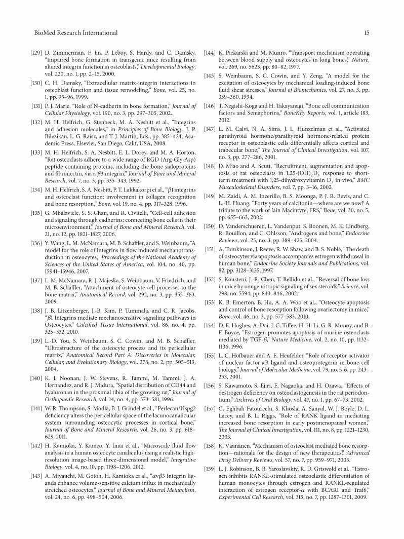
BioMed Research International 15
[129] D. Zimmerman, F. Jin, P. Leboy, S. Hardy, and C. Damsky,“Impaired bone formation in transgenic mice resulting fromaltered integrin function in osteoblasts,”Developmental Biology,vol. 220, no. 1, pp. 2–15, 2000.
[130] C. H. Damsky, “Extracellular matrix-integrin interactions inosteoblast function and tissue remodeling,” Bone, vol. 25, no.1, pp. 95–96, 1999.
[131] P. J. Marie, “Role of N-cadherin in bone formation,” Journal ofCellular Physiology, vol. 190, no. 3, pp. 297–305, 2002.
[132] M. H. Helfrich, G. Stenbeck, M. A. Nesbitt et al., “Integrinsand adhesion molecules,” in Principles of Bone Biology, J. P.Bilezikan, L. G. Raisz, and T. J. Martin, Eds., pp. 385–424, Aca-demic Press, Elsevier, San Diego, Calif, USA, 2008.
[133] M. H. Helfrich, S. A. Nesbitt, E. L. Dorey, and M. A. Horton,“Rat osteoclasts adhere to a wide range of RGD (Arg-Gly-Asp)peptide-containing proteins, including the bone sialoproteinsand fibronectin, via a 𝛽3 integrin,” Journal of Bone and MineralResearch, vol. 7, no. 3, pp. 335–343, 1992.
[134] M.H.Helfrich, S. A.Nesbitt, P. T. Lakkakorpi et al., “𝛽1 integrinsand osteoclast function: involvement in collagen recognitionand bone resorption,” Bone, vol. 19, no. 4, pp. 317–328, 1996.
[135] G. Mbalaviele, S. S. Chan, and R. Civitelli, “Cell-cell adhesionand signaling through cadherins: connecting bone cells in theirmicroenvironment,” Journal of Bone and Mineral Research, vol.21, no. 12, pp. 1821–1827, 2006.
[136] Y.Wang, L.M.McNamara,M. B. Schaffler, and S.Weinbaum, “Amodel for the role of integrins in flow induced mechanotrans-duction in osteocytes,” Proceedings of the National Academy ofSciences of the United States of America, vol. 104, no. 40, pp.15941–15946, 2007.
[137] L. M.McNamara, R. J. Majeska, S. Weinbaum, V. Friedrich, andM. B. Schaffler, “Attachment of osteocyte cell processes to thebone matrix,” Anatomical Record, vol. 292, no. 3, pp. 355–363,2009.
[138] J. B. Litzenberger, J.-B. Kim, P. Tummala, and C. R. Jacobs,“𝛽1 Integrins mediate mechanosensitive signaling pathways inOsteocytes,” Calcified Tissue International, vol. 86, no. 4, pp.325–332, 2010.
[139] L.-D. You, S. Weinbaum, S. C. Cowin, and M. B. Schaffler,“Ultrastructure of the osteocyte process and its pericellularmatrix,” Anatomical Record Part A: Discoveries in Molecular,Cellular, and Evolutionary Biology, vol. 278, no. 2, pp. 505–513,2004.
[140] K. J. Noonan, J. W. Stevens, R. Tammi, M. Tammi, J. A.Hernandez, and R. J. Midura, “Spatial distribution of CD44 andhyaluronan in the proximal tibia of the growing rat,” Journal ofOrthopaedic Research, vol. 14, no. 4, pp. 573–581, 1996.
[141] W. R.Thompson, S. Modla, B. J. Grindel et al., “Perlecan/Hspg2deficiency alters the pericellular space of the lacunocanalicularsystem surrounding osteocytic processes in cortical bone,”Journal of Bone and Mineral Research, vol. 26, no. 3, pp. 618–629, 2011.
[142] H. Kamioka, Y. Kameo, Y. Imai et al., “Microscale fluid flowanalysis in a human osteocyte canaliculus using a realistic high-resolution image-based three-dimensional model,” IntegrativeBiology, vol. 4, no. 10, pp. 1198–1206, 2012.
[143] A. Miyauchi, M. Gotoh, H. Kamioka et al., “𝛼v𝛽3 Integrin lig-ands enhance volume-sensitive calcium influx in mechanicallystretched osteocytes,” Journal of Bone and Mineral Metabolism,vol. 24, no. 6, pp. 498–504, 2006.
[144] K. Piekarski and M. Munro, “Transport mechanism operatingbetween blood supply and osteocytes in long bones,” Nature,vol. 269, no. 5623, pp. 80–82, 1977.
[145] S. Weinbaum, S. C. Cowin, and Y. Zeng, “A model for theexcitation of osteocytes by mechanical loading-induced bonefluid shear stresses,” Journal of Biomechanics, vol. 27, no. 3, pp.339–360, 1994.
[146] T. Negishi-Koga and H. Takayanagi, “Bone cell communicationfactors and Semaphorins,” BoneKEy Reports, vol. 1, article 183,2012.
[147] L. M. Calvi, N. A. Sims, J. L. Hunzelman et al., “Activatedparathyroid hormone/parathyroid hormone-related proteinreceptor in osteoblastic cells differentially affects cortical andtrabecular bone,” The Journal of Clinical Investigation, vol. 107,no. 3, pp. 277–286, 2001.
[148] D. Miao and A. Scutt, “Recruitment, augmentation and apop-tosis of rat osteoclasts in 1,25-(OH)
2D3response to short-
term treatment with 1,25-dihydroxyvitamin D3in vivo,” BMC
Musculoskeletal Disorders, vol. 7, pp. 3–16, 2002.[149] M. Zaidi, A. M. Inzerillo, B. S. Moonga, P. J. R. Bevis, and C.
L.-H. Huang, “Forty years of calcitonin—where are we now? Atribute to the work of Iain Macintyre, FRS,” Bone, vol. 30, no. 5,pp. 655–663, 2002.
[150] D. Vanderschueren, L. Vandenput, S. Boonen, M. K. Lindberg,R. Bouillon, and C. Ohlsson, “Androgens and bone,” EndocrineReviews, vol. 25, no. 3, pp. 389–425, 2004.
[151] A. Tomkinson, J. Reeve, R.W. Shaw, and B. S. Noble, “The deathof osteocytes via apoptosis accompanies estrogen withdrawal inhuman bone,” Endocrine Society Journals and Publications, vol.82, pp. 3128–3135, 1997.
[152] S. Kousteni, J.-R. Chen, T. Bellido et al., “Reversal of bone lossinmice by nongenotropic signaling of sex steroids,” Science, vol.298, no. 5594, pp. 843–846, 2002.
[153] K. B. Emerton, B. Hu, A. A. Woo et al., “Osteocyte apoptosisand control of bone resorption following ovariectomy in mice,”Bone, vol. 46, no. 3, pp. 577–583, 2010.
[154] D. E. Hughes, A. Dai, J. C. Tiffee, H. H. Li, G. R. Munoy, and B.F. Boyce, “Estrogen promotes apoptosis of murine osteoclastsmediated by TGF-𝛽,” Nature Medicine, vol. 2, no. 10, pp. 1132–1136, 1996.
[155] L. C. Hofbauer and A. E. Heufelder, “Role of receptor activatorof nuclear factor-𝜅B ligand and osteoprotegerin in bone cellbiology,” Journal ofMolecularMedicine, vol. 79, no. 5-6, pp. 243–253, 2001.
[156] S. Kawamoto, S. Ejiri, E. Nagaoka, and H. Ozawa, “Effects ofoestrogen deficiency on osteoclastogenesis in the rat periodon-tium,” Archives of Oral Biology, vol. 47, no. 1, pp. 67–73, 2002.
[157] G. Eghbali-Fatourechi, S. Khosla, A. Sanyal, W. J. Boyle, D. L.Lacey, and B. L. Riggs, “Role of RANK ligand in mediatingincreased bone resorption in early postmenopausal women,”The Journal of Clinical Investigation, vol. 111, no. 8, pp. 1221–1230,2003.
[158] K. Vaananen, “Mechanism of osteoclast mediated bone resorp-tion—rationale for the design of new therapeutics,” AdvancedDrug Delivery Reviews, vol. 57, no. 7, pp. 959–971, 2005.
[159] L. J. Robinson, B. B. Yaroslavskiy, R. D. Griswold et al., “Estro-gen inhibits RANKL-stimulated osteoclastic differentiation ofhuman monocytes through estrogen and RANKL-regulatedinteraction of estrogen receptor-𝛼 with BCAR1 and Traf6,”Experimental Cell Research, vol. 315, no. 7, pp. 1287–1301, 2009.
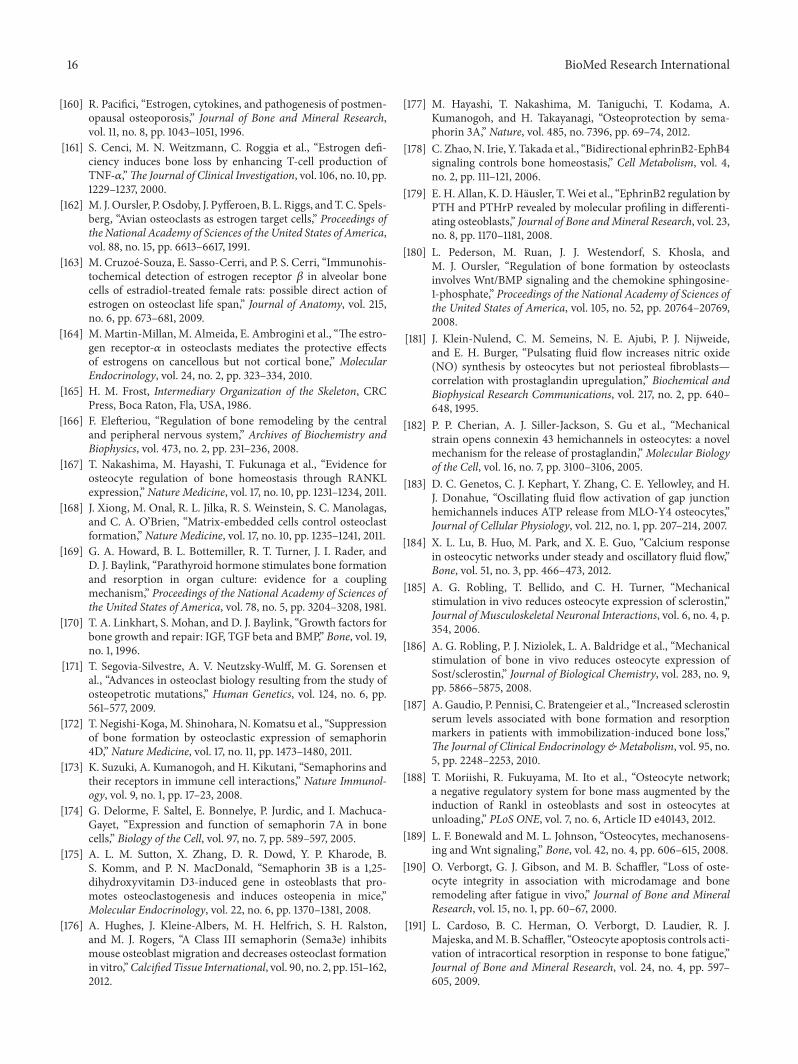
16 BioMed Research International
[160] R. Pacifici, “Estrogen, cytokines, and pathogenesis of postmen-opausal osteoporosis,” Journal of Bone and Mineral Research,vol. 11, no. 8, pp. 1043–1051, 1996.
[161] S. Cenci, M. N. Weitzmann, C. Roggia et al., “Estrogen defi-ciency induces bone loss by enhancing T-cell production ofTNF-𝛼,”The Journal of Clinical Investigation, vol. 106, no. 10, pp.1229–1237, 2000.
[162] M. J. Oursler, P.Osdoby, J. Pyfferoen, B. L. Riggs, andT. C. Spels-berg, “Avian osteoclasts as estrogen target cells,” Proceedings ofthe National Academy of Sciences of the United States of America,vol. 88, no. 15, pp. 6613–6617, 1991.
[163] M. Cruzoe-Souza, E. Sasso-Cerri, and P. S. Cerri, “Immunohis-tochemical detection of estrogen receptor 𝛽 in alveolar bonecells of estradiol-treated female rats: possible direct action ofestrogen on osteoclast life span,” Journal of Anatomy, vol. 215,no. 6, pp. 673–681, 2009.
[164] M.Martin-Millan, M. Almeida, E. Ambrogini et al., “The estro-gen receptor-𝛼 in osteoclasts mediates the protective effectsof estrogens on cancellous but not cortical bone,” MolecularEndocrinology, vol. 24, no. 2, pp. 323–334, 2010.
[165] H. M. Frost, Intermediary Organization of the Skeleton, CRCPress, Boca Raton, Fla, USA, 1986.
[166] F. Elefteriou, “Regulation of bone remodeling by the centraland peripheral nervous system,” Archives of Biochemistry andBiophysics, vol. 473, no. 2, pp. 231–236, 2008.
[167] T. Nakashima, M. Hayashi, T. Fukunaga et al., “Evidence forosteocyte regulation of bone homeostasis through RANKLexpression,” Nature Medicine, vol. 17, no. 10, pp. 1231–1234, 2011.
[168] J. Xiong, M. Onal, R. L. Jilka, R. S. Weinstein, S. C. Manolagas,and C. A. O’Brien, “Matrix-embedded cells control osteoclastformation,” Nature Medicine, vol. 17, no. 10, pp. 1235–1241, 2011.
[169] G. A. Howard, B. L. Bottemiller, R. T. Turner, J. I. Rader, andD. J. Baylink, “Parathyroid hormone stimulates bone formationand resorption in organ culture: evidence for a couplingmechanism,” Proceedings of the National Academy of Sciences ofthe United States of America, vol. 78, no. 5, pp. 3204–3208, 1981.
[170] T. A. Linkhart, S. Mohan, and D. J. Baylink, “Growth factors forbone growth and repair: IGF, TGF beta and BMP,” Bone, vol. 19,no. 1, 1996.
[171] T. Segovia-Silvestre, A. V. Neutzsky-Wulff, M. G. Sorensen etal., “Advances in osteoclast biology resulting from the study ofosteopetrotic mutations,” Human Genetics, vol. 124, no. 6, pp.561–577, 2009.
[172] T. Negishi-Koga, M. Shinohara, N. Komatsu et al., “Suppressionof bone formation by osteoclastic expression of semaphorin4D,” Nature Medicine, vol. 17, no. 11, pp. 1473–1480, 2011.
[173] K. Suzuki, A. Kumanogoh, and H. Kikutani, “Semaphorins andtheir receptors in immune cell interactions,” Nature Immunol-ogy, vol. 9, no. 1, pp. 17–23, 2008.
[174] G. Delorme, F. Saltel, E. Bonnelye, P. Jurdic, and I. Machuca-Gayet, “Expression and function of semaphorin 7A in bonecells,” Biology of the Cell, vol. 97, no. 7, pp. 589–597, 2005.
[175] A. L. M. Sutton, X. Zhang, D. R. Dowd, Y. P. Kharode, B.S. Komm, and P. N. MacDonald, “Semaphorin 3B is a 1,25-dihydroxyvitamin D3-induced gene in osteoblasts that pro-motes osteoclastogenesis and induces osteopenia in mice,”Molecular Endocrinology, vol. 22, no. 6, pp. 1370–1381, 2008.
[176] A. Hughes, J. Kleine-Albers, M. H. Helfrich, S. H. Ralston,and M. J. Rogers, “A Class III semaphorin (Sema3e) inhibitsmouse osteoblast migration and decreases osteoclast formationin vitro,”Calcified Tissue International, vol. 90, no. 2, pp. 151–162,2012.
[177] M. Hayashi, T. Nakashima, M. Taniguchi, T. Kodama, A.Kumanogoh, and H. Takayanagi, “Osteoprotection by sema-phorin 3A,” Nature, vol. 485, no. 7396, pp. 69–74, 2012.
[178] C. Zhao,N. Irie, Y. Takada et al., “Bidirectional ephrinB2-EphB4signaling controls bone homeostasis,” Cell Metabolism, vol. 4,no. 2, pp. 111–121, 2006.
[179] E. H. Allan, K. D. Hausler, T.Wei et al., “EphrinB2 regulation byPTH and PTHrP revealed by molecular profiling in differenti-ating osteoblasts,” Journal of Bone andMineral Research, vol. 23,no. 8, pp. 1170–1181, 2008.
[180] L. Pederson, M. Ruan, J. J. Westendorf, S. Khosla, andM. J. Oursler, “Regulation of bone formation by osteoclastsinvolves Wnt/BMP signaling and the chemokine sphingosine-1-phosphate,” Proceedings of the National Academy of Sciences ofthe United States of America, vol. 105, no. 52, pp. 20764–20769,2008.
[181] J. Klein-Nulend, C. M. Semeins, N. E. Ajubi, P. J. Nijweide,and E. H. Burger, “Pulsating fluid flow increases nitric oxide(NO) synthesis by osteocytes but not periosteal fibroblasts—correlation with prostaglandin upregulation,” Biochemical andBiophysical Research Communications, vol. 217, no. 2, pp. 640–648, 1995.
[182] P. P. Cherian, A. J. Siller-Jackson, S. Gu et al., “Mechanicalstrain opens connexin 43 hemichannels in osteocytes: a novelmechanism for the release of prostaglandin,”Molecular Biologyof the Cell, vol. 16, no. 7, pp. 3100–3106, 2005.
[183] D. C. Genetos, C. J. Kephart, Y. Zhang, C. E. Yellowley, and H.J. Donahue, “Oscillating fluid flow activation of gap junctionhemichannels induces ATP release from MLO-Y4 osteocytes,”Journal of Cellular Physiology, vol. 212, no. 1, pp. 207–214, 2007.
[184] X. L. Lu, B. Huo, M. Park, and X. E. Guo, “Calcium responsein osteocytic networks under steady and oscillatory fluid flow,”Bone, vol. 51, no. 3, pp. 466–473, 2012.
[185] A. G. Robling, T. Bellido, and C. H. Turner, “Mechanicalstimulation in vivo reduces osteocyte expression of sclerostin,”Journal of Musculoskeletal Neuronal Interactions, vol. 6, no. 4, p.354, 2006.
[186] A. G. Robling, P. J. Niziolek, L. A. Baldridge et al., “Mechanicalstimulation of bone in vivo reduces osteocyte expression ofSost/sclerostin,” Journal of Biological Chemistry, vol. 283, no. 9,pp. 5866–5875, 2008.
[187] A. Gaudio, P. Pennisi, C. Bratengeier et al., “Increased sclerostinserum levels associated with bone formation and resorptionmarkers in patients with immobilization-induced bone loss,”The Journal of Clinical Endocrinology &Metabolism, vol. 95, no.5, pp. 2248–2253, 2010.
[188] T. Moriishi, R. Fukuyama, M. Ito et al., “Osteocyte network;a negative regulatory system for bone mass augmented by theinduction of Rankl in osteoblasts and sost in osteocytes atunloading,” PLoS ONE, vol. 7, no. 6, Article ID e40143, 2012.
[189] L. F. Bonewald and M. L. Johnson, “Osteocytes, mechanosens-ing and Wnt signaling,” Bone, vol. 42, no. 4, pp. 606–615, 2008.
[190] O. Verborgt, G. J. Gibson, and M. B. Schaffler, “Loss of oste-ocyte integrity in association with microdamage and boneremodeling after fatigue in vivo,” Journal of Bone and MineralResearch, vol. 15, no. 1, pp. 60–67, 2000.
[191] L. Cardoso, B. C. Herman, O. Verborgt, D. Laudier, R. J.Majeska, andM. B. Schaffler, “Osteocyte apoptosis controls acti-vation of intracortical resorption in response to bone fatigue,”Journal of Bone and Mineral Research, vol. 24, no. 4, pp. 597–605, 2009.
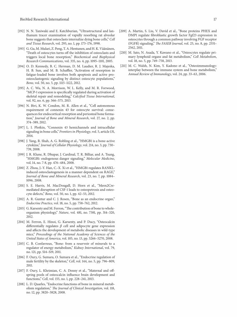
BioMed Research International 17
[192] N. N. Taniwaki and E. Katchburian, “Ultrastructural and lan-thanum tracer examination of vapidly resorbing rat alveolarbone suggests that osteoclasts internalize dying bone cells,” Celland Tissue Research, vol. 293, no. 1, pp. 173–176, 1998.
[193] G.Gu,M.Mulari, Z. Peng, T.A.Hentunen, andH.K.Vaananen,“Death of osteocytes turns off the inhibition of osteoclasts andtriggers local bone resorption,” Biochemical and BiophysicalResearch Communications, vol. 335, no. 4, pp. 1095–1101, 2005.
[194] O. D. Kennedy, B. C. Herman, D. M. Laudier, R. J. Majeska,H. B. Sun, and M. B. Schaffler, “Activation of resorption infatigue-loaded bone involves both apoptosis and active pro-osteoclastogenic signaling by distinct osteocyte populations,”Bone, vol. 50, no. 5, pp. 1115–1122, 2012.
[195] A. C. Wu, N. A. Morrison, W. L. Kelly, and M. R. Forwood,“MCP-1 expression is specifically regulated during activation ofskeletal repair and remodeling,” Calcified Tissue International,vol. 92, no. 6, pp. 566–575, 2013.
[196] N. Bivi, K. W. Condon, M. R. Allen et al., “Cell autonomousrequirement of connexin 43 for osteocyte survival: conse-quences for endocortical resorption and periosteal bone forma-tion,” Journal of Bone and Mineral Research, vol. 27, no. 2, pp.374–389, 2012.
[197] L. I. Plotkin, “Connexin 43 hemichannels and intracellularsignaling in bone cells,” Frontiers in Physiology, vol. 5, article 131,2014.
[198] J. Yang, R. Shah, A. G. Robling et al., “HMGB1 is a bone-activecytokine,” Journal of Cellular Physiology, vol. 214, no. 3, pp. 730–739, 2008.
[199] J. R. Klune, R. Dhupar, J. Cardinal, T. R. Billiar, and A. Tsung,“HMGB1: endogenous danger signaling,” Molecular Medicine,vol. 14, no. 7-8, pp. 476–484, 2008.
[200] Z. Zhou, J.-Y. Han, C.-X. Xi et al., “HMGB1 regulates RANKL-induced osteoclastogenesis in a manner dependent on RAGE,”Journal of Bone and Mineral Research, vol. 23, no. 7, pp. 1084–1096, 2008.
[201] S. E. Harris, M. MacDougall, D. Horn et al., “Meox2Cre-mediated disruption of CSF-1 leads to osteopetrosis and osteo-cyte defects,” Bone, vol. 50, no. 1, pp. 42–53, 2012.
[202] A. R. Guntur and C. J. Rosen, “Bone as an endocrine organ,”Endocrine Practice, vol. 18, no. 5, pp. 758–762, 2012.
[203] G. Karsenty andM. Ferron, “The contribution of bone towhole-organism physiology,” Nature, vol. 481, no. 7381, pp. 314–320,2012.
[204] M. Ferron, E. Hinoi, G. Karsenty, and P. Ducy, “Osteocalcindifferentially regulates 𝛽 cell and adipocyte gene expressionand affects the development of metabolic diseases in wild-typemice,” Proceedings of the National Academy of Sciences of theUnited States of America, vol. 105, no. 13, pp. 5266–5270, 2008.
[205] C. B. Confavreux, “Bone: from a reservoir of minerals to aregulator of energy metabolism,” Kidney International, vol. 79,no. 121, pp. S14–S19, 2011.
[206] F. Oury, G. Sumara, O. Sumara et al., “Endocrine regulation ofmale fertility by the skeleton,” Cell, vol. 144, no. 5, pp. 796–809,2011.
[207] F. Oury, L. Khrimian, C. A. Denny et al., “Maternal and off-spring pools of osteocalcin influence brain development andfunctions,” Cell, vol. 155, no. 1, pp. 228–241, 2013.
[208] L. D. Quarles, “Endocrine functions of bone in mineral metab-olism regulation,” The Journal of Clinical Investigation, vol. 118,no. 12, pp. 3820–3828, 2008.
[209] A. Martin, S. Liu, V. David et al., “Bone proteins PHEX andDMP1 regulate fibroblastic growth factor Fgf23 expression inosteocytes through a common pathway involving FGF receptor(FGFR) signaling,” The FASEB Journal, vol. 25, no. 8, pp. 2551–2562, 2011.
[210] M. Sato, N. Asada, Y. Kawano et al., “Osteocytes regulate pri-mary lymphoid organs and fat metabolism,” Cell Metabolism,vol. 18, no. 5, pp. 749–758, 2013.
[211] M. C. Walsh, N. Kim, Y. Kadono et al., “Osteoimmunology:interplay between the immune system and bone metabolism,”Annual Review of Immunology, vol. 24, pp. 33–63, 2006.

Submit your manuscripts athttp://www.hindawi.com
Hindawi Publishing Corporationhttp://www.hindawi.com Volume 2014
Anatomy Research International
PeptidesInternational Journal of
Hindawi Publishing Corporationhttp://www.hindawi.com Volume 2014
Hindawi Publishing Corporation http://www.hindawi.com
International Journal of
Volume 2014
Zoology
Hindawi Publishing Corporationhttp://www.hindawi.com Volume 2014
Molecular Biology International
GenomicsInternational Journal of
Hindawi Publishing Corporationhttp://www.hindawi.com Volume 2014
The Scientific World JournalHindawi Publishing Corporation http://www.hindawi.com Volume 2014
Hindawi Publishing Corporationhttp://www.hindawi.com Volume 2014
BioinformaticsAdvances in
Marine BiologyJournal of
Hindawi Publishing Corporationhttp://www.hindawi.com Volume 2014
Hindawi Publishing Corporationhttp://www.hindawi.com Volume 2014
Signal TransductionJournal of
Hindawi Publishing Corporationhttp://www.hindawi.com Volume 2014
BioMed Research International
Evolutionary BiologyInternational Journal of
Hindawi Publishing Corporationhttp://www.hindawi.com Volume 2014
Hindawi Publishing Corporationhttp://www.hindawi.com Volume 2014
Biochemistry Research International
ArchaeaHindawi Publishing Corporationhttp://www.hindawi.com Volume 2014
Hindawi Publishing Corporationhttp://www.hindawi.com Volume 2014
Genetics Research International
Hindawi Publishing Corporationhttp://www.hindawi.com Volume 2014
Advances in
Virolog y
Hindawi Publishing Corporationhttp://www.hindawi.com
Nucleic AcidsJournal of
Volume 2014
Stem CellsInternational
Hindawi Publishing Corporationhttp://www.hindawi.com Volume 2014
Hindawi Publishing Corporationhttp://www.hindawi.com Volume 2014
Enzyme Research
Hindawi Publishing Corporationhttp://www.hindawi.com Volume 2014
International Journal of
Microbiology


![Lamellar Bone is an Incremental Tissue Reconciling Enamel ... · as the scaling of bone mass to body mass is an axiom of vertebrate hard tissue biology [15]. Because enamel and bone](https://static.fdocuments.us/doc/165x107/5fa2f5bb05de466cf30f1103/lamellar-bone-is-an-incremental-tissue-reconciling-enamel-as-the-scaling-of.jpg)