U-18486 DTE Electric and DTE Gas Report on Billing Practices

Hindawi Publishing CorporationDiagnostic and Therapeutic EndoscopyVolume 2011, Article ID 478913, 10 pagesdoi:10.1155/2011/478913
Review Article
Diagnosis and Management of Cystic Lesions of the Pancreas
Niraj Jani,1 Murad Bani Hani,2 Richard D. Schulick,3 Ralph H. Hruban,4
and Steven C. Cunningham2
1 Department of Medicine, Saint Agnes Hospital, Baltimore, MD 21229, USA2 Department of Surgery, Saint Agnes Hospital, Baltimore, MD 21229, USA3 Department of Surgery, The Sol Goldman Pancreatic Cancer Research Center, The Johns Hopkins Hospital, Baltimore,MD 21287, USA
4 Department of Pathology, The Sol Goldman Pancreatic Cancer Research Center, The Johns Hopkins Hospital, Baltimore,MD 21231, USA
Correspondence should be addressed to Steven C. Cunningham, [email protected]
Received 9 May 2011; Revised 29 June 2011; Accepted 29 June 2011
Academic Editor: C. M. Wilcox
Copyright © 2011 Niraj Jani et al. This is an open access article distributed under the Creative Commons Attribution License,which permits unrestricted use, distribution, and reproduction in any medium, provided the original work is properly cited.
Pancreatic cysts are challenging lesions to diagnose and to treat. Determining which of the five most common diagnoses—pancreatic pseudocyst, serous cystic neoplasm (SCN), solid pseudopapillary neoplasm (SPN), mucinous cystic neoplasm (MCN),and intraductal mucinous papillary neoplasm (IPMN)—is likely the correct one requires the careful integration of many historical,radiographic, laboratory, and other factors, and management is markedly different depending on the type of cystic lesion of thepancreas. Pseudocysts are generally distinguishable based on historical, clinical and radiographic characteristics, and among theothers, the most important differentiation is between the mucin-producing MCN and IPMN (high risk for cancer) versus theserous SCN and SPN (low risk for cancer). EUS with FNA and cyst-fluid analysis will continue to play an important role indiagnosis. Among mucinous lesions, those that require treatment (resection currently) are any MCN, any MD IPMN, and BDIPMN larger than 3 cm, symptomatic, or with an associated mass, with the understanding that SCN or pseudocysts may beremoved inadvertently due to diagnostic inaccuracy, and that a certain proportion of SPN will indeed be malignant at the time ofremoval. The role of ethanol ablation is under investigation as an alternative to resection in selected patients.
1. Introduction
Pancreatic cysts are common in the general population.The reported incidence of asymptomatic cysts varies widely,largely due to differences in study design, ranging between0.7% and 24.3% [1–4]. The lowest estimate comes from astudy employing both single- and multidetector CT scannersand relying on original dictated reports as opposed torereview of images [1], while the highest estimates comefrom autopsy studies and studies including both symp-tomatic and asymptomatic patients [3, 4]. The incidenceof truly asymptomatic cysts in the general population isapproximately 2.6% [2]. In large series of pancreatic cysts[5], most (71%) cysts are largely asymptomatic and rangefrom benign to premalignant to malignant cysts. The mostuseful first dichotomy in the long differential diagnosis(Table 1) of pancreatic cysts is their classification as either
neoplastic or nonneoplastic. Nonneoplastic cysts includepseudocysts, retention cysts, and duplication cysts, whereasneoplastic cysts are further broadly classified as mucinousand nonmucinous cysts. The more common—and morecommonly malignant—mucinous neoplasms include pri-marily intraductal papillary mucinous neoplasm (IMPN)and mucinous cystic neoplasms (MCN), while nonmucinousneoplastic cysts include primarily serous cystic neoplasm(SCN), solid pseudopapillary neoplasm (SPN), and usuallysolid neoplasms with degenerative cystic changes [6, 7].Whereas most serous cystic neoplasms are not malignant,intraductal papillary mucinous neoplasms and mucinouscystic neoplasms can harbor an associated invasive carci-noma and should be treated as having malignant potential.
Differentiating among these cysts is challenging, and avariety of modalities—including imaging, cytology, and cystfluid analysis—are useful. The management of pancreatic
2 Diagnostic and Therapeutic Endoscopy
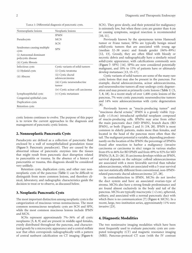
Table 1: Differential diagnosis of pancreatic cysts.
Nonneoplastic lesions Neoplastic lesions
IPMN
Pseudocysts MCN
SCN
Syndromes causing multiplecysts
SPN
(i) Autosomal dominantpolycystic disease
(ii) Cystic fibrosis
Infectious cysts Cystic variants of solid tumors
(i) Hydatid cysts (i) Cystic teratoma
(ii) Abscess(ii) Cystic ductaladenocarcinoma
(iii) Cystic neuroendocrinetumor
(iv) Cystic acinar cell carcinoma
Lymphoepithelial cysts (v) Cystic metastases
Congenital epithelial cysts
Duplication cysts
Retention cysts
cystic lesions continues to evolve. The purpose of this paperis to review the current approaches to the diagnosis andmanagement of pancreatic cystic lesions.
2. Nonneoplastic Pancreatic Cysts
Pseudocysts are defined as a collection of pancreatic fluidenclosed by a wall of nonepithelialized granulation tissue(Figure 5: Pancreatic pseudocyst). They are caused by theabnormal release of pancreatic enzymes into the tissuesthat might result from pancreatic duct disruption relatedto pancreatitis or trauma. In the absence of a history ofpancreatitis or trauma, this diagnosis should be consideredvery unlikely.
Retention cysts, duplication cysts, and other rare non-neoplastic cysts of the pancreas (Table 1) can be difficult todistinguish from more common lesions, and therefore cli-nical, laboratory, and radiographic characteristics guide thedecision to treat or to observe, as discussed below.
3. Neoplastic Pancreatic Cysts
The most important distinction among neoplastic cysts is thecategorization of mucinous versus nonmucinous. The mostcommon nonmucinous neoplastic cysts are SCN and SPN,while the most common mucinous lesions include IPMNand MCN.
SCNs represent approximately 7%–36% of all cysticneoplasms [5, 8, 9] and are present in middle-aged females,evenly distributed throughout the pancreas, and character-ized grossly by a microcystic appearance and a central stellatescar that often corresponds radiographically with a patternof central sunburst calcification on CT imaging (Figure 6:
SCN). They grow slowly, and their potential for malignancyis extremely low, but when these cysts are greater than 4 cmor causing symptoms, surgical resection is recommended[10, 11].
Previously known by the eponymous terms Hamouditumor or Franz tumor, SPNs are typically benign mixedsolid/cystic tumors that are associated with young age(median 32–38 years) and female gender (84%–89%)[12, 13]. Grossly, they are often filled with bloody ornecrotic debris and radiographically have a similarly mixedsolid/cystic appearance, with calcifications commonly seen(Figure 7: SPN) [14]. SPNs are now considered potentiallymalignant, and 10% to 15% of patients have or ultimatelydevelop metastases [13, 15–17].
Cystic variants of solid tumors are some of the many rarecystic lesions that may also be present in the pancreas. Forexample, ductal adenocarcinoma, acinar adenocarcinoma,and neuroendocrine tumors all may undergo cystic degener-ation and may present as primarily cystic lesions (Table 1) [5,7, 8, 18]. In a recent study of over 1,400 cystic lesions of thepancreas, 7% were cystic pancreatic neuroendocrine tumorsand 14% were adenocarcinomas with cystic degeneration[8].
Previously known as “mucin-producing tumor” and“mucinous ductal ectasia,” IPMN is a grossly visible (typ-ically ≥1.0 cm) intraductal epithelial neoplasm composedof mucin-producing cells. IPMNs may arise from eitherthe main pancreatic duct (MD IPMN), branch ducts (BDIPMN), or both (Figures 2 and 3) [19, 20]. IPMN is mostcommon in elderly patients, males more than females, andlocated in the head of the pancreas more often than thetail. The malignant potential is variable, depending predomi-nantly on the location of the IPMN: the percentage of IPMNsfound after resection to harbor a malignancy (invasivecarcinoma or carcinoma in situ) ranges in various studiesfrom 6% to 46% for BD IPMN and from 49% to 92% for MDIPMNs [5, 8, 21–26]. If carcinoma develops within an IPMN,survival depends on the subtype: colloid adenocarcinomasare associated with a more favorable survival than tubularadenocarcinomas, which are associated with a 5-year survivalrate not statistically different from conventional, non-IPMN-related pancreatic ductal adenocarcinoma [27, 28].
In contradistinction to IPMN, MCNs do not involvethe duct system and have an associated ovarian-type ofstroma. MCNs also have a strong female predominance andare found almost exclusively in the body and tail of thepancreas. MCNs are typically macrocystic (>2 cm), spheroid,solitary, and associated with a normal pancreatic duct withwhich there is no communication [7] (Figure 4: MCN). In arecent, large, two-institution series, approximately 11% wereinvasive [23].
4. Diagnostic Modalities
The two noninvasive imaging modalities which have beenmost frequently used to evaluate pancreatic cysts are com-puted tomography (CT) and magnetic resonance imaging(MRI). Pancreas-protocol CT scan (with the IV contrast

Diagnostic and Therapeutic Endoscopy 3
Imaging findings
Questionable findings
Consider resection
Good operative risk Poor operative risk Surveillanceb
Consider ablationc
Pancreas-protocol CT
EUS/FNA ± ERCP
Suspicious findingsa (e.g.,MD IPMN, size > 3 cm,
mural nodules, septations)
Reassuring findings (e.g.,very small, peripheral,
simple cysts)
Figure 1: Pancreatic cyst therapeutic algorithm. aAlso considered are nonimaging findings such as symptoms attributable to the cyst, rapidgrowth, and young age. bSurveillance may be performed initially at close intervals (e.g., 3 mo), and later spaced out to every 6, 12, or 24months. cNB: cyst ablation is largely experimental and not appropriate for main-duct IPMNs. Abbreviations: see text.
(a)
D1
(b)
Figure 2: Main-duct intraductal papillary mucinous neoplasm. (a)Typical CT (arrows) and (b) EUS (cross marks) appearance.
bolus timed for both arterial and venous phases and typicallywith water as the oral contrast to minimize artifacts arisingfrom denser contrast media) has become the preferred mod-ality to evaluate the pancreas due to its ease, relatively lowexpense, and diagnostic accuracy [29–31]. However, someauthors have argued that MRI with magnetic resonancecholangiopancreatography (MRCP) is the best noninvasivemethod for identifying the presence or absence of commu-nication between pancreatic cysts and the pancreatic ductalsystem [32]. While MRI/MRCP has the clear advantage overCT of not involving the use of ionizing radiation, it lacks the
(a)
1
(b)
Figure 3: Branch-duct intraductal papillary mucinous neoplasm.(a) CT and (b) EUS showing associated mass (cross marks).
ability to sample cyst fluid for analysis, which, as discussedbelow, can help distinguish between high-risk mucinous andlow-risk nonmucinous cysts.
Endoscopic retrograde cholangiopancreatography (ERCP)has been useful in cases of IPMN especially when combinedwith pancreatoscopy and/or intraductal ultra-sound. Hara etal. [33] found that lesions protruding more than 4 mm intothe pancreatic duct were malignant in 88% of cases. ERCP,although more invasive than MRCP, is very useful in definingthe communication of the cyst with the main pancreatic ductand provides another method for tissue acquisition. Due to
4 Diagnostic and Therapeutic Endoscopy
(a)
D1
(b)
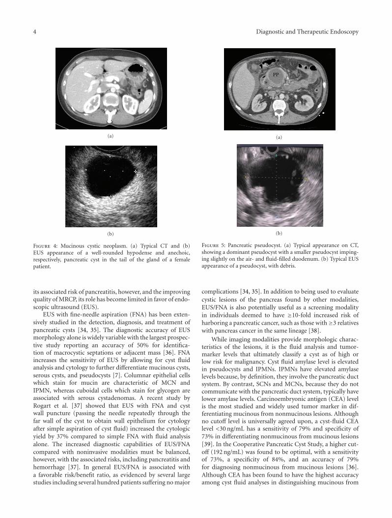
Figure 4: Mucinous cystic neoplasm. (a) Typical CT and (b)EUS appearance of a well-rounded hypodense and anechoic,respectively, pancreatic cyst in the tail of the gland of a femalepatient.
its associated risk of pancreatitis, however, and the improvingquality of MRCP, its role has become limited in favor of endo-scopic ultrasound (EUS).
EUS with fine-needle aspiration (FNA) has been exten-sively studied in the detection, diagnosis, and treatment ofpancreatic cysts [34, 35]. The diagnostic accuracy of EUSmorphology alone is widely variable with the largest prospec-tive study reporting an accuracy of 50% for identifica-tion of macrocystic septations or adjacent mass [36]. FNAincreases the sensitivity of EUS by allowing for cyst fluidanalysis and cytology to further differentiate mucinous cysts,serous cysts, and pseudocysts [7]. Columnar epithelial cellswhich stain for mucin are characteristic of MCN andIPMN, whereas cuboidal cells which stain for glycogen areassociated with serous cystadenomas. A recent study byRogart et al. [37] showed that EUS with FNA and cystwall puncture (passing the needle repeatedly through thefar wall of the cyst to obtain wall epithelium for cytologyafter simple aspiration of cyst fluid) increased the cytologicyield by 37% compared to simple FNA with fluid analysisalone. The increased diagnostic capabilities of EUS/FNAcompared with noninvasive modalities must be balanced,however, with the associated risks, including pancreatitis andhemorrhage [37]. In general EUS/FNA is associated witha favorable risk/benefit ratio, as evidenced by several largestudies including several hundred patients suffering no major
PP
∗
(a)
(b)
Figure 5: Pancreatic pseudocyst. (a) Typical appearance on CT,showing a dominant pseudocyst with a smaller pseudocyst imping-ing slightly on the air- and fluid-filled duodenum. (b) Typical EUSappearance of a pseudocyst, with debris.
complications [34, 35]. In addition to being used to evaluatecystic lesions of the pancreas found by other modalities,EUS/FNA is also potentially useful as a screening modalityin individuals deemed to have ≥10-fold increased risk ofharboring a pancreatic cancer, such as those with≥3 relativeswith pancreas cancer in the same lineage [38].
While imaging modalities provide morphologic charac-teristics of the lesions, it is the fluid analysis and tumor-marker levels that ultimately classify a cyst as of high orlow risk for malignancy. Cyst fluid amylase level is elevatedin pseudocysts and IPMNs. IPMNs have elevated amylaselevels because, by definition, they involve the pancreatic ductsystem. By contrast, SCNs and MCNs, because they do notcommunicate with the pancreatic duct system, typically havelower amylase levels. Carcinoembryonic antigen (CEA) levelis the most studied and widely used tumor marker in dif-ferentiating mucinous from nonmucinous lesions. Althoughno cutoff level is universally agreed upon, a cyst-fluid CEAlevel <30 ng/mL has a sensitivity of 79% and specificity of73% in differentiating nonmucinous from mucinous lesions[39]. In the Cooperative Pancreatic Cyst Study, a higher cut-off (192 ng/mL) was found to be optimal, with a sensitivityof 73%, a specificity of 84%, and an accuracy of 79%for diagnosing nonmucinous from mucinous lesions [36].Although CEA has been found to have the highest accuracyamong cyst fluid analyses in distinguishing mucinous from

Diagnostic and Therapeutic Endoscopy 5
(a)
D1
(b)
Figure 6: Serous cystic neoplasm: (a) CT and (b) EUS images bothshowing the central starburst calcification pattern characteristic ofserous cystic neoplasms.
nonmucinous cysts, other tumor markers have also beenpredictive. For instance, in the same Cooperative Study, cystfluid CA19-9 levels had a sensitivity of 68%, a specificity of62%, and an accuracy of 66% (P = 0.004) with a cutoffvalue of 2900 U/mL for differentiating nonmucinous frommucinous lesions [36]; corresponding numbers for CA72-4were 80%, 61%, and 72% (P = 0.001).
The demographic, historical, radiographic, gross, andcyst fluid analysis characteristics described above are sum-marized in Table 2 and are electronically available in aninteractive, online pancreatic cyst worksheet available athttp://pathology.jhu.edu/pancreas/professionals/ipmn.php[7].
4.1. Emerging Modalities. Loss-of-heterozygosity studies andDNA mutational analysis of cyst fluid have shown that thepresence of a point mutation in the KRAS gene is 96%specific in detecting a mucinous neoplasm, and when thereis a KRAS gene point mutation coupled with allelic lossat selected markers, there is a 96% specificity in detectingmalignancy (invasive versus in situ carcinoma not specified)[41]. However, this study has been rightfully criticized forharboring a selection bias resulting from the exclusion ofnonoperated patients from cyst fluid DNA analysis [42].Furthermore, there is poor correlation between cysts withhigh CEA levels and those with KRAS point mutations and
(a)
(b)
Figure 7: Solid pseudopapillary neoplasm: (a) Typical CT and (b)EUS appearance of solid and cystic components.
allelic loss. There are a number of other biomarkers that arecurrently under evaluation to predict risk in pancreatic cysts[43, 44].
Confocal laser endomicroscopy (CLE) [45] is an excitingemerging diagnostic modality that employs a low-powerlaser to illuminate tissue with subsequent detection light re-flected from the tissue through a small probe (pCLE) orneedle (nCLE). It is “confocal” because both illuminationand collection systems are aligned in the same focal plane[45]. While largely still experimental, nCLE has been usedsuccessfully in a porcine model to collect real-time, in vivopancreatic images at histologic resolutions and of acceptableimage quality [46].
5. Management
5.1. Nonneoplastic Cysts. Initially, the management of pseu-docysts is conservative since as many as 60% may completelyresolve spontaneously within a year [47]. As such, surveil-lance is the first-line therapy for noninfected pancreaticpseudocysts and may be done with US, CT, or MRI.Pseudocysts that either cause severe symptoms or are largeand refractory to surveillance should be drained percu-taneously, endoscopically, or surgically. The disadvantagesof percutaneous drainage include risk of infection, fistulaformation, and a low rate (21%) of resolution [48]. Open andlaparoscopic internal surgical management—including in-ternal and external drainage as well as resection—is effective,but is associated with 12%–35% complication rate, includinghemorrhage, infection, and fistulae, and a mortality rate

6 Diagnostic and Therapeutic Endoscopy
Table 2: Distinguishing features of pancreatic cystic lesions∗.
Typicalcharacteristics
IPMN MCN SCN PSEUDO SPN LEC cNET cPDAC
Age group Elderly MiddleMiddle-elderly
Any Young ElderlyMiddle-Elderly
Elderly
Gender >50% male 95% female >50% female >50% male80%–90%
female80% male 50% each >50% male
HistoryAsx; pain;± jaundice
Asx; Pain;nausea
Asx; VHL PancreatitisAsx; pain;
nauseaAsx
Asx; Fxnl;MEN
Asx; pain;± jaundice
% of allcysts∗∗∗
17%–40% 9%–28% 7%–36% 1%–19% 1%–13% <2% <8% 13%–16%
Location inpancreas
Head in 70%;multifocal
Body/Tail in95%
Anywhere Anywhere Anywhere Peripheral Anywhere Anywhere
Shape Ovoid Spheroid Ovoid Spheroid Ovoid Ovoid Spheroid Variable
Locularity Any Uni- or oligo-Oligo- or
multi-Uni-
Oligo- orMulti-
Oligo- Uni- Any
Duct com-munication
Common No No Common No No No Some
Calcification No NoCentral
sunburstNo Some No Some No
Cyst fluidappearance
Viscous, clear,muc.
Viscous, clear,muc.
Thin, clear,nonmuc.
Opaque,bloody/necroticdebris
Opaque,bloody/necroticdebris
Nonmuc.,crystalline
debrisNonmuc. Thin
HighCEA/Mucin∗∗
+ + − − − − − ±High Ca19-9 ± ± − − − − − ±High amylase + − − + − − − ±
EpitheliumColumnar,papillary
Columnar Cuboidal No epithelium
Poorlycohesive cellswith nuclear
grooves
Squamoid UniformGland-forming
Stroma Fibrotic Ovarian Fibrotic FibroticSometimeshyalinized
LymphoidSometimeshyalinized
Fibrotic
Abbreviations: IPMN: intraductal papillary mucinous neoplasm; MCN: mucinous cystic neoplasm; SC: serous cystadenoma; PSEUDO: pancreatic pseudocyst;SPN: solid-pseudopapillary neoplasm; LEC: lymphoepithelial cyst; cNET: cystic neuroendocrine tumor; cPDAC: pancreatic ductal adenocarcinoma with cysticdegeneration; VHL: von Hippel-Lindau disease; muc.: mucinous; Nonmuc: nonmucinous; Asx: asymptomatic; Fxnl: functional.∗∗∗Percentages references [8, 9, 22, 40].∗∗May be positive in cases of luminal contamination of endoscopic needle aspirate.NB: These data are derived generalizations of the literature, with the understanding that there is significant overlap among cyst types and there are inherentsampling errors associated with various tests; diagnostic and treatment decisions should not rely solely on the information presented in this paper.∗Table modified from [7] by Cunningham et al. Intraductal papillary mucinous neoplasms are differentiated from other pancreatic cystic lesions. World JGastrointest Surg 2010; 2(10): 331–336. An electronic worksheet version of this table is available at http://pathology.jhu.edu/pancreas/professionals/ipmn.php.
of 1% [49–51]. Endoscopic drainage has been reported toachieve a similarly high success rate but with lower rates ofcomplications, including bleeding, infection, perforation,and mild pancreatitis, which is generally self-limited [52, 53].Endoscopic drainage has therefore become the preferred mo-dality for draining cysts which have a mature wall and arewithin 1 cm of the gastrointestinal lumen. In a recent large re-trospective study by Ahn et al. [54], single-step EUS-guidedtransmural drainage and stent placement was effective in89% of patients with complete drainage, with an overall re-currence rate of 12% and minor complications in 11% ofpatients.
Unlike pancreatic pseudocysts, which are typically iden-tifiable as such based on historical, clinical, laboratory, and
radiographic information, other nonneoplastic cysts such asduplication cysts, retention cysts, congenital epithelial cysts,and lymphoepithelial cysts are rarer, and not easily diagnosedpreoperatively. As such, they are typically subjected to thevarious diagnostic modalities described above in an effort toclassify them correctly as low-risk versus high-risk cysts andthey are treated or surveilled accordingly.
5.2. Neoplastic Cysts
5.2.1. Indications for Resection. In the absence of randomizedcontrolled data to guide treatment recommendations, theSendai International Consensus Guidelines [21, 55], firstpublished online in 2005 by the International Association

Diagnostic and Therapeutic Endoscopy 7
of Pancreatology, identified several factors as relative indica-tions for resection of IPMN. These include a main-ductcomponent, diameter >3 cm, any solid component, andthe presence of symptoms attributable to the cyst, suchas abdominal pain, weight loss, and pancreatitis (Table 2).Rapid rate of growth of the cyst and young age (such that life-long surveillance would be prohibitively burdensome for thepatient) may be considered relative indications outside theSendai Guidelines. Resection recommended as the mainstayof treatment for lesions thought to have increased thepotential for harboring significant dysplasia or an associatedinvasive carcinoma, and indeed it is the only potentially cura-tive option for such lesions.
Unlike a cystic lesion thought to be IPMN, which maybe observed or resected, depending on the above-mentionedrisk factors, any lesion thought to be MCN should be resecteduntil data are available to better stratify these patients, if suchdata ever exist.
Regarding the serous lesions SCN and SPN, all lesionsknown to be SCN may be left in place and all those known tobe SPN should be resected. In reality, however, a given cysticlesion of the pancreas is not generally known to be one or theother with sufficient certainty, even despite all of the above-discussed diagnostic modalities. Therefore, each pancreato-logist and patient must together carefully weigh the risks andbenefits of resection and surveillance on a case-by-case basis(see Surveillance, below).
The chief difficulty is, of course, the fact that the only wayto achieve a definitive diagnosis in many cases of pancreaticcysts is to remove the cyst and subject it to pathologicevaluation. Although pancreatectomy is curative in mostcases of cystic lesions of the pancreas, it is associated witha perioperative morbidity rate of 30–60% [56–58] and amortality rate ranging from <1% to 2% [56–59]. In additionto complications associated with any operation in general,such as bleeding and infection, complications specific tothe resection of pancreatic lesions include pancreatic orbiliary fistula, delayed gastric emptying, and pancreaticinsufficiency, both exocrine and endocrine.
5.2.2. Cyst Ablation. In an effort to avoid a more inva-sive treatment and the associated complications, pancreaticcyst ablation has been suggested both as an experimentalapproach to treatment for pancreatic cysts in general and fortreatment of those patients specifically deemed unfit or at atoo high risk for a major operation (Figure 1) [60].
Although less commonly employed than resection, pan-creatic cyst ablation is an increasingly studied modality, typi-cally using EUS to guide injection of alcohol or other ablativeagents into the cyst cavity. Ethanol has the advantages ofbeing safe, inexpensive, readily available, and having thepotential to rapidly ablate the entire cyst wall epithelium.A 2005 pilot study using escalating doses (5% to 80%) ofethanol for 3- to 5-minute lavage [61] showed histologicalevidence of epithelial ablation in resected cysts. Patientsreported no symptoms at 2 hours, 72 hours, and 6–12months following the procedure, with no complicationsdetected [61], although theoretical complications include
acute pancreatitis, hemorrhage, intoxication, and abdominalpain.
In an effort to assess effectiveness as well as to furtherassess safety, DeWitt et al. [62] compared ethanol ablationto saline lavage in a randomized controlled trial including42 ethanol-lavaged and 17 saline-lavaged patients. Ethanollavage resulted in greater decrease in pancreatic cyst size(−43%), compared with saline (−11%), with similar safetyprofile [62]. Four patients underwent resection after lavageof mucinous cysts (2 who decided to drop out after lack ofresponse (one to saline and one to ethanol) and 2 whose cystfluid had atypical cells), and histology of resected pancreatashowed IPMNs in 3 and MCN in 1 patient; not surprisingly,there was more extensive ablation (50% to 100% of cystepithelium) in the ethanol group than the saline group (0%).Resolution by CT imaging was seen in 33% (12 of 36 cystslavaged with saline alone [1], ethanol alone [4], saline thenethanol [13], ethanol then ethanol [18]) [62].
Subsequent studies of EUS with ethanol ablation haveexpanded the field to include ablation of septated cysts(successful) [63], the addition of paclitaxil to increaseablative capacity of the lavage (62% of patients had completeresolution) [64], and longer (2 years) followup of ablatedpatients (no recurrence during second year) [65]. Althoughthese preliminary data suggested that ethanol ablation issafe and feasible, prospective randomized trials with longerfollowup in more patients comparing ablation with resectionare needed.
5.2.3. Surveillance. Patients too unfit to undergo resection orwhose cysts do not meet the above-mentioned Sendai criteriafor treatment may undergo surveillance (Figure 1). Indeed,not only a patient’s physiologic fitness for resection but alsoa patient’s goals must be considered. To this end, the Markovmodeling and nomograms have been used in a recent study[66] to assist patients with small asymptomatic BD IPMNswith decision making regarding the risks and benefits ofresection versus surveillance. The decision to resect or tosurveil depended on the patient’s age and comorbidities, thesize of the cyst, and whether the patient values quality orquantity of life more; that is, overall survival versus quality-adjusted survival [66]: those valuing primarily survival,irrespective of quality of life, would benefit most fromresection of lesions >2 cm. However, for patients valuingquality of life over longevity, a 3-cm threshold for resectionwould be more appropriate.
6. Summary
The diagnosis and management of cystic lesions of thepancreas is challenging and continues to evolve. The fivemost common diagnoses are pseudocysts, SCN, SPN, MCN,and IPMN. Pseudocysts are generally distinguishable basedon historical, clinical, and radiographic characteristics, leav-ing the most important differentiation being between themucin-producing (often malignant or premalignant), MCNand IPMN, and the serous (generally benign), SCN andSPN, cysts. EUS and FNA with cyst-fluid analysis have an

8 Diagnostic and Therapeutic Endoscopy
increasingly important role in diagnosis. Among mucinouslesions, those that require treatment (resection currently)are any MCN, any MD IPMN, and BD IPMN larger than3 cm, symptomatic, or with an associated mass. In the future,ethanol ablation may well supplant resection or at leastprovide an alternative treatment in selected patients.
References
[1] K. S. Spinelli, T. E. Fromwiller, R. A. Daniel et al., “Cysticpancreatic neoplasms: observe or operate,” Annals of Surgery,vol. 239, no. 5, pp. 651–659, 2004.
[2] T. A. Laffan, K. M. Horton, A. P. Klein et al., “Prevalence ofunsuspected pancreatic cysts on MDCT,” American Journal ofRoentgenology, vol. 191, no. 3, pp. 802–807, 2008.
[3] X. M. Zhang, D. G. Mitchell, M. Dohke, G. A. Holland, and L.Parker, “Pancreatic cysts: depiction on single-shot fast spin-echo MR images,” Radiology, vol. 223, no. 2, pp. 547–553,2002.
[4] W. Kimura, H. Nagai, A. Kuroda, T. Muto, and Y. Esaki,“Analysis of small cystic lesions of the pancreas,” InternationalJournal of Pancreatology, vol. 18, no. 3, pp. 197–206, 1995.
[5] C. R. Ferrone, C. Correa-Gallego, A. L. Warshaw et al.,“Current trends in pancreatic cystic neoplasms,” Archives ofSurgery, vol. 144, no. 5, pp. 448–454, 2009.
[6] O. Basturk, I. Coban, and N. V. Adsay, “Pancreatic cysts:pathologic classification, differential diagnosis, and clinicalimplications,” Archives of Pathology and Laboratory Medicine,vol. 133, no. 3, pp. 423–438, 2009.
[7] S. C. Cunningham, R. H. Hruban, and R. D. Schulick,“Differentiating intraductal papillary mucinous neoplasmsfrom other pancreatic cystic lesions,” World Journal of Gas-trointestinal Surgery, vol. 2, no. 10, pp. 331–336, 2010.
[8] S. Gaujoux, M. F. Brennan, M. Gonen et al., “Cystic lesionsof the pancreas: changes in the presentation and managementof 1,424 patients at a single institution over a 15-year timeperiod,” Journal of the American College of Surgeons, vol. 212,no. 4, pp. 590–600, 2011.
[9] N. V. Adsay, D. S. Klimstra, and C. C. Compton, “Cysticlesions of the pancreas. Introduction,” Seminars in DiagnosticPathology, vol. 17, no. 1, pp. 1–6, 2000.
[10] J. Le Borgne, L. De Calan, and C. Partensky, “Cystadenomasand cystadenocarcinomas of the pancreas: a multiinstitutionalretrospective study of 398 cases,” Annals of Surgery, vol. 230,no. 2, pp. 152–161, 1999.
[11] J. A. Wargo, C. Fernandez-del-Castillo, and A. L. Warshaw,“Management of pancreatic serous cystadenomas,” Advancesin Surgery, vol. 43, no. 1, pp. 23–34, 2009.
[12] J. M. Butte, M. F. Brennan, M. Gonen et al., “Solid pseu-dopapillary tumors of the pancreas. clinical features, surgicaloutcomes, and long-term survival in 45 consecutive patientsfrom a single center,” Journal of Gastrointestinal Surgery, vol.15, no. 2, pp. 350–357, 2010.
[13] S. Reddy, J. L. Cameron, J. Scudiere et al., “Surgical man-agement of solid-pseudopapillary neoplasms of the pancreas(Franz or Hamoudi Tumors): a large single-institutionalseries,” Journal of the American College of Surgeons, vol. 208,no. 5, pp. 950–957, 2009.
[14] S. Kawamoto, J. Scudiere, R. H. Hruban, C. L. Wolfgang, J.L. Cameron, and E. K. Fishman, “Solid-pseudopapillary neo-plasm of the pancreas: spectrum of findings on multidetectorCT,” Clinical Imaging, vol. 35, no. 1, pp. 21–28, 2011.
[15] R. C. G. Martin, D. S. Klimstra, M. F. Brennan, and K.
C. Conlon, “Solid-pseudopapillary tumor of the pancreas: asurgical enigma?” Annals of Surgical Oncology, vol. 9, no. 1,pp. 35–40, 2002.
[16] M. K. Chang, S. K. Kyung, S. C. Jin, H. Kim, J. L. Woo, andR. K. Byong, “Solid pseudopapillary tumor of the pancreassuggesting malignant potential,” Pancreas, vol. 32, no. 3, pp.276–280, 2006.
[17] S. G. Tipton, T. C. Smyrk, M. G. Sarr, and G. B. Thompson,“Malignant potential of solid pseudopapillary neoplasm of thepancreas,” British Journal of Surgery, vol. 93, no. 6, pp. 733–737, 2006.
[18] N. Jani, A. Khalid, N. Kaushik et al., “EUS-guided FNA diag-nosis of pancreatic endocrine tumors: new trends identified,”Gastrointestinal Endoscopy, vol. 67, no. 1, pp. 44–50, 2008.
[19] G. Kloppel, P. U. Heitz, C. Capella, and E. Solcia, “Pathologyand nomenclature of human gastrointestinal neuroendocrine(carcinoid) tumors and related lesions,” World Journal ofSurgery, vol. 20, no. 2, pp. 132–141, 1996.
[20] R. H. Hruban, M. B. Pitman, and D. S. Klimstra, Tumors of thePancreas, American Registry of Pathology and AFIP, 2007.
[21] M. Tanaka, S. Chari, V. Adsay et al., “International consensusguidelines for management of intraductal papillary mucinousneoplasms and mucinous cystic neoplasms of the pancreas,”Pancreatology, vol. 6, no. 1-2, pp. 17–32, 2006.
[22] C. Fernandez-Del Castillo, J. Targarona, S. P. Thayer et al.,“Incidental pancreatic cysts: clinicopathologic characteristicsand comparison with symptomatic patients,” Archives ofSurgery, vol. 138, no. 4, pp. 427–434, 2003.
[23] S. Crippa, C. Fernandez-del Castillo, R. Salvia et al., “Mucin-producing neoplasms of the pancreas: an analysis of distin-guishing clinical and epidemiologic characteristics,” ClinicalGastroenterology and Hepatology, vol. 8, no. 2, pp. 213–219,2010.
[24] T. A. Sohn, C. J. Yeo, J. L. Cameron et al., “Intraductal papillarymucinous neoplasms of the pancreas: an updated experience,”Annals of Surgery, vol. 239, no. 6, pp. 788–799, 2004.
[25] M. Kobari, S. I. Egawa, K. Shibuya et al., “Intraductalpapillary mucinous tumors of the pancreas comprise 2 clinicalsubtypes. Differences in clinical characteristics and surgicalmanagement,” Archives of Surgery, vol. 134, no. 10, pp. 1131–1136, 1999.
[26] T. Matsumoto, M. Aramaki, K. Yada et al., “Optimal manage-ment of the branch duct type intraductal papillary mucinousneoplasms of the pancreas,” Journal of Clinical Gastroenterol-ogy, vol. 36, no. 3, pp. 261–265, 2003.
[27] A. C. Yopp, N. Katabi, M. Janakos et al., “Invasive carcinomaarising in intraductal papillary mucinous neoplasms of thepancreas: a matched control study with conventional pancre-atic ductal adenocarcinoma,” Annals of Surgery, vol. 253, no. 5,pp. 968–974, 2011.
[28] G. A. Poultsides, S. Reddy, J. L. Cameron et al., “Histopatho-logic basis for the favorable survival after resection ofintraductal papillary mucinous neoplasm-associated invasiveadenocarcinoma of the pancreas,” Annals of Surgery, vol. 251,no. 3, pp. 470–476, 2010.
[29] S. Kawamoto, L. P. Lawler, K. M. Horton, J. Eng, R. H. Hruban,and E. K. Fishman, “MDCT of intraductal papillary mucinousneoplasm of the pancreas: evaluation of features predictive ofinvasive carcinoma,” American Journal of Roentgenology, vol.186, no. 3, pp. 687–695, 2006.
[30] C. A. Curry, J. Eng, K. M. Horton et al., “CT of primary cysticpancreatic neoplasms: can CT be used for patient triage andtreatment?” American Journal of Roentgenology, vol. 175, no.1, pp. 99–103, 2000.

Diagnostic and Therapeutic Endoscopy 9
[31] C. Procacci, C. Biasiutti, G. Carbognin et al., “Characteriza-tion of cystic tumors of the pancreas: CT accuracy,” Journalof Computer Assisted Tomography, vol. 23, no. 6, pp. 906–912,1999.
[32] I. Pedrosa and D. Boparai, “Imaging considerations in intra-ductal papillary mucinous neoplasms of the pancreas,” WorldJournal of Gastrointestinal Surgery, vol. 2, no. 10, pp. 324–330,2010.
[33] T. Hara, T. Yamaguchi, T. Ishihara et al., “Diagnosis andpatient management of intraductal papillary-mucinous tumorof the pancreas by using peroral pancreatoscopy and intraduc-tal ultrasonography,” Gastroenterology, vol. 122, no. 1, pp. 34–43, 2002.
[34] J. L. Frossard, P. Amouyal, G. Amouyal et al., “Performance ofendosonography-guided fine needle aspiration and biopsy inthe diagnosis of pancreatic cystic lesions,” American Journal ofGastroenterology, vol. 98, no. 7, pp. 1516–1524, 2003.
[35] R. Sedlack, A. Affi, E. Vazquez-Sequeiros, I. D. Norton, J. E.Clain, and M. J. Wiersema, “Utility of EUS in the evaluation ofcystic pancreatic lesions,” Gastrointestinal Endoscopy, vol. 56,no. 4, pp. 543–547, 2002.
[36] W. R. Brugge, K. Lewandrowski, E. Lee-Lewandrowski et al.,“Diagnosis of pancreatic cystic neoplasms: a report of thecooperative pancreatic cyst study,” Gastroenterology, vol. 126,no. 5, pp. 1330–1336, 2004.
[37] J. N. Rogart, D. E. Loren, B. S. Singu, and T. E. Kowalski, “Cystwall puncture and aspiration during EUS-guided fine needleaspiration may increase the diagnostic yield of mucinous cystsof the pancreas,” Journal of Clinical Gastroenterology, vol. 45,no. 2, pp. 164–169, 2011.
[38] R. E. Brand, M. M. Lerch, W. S. Rubinstein et al., “Advances incounselling and surveillance of patients at risk for pancreaticcancer,” Gut, vol. 56, no. 10, pp. 1460–1469, 2007.
[39] C. L. H. Snozek, R. C. Mascarenhas, and D. J. O’Kane, “Useof cyst fluid CEA, CA19-9, and amylase for evaluation ofpancreatic lesions,” Clinical Biochemistry, vol. 42, no. 15, pp.1585–1588, 2009.
[40] B. K. P. Goh, Y. M. Tan, P. C. Cheow et al., “Cystic lesions ofthe pancreas: an appraisal of an aggressive resectional policyadopted at a single institution during 15 years,” AmericanJournal of Surgery, vol. 192, no. 2, pp. 148–154, 2006.
[41] A. Khalid, M. Zahid, S. D. Finkelstein et al., “Pancreatic cystfluid DNA analysis in evaluating pancreatic cysts: a report ofthe PANDA study,” Gastrointestinal Endoscopy, vol. 69, no. 6,pp. 1095–1102, 2009.
[42] M. A. Anderson, R. S. Kwon, and J. M. Scheiman, “PANDAcyst-fluid analysis: eats, shoots and leaves?” GastrointestinalEndoscopy, vol. 69, no. 6, pp. 1103–1105, 2009.
[43] P. J. Allen, L. X. Qin, L. Tang, D. Klimstra, M. F. Brennan,and A. Lokshin, “Pancreatic cyst fluid protein expressionprofiling for discriminating between serous cystadenoma andintraductal papillary mucinous neoplasm,” Annals of Surgery,vol. 250, no. 5, pp. 754–759, 2009.
[44] A. V. Maker, N. Katabi, L. -X. Qin et al., “Cyst fluid interleukin-1β (IL1β) levels predict the risk of carcinoma in intraductalpapillary mucinous neoplasms of the pancreas,” ClinicalCancer Research, vol. 17, no. 6, pp. 1502–1508, 2011.
[45] ASGE-Technology-Committee, S. V. Kantsevoy, D. G. Adleret al., “Confocal laser endomicroscopy,” GastrointestinalEndoscopy, vol. 70, no. 2, pp. 197–200, 2009.
[46] V. Becker, M. B. Wallace, P. Fockens et al., “Needle-basedconfocal endomicroscopy for in vivo histology of intra-abdominal organs: first results in a porcine model (with
videos),” Gastrointestinal Endoscopy, vol. 71, no. 7, pp. 1260–1266, 2010.
[47] C. J. Yeo, J. A. Bastidas, A. Lynch-Nyhan, E. K. Fishman, M. J.Zinner, and J. L. Cameron, “The natural history of pancreaticpseudocysts documented by computed tomography,” SurgeryGynecology and Obstetrics, vol. 170, no. 5, pp. 411–417, 1990.
[48] E. Criado, A. A. De Stefano, T. M. Weiner, and P. F. Jaques,“Long term results of percutaneous catheter drainage ofpancreatic pseudocysts,” Surgery Gynecology and Obstetrics,vol. 175, no. 4, pp. 293–298, 1992.
[49] S. Bergman and W. S. Melvin, “Operative and nonoperativemanagement of pancreatic pseudocysts,” Surgical Clinics ofNorth America, vol. 87, no. 6, pp. 1447–1460, 2007.
[50] V. Usatoff, R. Brancatisano, and R. C. N. Williamson,“Operative treatment of pseudocysts in patients with chronicpancreatitis,” British Journal of Surgery, vol. 87, no. 11, pp.1494–1499, 2000.
[51] W. H. Nealon, E. Walser, C. J. Yeo, G. C. Vitale, and J.R. Potts, “Duct drainage alone is sufficient in the operativemanagement of pancreatic pseudocyst in patients with chronicpancreatitis,” Annals of Surgery, vol. 237, no. 5, pp. 614–622,2003.
[52] V. V. Gumaste and J. Aron, “Pseudocyst management: endo-scopic drainage and other emerging techniques,” Journal ofClinical Gastroenterology, vol. 44, no. 5, pp. 326–331, 2010.
[53] A. A. Aghdassi, J. Mayerle, M. Kraft, A. W. Sielenkamper,C. D. Heidecke, and M. M. Lerch, “Pancreatic pseudocysts—When and how to treat?” Journal of the International HepatoPancreato Biliary Association, vol. 8, no. 6, pp. 432–441, 2006.
[54] J. Y. Ahn, D. W. Seo, J. Eum et al., “Single-step EUS-Guidedtransmural drainage of pancreatic pseudocysts: analysis oftechnical feasibility, efficacy, and safety,” Gut and Liver, vol. 4,no. 4, pp. 524–529, 2010.
[55] M. Tanaka, “International consensus guidelines for the man-agement of IPMN and MCN of the pancreas,” Japanese Journalof Gastroenterology, vol. 104, no. 9, pp. 1338–1343, 2007.
[56] K. D. Lillemoe, S. Kaushal, J. L. Cameron, T. A. Sohn, H. A.Pitt, and C. J. Yeo, “Distal pancreatectomy: indications andoutcomes in 235 patients,” Annals of Surgery, vol. 229, no. 5,pp. 693–700, 1999.
[57] J. M. Winter, J. L. Cameron, K. A. Campbell et al., “1423pancreaticoduodenectomies for pancreatic cancer: a single-institution experience,” Journal of Gastrointestinal Surgery, vol.10, no. 9, pp. 1199–1211, 2006.
[58] M. L. DeOliveira, J. M. Winter, M. Schafer et al., “Assessmentof complications after pancreatic surgery: a novel gradingsystem applied to 633 patients undergoing pancreaticoduo-denectomy,” Annals of Surgery, vol. 244, no. 6, pp. 931–937,2006.
[59] Y. Vin, C. S. Sima, G. I. Getrajdman et al., “Managementand outcomes of postpancreatectomy fistula, leak, and abscess:results of 908 patients resected at a single institution between2000 and 2005,” Journal of the American College of Surgeons,vol. 207, no. 4, pp. 490–498, 2008.
[60] A. J. Goodman and F. G. Gress, “EUS-guided ethanol lavagefor pancreatic cysts: is it ready for prime time?” Gastrointesti-nal Endoscopy, vol. 72, no. 4, pp. 867–869, 2010.
[61] S. I. Gan, C. C. Thompson, G. Y. Lauwers, B. C. Bounds,and W. R. Brugge, “Ethanol lavage of pancreatic cystic lesions:initial pilot study,” Gastrointestinal Endoscopy, vol. 61, no. 6,pp. 746–752, 2005.
[62] J. DeWitt, K. McGreevy, C. M. Schmidt, and W. R. Brugge,“EUS-guided ethanol versus saline solution lavage for pancre-atic cysts: a randomized, double-blind study,” Gastrointestinal

10 Diagnostic and Therapeutic Endoscopy
Endoscopy, vol. 70, no. 4, pp. 710–723, 2009.[63] H. C. Oh, D. W. Seo, S. C. Kim et al., “Septated cystic tumors
of the pancreas: is it possible to treat them by endoscopicultrasonography-guided intervention?” Scandinavian Journalof Gastroenterology, vol. 44, no. 2, pp. 242–247, 2009.
[64] H. Oh, D. W. Seo, T. J. Song et al., “Endoscopicultrasonography-guided ethanol lavage with paclitaxel injec-tion treats patients with pancreatic cysts,” Gastroenterology,vol. 140, no. 1, pp. 172–179, 2011.
[65] J. Dewitt, C. J. Dimaio, and W. R. Brugge, “Long-term follow-up of pancreatic cysts that resolve radiologically after EUS-guided ethanol ablation,” Gastrointestinal Endoscopy, vol. 72,no. 4, pp. 862–866, 2010.
[66] B. M. Weinberg, B. M. R. Spiegel, J. S. Tomlinson, andJ. J. Farrell, “Asymptomatic pancreatic cystic neoplasms:maximizing survival and quality of life using markov-basedclinical nomograms,” Gastroenterology, vol. 138, no. 2, pp.531–540, 2010.

Submit your manuscripts athttp://www.hindawi.com
Stem CellsInternational
Hindawi Publishing Corporationhttp://www.hindawi.com Volume 2014
Hindawi Publishing Corporationhttp://www.hindawi.com Volume 2014
MEDIATORSINFLAMMATION
of
Hindawi Publishing Corporationhttp://www.hindawi.com Volume 2014
Behavioural Neurology
EndocrinologyInternational Journal of
Hindawi Publishing Corporationhttp://www.hindawi.com Volume 2014
Hindawi Publishing Corporationhttp://www.hindawi.com Volume 2014
Disease Markers
Hindawi Publishing Corporationhttp://www.hindawi.com Volume 2014
BioMed Research International
OncologyJournal of
Hindawi Publishing Corporationhttp://www.hindawi.com Volume 2014
Hindawi Publishing Corporationhttp://www.hindawi.com Volume 2014
Oxidative Medicine and Cellular Longevity
Hindawi Publishing Corporationhttp://www.hindawi.com Volume 2014
PPAR Research
The Scientific World JournalHindawi Publishing Corporation http://www.hindawi.com Volume 2014
Immunology ResearchHindawi Publishing Corporationhttp://www.hindawi.com Volume 2014
Journal of
ObesityJournal of
Hindawi Publishing Corporationhttp://www.hindawi.com Volume 2014
Hindawi Publishing Corporationhttp://www.hindawi.com Volume 2014
Computational and Mathematical Methods in Medicine
OphthalmologyJournal of
Hindawi Publishing Corporationhttp://www.hindawi.com Volume 2014
Diabetes ResearchJournal of
Hindawi Publishing Corporationhttp://www.hindawi.com Volume 2014
Hindawi Publishing Corporationhttp://www.hindawi.com Volume 2014
Research and TreatmentAIDS
Hindawi Publishing Corporationhttp://www.hindawi.com Volume 2014
Gastroenterology Research and Practice
Hindawi Publishing Corporationhttp://www.hindawi.com Volume 2014
Parkinson’s Disease
Evidence-Based Complementary and Alternative Medicine
Volume 2014Hindawi Publishing Corporationhttp://www.hindawi.com