REVIEW A practical approach to the genetic...
12
A practical approach to the genetic neuropathies Alexander M Rossor, Matthew R B Evans, Mary M Reilly MRC Centre for Neuromuscular Diseases, UCL Institute of Neurology and the National Hospital for Neurology and Neurosurgery, London, UK Correspondence to Professor Mary M Reilly, MRC Centre for Neuromuscular Diseases, UCL Institute of Neurology and the National Hospital for Neurology and Neurosurgery, Queen Square, London WC1N 3BG, UK; [email protected] Accepted 10 March 2015 Published Online First 21 April 2015 To cite: Rossor AM, Evans MRB, Reilly MM. Pract Neurol 2015;15:187–198. ABSTRACT Charcot–Marie–Tooth disease is the commonest inherited neuromuscular disease. It is characterised by degeneration of peripheral sensory and motor nerves and can be classified into axonal and demyelinating forms. This review provides a diagnostic approach to patients with suspected inherited neuropathy and an algorithm for genetic testing that includes recent advances in genetics such as next-generation sequencing. We also discuss important aspects of the long- term management of patients with inherited neuropathy. INTRODUCTION Genetic neuropathies encompass a range of diseases from those in which the neur- opathy is the sole or predominant feature of the disease to those in which the neur- opathy occurs as part of a multisystem disease, for example, Friedreich’s ataxia (table 1). Next-generation sequencing has allowed us to identify a third group where genes that normally cause a differ- ent neurological syndrome or a complex syndrome, for example, hereditary spastic paraparesis secondary to REEP1 or atlas- tin 1 mutations, can rarely cause an iso- lated neuropathy. We focus on those genetic neuropathies in which the neuropathy is the sole or predominant feature of the disease. This encompasses a group of diseases, collect- ively referred to as Charcot–Marie–Tooth disease (CMT) and related disorders. 1 The terms hereditary motor neuropathy and hereditary sensory (and autonomic) neuropathy refer to forms of CMT where the burden of the disease falls on either motor or sensory nerves and represent extremes of the CMT spectrum. We provide a structured approach to diagnosing a genetic neuropathy using recent advances in genetic sequencing technology and we also outline several management issues that arise in the long-term management of patients with CMT and related disorders. DIAGNOSIS The first step in diagnosing CMT and related disorders is to establish whether the patient has a neuropathy. This is usually obvious clinically as most patients present with length-dependent weakness and sensory loss that begins in the feet and then slowly ascends to the level of the knees before the hands become involved. The clinical impression should be confirmed with nerve conduction studies. It is noteworthy that in some congenital motor neuropathies, the com- pound muscle action potentials may be normal due to collateral sprouting and, in this scenario, a neuropathy may only be diagnosed with electromyography. IS IT GENETIC? The diagnosis of a genetic neuropathy may be obvious in large families with multiple affected family members but can be challenging in adopted individuals and those from small families. A slowly pro- gressive history over many years is the norm. The age of onset can usually be dated back to the first or second decade although one should be aware that for some axonal forms of CMT, such as those due to mutations in HSPB1, the age of onset can be as late as the fourth decade; 2 in other forms of CMT2, espe- cially the forms with as yet unidentified genes, the age of onset can be in the sixth or seventh decade. A developmental history is essential, specifically asking about the presence of contractures at birth, developmental milestones, ill-fitting shoes and poor performance in sport at school. Nerve conduction studies are particu- larly helpful for diagnosing demyelinating CMT and typically show homogenous slowing in the commonest subtype, Editor’s choice Scan to access more free content REVIEW Rossor AM, et al. Pract Neurol 2015;15:187–198. doi:10.1136/practneurol-2015-001095 187 on 4 June 2018 by guest. Protected by copyright. http://pn.bmj.com/ Pract Neurol: first published as 10.1136/practneurol-2015-001095 on 21 April 2015. Downloaded from
Transcript of REVIEW A practical approach to the genetic...
A practical approach to the geneticneuropathies
Alexander M Rossor, Matthew R B Evans, Mary M Reilly
MRC Centre for NeuromuscularDiseases, UCL Institute ofNeurology and the NationalHospital for Neurology andNeurosurgery, London, UK
Correspondence toProfessor Mary M Reilly,MRC Centre for NeuromuscularDiseases, UCL Institute ofNeurology and the NationalHospital for Neurology andNeurosurgery, Queen Square,London WC1N 3BG, UK;[email protected]
Accepted 10 March 2015Published Online First21 April 2015
To cite: Rossor AM,Evans MRB, Reilly MM. PractNeurol 2015;15:187–198.
ABSTRACTCharcot–Marie–Tooth disease is the commonestinherited neuromuscular disease. It ischaracterised by degeneration of peripheralsensory and motor nerves and can be classifiedinto axonal and demyelinating forms. This reviewprovides a diagnostic approach to patients withsuspected inherited neuropathy and an algorithmfor genetic testing that includes recent advancesin genetics such as next-generation sequencing.We also discuss important aspects of the long-term management of patients with inheritedneuropathy.
INTRODUCTIONGenetic neuropathies encompass a rangeof diseases from those in which the neur-opathy is the sole or predominant featureof the disease to those in which the neur-opathy occurs as part of a multisystemdisease, for example, Friedreich’s ataxia(table 1). Next-generation sequencing hasallowed us to identify a third groupwhere genes that normally cause a differ-ent neurological syndrome or a complexsyndrome, for example, hereditary spasticparaparesis secondary to REEP1 or atlas-tin 1 mutations, can rarely cause an iso-lated neuropathy.We focus on those genetic neuropathies
in which the neuropathy is the sole orpredominant feature of the disease. Thisencompasses a group of diseases, collect-ively referred to as Charcot–Marie–Toothdisease (CMT) and related disorders.1
The terms hereditary motor neuropathyand hereditary sensory (and autonomic)neuropathy refer to forms of CMTwherethe burden of the disease falls on eithermotor or sensory nerves and representextremes of the CMT spectrum.We provide a structured approach to
diagnosing a genetic neuropathy usingrecent advances in genetic sequencingtechnology and we also outline severalmanagement issues that arise in the
long-term management of patients withCMTand related disorders.
DIAGNOSISThe first step in diagnosing CMT andrelated disorders is to establish whetherthe patient has a neuropathy. This isusually obvious clinically as most patientspresent with length-dependent weaknessand sensory loss that begins in the feetand then slowly ascends to the level ofthe knees before the hands becomeinvolved. The clinical impression shouldbe confirmed with nerve conductionstudies. It is noteworthy that in somecongenital motor neuropathies, the com-pound muscle action potentials may benormal due to collateral sprouting and, inthis scenario, a neuropathy may only bediagnosed with electromyography.
IS IT GENETIC?The diagnosis of a genetic neuropathymay be obvious in large families withmultiple affected family members but canbe challenging in adopted individuals andthose from small families. A slowly pro-gressive history over many years is thenorm. The age of onset can usually bedated back to the first or second decadealthough one should be aware that forsome axonal forms of CMT, such asthose due to mutations in HSPB1, the ageof onset can be as late as the fourthdecade;2 in other forms of CMT2, espe-cially the forms with as yet unidentifiedgenes, the age of onset can be in thesixth or seventh decade. A developmentalhistory is essential, specifically askingabout the presence of contractures atbirth, developmental milestones, ill-fittingshoes and poor performance in sport atschool.Nerve conduction studies are particu-
larly helpful for diagnosing demyelinatingCMT and typically show homogenousslowing in the commonest subtype,
Editor’s choiceScan to access more
free content
REVIEW
Rossor AM, et al. Pract Neurol 2015;15:187–198. doi:10.1136/practneurol-2015-001095 187
on 4 June 2018 by guest. Protected by copyright.
http://pn.bmj.com
/P
ract Neurol: first published as 10.1136/practneurol-2015-001095 on 21 A
pril 2015. Dow
nloaded from

CMT1A, where the median or ulnar motor conduc-tion velocity is always <38 m/s.3 Patchy slowing andconduction block more suggestive an acquired inflam-matory neuropathy but can be seen in rare forms ofCMT (see below) so a careful history is critical.
CHAMELEONSAn acute or subacute onset, patchy neuropathy withconduction block strongly suggests an acquireddemyelinating neuropathy, such as chronic inflamma-tory demyelinating polyradiculoneuropathy. Over thelast 5 years, however, it has been recognised that for ahandful of genetic types of CMT (GJB1, MPZ,SH3TC2, SPTLC1 and FIG4) the neurophysiologymay suggest an acquired inflammatory neuropathy.4–8
This is most commonly encountered for CMTX1 dueto mutations in GJB1 and should be considered inpatients with treatment-resistant chronic inflammatorydemyelinating polyradiculoneuropathy.6
Other clinical markers that can help to differentiategenetic from acquired neuropathies include asymmet-ric weakness, cerebrospinal fluid (CSF) fluid examin-ation and MRI of nerve roots. While these remainuseful diagnostic tools, they are not absolute. Forexample, up to 7% of patients with CMT1A have aminor degree of asymmetry.9 Similarly, a CSF proteinof up to 1 g/L (but not >2 g/L) and the presence ofthickened nerve roots may occur in genetic demyelin-ating neuropathies such as CMT1A.10 11
If one takes a detailed history, almost all cases ofCMT have a chronic and progressive course. The oneexception is CMT due to homozygous or compoundheterozygous mutations in FIG4 in which patientsmay develop acute weakness and wasting of one limb
resembling motor neurone disease (often on the back-ground of a chronic demyelinating neuropathy).4 12
MIMICSDistal myopathies are a rare group of diseases that maybe difficult to differentiate clinically from the distalhereditary motor neuropathies. Electromyography isthe most useful investigation to distinguish the twoconditions, although there may be some clinical cluesin the upper limbs such as the early involvement of theintrinsic hand muscles in distal hereditary motor neur-opathy (dHMN) as opposed to the forearm flexors indistal myopathy.13 It is important to differentiate thedistal myopathies from dHMN as in the former cardiacscreening is indicated to identify a cardiomyopathy.14
Foot deformities such as pes cavus are common inmany genetic neuropathies; however, high arches arenot always pathological. High foot arches andhammer toes resembling pes cavus may also occur inlength-dependent muscle wasting from an acquiredand potentially treatable neuropathy.
DEFINING GENETIC NEUROPATHIES ACCORDINGTO PHENOTYPEHaving made a diagnosis of CMT and related disor-ders the next step is to classify the clinical phenotypeas this helps to direct genetic testing and to interpretnext generation sequencing data. Defining the clinicalphenotype involves asking three main questions:1. What is the likely mode of inheritance?2. Is the neuropathy demyelinating or axonal?3. Is the neuropathy predominantly sensory, motor or
mixed?
Table 1 Classification of the inherited neuropathies
1. The neuropathy is the sole or primary component of the disease
▸ CMT, HNPP, HSN, HMN, hereditary neuralgic amyotrophy
2. The neuropathy is part of a complex, multisystem disorder
Inherited ataxias ▸ Autosomal dominant: spinocerebellar ataxia▸ Autosomal recessive: Friedreich’s ataxia, vitamin E deficiency, ARSACS, AOA1
Inherited spastic paraplegia ▸ BSCL2, REEP1, Atlastin, KIF1A
Porphyrias ▸ AIP, variegate porphyria, hereditary coproporphyria
Disorders of lipid metabolism ▸ Lipoprotein deficiencies: Tangier’s disease, abetalipoproteinaemia, cerebrotendinous xanthomatosis▸ Leukodystrophies (metachromatic, Krabbe’s disease, adrenoleukodystrophy)▸ Peroxisomal disorders (Refsum’s disease, Fabry’s disease)▸ Sphingomyelin lipidoses and gangliosidoses
Mitochondrial disorders ▸ MNGIE, NARP, SANDO
Defective DNA repair/maintenance ▸ Xeroderma pigmentosum, Cockayne’s syndrome, ataxia telangiectasia
Other ▸ Neuroacanthocytosis, neurofibromatosis type 1 and 2, myotonic dystrophy, familial amyloid polyneuropathy
3. Multisystem disorders presenting as neuropathy and occasionally with neuropathy being the only sole manifestation
▸ Distal HMN due to REEP1 mutations▸ HSN1 secondary to atlastin 1 mutations.▸ Neuropathy secondary to MT-ATP 6 mutations.
AIP, acute intermittent porphyria; AOA1, ataxia with oculomotor apraxia type 1; ARSACS, autosomal recessive spinocerebellar ataxia of CharlevoixSaguenay; CMT, Charcot–Marie–Tooth disease; HMN, hereditary motor neuropathy; HNPP, hereditary neuropathy with liability to pressure palsies; HSN,hereditary sensory neuropathy; MNGIE, mitochondrial neurogastrointestinal encephalopathy syndrome; NARP, neuropathy, ataxia and retinitis pigmentosa;REEP1, receptor accessory protein 1; SANDO, sensory ataxia neuropathy dysarthria and ophthalmoplegia.
REVIEW
188 Rossor AM, et al. Pract Neurol 2015;15:187–198. doi:10.1136/practneurol-2015-001095
on 4 June 2018 by guest. Protected by copyright.
http://pn.bmj.com
/P
ract Neurol: first published as 10.1136/practneurol-2015-001095 on 21 A
pril 2015. Dow
nloaded from

Determining the mode of inheritance may be chal-lenging in small families and sporadic cases. When themode of inheritance is not obvious, as a general rule,autosomal dominant or de novo dominant inheritanceis more common in Northern Europe and America,whereas autosomal recessive inheritance is morecommon in countries where consanguineous marriageis more prevalent. Male-to-male transmission excludesX-linked inheritance. Strict maternal inheritance mayindicate a mitochondrial DNA mutation, which mayrarely cause CMT2.15
Having established the likely mode of inheritance, thenext step is to determine whether the neuropathy isdemyelinating or axonal using nerve conduction studies.Those with a conduction velocity of the median/ulnarnerve of <38 m/s are classified as demyelinating andthose >38 m/s as axonal.1 This cut-off is particularlyuseful for identifying patients with the commonest formof CMT1, CMT1A, in whom all individuals have con-duction velocities <38 m/s. There is also a third cat-egory, intermediate CMT, where the conductionvelocities are between 25 and 45 m/s.1 (See box 1).The third step in defining the phenotype is to deter-
mine the degree of motor and sensory involvement.Nerve conduction studies are essential as it iscommon for a patient with CMT to have significantsensory involvement on neurophysiology but minimalsymptoms and clinical signs on examination. This isparticularly relevant to axonal forms of CMT as itallows affected individuals to be classified as thosethat are motor predominant (hereditary motorneuropathy (HMN)), those that are sensory predom-inant (hereditary sensory neuropathy (HSN)) andthose with mixed motor and sensory involvement(CMT2). In reality, this is a spectrum and many of theHMN genes such as HSPB1 cause both HMN andCMT2; it is rare to find a patient with HSN whodoes not eventually develop weakness.2 5
Nerve biopsy now has almost no role in the evalu-ation of a patient with CMT and should be reservedfor complex patients referred to tertiary neuromuscu-lar clinics especially where there is clinical suspicionof an acquired and potentially treatable inflammatoryneuropathy.Having established the clinical phenotype, the next
step is to determine the causative gene. Although thismay seem daunting for a disease where there are over80 known genes, it is worth remembering that mostpatients with CMT (> 60% in the UK) have one offive gene mutations: the duplication of the 17pchromosome or mutations of PMP22, GJB1, MPZ andMFN217 18 (see figure 1). Of these, the 17p duplica-tion is the commonest, accounting for at least 90% ofCMT1.17 18 The commonest autosomal recessiveform of CMT seen in the UK is CMT4C due to muta-tions in SH3TC2. Table 2 provides a comprehensivesummary of all known genes in CMT and related dis-orders at the time of writing.
CMT1A (17p duplication)CMT1A is the commonest genetic subtype of CMT17 18
and is a fully penetrant disease on electrophysiologicalexamination.3 The clinical presentation is of distal limbweakness most evident in the lower limbs with reducedor absent deep tendon reflexes. Sensory symptoms areoften minimal although length-dependent sensory losson examination is a common feature and may affect allsensory modalities.19 Common reasons for presentationinclude delayed walking, inability to run despite previ-ously normal developmental milestones and the pres-ence of foot deformities.19
CMT1A is a slowly progressive disease and usuallyhas no effect on life span.20 Most patients withCMT1A remain fully ambulant but may require orth-otic support with increasing disease duration.The vast majority of cases of CMT1A are due to a
duplication of approximately 1.5 million base pairs onchromosome 17p11.1–p1221 containing the periph-eral myelin protein 22 (PMP22) gene.Point mutations in PMP22 are a much less common
cause of CMT than the 17p duplication (1%–5%) andmay cause CMT1A, hereditary neuropathy with liabil-ity to pressure palsies (HNPP) and a more severe formof CMT1 especially in de novo dominant cases.22
HNPP (17p deletion)Deletions of the same 1.5 million base pairs on chromo-some 17p duplicated in CMT1A result in HNPP.23
Patients with HNPP present with recurrent, severe pres-sure palsies often occurring after minimal injury. Thedisease is often obvious on nerve conduction studies,
Box 1 CMT categorisation
Charcot–Marie–Tooth disease (CMT) 1 refers to auto-somal dominant demyelinating CMT and CMT4 refers toautosomal recessive demyelinating CMT. Autosomal dom-inant axonal CMT is referred to as CMT2 and if recessiveas autosomal recessive-CMT2. If the neuropathy isaxonal and motor predominant, it is termed hereditarymotor neuropathy and if it is predominantly sensory it istermed hereditary sensory neuropathy. X-linked CMT isreferred to as CMTX. Intermediate CMT is referred to asdominant intermediate-CMT if dominant and recessiveintermediate-CMT if recessive. Dejerine–Sottas (or CMT3)and congenital hypomyelinating neuropathy are historicterms that describe congenital or early onset, severeinherited neuropathies. These are often due to de novodominant mutations in peripheral myelin protein 22,myelin protein zero and EGR2 and to avoid confusion itis probably easier to refer to them as severe early-onsetdemyelinating neuropathies (usually CMT1 or in rarecases of autosomal recessive inheritance, CMT4).16
Roussy–Levy syndrome is now an outdated term thatdescribes the association of CMT and tremor.
REVIEW
Rossor AM, et al. Pract Neurol 2015;15:187–198. doi:10.1136/practneurol-2015-001095 189
on 4 June 2018 by guest. Protected by copyright.
http://pn.bmj.com
/P
ract Neurol: first published as 10.1136/practneurol-2015-001095 on 21 A
pril 2015. Dow
nloaded from

which show a background neuropathy characterised byreduced sensory nerve action potentials, borderlinemotor and sensory nerve conduction velocities and con-duction block at sites of compression.24 As such, testingfor the 17p deletion is not recommended in isolatedcompressive mononeuropathies.The mainstay of management in HNPP involves
avoiding situations which may predispose to nervecompression. This includes avoiding excessive alcoholand ensuring meticulous intraoperative positioningduring surgery. It is currently unclear when orwhether to offer patients with a rapidly progressiveand symptomatic carpal tunnel syndrome and HNPPsurgical decompression. There have been case reportsof clinical improvement after surgery and it is ourusual practice to avoid surgery unless there is evidenceof progressive motor weakness or very severe, typicaland prolonged sensory symptoms.25
CMT1B (myelin protein zero (MPZ))Mutations in MPZ commonly result in an autosomaldominant neuropathy and are estimated to accountfor 5% of cases of CMT.26 MPZ mutations show con-siderable phenotypical heterogeneity ranging from asevere congenital demyelinating neuropathy with sig-nificant symptomatic large fibre sensory involvementthrough to a late-onset mild neuropathy. This hetero-geneity is also reflected in the neurophysiology whichmay show very slow nerve conduction velocity of<10 m/s through to normal conduction velocities.
CMT2A (mitofusin 2 (MFN2))Mutations in MFN2 are the commonest known causeof autosomal dominant CMT2.27 The age of onset isusually in the first decade with a classical phenotypeof foot deformity and distal weakness. The diseasecourse is usually aggressive in comparison with otherforms of CMT2 and CMT1 with many patientsrequiring a wheelchair by the second decade.Additional clinical features include the variable pres-ence of optic neuropathy and brisk reflexes.28 ManyMFN2 mutations are novel, many are de novo andthere are many polymorphisms all of which contributeto diagnostic difficulties.
CMTX1 (connexin 32, GJB1)CMTX1 is the second most common cause of CMTafter CMT1A.29 The disease can be considered to beX-linked dominant with both men and womenaffected but with men affected more severely.Disease onset in men is in the first or second decade
with distal weakness and loss of sensation. Positivesensory symptoms including pain are a commonfeature29 and patients may display the ‘split hand’phenomenon characterised by increased wasting andweakness of the abductor pollicis brevis comparedwith the first dorsal interosseous muscle.The nerve conduction velocities in CMTX1 usually
show a patchy demyelinating neuropathy although inwomen the conduction velocities may be in theaxonal range.29
Figure 1 Genetic diagnoses in Charcot–Marie–Tooth disease (CMT) and related disorders in patients attending a specialist CMTclinic in the UK (inherited neuropathy clinic, National Hospital for Neurology and Neurosurgery, Queen Square).17 It can be seen thatfor the majority of patients with CMT2, hereditary sensory neuropathy (HSN) and hereditary motor neuropathy (HMN) the underlyinggenetic defect is unknown. BSCL, Berardinelli-Seip congenital lipodystrophy; GARS, glycyl tRNA synthetase; HSPB8, heat shockprotein 22kDa protein 8; MFN, mitofusin 2; MPZ, myelin protein zero; NGFB, nerve growth factor β; PMP, peripheral myelin protein;SPTLC, serine palmitoyltransferase, long-chain; SMN, survival of motor neuron.
REVIEW
190 Rossor AM, et al. Pract Neurol 2015;15:187–198. doi:10.1136/practneurol-2015-001095
on 4 June 2018 by guest. Protected by copyright.
http://pn.bmj.com
/P
ract Neurol: first published as 10.1136/practneurol-2015-001095 on 21 A
pril 2015. Dow
nloaded from
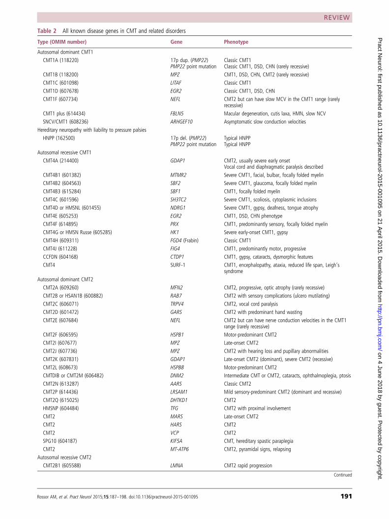
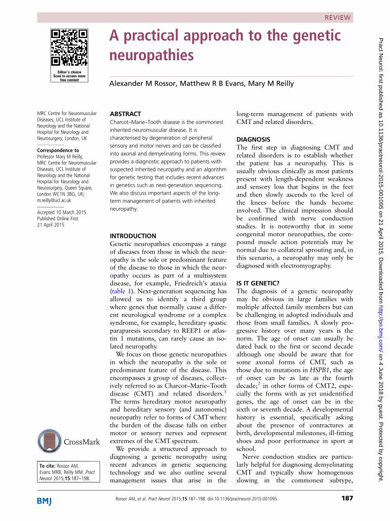
Table 2 All known disease genes in CMT and related disorders
Type (OMIM number) Gene Phenotype
Autosomal dominant CMT1
CMT1A (118220) 17p dup. (PMP22)PMP22 point mutation
Classic CMT1Classic CMT1, DSD, CHN (rarely recessive)
CMT1B (118200) MPZ CMT1, DSD, CHN, CMT2 (rarely recessive)
CMT1C (601098) LITAF Classic CMT1
CMT1D (607678) EGR2 Classic CMT1, DSD, CHN
CMT1F (607734) NEFL CMT2 but can have slow MCV in the CMT1 range (rarelyrecessive)
CMT1 plus (614434) FBLN5 Macular degeneration, cutis laxa, HMN, slow NCV
SNCV/CMT1 (608236) ARHGEF10 Asymptomatic slow conduction velocities
Hereditary neuropathy with liability to pressure palsies
HNPP (162500) 17p del. (PMP22)PMP22 point mutation
Typical HNPPTypical HNPP
Autosomal recessive CMT1
CMT4A (214400) GDAP1 CMT2, usually severe early onsetVocal cord and diaphragmatic paralysis described
CMT4B1 (601382) MTMR2 Severe CMT1, facial, bulbar, focally folded myelin
CMT4B2 (604563) SBF2 Severe CMT1, glaucoma, focally folded myelin
CMT4B3 (615284) SBF1 CMT1, focally folded myelin
CMT4C (601596) SH3TC2 Severe CMT1, scoliosis, cytoplasmic inclusions
CMT4D or HMSNL (601455) NDRG1 Severe CMT1, gypsy, deafness, tongue atrophy
CMT4E (605253) EGR2 CMT1, DSD, CHN phenotype
CMT4F (614895) PRX CMT1, predominantly sensory, focally folded myelin
CMT4G or HMSN Russe (605285) HK1 Severe early-onset CMT1, gypsy
CMT4H (609311) FGD4 (Frabin) Classic CMT1
CMT4J (611228) FIG4 CMT1, predominantly motor, progressive
CCFDN (604168) CTDP1 CMT1, gypsy, cataracts, dysmorphic features
CMT4 SURF-1 CMT1, encephalopathy, ataxia, reduced life span, Leigh’ssyndrome
Autosomal dominant CMT2
CMT2A (609260) MFN2 CMT2, progressive, optic atrophy (rarely recessive)
CMT2B or HSAN1B (600882) RAB7 CMT2 with sensory complications (ulcero mutilating)
CMT2C (606071) TRPV4 CMT2, vocal cord paralysis
CMT2D (601472) GARS CMT2 with predominant hand wasting
CMT2E (607684) NEFL CMT2 but can have nerve conduction velocities in the CMT1range (rarely recessive)
CMT2F (606595) HSPB1 Motor-predominant CMT2
CMT2I (607677) MPZ Late-onset CMT2
CMT2J (607736) MPZ CMT2 with hearing loss and pupillary abnormalities
CMT2K (607831) GDAP1 Late-onset CMT2 (dominant), severe CMT2 (recessive)
CMT2L (608673) HSPB8 Motor-predominant CMT2
CMTDIB or CMT2M (606482) DNM2 Intermediate CMT or CMT2, cataracts, ophthalmoplegia, ptosis
CMT2N (613287) AARS Classic CMT2
CMT2P (614436) LRSAM1 Mild sensory-predominant CMT2 (dominant and recessive)
CMT2Q (615025) DHTKD1 CMT2
HMSNP (604484) TFG CMT2 with proximal involvement
CMT2 MARS Late-onset CMT2
CMT2 HARS CMT2
CMT2 VCP CMT2
SPG10 (604187) KIF5A CMT, hereditary spastic paraplegia
CMT2 MT-ATP6 CMT2, pyramidal signs, relapsing
Autosomal recessive CMT2
CMT2B1 (605588) LMNA CMT2 rapid progression
Continued
REVIEW
Rossor AM, et al. Pract Neurol 2015;15:187–198. doi:10.1136/practneurol-2015-001095 191
on 4 June 2018 by guest. Protected by copyright.
http://pn.bmj.com
/P
ract Neurol: first published as 10.1136/practneurol-2015-001095 on 21 A
pril 2015. Dow
nloaded from

Table 2 Continued
Type (OMIM number) Gene Phenotype
CMT2B2 (605589) MED25 Classic CMT2
NMAN (137200) HINT1 Neuromyotonia and axonal neuropathy, motor predominant
CMT2R (615490) TRIM2 Infantile-onset CMT2
AR-CMT2 IGHMBP2 CMT2
AR-CMT2 HSJ1 CMT2
X-linked CMT
CMTX1 (302800) GJB1 Males CMT1 (patchy NCV); females CMT2
CMTX4 or Cowchock’o syndrome (310490) AIFM1 CMT2, infantile onset, developmental delay, deafness, learningdifficulties
CMTX5 (311070) PRPS1 CMT2, deafness, optic atrophy
CMTX6 (300905) PDK3 CMT2
Dominant intermediate CMT
CMTDIB or CMT2M (606482) DNM2 Intermediate CMT or CMT2, cataracts, ophthalmoplegia, ptosis
CMTDIC (608323) YARS Intermediate CMT
CMTDID (607791) MPZ Intermediate CMT
CMTDIE (614455) IFN2 Intermediate CMT, focal segmental glomerulosclerosis,end-stage renal failure
CMTD1F (615185) GNB4 Intermediate CMT
Recessive intermediate CMT
CMTRIA (608340) GDAP1 Intermediate CMT
CMTRIB (613641) KARS Intermediate CMT, learning difficulty, vestibular schwannoma
CMTRIC (615376) PLEKHG5 Intermediate CMT, SMA
CMTRID (616039) COX6A1 Intermediate CMT, onset first decade
Hereditary motor neuropathy
HMN2A (158590) HSPB8 Classical HMN, dominant
HMN2B (608634) HSPB1 Classical HMN, dominant
HMN2C (613376) HSPB3 Classical HMN, dominant
HMN2D (615575) FBXO38 Classical HMN, dominant
HMN with pyramidal features or ALS4 (602433) SETX HMN with pyramidal signs, dominant
DSMA5 (614881) DNAJB2 (HSJ1) Classical HMN, recessive
HMN5A (600794) or SPG17 (270685) BSCL2 Predominant hand wasting, Silver syndrome but can havesensory involvement as in CMT2D, dominant
HMN5A (600794) GARS Predominant hand wasting, dominant
HMN5B (614751) or SPG31 (610250) REEP1 Predominant hand wasting, pyramidal signs, dominant
HMN6 or SMARD1 (604320) IGHMBP2 Infantile onset, respiratory distress, recessive
SMARD2 or SMAX LAS1L Infantile onset, respiratory distress, X-linked recessive
HMN7A (158580) SLC5A7 Classical HMN, vocal cord palsy, dominant
HMN7B (607641) DCTN1 HMN, bulbar and facial weakness, dominant
SMAX3 (300489) ATP7A Classical HMN, X-linked
SMALED (158600) DYNC1H1 Congenital, contractures, lower-limb predominant, pyramidalsigns, cortical migration defects, learning difficulties, dominant
SMALED2 (615290) BICD2 Congenital, contractures, lower-limb predominant, pyramidalsigns, dominant
PNMHH (614369) MYH14 Typical HMN, distal myopathy, hoarseness, hearing loss,dominant
SPSMA (181405) TRPV4 HMN, scapular winging, vocal cord palsy, dominant
HMN AARS Typical HMN, dominant
HMN HINT1 HMN with neuromyotonia, recessive
Hereditary sensory neuropathy (also called Hereditary sensory and autonomic neuropathy (HSAN))
HSAN1A (162400) SPTLC1 HSN with sensory complications (ulcero mutilating), dominant
HSAN1C (613640) SPTLC2 HSN with sensory complications (ulcero mutilating), dominant
CMT2B (600882) RAB7 HSN with sensory complications (ulcero mutilating), dominant
Continued
REVIEW
192 Rossor AM, et al. Pract Neurol 2015;15:187–198. doi:10.1136/practneurol-2015-001095
on 4 June 2018 by guest. Protected by copyright.
http://pn.bmj.com
/P
ract Neurol: first published as 10.1136/practneurol-2015-001095 on 21 A
pril 2015. Dow
nloaded from

Connexin 32 is also expressed in the central nervoussystem and may cause white matter lesions on brainMRI that are usually but not always asymptomatic.29
CMT4C (SH3TC2)Homozygous or compound heterozygous mutationsin SH3TC2 are the commonest cause of autosomalrecessive demyelinating CMT (CMT4) in the UK andshould be considered in sporadic cases of demyelinat-ing CMT. Affected individuals present in the firstdecade of life and scoliosis is a common feature andmay be severe enough to warrant surgery.7 Mediannerve conduction velocities range from 4 to 37 m/s.30
GENETIC TESTING IN CMT, THE OLD AND THENEWGenetic testing for CMT and many other neurologicalconditions has traditionally involved sequential testingof individual genes using Sanger sequencing. In thisscenario, the most promising candidate gene aftercareful phenotyping is analysed and if negative thenext most likely candidate is tested. This method istime and cost effective for CMT1A due to the 17pduplication but can be very frustrating, expensive andultimately futile in axonal CMT, where each causativegene is individually rare and in as many as 75% ofcases the disease gene is unknown (see figure 1).17
The advent of multiple parallel or next generationsequencing has transformed the approach to genetictesting in CMT.31 The technology allows the masssequencing of a selection of genes (panels), the exome
(containing only the protein encoding sequences) orthe whole genome in a matter of days. There is a com-promise, however, whereby for a given cost, either asmall number of genes can be screened with goodcoverage (depth) or a large number of genes, forexample, a whole exome can be screened but with lessread depth so that the chance of missing a pathogenicmutation increases. It is our current practice to screenfor mutations in patients with CMT and related disor-ders using targeted panels except in the case of CMT1where we advocate testing for the 17p duplication firstas it is cheap and the hit rate is high (see figure 2). Thisis a rapidly advancing field, however, and it is likelythat as the technology advances and the cost falls,whole exome (and eventually whole genome) sequen-cing will replace disease-specific panels.We are aware that some clinical geneticists, faced
with a patient with clinically evident CMT, proceeddirectly to testing for the 17p duplication (as it is thecommonest cause) and only undertake neurophysi-ology if this is negative. We do not advocate thisapproach for two main reasons (except perhaps inchildren where neurophysiology may be poorly toler-ated and the genetic diagnosis of CMT1A has beenconfirmed in another family member). First, eventhough CMT1A is the commonest cause of CMT, itstill only accounts for <40% of all cases of inheritedneuropathy and so testing for the 17p duplicationwithout differentiating demyelinating from axonaltypes equates to a significant unnecessary expenditure.Second, a diagnosis of a particular type of CMT
Table 2 Continued
Type (OMIM number) Gene Phenotype
HSN1D (613708) or SPG3A (182600) ATL1 HSN with sensory complications (ulcero mutilating), spasticity,dominant
HSN1E (614116) DNMT1 HSN, hearing loss, dementia, dominant
HSN1F (615632) ATL3 HSN, bone destruction, dominant
HSAN2A (201300) WNK1 HSN with sensory complications (ulcero mutilating), recessive
HSAN2B or HSAN1B (613115) FAM134B HSN with sensory complications (ulcero mutilating), recessive
HSN2C (614213) or SPG30 (610357) KIF1A HSN with sensory complications (ulcero mutilating), recessive
HSAN3, familial dysautonomia or Rileymia or Rileyt IKBKAP Ashkenazi Jewish, autonomic dysfunction, HSN, absentfungiform papillae, recessive
Insensitivity to pain (24300), paroxysmal extreme paindisorder (167400), primary erythermalgia (133020),small-fibre neuropathy
SCN9A Recessive: insensitivity to painDominant: paroxysmal extreme pain disorder, primaryerythermalgia, small fibre neuropathy
CIPA or HSAN4 (256800) NTRK1 Congenital insensitivity to pain with anhydrosis, recessive
HSAN5 (608654) NGF-B Insensitivity to pain, recessive
HSAN6 (614653) DST Ashkenazi Jewish, autonomic dysfunction, HSN, absentfungiform papillae, death by age 2, recessive
HSAN7 (615548) SCN11A Congenital insensitivity to pain with hyperhidrosis andgastrointestinal dysfunction, dominant
HSAN and dementia PRNP Autonomic dysfunction, sensory loss, dementia, dominant
Hereditary sensory neuropathy with spastic paraplegia(256840)
CCT5 HSN with sensory complications (ulcero mutilating) and spasticparaplegia, recessive
CHN, congenital hypomyelinating neuropathy; CMT, Charcot–Marie–Tooth disease; DSD, Dejerine–Sottas disease; HMN, hereditary motor neuropathy;HNPP, hereditary neuropathy with liability to pressure palsies; HSN, hereditary sensory neuropathy; MCV, motor conduction velocity; NCV, nerve conductionvelocity; SMA, spinal muscular atrophy; SNCV, slowed nerve conduction velocity.
REVIEW
Rossor AM, et al. Pract Neurol 2015;15:187–198. doi:10.1136/practneurol-2015-001095 193
on 4 June 2018 by guest. Protected by copyright.
http://pn.bmj.com
/P
ract Neurol: first published as 10.1136/practneurol-2015-001095 on 21 A
pril 2015. Dow
nloaded from
depends on validating the genetic results and one ofthe important tools in validation is whether thephenotype fits. As the clinical features of differentgenetic types of CMT are often very similar, neuro-physiology is an essential part of the phenotyping(box 2).
CHALLENGES OF NEXT-GENERATIONSEQUENCING FOR THE PRACTISINGNEUROLOGIST: DETERMINING THEPATHOGENICITY OF A NOVEL MUTATIONOne of the main challenges of next generationsequencing technology is in the bioinformatics ana-lysis and the interpretation of the large number ofgenetic variants in known pathogenic genes. Theaverage person has 400 potentially pathogenic var-iants in their exome.33 Thus, when a clinician requestsa CMT panel encompassing up to 50 genes, it is usualto find several potentially pathogenic variants in morethan one gene. Determining which one is the
pathogenic variant is a new skill that is increasinglybeing asked of the clinical neurologist. It is our prac-tice to approach this question by evaluating (1) theclinical phenotype, (2) segregation of the mutationwith the disease and (3) the molecular properties andfrequency of the mutation in healthy controls.
PhenotypeWhen first evaluating a potentially pathogenic mutation,it is important to determine whether the patient’sphenotype fits with what has already been described forthe gene. For example, a novel missense mutation inMFN2 (a gene with a large number of polymorphisms)is unlikely to be the cause of a demyelinating neur-opathy. If a mutation has previously been published forthe patient’s phenotype then this often but not alwaysprovides further evidence for the pathogenicity of amutation. The caveat to this is that many publishedgenes and mutations have only been described in singlefamilies and doubt therefore remains as to their truepathogenicity. With next-generation sequencing, we arealso seeing CMTcaused by genes that traditionally causea different phenotype, for example, REEP1 causingdistal hereditary motor neuropathy rather than heredi-tary spastic paraparesis so the broadening phenotypesseen with different genes needs to be kept in mind.
Segregation in familiesPerhaps the most useful test for determining thepathogenicity of a mutation is to determine whetherthe mutation segregates with the disease. This can betime consuming and requires both affected andunaffected family members to be examined oftenincluding performing neurophysiology and for furtherDNA to be collected and tested. As some forms ofaxonal neuropathy may manifest after the fourthdecade, care must be taken in labelling a familymember as unaffected. In most cases it is necessary to
Figure 2 Suggested algorithm for genetic testing in Charcot–Marie–Tooth disease (CMT) and related disorders in the age ofdisease-specific gene panels. *Motor nerve conduction velocity. HMN, hereditary motor neuropathy; HSN, hereditary sensoryneuropathy.
Box 2 Practical limitations of next-generationsequencing
Next-generation sequencing technology currently cannotreliably detect large exonic duplications and deletionssuch as the 17p duplication (Charcot-Marie-Tooth disease(CMT) 1A) and deletion (hereditary neuropathy withliability to pressure palsies). Multiplex ligation-dependentprobe amplification (MLPA) is the investigation of choicefor detecting such genomic rearrangements. MPLA isalso useful for the detection of exonic deletions in otherCMT genes, for example, in the UK, there is a commonfounder deletion in mitofusin 2 that, in association witha point mutation on the other allele, is a cause of auto-somal recessive CMT2.32
REVIEW
194 Rossor AM, et al. Pract Neurol 2015;15:187–198. doi:10.1136/practneurol-2015-001095
on 4 June 2018 by guest. Protected by copyright.
http://pn.bmj.com
/P
ract Neurol: first published as 10.1136/practneurol-2015-001095 on 21 A
pril 2015. Dow
nloaded from

reserve judgement on unaffected individuals unlessvery elderly or in a family with a large number ofaffected individuals with a similar early age of onset.
Molecular and epidemiological properties of the mutationSeveral predictive programmes, freely available online(eg, Sibyl, PON-P2, Predict SNP, META-SNP) aim topredict the pathogenicity of a missense mutation.While these programmes can help, it is worth remem-bering that for many known pathogenic mutations(eg, pathogenic HSPB1 mutations) some of these pro-grammes have failed to predict pathogenicity.An additional step in determining the pathogenicity
of a mutation is to discover whether the substitutedamino acid is conserved across species as mutations inamino acids that are not conserved are less likely to bepathogenic. This analysis can be easily performedusing the freely available polyphen-2 software (http://genetics.bwh.harvard.edu/pph2/). Finally, searchingfor novel mutations on public databases of singlenucleotide polymorphisms (eg, the exome variantserver) will reveal whether the novel variant is presentin ‘healthy controls’. The presence of a variant in oneof these databases should not be taken as absolute evi-dence that the variant is non-pathogenic as it is likelythat several pathogenic variants have been miscate-gorised as single nucleotide polymorphisms. Theincreasing information about polymorphisms in differ-ent ethnic groups being gained from next-generationsequencing will greatly help in the future to determinewhether a mutation is pathogenic.
MANAGEMENTWhile achieving a genetic diagnosis is an importantpart of the clinical evaluation of a patient with agenetic neuropathy there are several other clinicalaspects to be addressed during the outpatientconsultation.
Foot careCMT and related disorders share many similarities todiabetic polyneuropathy. It is our practice to provideour patients with general advice on foot care and torefer all patients with significant sensory involvementto a chiropodist in order to prevent foot ulcers.
Orthopaedic aspects of CMTThere are three main reasons for orthopaedic inter-vention in CMT and related disorders. These include(1) scoliosis, (2) hip dysplasia and (3) foot and anklesurgery.34 Scoliosis occurs in 26%–37% of patientswith CMT but rarely requires surgical interventionunless there is rapid progression or the degree ofdeformity extends beyond 45°.The prevalence of hip dysplasia in CMT and related
disorders is about 8%35 and is more common inCMT1. X-rays of the hips and pelvis should thereforebe requested in patients with a significant deterior-ation in their gait or if transitioning from paediatric toadult services without previous imaging.
Foot and ankle manifestationsThere are three common foot deformities in CMTand related disorders: claw toes, forefoot (pes) cavusand hind foot varus (ankle inversion; see figure 3).For any patient with foot deformity or weakness werecommend referral to a physiotherapist and/or ortho-tist with an interest in CMT.
Conservative management of foot deformity in CMTThe non-operative management of foot deformity inCMT includes gastrocnemius stretching exercises andthe provision of insoles or ankle–foot orthoses toreduce foot pain and to improve ambulation.36 Thereis a wide variety of ankle–foot orthoses offering dif-ferent degrees of support and rigidity (figure 4).
Figure 3 Common foot deformities in Charcot–Marie–Tooth disease and related disorders. (A) Claw toes, (B) pes cavus and (C)hind foot varus deformities of the feet.
REVIEW
Rossor AM, et al. Pract Neurol 2015;15:187–198. doi:10.1136/practneurol-2015-001095 195
on 4 June 2018 by guest. Protected by copyright.
http://pn.bmj.com
/P
ract Neurol: first published as 10.1136/practneurol-2015-001095 on 21 A
pril 2015. Dow
nloaded from

Surgical management of foot deformity in CMTIf conservative management of foot deformity isunsuccessful, it may be necessary to consider surgicalintervention. There are three main types of foot oper-ation for patients with CMT aimed at improvingambulation. They are soft tissue corrections, osteoto-mies and fusions (see figure 5).34
DrivingIn any patient with peripheral neuropathy and signifi-cant weakness or sensory involvement, it is importantto remind them of the need to inform the drivingauthorities of their condition. For those patients inwhom driving is becoming increasingly problematic,they may be directed to the regional driving assess-ment centres (http://www.rdac.co.uk/) who offer aself-funded disability driving assessment.
Genetic counsellingThere are several scenarios in CMT that require specialconsideration before offering genetic testing. First, pre-symptomatic testing of children aged <18 years at riskof developing CMT is not usually performed. Ourcurrent practice is to only offer testing of at risk childrenwhen they are thought to be affected while acknowledg-ing that the early symptoms may be very subtle with asuspicion of minimal walking difficulties. In this earlysymptomatic scenario, parents sometimes opt for afollow-up appointment to monitor the child rather than
further tests at this stage. In children who are clinicallyaffected, we usually advocate undertaking neurophysi-ology first and proceed to appropriate genetic testing ifthe neurophysiology is abnormal. In a family where thegenetic diagnosis is known to be CMT1A, especially inyoung children where neurophysiology may not be tol-erated as well, it is appropriate to proceed directly totesting for the chromosome 17 duplication.Second, a relatively common scenario in both spe-
cialist neuromuscular and general neurology clinics isfor a patient with CMT to be referred from primarycare as they wish to start a family. Some of thesepatients wish to undergo antenatal testing or preim-plantation genetic diagnosis. Genetic counselling forprenatal diagnosis of CMT is becoming increasinglycomplex. In the case of the common pathogenicmutations such as the 17p duplication, identifying acarrier prenatally is straightforward. Unfortunately,many mutations identified in routine genetic testingare novel, and it can be difficult trying to decide ifthey are pathogenic or simply polymorphisms. In theUK, each individual causative gene must be approvedby a panel before it can be offered for preimplantationgenetic diagnosis.
SUMMARYMost general neurologists will come across CMT andrelated disorders in their daily practice. A positive
Figure 4 Various lower limb ankle orthoses used in the management of distal lower limb muscle weakness in patients withinherited neuropathies. (A) Push Aequi ankle brace; (B) injection-moulded polyethylene Swedish ankle–foot orthosis; (C) siliconeankle–foot orthosis; (D) matrix max carbon fibre ankle–foot orthosis; (E) rigid ankle–foot orthosis; (F) foot-up ankle–foot orthosis.
REVIEW
196 Rossor AM, et al. Pract Neurol 2015;15:187–198. doi:10.1136/practneurol-2015-001095
on 4 June 2018 by guest. Protected by copyright.
http://pn.bmj.com
/P
ract Neurol: first published as 10.1136/practneurol-2015-001095 on 21 A
pril 2015. Dow
nloaded from

family history and slowly progressive disease courseare the strongest clues to a genetic aetiology.Genetic testing in CMT has been transformed by
the introduction of next-generation sequencing.CMT1A due to the 17p duplication remains the com-monest type of CMT and should be tested first in anypatient with sporadic or autosomal dominant CMT1before proceeding to panel or whole exome testing.The new challenge in CMT and related disorders is
in determining the true pathogenic mutation among ahandful of novel variants identified in several knowndisease genes. This can be time consuming andrequires a detailed clinical assessment to define thephenotype and where possible the evaluation ofaffected and unaffected family members.Although there are no treatments for CMT and
related disorders, there is much that can be done toimprove a patient’s quality of life. Physiotherapists,
orthotists and podiatrists can offer advice on orthoticsand stretching exercises to prevent Achilles tendoncontractures; where there is progressive foot deform-ity or pain, an orthopaedic foot surgeon with experi-ence in CMT may be consulted.
ONLINE RESOURCESSibyl: http://bioinformatics.ua.pt/sibylPON-P2: http://structure.bmc.lu.se/PON-P2/Predict SNP: http://loschmidt.chemi.muni.cz/predictsnpMeta-SNP: http://snps.biofold.org/meta-snp/
Acknowledgements MMR is grateful to the Medical ResearchCouncil (MRC), MRC Centre grant (G0601943), and theNational Institutes of Neurological Diseases and Stroke andoffice of Rare Diseases (U54NS065712) for their support. Weare grateful to Shayan Nosrat Jogan for designing andproducing figure 5.
Contributors MMR had the original idea for the manuscriptand edited the initial draft. AMR wrote the first draft andMRBE designed the figures and edited the first draft.
Funding Medical Research Council; National Institute ofNeurological Disorders and Stroke. AMR has been in receiptof fellowship funding from the National Institutes ofNeurological Diseases and Stroke and office of Rare Diseases(U54NS065712) and an IPSEN clinical research fellowship.The INC (U54NS065712) is a part of the NCATS RareDiseases Clinical Research Network (RDCRN). RDCRN is aninitiative of the Office of Rare Diseases Research (ORDR),NCATS, funded through a collaboration between NCATS andthe NINDS. This research was also supported by the NationalInstitute for Health Research University College LondonHospitals Biomedical Research Centre.
Competing interests None declared.
Provenance and peer review Commissioned; externally peerreviewed. This paper was reviewed by Gareth Llewelyn,Cardiff, UK.
REFERENCES1 Reilly MM, Murphy SM, Laura M. Charcot-Marie-Tooth
disease. J Peripher Nerv Syst 2011;16:1–14.2 Houlden H, Laura M, Wavrant-De Vrieze F, et al. Mutations
in the HSP27 (HSPB1) gene cause dominant, recessive, andsporadic distal HMN/CMT type 2. Neurology 2008;71:1660–8.
3 Birouk N, Gouider R, Le Guern E, et al. Charcot-Marie-Toothdisease type 1Awith 17p11.2 duplication. Clinical andelectrophysiological phenotype study and factors influencingdisease severity in 119 cases. Brain 1997;120(Pt 5):813–23.
4 Cottenie E, Menezes MP, Rossor AM, et al. Rapidly progressiveasymmetrical weakness in Charcot-Marie-Tooth disease type 4Jresembles chronic inflammatory demyelinating polyneuropathy.Neuromuscul Disord 2013;23:399–403.
5 Houlden H, King R, Blake J, et al. Clinical, pathological andgenetic characterization of hereditary sensory and autonomicneuropathy type 1 (HSAN I). Brain 2006;129(Pt 2):411–25.
6 Michell AW, Laura M, Blake J, et al. GJB1 gene mutations insuspected inflammatory demyelinating neuropathies notresponding to treatment. J Neurol Neurosurg Psychiatry2009;80:699–700
7 Houlden H, Laura M, Ginsberg L, et al. The phenotype ofCharcot-Marie-Tooth disease type 4C due to SH3TC2mutations and possible predisposition to an inflammatoryneuropathy. Neuromuscul Disord 2009;19:264–9.
8 Murphy SM, Laura M, Blake J, et al. Conduction block andtonic pupils in Charcot-Marie-Tooth disease caused by a myelin
Figure 5 Common foot operations for Charcot–Marie–Toothdisease and related disorders. (A) Various stages of a tendontransfer operation for pes cavus (soft tissue correction) in whichthe posterior tibialis tendon is transferred to the peroneus brevistendon, thereby strengthening both the dorsiflexion andeversion forces on the foot and correcting the foot drop andhind foot varus deformity. Other soft tissue correctionprocedures include Achilles tendon lengthening. (B) Procedurefor a triple arthrodesis in which the subtalar, calcaneocuboidand talonavicular joints are fused permitting some movement atthe ankle. (C) Procedure of a calcaneal osteotomy required tocorrect a fixed hind foot varus deformity.
REVIEW
Rossor AM, et al. Pract Neurol 2015;15:187–198. doi:10.1136/practneurol-2015-001095 197
on 4 June 2018 by guest. Protected by copyright.
http://pn.bmj.com
/P
ract Neurol: first published as 10.1136/practneurol-2015-001095 on 21 A
pril 2015. Dow
nloaded from

protein zero p.Ile112Thr mutation. Neuromuscul Disord2011;21:223–6.
9 Pelayo-Negro AL, Carr AS, Laura M, et al. An observational studyof asymmetry in CMT1A. J Neurol Neurosurg Psychiatry 2014.
10 Liao JP, Waclawik AJ. Nerve root hypertrophy in CMT type1A. Neurology 2004;62:783.
11 Ishigami N, Kondo M, Nakagawa M. [Case of Charcot-Marie-Tooth disease type 1Awith increased cerebrospinal fluidproteins and nerve root hypertrophy]. Rinsho Shinkeigaku2008;48:419–21.
12 Nicholson G, Lenk GM, Reddel SW, et al. Distinctive geneticand clinical features of CMT4J: a severe neuropathy caused bymutations in the PI(3,5)P phosphatase FIG4. Brain 2011;134(Pt 7):1959–71.
13 Udd B. Molecular biology of distal muscular dystrophies–sarcomeric proteins on top. Biochim Biophys Acta2007;1772:145–58.
14 Horowitz SH, Schmalbruch H. Autosomal dominant distalmyopathy with desmin storage: a clinicopathologic andelectrophysiologic study of a large kinship. Muscle Nerve1994;17:151–60.
15 Pitceathly RD, Murphy SM, Cottenie E, et al. Geneticdysfunction of MT-ATP6 causes axonal Charcot-Marie-Toothdisease. Neurology 2012;79:1145–54.
16 Baets J, Deconinck T, De Vriendt E, et al. Genetic spectrum ofhereditary neuropathies with onset in the first year of life.Brain 2011;134(Pt 9):2664–76.
17 Murphy SM, Laura M, Fawcett K, et al. Charcot-Marie-Toothdisease: frequency of genetic subtypes and guidelines for genetictesting. J Neurol Neurosurg Psychiatry 2012;83:706–10.
18 Saporta AS, Sottile SL, Miller LJ, et al. Charcot-Marie-Toothdisease subtypes and genetic testing strategies. Ann Neurol2011;69:22–33.
19 Thomas PK, Marques W Jr, Davis MB, et al. The phenotypicmanifestations of chromosome 17p11.2 duplication. Brain1997;120(Pt 3):465–78.
20 Shy ME, Chen L, Swan ER, et al. Neuropathy progression inCharcot-Marie-Tooth disease type 1A. Neurology 2008;70:378–83.
21 Raeymaekers P, Timmerman V, Nelis E, et al. Duplication inchromosome 17p11.2 in Charcot-Marie-Tooth neuropathytype 1a (CMT 1a). The HMSN Collaborative Research Group.Neuromuscul Disord 1991;1:93–7.
22 Russo M, Laura M, Polke JM, et al. Variable phenotypes areassociated with PMP22 missense mutations. NeuromusculDisord 2011;21:106–14.
23 Chance PF, Alderson MK, Leppig KA, et al. DNA deletionassociated with hereditary neuropathy with liability to pressurepalsies. Cell 1993;72:143–51.
24 Chance PF. Inherited focal, episodic neuropathies: hereditaryneuropathy with liability to pressure palsies and hereditaryneuralgic amyotrophy. Neuromolecular Med 2006;8:159–74.
25 Earle N, Zochodne DW. Is carpal tunnel decompressionwarranted for HNPP? J Peripher Nerv Syst 2013;18:331–5.
26 Ikegami T, Ikeda H, Mitsui T, et al. Novel mutation of themyelin Po gene in a pedigree with Charcot-Marie-Tooth diseasetype 1B. Am J Med Genet 1997;71:246–8.
27 Verhoeven K, Claeys KG, Zuchner S, et al. MFN2 mutationdistribution and genotype/phenotype correlation inCharcot-Marie-Tooth type 2. Brain 2006;129(Pt 8):2093–102.
28 Zuchner S, Mersiyanova IV, Muglia M, et al. Mutations in themitochondrial GTPase mitofusin 2 cause Charcot-Marie-Toothneuropathy type 2A. Nat Genet 2004;36:449–51.
29 Kleopa KA, Scherer SS. Molecular genetics of X-linkedCharcot-Marie-Tooth disease. Neuromolecular Med2006;8:107–22.
30 Dubourg O, Azzedine H, Verny C, et al. Autosomal-recessiveforms of demyelinating Charcot-Marie-Tooth disease.Neuromolecular Med 2006;8:75–86.
31 Rossor AM, Polke JM, Houlden H, et al. Clinical implicationsof genetic advances in Charcot-Marie-Tooth disease. Nat RevNeurol 2013;9:562–71.
32 Carr AS, Polke JM, Wilson J, et al. MFN2 deletion foundermutation in the UK population. In press.
33 Wright CF, Middleton A, Burton H, et al. Policy challenges ofclinical genome sequencing. BMJ 2013;347:f6845.
34 Yagerman SE, Cross MB, Green DW, et al. Pediatric orthopedicconditions in Charcot-Marie-Tooth disease: a literature review.Curr Opin Pediatr 2012;24:50–6.
35 Walker JL, Nelson KR, Heavilon JA, et al. Hip abnormalities inchildren with Charcot-Marie-Tooth disease. J Pediatr Orthop1994;14:54–9.
36 Burns J, Crosbie J, Ouvrier R, et al. Effective orthotic therapyfor the painful cavus foot: a randomized controlled trial. J AmPodiatr Med Assoc 2006;96:205–11.
REVIEW
198 Rossor AM, et al. Pract Neurol 2015;15:187–198. doi:10.1136/practneurol-2015-001095
on 4 June 2018 by guest. Protected by copyright.
http://pn.bmj.com
/P
ract Neurol: first published as 10.1136/practneurol-2015-001095 on 21 A
pril 2015. Dow
nloaded from