REVIEW: “A LITTLE DRAGON-DRANCUCULUS - Ijates · REVIEW: “A LITTLE DRAGON-DRANCUCULUS ......
16
101 | Page REVIEW: “A LITTLE DRAGON-DRANCUCULUS MEDINENSIS” A SURPRISE IN THE MICROBIAL WORLD Sonu Mishra 1 , Virendra S. Gomase 2 1,2 Department of Biotechnology, Mewar University, Chittorgarh, (India) ABSTRACT “Skin” the physical barrier which protects everyone from the external exposed environment.This review aim to discuss about the issue that, what happen when this barrier is used to get rid of a foreign body unnaturally by the parasite .Drancuculus medinensis have been well known and documented from thousands of years. The life cycle wheels of this parasite comprised of six development stages , which chooses a year to complete .Through worm pushes her way , which results in break open the blister to expel her larvae in external environment. At this point of time, the host’s immune system begins to respond against infection.The inflammatory reaction, severe pain in infected site and crippling effects last for months and can become permanents, depending on where the worm emerges .The most interesting characteristics of this nematode infection is that, the in spite of having strong defensive immune system mechanisms of human this organism gets stay and grow for a year without any kind of recognition and realization by host.There is the probability that, this pathogen might be down-regulating the host’s immune response and adapt very quickly to the responses thrown by the host’s body.This organism can even use what body sends out to locate them to there advantage by disguising themselves as part of the host’s body, or using the antibodies as a food source. This review focuses on the biological immunological information about the Dracunculiasis and also focus on the detection, diagnostics and treatment measure taken till date. We have also discussed in brief about the epidemiology of the disease, its social and economical impacts during outbreaks and theinitiative eradication program.It also summarizes the broader benefits uniquely linked to interventions against dracunculiasis. The latest biotechnology and bioinformatics techniques can be used to investigate this disease in more comprehensive way to develop the synthetic vaccine or drugs against this disease, to eradicate it completely. Keywords: GWD,dracunculiasis Antigen, Immunoproteomics, Antigen, Antibody, Bioinformatics, Epitopes. I. INTRODUCTION OF DRACUNCULIASIS Dracunculiasis/ dracuncalosismost commonly known as Guinea warm disease .The parasite agent is Dracunculusmedinesis. These nematodes (round worm) is the largest tissue parasite and only species within the genus Dracunculuswhich infect humans.This bears the characteristics of the water borne filarial nematode.There are other human filarial nematods which is responsible for subcutaneous filariasis like Loa loa (the African eyeworm), mansonellastreptocercaandOnchocerca volvulus (river blindness).The first known mention of the disease worm in the Turin Papyrus in the fifteenth century BC by the Egyptian; it has been since described by ancient Greek, Roman,Arab,Persian and Indian physicians [Groove D.1900].The common name of Dracunculiasis is derived its prevalence on the Gulf of Guinea[Palmer P,Reeder M.2001].The single uterus of
Transcript of REVIEW: “A LITTLE DRAGON-DRANCUCULUS - Ijates · REVIEW: “A LITTLE DRAGON-DRANCUCULUS ......

101 | P a g e
REVIEW: “A LITTLE DRAGON-DRANCUCULUS
MEDINENSIS” A SURPRISE IN THE MICROBIAL
WORLD
Sonu Mishra1, Virendra S. Gomase
2
1,2Department of Biotechnology, Mewar University, Chittorgarh, (India)
ABSTRACT
“Skin” the physical barrier which protects everyone from the external exposed environment.This review aim to
discuss about the issue that, what happen when this barrier is used to get rid of a foreign body unnaturally by
the parasite .Drancuculus medinensis have been well known and documented from thousands of years. The life
cycle wheels of this parasite comprised of six development stages , which chooses a year to complete .Through
worm pushes her way , which results in break open the blister to expel her larvae in external environment. At
this point of time, the host’s immune system begins to respond against infection.The inflammatory reaction,
severe pain in infected site and crippling effects last for months and can become permanents, depending on
where the worm emerges .The most interesting characteristics of this nematode infection is that, the in spite of
having strong defensive immune system mechanisms of human this organism gets stay and grow for a year
without any kind of recognition and realization by host.There is the probability that, this pathogen might be
down-regulating the host’s immune response and adapt very quickly to the responses thrown by the host’s
body.This organism can even use what body sends out to locate them to there advantage by disguising
themselves as part of the host’s body, or using the antibodies as a food source. This review focuses on the
biological immunological information about the Dracunculiasis and also focus on the detection, diagnostics and
treatment measure taken till date. We have also discussed in brief about the epidemiology of the disease, its
social and economical impacts during outbreaks and theinitiative eradication program.It also summarizes the
broader benefits uniquely linked to interventions against dracunculiasis. The latest biotechnology and
bioinformatics techniques can be used to investigate this disease in more comprehensive way to develop the
synthetic vaccine or drugs against this disease, to eradicate it completely.
Keywords: GWD,dracunculiasis Antigen, Immunoproteomics, Antigen, Antibody, Bioinformatics, Epitopes.
I. INTRODUCTION OF DRACUNCULIASIS
Dracunculiasis/ dracuncalosismost commonly known as Guinea warm disease .The parasite agent is
Dracunculusmedinesis. These nematodes (round worm) is the largest tissue parasite and only species within the
genus Dracunculuswhich infect humans.This bears the characteristics of the water borne filarial nematode.There
are other human filarial nematods which is responsible for subcutaneous filariasis like Loa loa (the African
eyeworm), mansonellastreptocercaandOnchocerca volvulus (river blindness).The first known mention of the
disease worm in the Turin Papyrus in the fifteenth century BC by the Egyptian; it has been since described by
ancient Greek, Roman,Arab,Persian and Indian physicians [Groove D.1900].The common name of
Dracunculiasis is derived its prevalence on the Gulf of Guinea[Palmer P,Reeder M.2001].The single uterus of

102 | P a g e
female GWD parasite makes it different from other filarial parasite ,where as filarial have two.The outbreak of
this disease is generally somehow associated with places where there is a lack of clean drinking water eg. Step
wells in India, covered cisterns in Iran, and ponds in Ghana .The life cycle usually involves copepod
intermediate host.They are parasite in the connective tissue or coelom of vertebrates.
1.1 Detection methods of Pathogen (GWD)
The realization of this disease becomes difficult because of its year and half incubation life cycle within the host
body without showing any significant remarks and symptoms. After successful completion of the incubation
period the mature female worm come towards the skin and startthe formation of a small round bulge on the
skinby secreting an irritating chemical. This blisterappearance becomes the red flag of GWD infection. The
blister rapture and the larvae starts releasing, this is only the point when the host‘s immune system recognizes
themas parasite and as foreign and then an allergic reactionbegins. The symptoms expressions of the infection
are highfever, redness, swelling, and pain (at the site of the worm‘s location on the skin, usually on the lower
extremities) [Muller, (1979); Molyneux et al., (2004)]. Theadditional systemic symptoms include vomiting,
nausea, diarrhea and dizziness. These symptoms can persist for several days and later on begin to diminish when
the worm is manually forced out by slowly winding it onto a stick over several weeks to months [Molyneux, D.,
D. Hopkins, N. Zagaria. 2004].Sadly, but there is no other way of detecting this guinea worm disease other than
the noticing a blister somewhere on the lower extremities. Till date thereis no confined assay tests has been
design for its early onset stages detection.
1.2 Classification, Morhology and Biology of D. medinensis
The filarial nematode Dracunculus genus belongs to the family:Dracunculidae,superfamily: Dracunculoidea,
order: spirurida, class: chromadorea,phylum: Nematoda.The Dracunculidaefamily consists of two genera
Dracunculusand Micropleura. Furthermore, Dracunculus genus can be divided differently into 12
species:Dracunculusbrasiliensis,Dracunculusdanomensis,Dracunculusdoi,Dracunculusfaelleborni,Dracunc
ulusglobocephalus,Dracunculushoudemeri,Dracunculusinsignis,Dracunculuslutrae,Dracunculusmedinensis
,Dracunculus mulbus,Dracunculusoesophageus, Dracunculusophidensis[Muller ,R., 1971; Muller,R., 1979;
Jones,h.I., and Mulder, E.,2007;Moravee,F, and Santos C.P, 2009].From all the Dracunculusspecies ,only
D.medinesis (a little dragon from Medina ) infects humans,causes“Guinea Worm Disease”.Whereas, other
Dracunculusspecies generally resides in the internal tissues and body cavities of non-human mammals and
reptiles( snake and turtles)[Bimi,L., et al., 2005].Dracunculusmedinensis taxon closely related to the Filarioidea
under the order Spiruida .These two super-families members share similar type of general morphology and all
are tissue dwelling parasites,ovoviviparous, and their intermediate hosts is arthropod.Frequently, these two taxa
discussed broadly in filarial nematode group[HotezPJ., 2013].Dracunculusspecies can be described differently
from true filarial nematodes based on certain specific morphological features, molecular phylogenies and life
cycle .The relationship between Dracunculusspp. and other spirurids (B.malayi and W.bancrofti) can be
distinguish based on the phylogenetic analysis of 18srRNA gene sequence .It has been seen that in spite of
having similar morphological characteristics D. oesophageusreprents different species from human Guinea
worm. The dracunculiasisprecisely denoted as both a disease of poverty and cause of poverty. Sometimes, due
to outbreaks of this peculiar disease, large number of village population get into trap, which ultimately results in
103 | P a g e
agricultural productivity, maternal and child health and the school attendance [Ruiz-
TibenE,HopkinsDR.,2006].In numberof all neglected tropical disease, it has a low mortality, but morbidity is
considerably high due to huge disabilities which was physically and economically devastating[Ramakrishna
J,Brieger WR, Adeniyi JD, Kale OO.(2006-2007);FeaseyN,Wansbrough-Jones M
,MabeyDC,SolomonAW.Neglected tropical disease.Br Med Bull 2010;93:179-200].The adult female worm
measures upto one meter in length, whereas, the male measures about 2cm[Molyneux, D., D. Hopkins, N.
Zagaria., 2004].
1.3 Properties of Dracunculus medinensis Environments
Dracunculusmedinensis thrivesin fresh water habitats usually in stagnant waters such as ponds, reservoirs, dried
up pools in riverbeds, and dug up water holes. Reason behind to thrives in these kind of environment is that ,that
the water never gets disturbed or moved and the larvae can able to multiply in the stagnant waters without any
external disturbance. This kind of natural territory are mostly the reservoirs for copepods- the vector of
Dracunculusmedinensis . The copepods eat the larvae and get infected with the disease they carry. The infected
larvae survival is only last for an about only three days in the water before it dies and doesn‘t harm to the
copepods even if they eat the dead larvae. The known reported cases of dracunculiasis in sub-Saharan Africa
where the climate is found dry at least part of the year,which is the most favorable environment for worm
[Greenaway, 2004]. This parasite can be ranked as a xenophile ,an obligate thermophile, because of its habitat
which has mostly observed in the dry, warm places like - sub-Saharan Africa.
II. BIOLOGY OF THE PARASITE
The stagnant contaminated drinking water with infected copepode is the transmission source of the
Dracunculiasis .The cyclops(The small water fleas)are the one which act as the intermediate host of
pathogenesis of GWD. The moment when populationconsume this unfiltered water from stagnant pods or open
a shallow well which is contaminated with infected copepods withpathogenicDraculiasislarvae.After ingestion,
the cyclops dies by the stomach digestive juices which interns results in larvae release. This larvae penetrate the
digestive wall into the body cavityand get entry in abdominal cavity and retroperitoneal space .The larvae
develop and mature into adults ;after copulationthe ovoviviparous female grows upto 1 meter(60-100cm/3 feet)
length, but only 1 to 2mm thickness in the subcutaneous tissue of skin and have survival upto 12 to 19 months
.Whereas male dies soon after copulation with few months[Greenaway C., 2004].No symptoms manifest till a
year or so .With 12 to 14 months after infection female worm .By the time produced millions of eggs in its
uterus,and it gravid with microfilariae migrateand predominantly localized in the lower extremities(80-
90%).After an incubation period the female worm release which induces a painful blister (1 to 6cm diameter )
on the skin of lower limbs ; the person develop a slight fever , local skin redness , swelling and severe pruritus
around the blister .The presence of other symptoms like diarrhea, nausea, vomiting and dizziness has been seen
.The blister burst within 1 to 3 days and female worms one or more slowely comes out from the wounds which
causes an excoriating burning sensation and pain [Miillner A, Helfer A ,KotlyarD.Oswald J,
EfferthT(2011)].Immersing or pouring water over the blister provide pain relieve. But this the moment that adult
female exposed to the external environment [Ruiz-Tiben E, Hopkins DR.(2006].And while emerging the limbs

104 | P a g e
in open water sources it recognizes the temperature difference and releases the milky white liquid in the water
which contains millions of immature larvae , when larvae released in water are ingested by copepods where they
mount twice and become infective larvae within two weeks [IriemenamNC,Oyibo WA, Fagbenro-BeyiokuAF.(
2008].The cycle begins a new shift if the contaminateds drinking water is ingested by any individuals.[ Hopkins
DR,Ruiz-Tiben E, Downs P,Withers PC jr,Roy S. (2008); Muller R.(1985)][Fig. 6& Table:1].Once the blister
comes in contact with the water it bursts and kill the worm and the infected larvae comes out its mouth into the
water and the host wound. This is only the time when immune system of the host body recognizes it as the
foreign body and start producing the immune responses against it by sending eosinophils, basophils, and other
specialized granular cells to the scene (where the larvae have been released). They release toxic chemicals in
attempt to destroy the invader. B- Cells then make antibodies specifically for the parasite. The antibodies then
attach to the invader and act as signals and communicate to other immune system cells to destroy them. There is
some evidence which suggest that the antibodies are used as a food source, and that the worms are able to
trigger the immune system to make more antibodies. They can even disguise themselves as part of the host‘s
body by displaying different proteins on their surfaces that identify them as part of the host [Molyneux, 2004]. It
is a pretty nasty parasite, and investigations on still being made to really understand the mechanisms of this
organism.The treatment measures includes the extraction of the worm manually and painkiller medication to
ease the pain. It can take months to recover from the extracting of the worm mainly because one‘s need to be
very carefully while extracting the worm slowly. With this fevers and wells and pain and the other symptoms
with accompany the process until the worm is completely out. Secondary bacterial infections are also a problem
with the lesions; the bacterial infections cause increased pain and may lead to locked joints or permanent
crippling [Molyneux, 2004]. The infection is resolved and back to normal when the worm is completely
extracted. As the body recovers it is important not infect the wound, and to properly filter water when using it.
The guinea worm disease is not communicable, so the chances of contracting the disease are at none from
person to person. Treatment measures in Africa are being practiced to reduce the number of cases as well as
trying to eliminate this preventable disease.
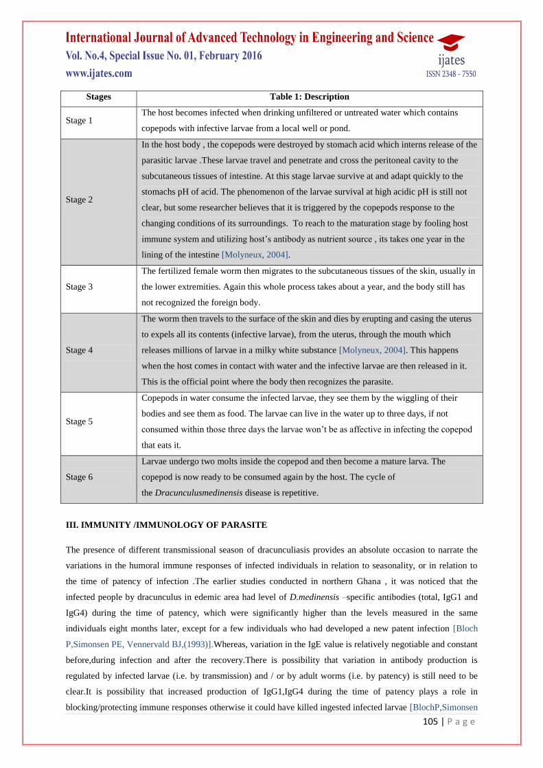
Table 1-Life cycle stages of GWD
105 | P a g e
Stages Table 1: Description
Stage 1 The host becomes infected when drinking unfiltered or untreated water which contains
copepods with infective larvae from a local well or pond.
Stage 2
In the host body , the copepods were destroyed by stomach acid which interns release of the
parasitic larvae .These larvae travel and penetrate and cross the peritoneal cavity to the
subcutaneous tissues of intestine. At this stage larvae survive at and adapt quickly to the
stomachs pH of acid. The phenomenon of the larvae survival at high acidic pH is still not
clear, but some researcher believes that it is triggered by the copepods response to the
changing conditions of its surroundings. To reach to the maturation stage by fooling host
immune system and utilizing host‘s antibody as nutrient source , its takes one year in the
lining of the intestine [Molyneux, 2004].
Stage 3
The fertilized female worm then migrates to the subcutaneous tissues of the skin, usually in
the lower extremities. Again this whole process takes about a year, and the body still has
not recognized the foreign body.
Stage 4
The worm then travels to the surface of the skin and dies by erupting and casing the uterus
to expels all its contents (infective larvae), from the uterus, through the mouth which
releases millions of larvae in a milky white substance [Molyneux, 2004]. This happens
when the host comes in contact with water and the infective larvae are then released in it.
This is the official point where the body then recognizes the parasite.
Stage 5
Copepods in water consume the infected larvae, they see them by the wiggling of their
bodies and see them as food. The larvae can live in the water up to three days, if not
consumed within those three days the larvae won‘t be as affective in infecting the copepod
that eats it.
Stage 6
Larvae undergo two molts inside the copepod and then become a mature larva. The
copepod is now ready to be consumed again by the host. The cycle of
the Dracunculusmedinensis disease is repetitive.
III. IMMUNITY /IMMUNOLOGY OF PARASITE
The presence of different transmissional season of dracunculiasis provides an absolute occasion to narrate the
variations in the humoral immune responses of infected individuals in relation to seasonality, or in relation to
the time of patency of infection .The earlier studies conducted in northern Ghana , it was noticed that the
infected people by dracunculus in edemic area had level of D.medinensis –specific antibodies (total, IgG1 and
IgG4) during the time of patency, which were significantly higher than the levels measured in the same
individuals eight months later, except for a few individuals who had developed a new patent infection [Bloch
P,Simonsen PE, Vennervald BJ,(1993)].Whereas, variation in the IgE value is relatively negotiable and constant
before,during infection and after the recovery.There is possibility that variation in antibody production is
regulated by infected larvae (i.e. by transmission) and / or by adult worms (i.e. by patency) is still need to be
clear.It is possibility that increased production of IgG1,IgG4 during the time of patency plays a role in
blocking/protecting immune responses otherwise it could have killed ingested infected larvae [BlochP,Simonsen

106 | P a g e
E.PAUL,(1998)].The recent studies on response profile of guinea worm –specific cellular cytokines and
reactivity evaluation of parasite-specific antibody subclass shows that the antigen-specific depression found
similar for IFN-gamma and T-helper type2 cytokines IL-10 production in patent, post-patent and control
individual .Whereas the Il-5 production was always the highest in control[Knopp S, Amegbo IK, Hamm
DM,Schulz-Key H,BanlaM,Soboslay PT.(2008)].While examine for presence and localization of human serum
albumin and immunoglobulins by immunoelectrophoretic technique, it‘s found that anti-human albumin
antibodies reacted to the both stages of parasite which was recovered from infected individuals. Moreover,
antigens mimicking human albumin and human immunoglobulins(isotypes Ig G) were found on the surface of
adult female worm while conducting investigation through direct fluorescence . This investigation suggest that
there might be possibility that due to occurrence of host-like compound on the parasites may be the reason for
the parasite survival in host body and adaptation[Bloch P,LundM,VennervaldBJ,Simonsen PE(1999)].There are
different types of immunodiagnostic methods has been used to detect the antibody response of the pre-patent
and patent D.medinensis infected individual .There are the possibility to detect the early asymptomatic infection
six month in prior before worm emerges from the infected individual through ELISA immunodiagnostic
method [BapnaS,Eenapurkar DM(1996].The other specific and sensitive methods like dot-enzyme-linked
immunosorbent assay(dot-ELISA) [Prakash D,Parab PB, Sharma RN.(1993)] , The Falcon assay screening test-
enzyme-linked immunosorbent assay(FAST-ELISA) and the enzyme-linked immunoelectrotransfer blot(EITB)
in order to assess adult worm antigen and their potential value. [FagbemiBO,Hillyer GV(1990)],
Immunoelectroblotting and ELISA to identify non cross –reacting antigenic compound proven to be possible
immunodiagnostic method for the detection of infection [Garate T, Kliks MM , Cabrera Z,Parkhouse
RM(1990)].The usual property of dracunculiasis compare to other parasite there is less facts on the acquired
immunity .It is difficult to detect by most standard tests such as blood and skin biopsy in prior to rupturing of
blister.It is important to understand that how this parasite conceal themselves for more than an year incubation
period in spite of having strong human immune defense system .The usual symptoms which arise from the
parasite infection are the result of the interactions between the immune system and the parasite.
3.1 The expression of Immune System against the parasitic infection
Firstly , through inflammatory responses by the esinophils, basophils and other specialized granular cells
release their toxic chemical to attempt to destroy the invader. Later on after activation of B cells starts
making antibodies against the specific parasite .These antibodies get attached to invader and act like a
signal flare, and communicate the signals to other immune cells to destroy the foreign antigen .The
immune cells like phagocytes, which travel throughout the body to phagocytos the foreign body. Which
are not recognized as body belonging .These cells are much more effective at destroying any
worms/parasite that labeled by antibodies.
Secondly,there are proteins like complement protein which make up complement system that are able to
recognize some general characteristics.This protein rarely capable to kill targets by forming a membrane
attack complex. The amazing partis the interaction of worm and immune system .Both play hide and seek
for their own mutual benefits .There is the probability by which the worm hide themselves from immune
system by degrading the antibodies that attach to them.

107 | P a g e
Some evidence suggests that these worms used antibodies as their food source and that the worms are able to
triggerthe immune system to produce large amount of antibodies.The parasites identify themselves through
displaying different proteins on their surfaces which is recognized as a part of host. The parasite approach to
hide them from host immune system: (1) They avoid the complement proteins from attaching to their surface;
(2) They avoid killer protein to destroy them by releasing different types of molecules that act as decoys; (3)
They also produce other proteins which protect them from phagocytosis.Although the process of
accomplishment is still unknown.Nematodes generally infect the digestive system, but mostly the infections are
asymptomatic, which signifies that these worms are capable of down regulation of host‘s immune responses
against them.The analysis on adult guinea worm was conducted and found that there were morphine and its
active opiate, alkaloid metabolite morphine-6-glucoronide (M6G).There might be a chance that this substances
is utilized byD.medinensis to evade immune system.The lack of immune response to the living host is due to
release of opiate chemical.Morphine release from parasitic nematodes cause immune cell inactivation, and when
the worm recognized the immune cells it is triggered to produce the morphine.This can be one of the way by
which parasite can suppress the immune system[Zhu,W., et. Al (2002)]. The other study conducted via ELISA
and western blot technique is used to identify and detect the antibody responses of total and isotypes of
IgG1,IgM,IgA and IgE).to conduct this test sera is collected from infected person at early and late in the peak
transmission period and as well as from persons with signs of the disease and after the infection had cleared.The
ELISA absorbance values obtained from sera of the same individualvaried between the two transmission
seasons with the highest titers were obtained towards the end of the peak transmission period .Western blots
were obtained while detecting the isotype of IgG4 antibodies.This study suggest that parasites use antigenic
cloaking, that the reason that response of antibody not observed until the larvae are about to release from the
infected person which makes difficult to detect at early onset of the disease.The antigenic cloaking is observed
when Guinea worm binds to the hosts proteins to its surface, so that the host‘s immune system will identify it as
self.This investigation prove the theory that when the parasite first gets entry to the host it is hidden from the
host immune system completely and with respect to the time (progression)progress the host body starts realizing
the foreign body.Although the worm is good at cloaking itself, and immune system starts to breakdown
[Bloch,P., Simonsen,P.E.(1998)].―Autoimmunity/Allergy and hypersensitivity‖ Journals, discuss about the
strengthen ofimmune system response and the obviousness of most common symptoms , When IgE binds to
FcY receptors on the surface of mast cells, basophils and eosinophils which causes smooth muscle contraction
of surrounding the airways and the gut.The strong muscular contractions leads to expel the parasites from the
gut or airways which in terns increased local blood flow and can help to flush parasites out from the body with
the combination action of IgE, mast cell , basophils, and eosinophils.Eosinophils utilize its FcE receptors for
direct action against multicellular parasites .Whereas phagocytic cells cannotphagocytos the whole worm
because of its large size in spite of large number of phagocytes. The mast cells responses is most frequently seen
as a disadvantage in humans who rarely encounter parasites, because its triggers allergies and asthma.This is
part of the Hygiene Hypothesis.Individual with these conditions produces IgE in responses to relatively
harmless substances called allergens.Some individual produces very specific IgE which can leads to massive
degranulation of mast cell, which show anaphylaxis [Capton.M,Kinet,J.(2005)].

108 | P a g e
IV. CLINICAL ASPECTS OF DRACUNCULIASIS
Clinical manifestation and pathogenesis pre-emergent female worms can move very easily through the
connective tissues, while emerges to the surface, a few larvae gets released into the sub-dermis through a rupture
at the anterior end. The blister which burst later in few days gives a shallow ulcer and a marked inflammatory
response against the cuticle of the entire worm, which prevent its removal. The bacteriologically sterile blister
fluid contains larvae surrounded principally by polymorphonuclear neutrophils with macrophages,lymphocytes,
and eosinophils [Muller R.(1976)] The thousands of larvae when expels at the end of worms dries up, and this
process is repeated a few times with the complete worm being extruded in a few weeks andsoon after that the
lesion heals quickly. It has been observed in most of the cases the unfortunate secondarily infection and patients
incapacitated severely.Ghana study suggest that 28% of patients continue in pain for 12 to 18 months after
emergence of worm and 0.5% had suffered from permanent physical impairment due to ―locked‖ knees or other
joints related issues[Hours,M., and S.aincross.(1994)] .The study in Benin suggest that there eas 0.3% mortality
from tetanus and septicemia [Chippaux,J.P., and A.Massougbodji.(1991)].In few cases female worms burst in
the tissues which results in large pus-filled abscess and severe cellulitis, whereas, infertile females or males
elicit a slight inflammatory reaction and sometimes calcify, showing up on a roentgenogram.In the
Dracunculiasis parasitic infection very little evidence of acquired immunity has observed and there are
possibilities of number of reinfection of the same individual. The response to the extrusion of larvae is
indicative of an Arthus reaction followed by a delayed hypersensitivity response [Muller R.(1976)]
V. DIAGNOSIS OF DRACUNCULIASIS
In the endemicity area patients have no doubt about the diagnosis when, or just before the blister from the local
itching and then sharp pain and often general allergic symptoms including urticarial follow. When the blister
burst, cold water encourages release of larvae, which can be observed under low power microscope. Due to lack
of pre-paten serum samples it‘s difficult to go ahead with immunodiagnostic methods and it not useful in
practice because prepaten infection detection is notpossible.Enzyme –linked –immunosorbent assay can be used
to detect the antibodies using whole worm antigen of infected individuals.Immunoglobin G4 is the most specific
reaction can be used for disease diagnosis [Bloch , P., P.E.Simonsen, and B.J.Vennervald.(1993)] . This test can
detect prepatent infections upto 6 months before emergence [Bapna,S., and D.M.Renapurkar.(1996)], in which
case it could have practical important. The presence of circulating antigen evidences has not been detected
[Bloch,P.,B.J.Vennervald, and P.E.Simonsen.(1998)].
VI. MEDICAL CARE OF DRACUNCULIASIS
Till date there is no accurate and efficient curative drug of vaccine is available against dracunculiasis[Muller
R.1985].Immunity is not developed by the infected individual [Cairncross S, Muller R, Zagaria N.(2002); Issaka
–Tinogah A, magnussenp,BlochP,Yakubu A.(1994);Greenway C. (2004)].The ‗traditional treatment‖ i.e-pulling
out the worm gradually and manually by winding the worm a few inches/centimeters on a small wooden stick
each day, which is usually a very distress procedure [Hopkins Dr, Ruiz-Tiben E, Downs P , Withers PC Jr, Roy
S .(2008)]. The traditional treatment customarily taken weeks to months to shed the complete length worm,
109 | P a g e
during this period the infected individual become often severely incapacitated. Furthermore, approximately
every infected individual become victim/sufferer of secondary bacterial infection. The untreated lesions may
cause several complication without antibiotics treatments alike erysipelas/cellutitis, abscesses, sepsis,septic
arthritis and even trismus (lock jaw caused by tetanus infection)[Iriemenam NC ,OyiboWA,Fagbenro-
BeyiokuAF(2008)].The infected patients needs to be careful and avoid breakdown of the worm in manual
extraction procedure , by any chance of worm breaking can leads to intense inflammation because the left out
part of dead worm disintegrated in the affected limbs .Treatment includes winding up worms out on stick,
combined with clean dressing and antibiotic ointment to prevent from secondary bacterial infection and pain
killer drug [Magnussen,p.,A.Yakubu, and P.Bloch(1994)]. Till date there is no evidence of any
chemotherapeutic agent has a direct action against guinea worms, although various benzimidazoles may have an
anti- inflammatory action, aiding elimination [Nwoke, B. E. (1992)].Similarly, aspirin is equally effective
(Muller,unpublished). Study suggests, the effectiveness of Ivermectin against other nematodes but it has no
effect on this organism nor in experimental infections [Eberhard, M. L., F. H. Brandt, and A. Hightower. (1990);
Issaka-Tinorgah, A., P. Magnussen, P. Bloch, and A. Yakubu. (1994)].Chippaux[Chippaux, J. P. (1991)] found
that treatment with mebendazole was associated with aberrant migration of the worms, which were more likely
than usual to emerge at places other than the lower limbs.
6.1 Precaution Measures
By avoiding contaminated source of drinking water
Filtering unsafe water with cloth and fine-mesh strainers before consumption
Use of drinking water from improved sources and controlling the vectors of transmission.
VII. EPIDEMIOLOGY, ECONOMICAL AND SOCIAL IMPACT DUE TO
DRACUNCULIASIS
Dracunculiasis affect the masses / people that depend on contaminated drinking water sources, stagnant water
source such as ponds.The 15% to 70% of the whole population gets affected by Dracunculiasis who depend on
the stagnant water as the water sources. This is quite threaten, slow down the economic development and life
style of infected individuals [OjoTB,OjoKK. (2011)].Reason is that it mainly affects the most productive people
(12-50 years old), sustaining the disease-provertycycle.The evidence of the incidence variation has been seen
between gender,age, occupation usually identified based on their water resource that from where they use water
for drinking purpose.This disease transmission has got a seasonal pattern and closely related to rainfall .In arid
areas; people get infected during the rainy season, when surface water is available. Along with this its infections
has also seen in the wet regions during the dry season, when the consuming water source become scarce and
stagnant.The disease transmission pattern and seasonal clinical manifestations often coincides with harvest or
planting seasons and significantly affects productivity of agricultural product.For an example in Nigeria, due to
this disease an approximately 11.6% decrease in total rice production. Estimately, the infected individual loose
100days of works per year, school attendance gets affect due to GWD. The world bank has estimated that the
economicsrate of return on the investment in GWEP will be about 29% per year once the disease is
eradicated[RamkrishnaJ.BriegerWR,Adeniyi JD, Kale OO.(1985-86)].

110 | P a g e
7.1 Socioeconomics Impact
In current scenario, the understanding has been development the only biological and technical feasibility is not
the only criterion to consider before launching an eradication program, simultaneously costs and benefits are no
less important[Dowdle, W. R., and D. R. Hopkins. ed. (1998)].The condition benefits of dracunculiasis in
contrast to those of smallpox and polio will accrue almost exclusively to the population in which the disease is
endemic[Aylward, B., K. A. Hennessey, N. Zagaria, J.-M. Olivé, and S. Cochi. (2000)].Earlier the
dracunculiasis cases went unreported due to number of reasons.Most health centers had very little to offer the
patient besides palliative treatment; most patients live in poor, remove rural areas and are hindered by their
disease from walking to a health facility; and most recover spontaneously after expulsion of the worm .Nerthe
less , the endemicity of affected area and is social , economical and educational I consequences and the cost
incurred by the individuals, households, and communities suffer from it and can be substantial.
7.2 Economic Impact
The economic impact can be estimated by disease is multiplied the number of days of labour lost by the mean
value of production per day /by the wage rate.A study in Nigeria based on the survey of 87 households,
concluded that the three states of southern Nigeria of rice- growing area substained an $ 20 million annual loss
due to Dracunculiasis[de Rooy, C., and L. D. Edungbola.(1988)].But this simple arguments, mobilized the
senior politician of Nigeria for the disease eradication[Edungbola, L. D, et al.,(1992)].This type of calculation
method has been argued previously used as oversimplified approach and is likely to overestimate the coast
[Guiguemdé, T. R., et al.,(1986);Paul, J. E. (1988)] as it doesn‘t permits for the various coping strategies by
which households respond to illness(such as abandoning other tasks and using additional labour) found to be
common in peasant farming [Brieger, W. R., S. Watts, and M. Yacoob. (1989);Chippaux, J. P., A. Banzou, and
K. Agbede. (1992)].
7.3 Disability of Infected individual
Dracunculiasis is rarely found fatal; the generous study conducted in India on medical records suggest 0.1%
[Imtiaz, R., D. R. Hopkins, and E. Ruiz-Tiben. (1990) ;Rao, C. K., and G. V. M. Reddy. (1965) ;Singh, J., and
N. G. S. Raghavan. (1957)].The social Impact of Dracunculuiasis is majorly attributable to the temporary
disability suffered by the patient.The study conducted in Nigeria [Bhatt, A. N., and K. H. Palan. (1978);Smith,
G. S., D. Blum, S. R. A. Huttly, N. Okeke, B. R. Kirkwood, and R. G. Feachem. (1989)]. Suggest that 58 to
76% of Guinea worm disease suffering patients are unable to leave their beds for approximately a month during
and after emergence of the worm.The secondary infection of the lesion is the more severe and protracted
disability had observed in half of the cases [Nwosu, A. B. C., E. O. Ifezulike, and A. O. Anya. (1982); Wurapa,
F. K., D. W. Belcher, and W. B. Ward. (1975)].This disease impact is leads to temporary disability and
reinforcement by the patency period of worm transmission,often peaking at the stage of the agricultural year
when the labour is in maximum demand.Indeed , it has been claimed that the effect of the disease on agricultural
productivity can be identified in satellite photographs [Ahearn, S. C., and C. de Rooy. (1996)].‗The disease of
the empty granary‘ is referred by the Dogon people of Mali [World Health Organization. (1998)].The Guinea
worm disease effect doesn‘t end when the worm comes out and the sufferer returns to their normal life style.The
study in Ghana [Hours, M., and S. Cairncross. (1994)] suggest that between 12 to 18 months after the
111 | P a g e
emergence of worms, 34% of patient find difficulty in performing everyday activities, reason may be due to pain
attributable by its location and the date of onset to the episode of Guinea worm disease.But this types of
disability is not necessarily to be permanents, it extends beyond the incapacity occurring worm emergence.
VIII. THE INITIATIVE ERADICATION PROGRAMS
Dracunculiasis is the one of the promising candidate for the successful eradication after its first pointed
registered report [Muller, R. (1979)].It‘s prevention in seasonal campaigns permits a more intensive focus
because of its markedly seasonal out breaks. The extensive effort for its prevention and control was taken care
by the members of the Centers for Disease Controland Prevention(CDC),who began the advocacy campaign in
1980 and sustained over more than a decades and highly succeeded in convincing former U.S. president Jimmy
Carter, the United Nations Children‘s Fund (UNICEF) Executive Board, the 1989 African Regional Committee
of the World Health Organization (WHO), and the 1990 World Summit for Children to take up the challenge.
The World Health Assembly in 1991 declared the eradicating dracunculiasis by end of 1995.In order to establish
the eradication programe and to advocate about the disease the efforts needed to be replicated by each country
[Edungbola, L. D., et al.,(1992)] .India taken the first initiative step in 1982 for eradication of GWD. Later on,
Pakistan, Ghana, Nigeria, and Cameroon in 1990 taken the initiative of the eradication programe .In duration of
5 years, all the other known countries of endemicity also established national eradication programs, and
substantial and progressive reductions in disease incidence were recorded each year, particularly at the
beginning of the campaign. UNICEF and WHO maintained a joint technical team based in Ouagadougou,
Burkina Faso, to provide technical support to national program coordinators in the region and to external
support agencies(From 1992 to 1996).The resultant of this type of extensive eradication program from different
supported agencies and the organization led to a remarkable decline of the number of cases almost 98%, from
an estimated 3.3 millionworldwide in 1986 [Watts, S. J. (1987)] to only 75,223 cases reported in 2000.In 2000,
Only 14 countries, all in Africa, reported indigenous case [WHO(2001)].The number of dracunculiasis cases
reported worldwide in 2013 declined by 73% compared with 2012, and by 71% during January–June 2014
compared with January–June 2013. Transmission remains endemic in four countries, with South Sudan
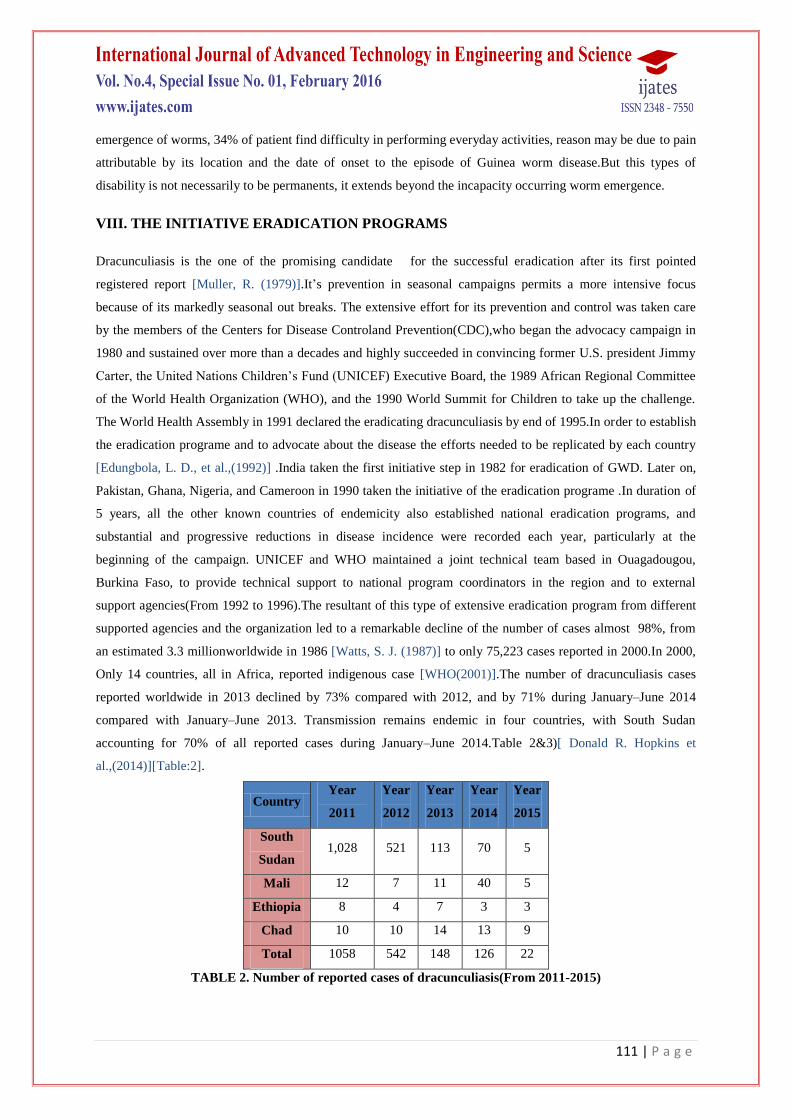
accounting for 70% of all reported cases during January–June 2014.Table 2&3)[ Donald R. Hopkins et
al.,(2014)][Table:2].
Country Year
2011
Year
2012
Year
2013
Year
2014
Year
2015
South
Sudan 1,028 521 113 70 5
Mali 12 7 11 40 5
Ethiopia 8 4 7 3 3
Chad 10 10 14 13 9
Total 1058 542 148 126 22
TABLE 2. Number of reported cases of dracunculiasis(From 2011-2015)

112 | P a g e
8.1 Interventions
In the direction of the dracunculiasis prevention it is necessary to select and develop the most effective
interventions andsimultaneously the apt of field experience is more reliable than only having the theory or basic
biology .A number of intervention would seems to be taken into the consideration:
(i) Supply of a safe water
(ii) To remove Cyclops, one‘s should drinking filtrated water
(iii)Investigation and identification of the active cases patients and its proper management
(iv) Advocating and aware patients to avoid contact with ponds
(v) Shooting down or withdrawing cyclops from ponds
IX. FUTURE PROSPECTIVE
There is the need for more timely and comprehensive methods of monitoring and control of the infectious
disease. The Identification, screening, classification and high throughput screening of nucleotide and protein
sequences of nematode protein needs to be carried out to analyze the toxin‘s sequences which is responsible for
the disease progression using computational biology, advanced biological databases and bioinformatics.
Advanced database management system (DBMS) will use for data mining and data manipulation. It leverages
from recent breakthroughs in high-throughput molecular profiling of pathogen and text mining as well as upon
the growing electronic sources of knowledge about the molecular epidemiology of pathogens with epidemic
potential.The pathogen profiling and biosurveillance focused text mining tools can enable integrated infectious
disease outbreak detection and response environments based upon bioinformatics knowledge models and
measured by outcomes including the accuracy and timeliness of outbreak detection.This concept can be used for
effectively in public health and disease management .To find the high throughput screening of sequence,
sequence analysis, computational modeling and patterns comparison of protein, expression analysis, functional
annotation and target confirmation for disease profiling .Identification and high throughput screening
ofsequence that are involve in disease progression and may be novel therapeutic target for disease. It is plausible
that there are minor differences in toxin/protein expression profile based on the disease aggressiveness, which
may be exploited for development of biomarkers for different stages of disease. The proposed study will
establish that modulation of expression profiles of disease related proteins and may be better therapeutic
approach for disease treatment. Bioinformatics is being increasingly used to support target validation by pro-
viding functionally predictive information mined from databases and experimental datasets using variety
computational tools .Application of structural and functional genomics and proteomics approaches allow high-
throughput expression profiling in human disease Presently available immunoproteomics validation techniques
and strategies can play an import role diagnosis and developing a therapeutic methods ,drug or vaccine against
numerous pathogen /disease.Drug discovery depends on well integrated data management in order to identify
drug targets and in determining drug-gene interaction. By determines the molecular processes involved in
disease progression incisively and ultimately brings a new prospect in the field of therapeutic targets and
treatments.

113 | P a g e
9.1 Abbreviations
Cyclops: The small water fleas
Dot-ELISA: dot-enzyme-linked immunosorbent assay
FAST-ELISA): Falcon assay screening test-enzyme-linked immunosorbent assay
EITB: Enzyme-linked immunoelectrotransfer blot
M6G: morphine-6-glucoronide
GWEP-Guinea worm eradication program
CDC: Centers for Disease Controland Prevention
WHO: World Health Organization
UNICEF: United Nations Children‘s Fund
DBMS: database management system
REFERENCES
[1]. Groove D.(1900).A history of human helminthology.Wallinford,Oxfordshire,UK:CAB international.
[2]. Palmer P,Reeder M.(2001).The imaging of tropical disease. seehttp://tmcr.usuhs.mil/tmcr/chapter
27/intro.htm(accessed 27 september 2012
[3]. Muller, R. (1979). Guinea worm disease: epidemiology, control, and treatment. Bulletin of the World
Health Organization, 57: 683-689.
[4]. Molyneux DH.(2004).‖Neglected‖ diseases but unrecognized successes:challenges and opportunities for
infectious disease control.Lancet 364,380-383.
[5]. Molyneux, D., D. Hopkins, N. Zagaria. (2004). Disease eradication, elimination and control: the need for
accurate and consistent usage. TRENDS in Parasitology.
[6]. Muller ,R., (1971).Dracunculusanddracuneuliasis.adv. Parasitol., 9,73.
[7]. Muller,R., (1979).Guinea worm disease: epidemiology, control and treatment .Buel.WHO,57.683.
[8]. Jones,h.I., and Mulder, E., (2007).Dracunculusmulbusn.sp.(nematode:Spirurida) from water Python
Liasisfuscus(sperpentes:Boidae) in northern .Australia .Syst. Parasitol., 66.195.
[9]. Moravee F, and Santos C.P.(2009). Dracunculusbrasiliensissp.n.(Nematoda:Dracunculidae)from the
anaconda.Eunectesmurinus (Ophidia: Boidae).Parasitol.Res., 104,589.
[10]. Bimi,L., et al., (2005).Ginea worm infection of urinary bladder manifesting as obstructureuropathy in rural
Maharastra.Trop.D Oct ., 35,242
[11]. HotezPJ,( 2013):Forgotten people, Forgotten Diseases.2ndedition.Washington DC: ASM Press;
[12]. Ruiz-TibenE,HopkinsDR.(2006).Dracunculiasis (Guinea worm disease) eradication. AdvParasitol .1:275-
309.
[13]. Ramakrishna J,Brieger WR, Adeniyi JD, Kale OO.(2006-2007).Illness behavior in guineaworm
disease.1985-86.Int Q community Health Edu 2006-2007;26(2);127-140
[14]. FeaseyN,Wansbrough-Jones M ,MabeyDC,SolomonAW.Neglected tropical disease.Br Med Bull
2010;93:179-200.
[15]. Molyneux, D., D. Hopkins, N. Zagaria.( 2004). Disease eradication, elimination and control: the need for
accurate and consistent usage. TRENDS in Parasitology, 20: 347-351.

114 | P a g e
[16]. Greenaway, C. 2004. Dracunculiasis (Guinea worm Disease). Canadian Medical Association Journal, 170:
495-500
[17]. Miillner A, Helfer A ,KotlyarD.Oswald J, EfferthT(2011).Chemistry and pharmacology of neglected
helminthic disease.Curr Med Chem;18(5): 767-789.
[18]. Ruiz-Tiben E, Hopkins DR.(2006).Dracunculiasis (Guinea worm disease) eradication
.AdvParasitol;61:275-309.
[19]. IriemenamNC,Oyibo WA, Fagbenro-BeyiokuAF.( 2008).Dracunculiasis – The saddle is virtually
ended.Parasitol Res;102(3): 343-347.
[20]. Hopkins DR,Ruiz-Tiben E, Downs P,Withers PC jr,Roy S. (2008).Dracunculiasis eradication: neglected
no longer .Am J Trop Med Hyg.;79(4): 474-479.
[21]. Muller R.(1985) .Life cycle of DracunculusMedinesis .In workshop on opportunities for control of
dracunculiasis : contaminated papers, washinton,DC:National Academy Press.
[22]. Bloch P,Simonsen PE, Vennervald BJ,(1993).The antibody response to Dracunculusmedinensis in an
endemic human population of northern Ghana.Helminthol 67:37-48.
[23]. BlochP,Simonsen E.PAUL,(1998).Immunoepidemiology of Dracunculusmedinensis infections
II.Variation in antibody respomses in relation to transmission season and patency.Am J. Trop.
Med.Hyg;59(6),985-990
[24]. Knopp S, Amegbo IK, Hamm DM,Schulz-Key H,BanlaM,Soboslay PT.(2008).Antibody and cytokine
responses in Dracunculusmedinensis patients at distinct states of infection.Trans R Soc Trop Med
Hyg,102(3):277-83]
[25]. Bloch P,LundM,VennervaldBJ,Simonsen PE(1999).Human serum albumin and immunoglobulin on
Dracunculusmedinensis.Acta Trop;73(2):135-41.
[26]. BapnaS,Eenapurkar DM(1996).Immunodiagnostic of early dracunculiasis.JCommun Dis.,28(1):33-37.
[27]. Prakash D,Parab PB, Sharma RN.(1993).Immunodiagnosis of dracuncuiasis by dot-ELISA.Ann Trop Med
Parasitol.87(2):195-9.
[28]. FagbemiBO,Hillyer GV(1990).Immunodiagnosis of dracunculiasis by Falcon assay screening test-
enzyme-linked immunosorbent assay(FAST-ELISA) and by enzyme-linked immunoelectrotransfer
blot(EITB) technique.Am J TropMed Hyg.43(6):665-8.
[29]. Garate T, Kliks MM , Cabrera Z,Parkhouse RM(1990).Specific and cross-reacting antibodies in human
responses to Onchocerca volvulus and Dracunculusmedinensis infections .Am J Trop Med
Hyg.,42(2):140-7.
[30]. Zhu,W., et. Al (2002).Dracunculusmedinensis and Schistosomamansoni contain opiate alkaloids.Annals of
Tropical Medicine and parasitology.96(3)pp.309-316.
[31]. Bloch,P., Simonsen,P.E.(1998).Studies on immunodiagnosis of Dracunculiasis .Detection of specific
serum antibodies.ActaTropica 70(1).pg.73-86.
[32]. Capton.M,Kinet,J.(2005).Infections and allergy-helminthes, hygiene and host immune regulation
Autoimmunity/Allergy and hyhypersensitivity.17(6).pp.656-661.

115 | P a g e
[33]. Muller R.(1976).The pathology of experimental Dracunculus infection and its relevance to
chemotherapy,pp-133-148.In E.J.L Souls by (ed.), pathophysiology of parasitic infection Academic
Press,NewYork,N.Y.
[34]. Hours,M., and S.aincross.(1994).Long-term disability due to guinea worm
disease.Trans.R.Soc.Trop.Med.Hyg.88:559-560.
[35]. Chippaux,J.P., and A.Massougbodji.1991.Evaluation Clinique et epidemiologique de la dracunculose au
Benin.Med.Trop.51:269-274.
[36]. Bloch,P.,B.J.Vennervald, and P.E.Simonsen(1998)Studies on immunodiagnosis of dracunculiasis II-search
for circulating antigen.Acta Trop.70:303-315.
[37]. Cairncross S, Muller R, Zagaria N.(2002). Dracunculiasis (Guinea worm disease) and the eradication
initiative.Clin. Microbiol.Rev.15,223-246.
[38]. Issaka –Tinogah A, magnussenp,BlochP,Yakubu A.(1994). Lack of effect of ivermectin on prepatent
guinea – worm: a single- blind, placebo-controlled trial .trans. R.Soc.Trop.Med. 88,346-348.
[39]. Magnussen,p.,A.Yakubu, and P.Bloch(1994).The effect of antibiotic and hydrocortisone containg
ointments in preventing secondary infection in guinea worm disease.Am.J.Trop.Med.Hyg.57.797-799.
[40]. Nwoke, B. E. 1992. Behavioural aspects and their possible uses in the control of dracontiasis (guinea-
worm) in Igwun river basin area of Imo State, Nigeria. Angew. Parasitol. 33:205–210.
[41]. Eberhard, M. L., F. H. Brandt, and A. Hightower. 1990. Chemoprophylacticdrug trials for treatment of
dracunculiasis using the Dracunculusinsignisferret model. J. Helminthol. 64:79–86.
[42]. Chippaux, J. P. 1991. Mebendazole treatment of dracunculiasis. Trans. R. Soc. Trop. Med. Hyg. 85:280.
[43]. OjoTB,OjoKK. (2011).Nigeria‘sdracunculiais eradication triuph and the need for
caution.Jinfect.Devctries; 5(12):901-902.
[44]. RamkrishnaJ.BriegerWR,Adeniyi JD, Kale OO.(1985-86).Illness behavior in guinea worm disease.Int. Q
community Health Educ 2006-2007;26(2):127-140.
[45]. Dowdle, W. R., and D. R. Hopkins. ed. 1998. Report of the Dahlem Workshop on the Eradication of
Infectious Diseases. John Wiley & Sons, Chichester, England.
[46]. Aylward, B., K. A. Hennessey, N. Zagaria, J.-M. Olivé, and S. Cochi. 2000. When is a disease eradicable?
100 years of lessons learned. Am. J. Public Health 90(10):1–10.
[47]. de Rooy, C., and L. D. Edungbola. 1988. Guinea worm control as a major contributor to self-sufficiency in
rice production in Nigeria. UNICEF working document. United Nations Children‘s Fund, New York, N.Y.
[48]. Edungbola, L. D., P. C. Withers, Jr., E. I. Braide, O. O. Kale, L. O. Sadiq,B. C. Nwobi, T. Alakija, P.
McConnon, and D. R. Hopkins. 1992. Mobilizationstrategy for guinea worm eradication in Nigeria. Am. J.
Trop. Med.Hyg. 47:529–538.
[49]. Guiguemdé, T. R., F. Orivel, B. Millot, A. R. Gbary, and J.-B. Ouédraogo. 1986. Le poids socio-
économique de la dracunculose; méthode de calcul du coûtéconomiquechiffré de la maladie. Étud. Méd
3:113–124.
[50]. Paul, J. E. 1988. A field test report of implementation planning and a cost-benefit model for Guinea worm
eradication in Pakistan. WASH field report no. 231, March 1988. WASH Project for U.S. Agency for
International Development, Arlington, Va.]
116 | P a g e
[51]. Brieger, W. R., S. Watts, and M. Yacoob. 1989. Guinea worm, maternal morbidity and child health. J.
Trop. Paediatr. 35:285–288.
[52]. Chippaux, J. P., A. Banzou, and K. Agbede. 1992. Social and economic impact of dracunculosis; a
longitudinal study carried out in 2 villages in Benin. Bull. W. H. O. 70:73–78.
[53]. Imtiaz, R., D. R. Hopkins, and E. Ruiz-Tiben. 1990. Permanent disability from dracunculiasis. Lancet
336:630.
[54]. Rao, C. K., and G. V. M. Reddy. 1965. Dracontiasis in West Godavari and Kurnool Districts, Andhra
Pradesh. Bull. Indian Soc. MalariaCommun.Dis. 2:275–295.
[55]. Singh, J., and N. G. S. Raghavan. 1957. Dracontiasis in India, its public health importance and its control.
Bull. Natl. Soc. India Malaria Mosquito- Borne Dis. 5:141–158]
[56]. Bhatt, A. N., and K. H. Palan. 1978. Guinea-worm infection in BanaskanthaDistrict of Gujarat—some
important epidemiological aspects. Indian J. Med. Sci. 32:1–4.
[57]. Smith, G. S., D. Blum, S. R. A. Huttly, N. Okeke, B. R. Kirkwood, and R. G. Feachem. 1989. Disability
from dracunculiasis: effect on mobility. Ann. Trop. Med. Parasitol. 83:151–158]
[58]. Nwosu, A. B. C., E. O. Ifezulike, and A. O. Anya. 1982. Endemic dracontiasisin Anambra State of
Nigeria; geographic distribution, clinical features, epidemiology and socio-economic impact of the disease.
Ann. Trop. Med.Parasitol. 76:187–200.
[59]. Wurapa, F. K., D. W. Belcher, and W. B. Ward. 1975. A clinical picture of guinea worm disease in
southern Ghana. Ghana Med. J. 14:10–15.
[60]. Ahearn, S. C., and C. de Rooy. 1996. Monitoring the effects of dracunculiasisremediation on agricultural
productivity using satellite data. Remote Sens. 17:917–929.
[61]. World Health Organization. (1998). Yoro, the empty granary. Film (35 mm)and videotape. Coproduction
by WHO/ORSTOM/ENMP, Geneva, Switzerland
[62]. Hours, M., and S. Cairncross. (1994). Long-term disability due to guineaworm disease. Trans. R. Soc.
Trop. Med. Hyg. 88:559–560.
[63]. Watts, S. J. 1987. Dracunculiasis in Africa in 1986: its geographic extent, incidence, and at-risk
population. Am. J. Trop. Med. Hyg. 37:119–125
[64]. World Health Organization. 2001. Dracunculiasis—global surveillance summary, 2000. Wkly. Epidemiol.
Rec. 76(18):133–139
[65]. Donald R. Hopkins, Ernesto Ruiz-Tiben, , Mark L. Eberhard, Sharon L. Roy. CDC. Morbidity and
Mortality Weekly Report (MMWR). Progress Toward Global Eradication of Dracunculiasis (January
2013–June 2014).Weekly November 21, 2014 / 63(46);1050-1054