Reversible - PNAS · Proc. Nati. Acad. Sci. USA Vol. 89, pp. 5462-5466, June 1992 Medical Sciences...
5
Proc. Nati. Acad. Sci. USA Vol. 89, pp. 5462-5466, June 1992 Medical Sciences Reversible in vitro growth of Alzheimer disease ,8-amyloid plaques by deposition of labeled amyloid peptide JOHN E. MAGGIO*t, EVELYN R. STIMSON*, JOSEPH R. GHILARDIt, CLARK J. ALLENt, CHARLES E. DAHL*, DAVID C. WHITCOMB§, STEVEN R. VIGNA¶, HARRY V. VINTERSII, MARK E. LABENSKIt, AND PATRICK W. MANTYHtt *Department of Biological Chemistry and Molecular Pharmacology, Harvard Medical School, Boston, MA 02115; Departments of CCell Biology and tMedicine, Duke University Medical Center, Durham, NC 27710; I'Department of Pathology, University of California, Los Angeles, CA 90024; and *Molecular Neurobiology Laboratory (151), Veteran's Administration Medical Center, Minneapolis, MN 55417, and Department of Psychiatry, University of Minnesota, Minneapolis, MN 55455 Communicated by Elkan Blout, February 25, 1992 (received for review January 9, 1992) ABSTRACT The salient pathological feature of Azhelmer diseae (AD) is the presence of a high density of amyloid plaques in the brain tissue of victims. The plaques are predominantly composed of human 3-amyloid peptide (PA4), a 40-mer whose neurotoxicity is related to its aggregation. Radioiodinated human ,BA4 is rapidly deposited in vitro from a dilute (<10 pM) solution onto neuritic and diffuse plaques and cerebrovascular amyloid in AD brain tissue, whereas no deposition is detectable in tissue without preformed plaques. This growth of plaques by deposition of radiolabeled ,BA4 to plaques is reversible, with a dissodation half-time of '4 h. The fraction of grey matter occupied by plaques that bind radiolabeled ,BA4 in vitro is dramaticafly larger in AD cortex (23 ± 11%) than in age- matched normal controls (<2%). In contrast to the human peptide, rat/mouse BA4 (differing at three positions from human flA4) does not affect the deposition of radiolabeled human SA4. lA4 has no detectable interaction with tachykihn receptors in rat or human brain. The use of radioiodinated 3A4 provides an in vitro system for the qumtitative evaluation of agents or conditions that may inhibit or enhance the growth or dissolution of AD plaques. This reagent also provides an extremely sensitive method for visualizing various types of amyloid deposits and a means for characterizing and locating sites of amyloid peptide binding to cells and tissues. Alzheimer disease (AD), a widespread progressive dementia affecting a significant fraction of the elderly, is characterized by the presence in brain of numerous extracellular protein- aceous deposits called amyloid on the basis of their staining properties. The extracellular amyloid is deposited both at neuropil and vascular sites, and the density of these deposits in the cerebral cortex and blood vessels correlates positively with the degree of dementia. The principal component of both neuritic and vascular amyloid is ,B-amyloid peptide (13A4), a hydrophobic peptide of 39-43 amino acids that is encoded by a gene for a much larger protein termed the amyloid precursor protein. Neither the processing of amyloid precursor protein to ,BA4 nor the genesis of the amyloid deposits is well under- stood. The characteristics of f3A4 deposition and the factors that affect it remain key questions in the pathology of AD (1-8). Recent work (9-16) has suggested that fA4 has biological activities of its own, and the 42-mer or its fragments have been reported to be trophic, toxic, and/or amnestic at various concentrations. BA4 has a considerable tendency to form insoluble aggregates under various conditions (17-22) and evidence has been presented that the neurotoxicity of 83A4 is related to the aggregation process (13, 14). A sequence similarity (Table 1) between residues 25-35 of 83A4 and the tachykinin neuropeptides (23-25) led to an investigation of the latter and the finding that tachykinins can block and tachykinin antagonists can mimic some of the biological activities of 83A4 on rat embryonic hippocampal neurons in primary culture (15), which suggested an interaction of (A4 with tachykinin receptors. Furthermore, direct injection of BA4-(1-40) into the cerebral cortex or hippocampus of adult rats was reported (16) to have neurotoxic effects that are blocked by coadministration of the mammalian tachykinin substance P in a dose-dependent manner. To explore the aggregation properties of human ,BA4, the sites at which the peptide is bound, and what factors may affect this binding, we have synthesized a 1251-radiolabeled amyloid peptide [1251I-pA4-(1-40)] and developed a method to characterize and localize the sites with which it interacts in homogenates or thin sections of normal or AD tissue. MATERIALS AND METHODS Synthetic Peptides. Peptides were purchased (Bachem) or synthesized by conventional solid-phase fluoren-9-ylmeth- oxycarbonyl (Fmoc) chemistry in dimethylformamide. Acti- vation was by diisopropylcarbodiimide and hydroxybenzo- triazole and Fmoc groups were removed with piperidine. Peptides were purified to near homogeneity (>95%) by reversed-phase HPLC (RP-HPLC) with a gradient of aceto- nitrile in 0.01 M trifluoroacetic acid and a C18 column. All peptides were characterized by RP-HPLC and amino acid analysis. Peptides were stored at -20°C Iyophilized or in solution at 1 mM in the solvents in which they were eluted from the HPLC column with 1% 2-mercaptoethanol added as antioxidant. Under these conditions, no evidence of oxida- tion, aggregation, or degradation was observed over months of storage. Except as otherwise noted, all peptides are based on the human ,BA4 sequence (Table 1). The analogous peptide in rat and mouse [Gly5,Phe'O,Arg ifBA4-(1-40) differs at three positions within this sequence. Dutch ,BA4, [Gln22]fA4- (1-40), which differs at one position within this sequence, is the amyloid peptide of cerebrovascular deposits of hereditary cerebral hemorrhage with amyloidosis-Dutch type (1-4). Radloiodination. Peptides containing tyrosine were radio- labeled by oxidative radioiodination using Na125I and chlo- ramine T and separated from free iodide by reverse-phase adsorption. Peptides not containing tyrosine were first acy- lated with the N-hydroxysuccinimide ester of 4-hydroxyphe- nylpropionic acid and then oxidatively radioiodinated as above. Labeled peptides containing methionine were then reduced from sulfoxide to native form with 2-mercaptoeth- Abbreviations: AD, Alzheimer disease; ,BA4, ,B-amyloid peptide; RP-HPLC, reversed-phase HPLC; 125I-PA4-(1-40), 1251-labeled ,BA4-(1-40). tTo whom reprint requests should be addressed. 5462 The publication costs of this article were defrayed in part by page charge payment. This article must therefore be hereby marked "advertisement" in accordance with 18 U.S.C. §1734 solely to indicate this fact.
Transcript of Reversible - PNAS · Proc. Nati. Acad. Sci. USA Vol. 89, pp. 5462-5466, June 1992 Medical Sciences...

Proc. Nati. Acad. Sci. USAVol. 89, pp. 5462-5466, June 1992Medical Sciences
Reversible in vitro growth of Alzheimer disease ,8-amyloid plaquesby deposition of labeled amyloid peptideJOHN E. MAGGIO*t, EVELYN R. STIMSON*, JOSEPH R. GHILARDIt, CLARK J. ALLENt, CHARLES E. DAHL*,DAVID C. WHITCOMB§, STEVEN R. VIGNA¶, HARRY V. VINTERSII, MARK E. LABENSKIt,AND PATRICK W. MANTYHtt
*Department of Biological Chemistry and Molecular Pharmacology, Harvard Medical School, Boston, MA 02115; Departments of CCell Biology and tMedicine,Duke University Medical Center, Durham, NC 27710; I'Department of Pathology, University of California, Los Angeles, CA 90024; and *MolecularNeurobiology Laboratory (151), Veteran's Administration Medical Center, Minneapolis, MN 55417, and Department of Psychiatry,University of Minnesota, Minneapolis, MN 55455
Communicated by Elkan Blout, February 25, 1992 (received for review January 9, 1992)
ABSTRACT The salient pathological feature of Azhelmerdiseae (AD) is the presence ofa high density ofamyloid plaquesin the brain tissue of victims. The plaques are predominantlycomposed of human 3-amyloid peptide (PA4), a 40-mer whoseneurotoxicity is related to its aggregation. Radioiodinatedhuman ,BA4 is rapidly deposited in vitro from a dilute (<10 pM)solution onto neuritic and diffuse plaques and cerebrovascularamyloid in AD brain tissue, whereas no deposition is detectablein tissue without preformed plaques. This growth of plaques bydeposition of radiolabeled ,BA4 to plaques is reversible, with adissodation half-time of '4 h. The fraction of grey matteroccupied by plaques that bind radiolabeled ,BA4 in vitro isdramaticafly larger in AD cortex (23 ± 11%) than in age-matched normal controls (<2%). In contrast to the humanpeptide, rat/mouse BA4 (differing at three positions fromhuman flA4) does not affect the deposition of radiolabeledhuman SA4. lA4 has no detectable interaction with tachykihnreceptors in rat or human brain. The use of radioiodinated 3A4provides an in vitro system for the qumtitative evaluation ofagents or conditions that may inhibit or enhance the growth ordissolution of AD plaques. This reagent also provides anextremely sensitive method for visualizing various types ofamyloid deposits and a means for characterizing and locatingsites of amyloid peptide binding to cells and tissues.
Alzheimer disease (AD), a widespread progressive dementiaaffecting a significant fraction of the elderly, is characterizedby the presence in brain of numerous extracellular protein-aceous deposits called amyloid on the basis of their stainingproperties. The extracellular amyloid is deposited both atneuropil and vascular sites, and the density of these depositsin the cerebral cortex and blood vessels correlates positivelywith the degree of dementia. The principal component of bothneuritic and vascular amyloid is ,B-amyloid peptide (13A4), ahydrophobic peptide of 39-43 amino acids that is encoded bya gene for a much larger protein termed the amyloid precursorprotein. Neither the processing of amyloid precursor proteinto ,BA4 nor the genesis of the amyloid deposits is well under-stood. The characteristics of f3A4 deposition and the factorsthat affect it remain key questions in the pathology of AD(1-8).Recent work (9-16) has suggested that fA4 has biological
activities of its own, and the 42-mer or its fragments havebeen reported to be trophic, toxic, and/or amnestic at variousconcentrations. BA4 has a considerable tendency to forminsoluble aggregates under various conditions (17-22) andevidence has been presented that the neurotoxicity of 83A4 isrelated to the aggregation process (13, 14). A sequence
similarity (Table 1) between residues 25-35 of 83A4 and thetachykinin neuropeptides (23-25) led to an investigation ofthe latter and the finding that tachykinins can block andtachykinin antagonists can mimic some of the biologicalactivities of 83A4 on rat embryonic hippocampal neurons inprimary culture (15), which suggested an interaction of (A4with tachykinin receptors. Furthermore, direct injection ofBA4-(1-40) into the cerebral cortex or hippocampus of adultrats was reported (16) to have neurotoxic effects that areblocked by coadministration of the mammalian tachykininsubstance P in a dose-dependent manner.To explore the aggregation properties of human ,BA4, the
sites at which the peptide is bound, and what factors mayaffect this binding, we have synthesized a 1251-radiolabeledamyloid peptide [1251I-pA4-(1-40)] and developed a method tocharacterize and localize the sites with which it interacts inhomogenates or thin sections of normal or AD tissue.
MATERIALS AND METHODSSynthetic Peptides. Peptides were purchased (Bachem) or
synthesized by conventional solid-phase fluoren-9-ylmeth-oxycarbonyl (Fmoc) chemistry in dimethylformamide. Acti-vation was by diisopropylcarbodiimide and hydroxybenzo-triazole and Fmoc groups were removed with piperidine.Peptides were purified to near homogeneity (>95%) byreversed-phase HPLC (RP-HPLC) with a gradient of aceto-nitrile in 0.01 M trifluoroacetic acid and a C18 column. Allpeptides were characterized by RP-HPLC and amino acidanalysis. Peptides were stored at -20°C Iyophilized or insolution at 1 mM in the solvents in which they were elutedfrom the HPLC column with 1% 2-mercaptoethanol added asantioxidant. Under these conditions, no evidence of oxida-tion, aggregation, or degradation was observed over monthsof storage. Except as otherwise noted, all peptides are basedon the human ,BA4 sequence (Table 1). The analogous peptidein rat and mouse [Gly5,Phe'O,Arg ifBA4-(1-40) differs atthree positions within this sequence. Dutch ,BA4, [Gln22]fA4-(1-40), which differs at one position within this sequence, isthe amyloid peptide ofcerebrovascular deposits ofhereditarycerebral hemorrhage with amyloidosis-Dutch type (1-4).
Radloiodination. Peptides containing tyrosine were radio-labeled by oxidative radioiodination using Na125I and chlo-ramine T and separated from free iodide by reverse-phaseadsorption. Peptides not containing tyrosine were first acy-lated with the N-hydroxysuccinimide ester of 4-hydroxyphe-nylpropionic acid and then oxidatively radioiodinated asabove. Labeled peptides containing methionine were thenreduced from sulfoxide to native form with 2-mercaptoeth-
Abbreviations: AD, Alzheimer disease; ,BA4, ,B-amyloid peptide;RP-HPLC, reversed-phase HPLC; 125I-PA4-(1-40), 1251-labeled,BA4-(1-40).tTo whom reprint requests should be addressed.
5462
The publication costs of this article were defrayed in part by page chargepayment. This article must therefore be hereby marked "advertisement"in accordance with 18 U.S.C. §1734 solely to indicate this fact.
Proc. Natl. Acad. Sci. USA 89 (1992) 5463
Table 1. Peptide sequencesPeptide Sequence
10 20 30 40Human BA4 H-DAEFRHDSGYEVHHQKLVFFAEDVGSNKGAIIGLMVGGVVIAT-OH
10 20 30 40Rat 3A4 H-DAEFGHDSGFEVRHQKLVFFAEDVGSNKGAIIGLMVGGVVIAT-OH
10 20 30 40Dutch 3A4 H-DAEFRHDSGYEVHHQKLVFFAQDVGSNKGAIIGLMVGGVVIAT-OH
3013A4-(25-35)-OH H-GSNKGAIIGLM-OH
30/3A4-(25-35)-NH2 H-GSNKGAIIGLM-NH2Substance P H-RPKPQQFFGLM-NH2Neurokinin A (substance K) H-HKTDSFVGLM-NH2Neurokinin B (neuromedin K) H-DMHDFFVGLM-NH2Sequences of amyloid and tachykinin peptides. H-, free amino group; -OH, free carboxyl group; -NH2, carboxyamide.
Sequences are numbered from the N termini with positions indicated by superscripts.
anol or dithiothreitol. The iodinated peptides were purified byRP-HPLC to essentially quantitative specific activity (-2000Ci/mmol; 1 Ci = 37 GBq) and stored as above at <2 x 10⁵dpm/,ul (26-28). The RP-HPLC separations employed shal-low acetonitrile gradients that cleanly separate sulfoxidefrom reduced methionine and iodinated from noniodinatedforms of the peptides. It should be noted that the radioio-dinated peptides are not precisely identical in chemicalstructure to the native peptides.
Tissue. Brain tissue was obtained from normal and ADpatients (Table 2) <14 h postmortem, frozen on dry ice aftercollection, and stored below -20°C until use. For filterbinding studies, tissue homogenates were prepared by themethod of Too and Hanley (29). Briefly, tissue was homog-enized by a Polytron, washed several times with centrifuga-tion in a buffer containing protease inhibitors, and then storedbelow -20°C until use. For autoradiographic binding studies,unfixed tissue was serially sectioned at 15 ,um and thaw-mounted onto gelatin-coated slides. Slide-mounted sectionswere stored at -20°C in closed boxes over desiccant for <3months before use. Rat brain tissue was removed immedi-ately after sacrifice by decapitation, frozen on dry ice aftercollection, and stored below -20°C until use.Homogenate Binding. Membranes (equivalent to =30 mg of
tissue) were resuspended in 0.5 ml of 50mM Tricine (pH 7.5)containing bovine serum albumin (1 mg/ml), 10 mM MnC12,0.004% bacitracin, 0.0002% chymostatin, 0.0004% leupeptin,and 0.1% dimethyl sulfoxide for 30 min prior to addition ofradioligand at 10 pM to 1 nM and various concentrations ofunlabeled peptides in the same buffer. After incubating 2 h atroom temperature, the homogenates (final volume, 0.575 ml)were filtered through glass fiber filters (Whatman GF/Dpresoaked for at least 2 h in bovine serum albumin at 1 mg/ml)and rinsed with 25 mM Tricine (pH 7.5). After washing twicewith 25 mM Tricine (pH 7.5) at room temperature, radioac-
Table 2. Patient informationDiagnosis Sex and no. Age, years % plaques in cortexAD 6F, 4M 74.4 ± 6.29 22.7 ± 11.2
Normal(63-84)
4F, 3M 64.1 + 6.12(59-75)
(6.4-42.0)<1.8
(<0.2-1.8)Summary of the clinicopathological features of the control (N = 7)
and AD (N = 10) patients whose tissues (frontal and/or temporalcortex) were used for the present study. The percent of total greymatter occupied by plaques that bound 1251-fA4-(1-40) is noted.Averages: AD, 22.7 11.2%; normal, <1.8% (P < 0.01). Diagnosiswas based on clinical, neuropsychological, and neuropathologicalcriteria or on clinical assessment by the primary physician andneuropathological examination. Data are presented as mean ± SDwith the range in parentheses. Six of seven brains diagnosed normalshowed <0.2% of the cortex occupied by plaques that bound125I-,A4.(1-40). No relationship between plaque density and post-mortem interval (range <1 to 13.5 h) was noted.
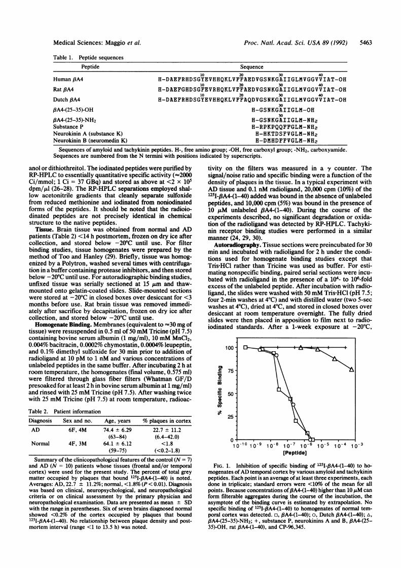
tivity on the filters was measured in a y counter. Thesignal/noise ratio and specific binding were a function of thedensity of plaques in the tissue. In a typical experiment withAD tissue and 0.1 nM radioligand, 20,000 cpm (10%6) of the125I-pA4-(1-40) added was bound in the absence of unlabeledpeptides, and 10,000 cpm (5%) was bound in the presence of10 ,uM unlabeled f3A4-(1-40). During the course of theexperiments described, no significant degradation or oxida-tion of the radioligand was detected by RP-HPLC. Tachyki-nin receptor binding studies were performed in a similarmanner (24, 29, 30).
Autoradiography. Tissue sections were preincubated for 30min and incubated with radioligand for 2 h under the condi-tions used for homogenate binding studies except thatTris HCl rather than Tricine was used as buffer. For esti-mating nonspecific binding, paired serial sections were incu-bated with radioligand in the presence of a 104- to 106-foldexcess of the unlabeled peptide. After incubation with radio-ligand, the slides were washed with 50 mM Tris-HCI (pH 7.5;four 2-min washes at 4°C) and with distilled water (two 5-secwashes at 4°C), dried at 4°C, and stored in closed boxes overdesiccant at room temperature overnight. The fully driedslides were then placed in apposition to film next to radio-iodinated standards. After a 1-week exposure at -20°C,
100
.E75
50CL
co-;
i
50
-101 10-9 1o-8 10-7 1 0 6 1 0 5 1 0-4 1 0-3[Peptidel
FIG. 1. Inhibition of specific binding of 1251-,8A4-(1-40) to ho-mogenates ofAD temporal cortex by various amyloid and tachykininpeptides. Each point is an average of at least three experiments, eachdone in triplicate; standard errors, were <1011o of the mean for allpoints. Because concentrations of,BA4-(1-40) higher than 10 ,uM canform filterable aggregates during the course of the incubation, theasymptote of the binding curve is estimated by extrapolation. Nospecific binding of 125I-,8A4(1-40) to homogenates of normal tem-poral cortex was detected. o, )A4-(1-40); o, Dutch ,PA4(1-40); A,/3A4-(25-35)-NH2; +, substance P. neurokinins A and B. ,PA4(25-35)-OH, rat BA4-(1-40), and CP-96,345.
Medical Sciences: Maggio et al.
5464 Medical Sciences: Maggio et al.
the film was developed, fixed, and washed. Sections werelater dipped in photographic emulsion for higher-resolutionautoradiography and/or counterstained by standard proce-dures with thioflavin S, hematoxylin/eosin, or antibodies forimmunohistochemistry. Autoradiography of tachykinin re-ceptors was performed as described (24, 30).Time Course Experiments. Serial sections were incubated in
the presence of 0.1 nM 125I-/3A4-(1-40) for various times.Sections incubated 2 h were then transferred to a solutioncontaining no ,8A4 for further incubation. At various times, thesections were removed from the incubation medium andprocessed for autoradiography as above; the amount of radio-ligand bound to plaques was later quantified by densitometry.
RESULTSAD vs. Normal Tissue. Whereas there was essentially no
displaceable binding of 125I-13A4-(1-40) to normal tissue sec-tions or homogenates (data not shown), there was significant
I
I
_
Proc. Nati. Acad. Sci. USA 89 (1992)
specific binding to AD tissue (Figs. 1 and 2). This binding toAD tissue was not saturable. In severe cases of the disease,more than a third of the volume of the affected cortical areasexamined was occupied by plaques capable of binding theradioligand; in contrast, no such deposits were detected inmost of the control tissues examined (Table 2). No displace-able binding was observed when plaques were absent (i.e., innormal human or rat tissue), and autoradiography of thehomogenate confirmed that the highest density of bindingsites in AD tissue homogenates was on intact plaques.
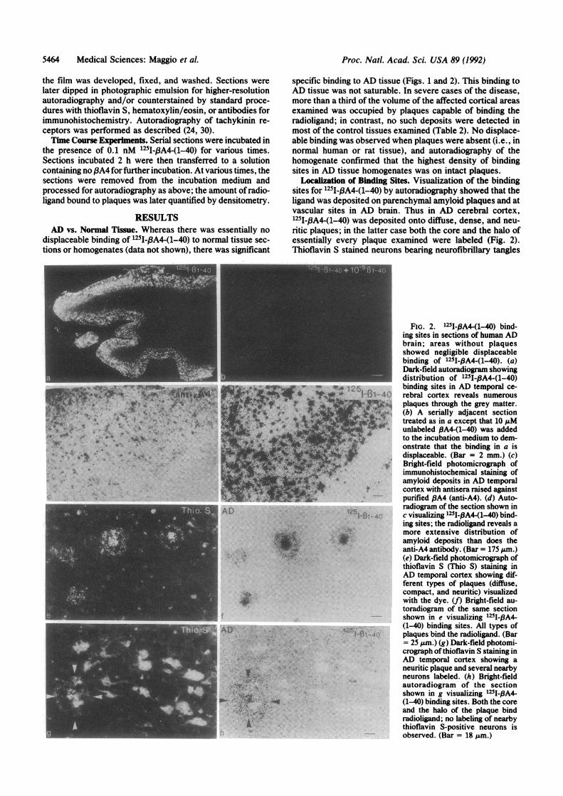
Localization of Binding Sites. Visualization of the bindingsites for 1251I-3A4-(1-40) by autoradiography showed that theligand was deposited on parenchymal amyloid plaques and atvascular sites in AD brain. Thus in AD cerebral cortex,1251-f3A4-(1-40) was deposited onto diffuse, dense, and neu-ritic plaques; in the latter case both the core and the halo ofessentially every plaque examined were labeled (Fig. 2).Thioflavin S stained neurons bearing neurofibrillary tangles
FIG. 2. 125I-BA4-(1-40) bind-ing sites in sections of human ADbrain; areas without plaquesshowed negligible displaceablebinding of 1251-PA4-(1-40). (a)Dark-field autoradiogram showingdistribution of 125I-,BA4-(1-40)
'A 125 binding sites in AD temporal ce-ifi,- 1-131- 40 rebral cortex reveals numerous
7I5NS plaques through the grey matter.(b) A serially adjacent section
kS** treated as in a except that 10 ,uMunlabeled PA4-(1-40) was addedto the incubation medium to dem-onstrate that the binding in a isdisplaceable. (Bar = 2 mm.) (c)Bright-field photomicrograph ofimmunohistochemical staining ofamyloid deposits in AD temporalcortex with antisera raised againstpurified ,BA4 (anti-A4). (d) Auto-radiogram of the section shown in
1-1r-40 c visualizing 125I-,A4-(1-40) bind-ing sites; the radioligand reveals amore extensive distribution of
SS,.1>,r amyloid deposits than does theanti-A4 antibody. (Bar = 175 Mm.)(e) Dark-field photomicrograph ofthioflavin S (Thio S) staining inAD temporal cortex showing dif-ferent types of plaques (diffuse,compact, and neuritic) visualizedwith the dye. (f) Bright-field au-toradiogram of the same sectionshown in e visualizing 125I-IA4-
,;25; (1-40) binding sites. All types ofI-'1 -4C plaques bind the radioligand. (Bar
=25 Mm.) (g) Dark-field photomi-crograph ofthioflavin S staining inAD temporal cortex showing aneuritic plaque and several nearbyneurons labeled. (h) Bright-fieldautoradiogram of the sectionshown in g visualizing 125I-,BA4-(1-40) binding sites. Both the coreand the halo of the plaque bindradioligand; no labeling of nearbythioflavin S-positive neurons isobserved. (Bar = 18 Mm.)

Proc. Natl. Acad. Sci. USA 89 (1992) 5465
~ ~ 2
^<~~~~~~TI,,T4110' o 'w e_,9
d
FIG. 3. 125I-13A4-(1-40) bind-ing sites in the cerebral vascula-ture of human AD brain. (a) Dark-field autoradiogram showingplaques in the parenchyma andamyloid in blood vessel (BV) wallsin AD temporal cortex. (b) A se-rially adjacent section treated as ina except that 10 1uM unlabeled,fA4-(1-40) was added to the in-
1-40 cubation medium to demonstratethat the binding in a is displace-
A able. (Bar = 280 ,um.) (c) Bright-field photomicrograph ofa sectionstained with hematoxylin/eosin(H&E) showing a cerebral artery.(d) Bright-field autoradiogram ofthe section shown in c visualizing
p "125I-,A4-(1-40) binding sites; thehighest density of binding sites isassociated with the tunica media(TM). TA, tunica adventitia; L,lumen; TI, tunica intima. (Bar =
- 40 um.)
outside the plaques and the plaques themselves, but deposi-tion of the radioligand was restricted to the plaques alone.The sensitivity of detection of plaques with the radioligandfar exceeds that of the antibodies we have examined (Fig. 2).Amyloid deposits were visualized by 1251-PA4-(1-40) dep-
osition in 5-20o of intra- and extraparenchymal blood ves-sels in AD brain (Fig. 3). The cerebrovascular deposits wereconsistently labeled more intensely than cerebral plaqueswithin the same section. In vessels seen in transverse section(Fig. 3), the deposition of 8A4 radioligand was not uniformbut rather was concentrated in a part of the vessel-apparently, the smooth muscle of the tunica media; theendothelium was not labeled.
Competition Binding Experiments. All experiments on tissuesections or homogenates included tubes containing identicalsolutions in the absence of tissue that were monitored foraggregate formation by both filtration and centrifugation.These parallel experiments showed no evidence ofaggregationunder the conditions described. Deposition of 125I-PA4-(1-40)onto plaques in both homogenates and sections ofAD cerebralcortex is significantly attenuated by excess unlabeled ,A4-(1-40) and Dutch fBA4-(1-40) (Figs. 1 and 2). /3A4-(25-35)-NH2also competes at much higher concentrations, whereas the freeacid f3A4-(25-35)-OH has no detectable activity in this assay.Rat fA4-(1-40) had no effect on the deposition of 125I-.BA4-(1-40) onto plaques. No significant differences in f3A4 depo-sition were noted between homogenates and sections of ADtissue or between neuritic and diffuse plaques.The mammalian tachykinins (23), substance P and neuro-
kinins A and B, and a nonpeptide NK1 tachykinin receptorantagonist, CP-96,345 (25), did not inhibit the deposition ofradiolabeled 3A4 onto AD plaques over a broad range ofconcentration (Fig. 1).
Saturable binding sites for tachykinin radioligands selec-tive for NK1 and NK3 tachykinin receptor subtypes (30) werepresent at similar densities in both AD and normal braintissue (data not shown). Cell types expressing tachykininbinding sites were not identified. Unlabeled tachykininsdisplaced their radiolabeled analogues at nanomolar concen-trations in both tissues. In contrast, there was no displace-ment of any of the tachykinin radioligands by A4-(1-40), rat,BA4-(1-40), or Dutch B3A4-(1-40) at up to 30 ,uM. f3A4-(1-40)also failed to displace radiolabeled tachykinins from theirbinding sites in rat (30) brain homogenates and sections.
Time Course of P3A4-(1-40) Deposition. As quantitated bydensitometry of autoradiograms, radiolabeled PA4 deposi-tion from solution onto amyloid plaques was linear for at leastseveral hours. When plaques onto which 125I-f3A4-(1-40) hadbeen deposited for 2 h were incubated in the absence ofamyloid peptide, the radiolabeled peptide dissociated fromthe plaques (Fig. 4). Thus deposition of 1251-pA4-(1-40) ontoplaques is reversible.
DISCUSSIONIn Vitro Growth of AD Amyloid. Radiolabeled /3A4-(1-40)
binds specifically, reversibly, and with high affinity to sites inAD tissue but not normal tissue (Figs. 1-4). The observation
200
150
I-100
50
04-0.0 0.5 1.0 1.5 2.0 2.5 3.0 3.5 4.0
Time (Hrs)
FIG. 4. Time course of 1251-PA4-(1-40) association onto anddissociation from AD plaques in tissue sections of human AD frontalcortex. Periods as long as 4 h are shown, and a 24-h time course isshown in the Inset. Saturation of the film prevented accuratequantitation of the 24-h time point for association; the 24-h pointshown is a lower limit. Error bars represent the SEM of at least threedeterminations. Analysis of the time course of dissociation yields a
dissociation half-time of 1 h under the described conditions.
TM H&E
JTA
Medical Sciences: Maggio et al.

5466 Medical Sciences: Maggio et al.
that binding is not saturable suggests that the vast majority ofthe sites to which the radioligand binds are not receptors inthe usual sense. Instead, the characteristics of this bindingare consistent with a steady-state equilibrium between fiA4on the tissue amyloid plaques and in solution. Thus 1251-#A4-(1-40) at picomolar concentrations is reversibly depositedonto AD plaques and dissociates when the plaques areexposed to a solution containing no B3A4 (Fig. 4). The precisenature of the plaque component to which the radioligandbinds is not addressed by these experiments.Under these experimental conditions, the in vitro growth of
plaques by deposition of 125I-/3A4-(1-40) proceeds at thelowest concentration of radioligand that can be convenientlyquantitated (1-10 pM). The affinity of (3A4 for the plaques issufficiently high that deposition is a favorable process evenat such dilute concentrations.Both the classic senile plaques and the diffuse nonneuritic
deposits in the cortical grey matter and the vascular amyloiddeposits within the walls of the cerebral arteries are capableof in vitro growth by addition of8A4 from dilute solution. Useof radiolabeled f3A4 detects tissue amyloid with great sensi-tivity and emphasizes the great density ofamyloid deposits inAD vs. normal tissue (Table 2).
.8A4 and Tachykinins. The tachykinin neuropeptides aredefined by their C terminus, -Phe-Aaa-Gly-Leu-Met-NH2,where Aaa is an aromatic or aliphatic residue. This conservedC-terminal sequence, including the terminal carboxyamide,is required for essentially all the biological activities of thepeptides in the tachykinin family.
Various tachykinin agonists and antagonists had no de-tectable effect on in vitro growth of AD amyloid (Fig. 1).Since several experiments failed to detect any interactionbetween fBA4 and various tachykinin receptors found innormal human and AD brain and in rat brain, 3A4 apparentlydoes not interact with tachykinin receptors under theseconditions (data not shown). These results are consistentwith the reported structure-activity studies among the ta-chykinin peptide family that a C-terminal amide is requiredfor activity (23) and argue against the hypothesis (15) that thereported biological activity of PA4 (9-16) results from itsinteraction with tachykinin receptors.
Aggregation of pA4 in the Absence of Tissue. Under theconditions of the tissue binding assay described above, thereis no detectable aggregation ofpA4 in the absence ofplaques.At higher concentrations or in solvents other than thosedescribed above, PA4-(1-40) and certain of its fragments canalso form aggregates (17-21) in the absence of nucleatingplaques, and the radiolabeled fA4 can be used as a sensitiveand convenient monitor of such aggregation. The rate ofaggregation ofPA4 in tissue free systems is highly dependenton experimental conditions (data not shown).
Implications for AD. The avidity of PA4 for amyloidplaques implies that once a nucleating aggregate of BA4 hasformed, even extremely low concentrations of jA4 willsupport its growth. The reversibility ofplaque growth in vitrosuggests a steady-state equilibrium between fA4 in plaquesand in solution. The in vitro studies described here areconsistent with the hypothesis that the plaques themselves(neuritic, diffuse, and vascular) can grow in vivo in thepresence of traces of PA4. A similar situation in vivo wouldimply that an alteration in the processing or degradation ofPA4 that increased the extracellular concentration of j3A4could play a critical role in the genesis of AD (1) and thatplaque growth in the brain may be reversible. The sponta-neous aggregation ofBA4 under certain conditions is likely tobe related to the early nucleating events of plaque formationand thus to the neurotoxicity of,A4 (13-22) and the pathol-ogy of the disease. The use of radiolabeled 3A4 provides aconvenient model of these processes for further investigationof the role of fA4 in AD. The present study suggests a
sensitive and quantitative assay for evaluation of agents andconditions that may alter the growth of amyloid plaques andaid in the search for effective treatments of AD.We thank R. M. Snider for providing CP-96,345. Tissue was
provided by the Kathleen Price Bryan Brain Bank of the Joseph andKathleen Bryan Alzheimer's Disease Research Center, Duke Uni-versity Medical Center, supported by the National Institutes ofHealth under Grant AG05128, and by W. H. Frey II of the Alz-heimer's Disease Treatment and Research Center of St. Paul. Thiswork was supported by the National Institutes of Health underGrants NS22961 (J.E.M.), GM15904 (J.E.M.), NS23970 (P.W.M.),NS26312 (H.V.V.), and AG10123 (H.V.V.), and a Veterans Admin-istration Merit review (P.W.M.).
1. Selkoe, D. J. (1991) Neuron 6, 487-498.2. Selkoe, D. J. (1990) Science 248, 1058-1060.3. Milier-Hill, B. & Beyreuther, K. (1989) Annu. Rev. Biochem.
58, 287-307.4. Katzman, R. & Saitoh, T. (1991) FASEB J. 5, 278-286.5. Goldgaber, D., Lerman, J. I., McBride, 0. W., Saffioti, U. &
Gajdusek, D. C. (1987) Science 235, 877-880.6. Kang, J., Lemaire, H. G., Unterbeck, A., Salbaum, J., Mas-
ters, L., Grzeschik, K. H., Multhaup, G., Beyreuther, K. &Muller-Hill, B. (1987) Nature (London) 325, 733-736.
7. Tanzi, R. E., Guselia, J. F., Watkins, P. C., Bruns, G. A. P.,St. George-Hyslop, P., Van Keuren, M. L., Patterson, S. P.,Pagan, S., Kurnit, D. M. & Neve, R. L. (1987) Science 235,880-884.
8. Robakis, N. K., Ramakrishna, N., Wolfe, G. & Wisniewski,H. M. (1987) Proc. Nat!. Acad. Sci. USA 84, 4190-4194.
9. Whitson, J. S., Selkoe, D. J. & Cotman, C. W. (1989) Science243, 1488-1490.
10. Koh, J.-Y., Yang, L. L. & Cotman, C. W. (1990) Brain Res.533, 315-320.
11. Flood, J. F., Morley, J. E. & Roberts, E. (1991) Proc. Nat!.Acad. Sci. USA 88, 3363-3366.
12. Yankner, B. A., Dawes, L. R., Fisher, S., Villa-Komaroff, L.,Oster-Granite, M. L. & Neve, R. L. (1989) Science 245, 417-420.
13. Pike, C. J., Walencewitz, A. J., Glabe, C. G. & Cotman, C. W.(1991) Eur. J. Pharmacol. 207, 367-368.
14. Pike, C. J., Walencewitz, A. J., Glabe, C. G. & Cotman, C. W.(1991) Brain Res. 563, 311-314.
15. Yankner, B. A., Duffy, L. K. & Kirchner, D. A. (1990) Sci-ence 250, 279-282.
16. Kowall, N. W., Beal, M. F., Busciglio, J., Duffy, L. K. &Yankner, B. A. (1991) Proc. Nat!. Acad. Sci. USA 88, 7247-7251.
17. Castano, E. M., Ghiso, J., Prelli, F., Gorevic, P. D., Mighell,A. & Frangione, B. (1986) Biochem. Biophys. Res. Commun.141, 782-789.
18. Kirshner, D. A., Inouye, H., Duffy, L. K., Sinclair, A., Lind,M. & Selkoe, D. J. (1987) Proc. Nat!. Acad. Sci. USA 84,6953-6957.
19. Hilbich, C., Kisters-Woike, B., Reed, J., Masters, C. L. &Beyreuther, K. (1991) J. Mol. Biol. 218, 149-163.
20. Halverson, K., Fraser, P. E., Kirshner, D. A. & Lansbury,P. T., Jr. (1990) Biochemistry 29, 2639-2644.
21. Barrow, C. J. & Zagorski, M. G. (1991) Science 253, 179-182.22. Quon, D., Wang, Y., Catalano, R., Scardina, J. M., Murakami,
K. & Cordell, B. (1991) Nature (London) 352, 239-241.23. Maggio, J. E. (1988) Annu. Rev. Neurosci. 11, 13-28.24. Mantyh, P. W., Johnson, D. J., Boehmer, C. G., Catton,
M. D., Vinters, H. V., Maggio, J. E., Too, H.-P. & Vigna,S. R. (1989) Proc. Nat!. Acad. Sci. USA 86, 5193-5197.
25. Snider, R. M., Constantine, J. W., Lowe, J. A., III, Longo,K. P., Lebel, W. S., Woody, H. A., Drozda, S. E., Desai,M. C., Vinick, F. J., Spencer, R. W. & Hess, H.-J. (1991)Science 251, 435-437.
26. Hunter, W. M. & Greenwood, F. C. (1%2) Nature (London)194, 495-496.
27. Bolton, A. E. & Hunter, W. M. (1973) Biochem. J. 133,5 29-538.28. Too, H.-P. & Maggio, J. E. (1991) Methods Neurosci. 6,
232-247.29. Too, H.-P. & Hanley, M. R. (1988) Biochem. J. 252, 545-551.30. Mantyh, P. W., Gates, T. S., Mantyh, C. R. & Maggio, J. E.
(1989) J. Neurosci. 9, 258-279.
Proc. Nad. Acad Sci. USA 89 (1992)