Revealing the kinetics of Leishmania chagasi infection in ... · Revealing the kinetics of...
6
Revealing the kinetics of Leishmania chagasi infection in the male genital system of hamsters Quintal et al. Quintal et al. Infectious Diseases of Poverty (2016) 5:29 DOI 10.1186/s40249-016-0122-0
Transcript of Revealing the kinetics of Leishmania chagasi infection in ... · Revealing the kinetics of...
Revealing the kinetics of Leishmania chagasiinfection in the male genital system of hamstersQuintal et al.
Quintal et al. Infectious Diseases of Poverty (2016) 5:29 DOI 10.1186/s40249-016-0122-0

SHORT REPORT Open Access
Revealing the kinetics of Leishmaniachagasi infection in the male genitalsystem of hamstersAmanda P. N. Quintal1, Bruna C. Borges1, Paula C. Brígido1, Rebecca T. Silva1, Ana F. Notário1, Marlus A. Santos1,Maria A. de Souza1, Fernanda G. O. Nascimento2, Antônio V. Mundim2, Guilherme M. J. Costa3,André B. Vasconcelos4 and Claudio V. da Silva1,5*
Abstract
Background: Leishmaniasis causes alterations and lesions in the genital system, which leads to azoospermia andtesticular atrophy in animals during the chronic phase of the infection. The aim of this study was to reveal thekinetics of Leishmania chagasi infection in the genital system of male golden hamsters (Mesocricetus auratus).
Methods: Animals were intraperitoneally inoculated with amastigotes from L. chagasi. At different time pointsanimals were euthanized and genital organs processed for histo-pathological, qPCR, cytokines and testosteronedetection assays.
Results: Our results showed a high parasite load in testis, followed by an increase of pro-inflammatory cytokinesIL1-β, TNF-α and IFN-γ, and testosterone. Subsequently, IL-4 expression was upregulated and basal parasitepersistence in testis was observed using the experimental approach.
Conclusion: Extracellular amastigotes migrated to the epididymis posing as a potential major factor ofparasite persistence and venereal transmission of L. chagasi infection in hamsters.
Keywords: Leishmania chagasi, Extracellular amastigotes, Testis, Epididymis, Immune response, Testosterone
Multilingual abstractsPlease see Additional file 1 for the translations of ab-stract into the six official working languages of theUnited Nations.
FindingsLeishmania chagasi is an intracellular protozoan thatcauses visceral leishmaniasis (VL) [1], a potentially fatalhuman disease that infects the macrophages in thespleen and liver, leading to splenomegaly and hepato-megaly [2]. It is estimated that globally 200,000 to400,000 new cases and 20,000 to 30,000 deaths occureach year [3].
Visceral leishmaniasis causes alterations and lesions inthe genital system [2, 4, 5]. Other authors have previouslydemonstrated the presence of the Leishmania parasite inurine, semen, and reproductive organs of dogs [5–9]. Inaddition, the possibility of venereal transmission in dogsnaturally infected by L. chagasi has been proposed [5]. Inthis study, we aimed to verify the kinetics of L. chagasiinfection in the genital organs of male golden hamsters.
MethodsAnimals and parasitesMale golden hamsters (Mesocricetus auratus) aged 6 to8 weeks were kept under standard conditions on a 12-hlight, 12-h dark cycle in a temperature-controlled room(25 ± 2 °C), with food and water available ad libitum.L. chagasi (MHON/BR/1972/LD) strain was main-
tained in vivo by inoculation into peritoneal cavity ofhamsters. Six weeks post-infection, the hamsters wereeuthanized and amastigote forms were recovered from
* Correspondence: [email protected] de Ciências Biomédicas, Universidade Federal de Uberlândia,Uberlândia, Minas Gerais, Brasil5Departamento de Imunologia – Instituto de Ciências Biomédicas,Universidade Federal de Uberlândia, Campus Umuarama, Uberlândia, MG,BrasilFull list of author information is available at the end of the article
© 2016 Quintal et al. Open Access This article is distributed under the terms of the Creative Commons Attribution 4.0International License (http://creativecommons.org/licenses/by/4.0/), which permits unrestricted use, distribution, andreproduction in any medium, provided you give appropriate credit to the original author(s) and the source, provide a link tothe Creative Commons license, and indicate if changes were made. The Creative Commons Public Domain Dedication waiver(http://creativecommons.org/publicdomain/zero/1.0/) applies to the data made available in this article, unless otherwise stated.
Quintal et al. Infectious Diseases of Poverty (2016) 5:29 DOI 10.1186/s40249-016-0122-0

their spleens. The spleens were removed, macerated in4 mL of phosphate-buffered saline (PBS), and centri-fuged to remove residual tissue. The purified amastigoteswere counted in bright light microscope and used toinfect animals for the experimental procedures.
Experimental infectionForty-two male golden hamsters were split into six groupsof seven hamsters each. Animals were intraperitoneallyinoculated with 1 × 10⁶ L. chagasi amastigotes, and eu-thanasia was conducted seven, 10, 13, 16, and 19 weekspost-infection. At each of these times, the spleens, livers,testis, and epididymis were removed and split into threeequal parts for polymerase chain reaction (PCR), cytokine,testosterone detection, and histological procedures.
Histological samplesParaffin-embedded epididymis samples were used in in-direct immunofluorescence as follows: First, paraffin wasremoved with xylene and alcohol, then it was treatedwith 50 mM of ammonium chloride for one hour, and fi-nally it was blocked with albumin (one egg white qsq in100 ml of distilled water) for 20 min and with skimmedmilk overnight. After this, the samples were incubatedwith rabbit polyclonal antibody of anti-L. chagasi dilutedin PGN-saponin (PBS + gelatin + azide) (1: 100) overnight.Finally, the samples were incubated with mouse anti-rabbit IgG Alexa Fluor® 488 (Thermo Fisher Scientific,USA) conjugated antibody (1:200) and TO-PRO®-3 stain(Life Technologies, USA) diluted in PGN-saponin (1:500)for one hour and analyzed using a confocal microscope.
Conventional and qPCRPrimers 13A (5’ - GGG GTG GAG TCT GGG CGT – 3’)and 13B (5’ - ATT TTA CAC CAA CCC CCA GTT – 3’)were used in conventional and real-time PCR (qPCR)detection. The procedures were performed as describedelsewhere [10, 11].
Cytokines and testosterone detectionEnzyme-linked immunosorbent assay (ELISA) was usedto detect the IFN-γ, TNF-α, I-L4, and IL1-β cytokines(BD OpTEIA™, BD Bioscience, San Diego, CA, USA),and testosterone (Testosterone ELISA Kit, CaymanChemical Company, USA) in the testicle samples. Theprocedures were performed according to the manufac-turers’ instructions.
Statistical analysisStatistical analysis was performed using GraphPad Prism6.0 (GraphPad Software Inc., San Diego, CA, USA). Datawere expressed as mean ± standard deviation. An ana-lysis of variance (ANOVA) was performed followed byBonferroni post-test. P <0.05 was considered significant.
EthicsMaintenance and care of the animals complied with theguidelines of the laboratory of the Animal Ethics Commit-tee from University of Uberaba. Animal euthanasia wasperformed in accordance with the American VeterinaryMedical Association Guidelines for the Euthanasia ofAnimals. The research was approved by the Ethics Com-mittee for Animal Experimentation of the University ofUberaba (process CEEA 016004/2014).
Results and DiscussionAfter confirming animal infection by conventional PCRof DNA extracted from the livers and spleens of thehamsters (data not shown), we evaluated the parasiteload in the testis and epididymis. We observed a highparasite load in the testis of infected animals by weekseven post-infection (see Fig. 1a). The intensity of tissueparasitism correlated to pro-inflammatory cytokines ex-pression. (see Fig. 1b, c, and d). Interestingly, high levelsof pro-inflammatory cytokines were followed by an in-creased expression of testosterone (see Fig. 1f ). Testos-terone seems to play an important anti-inflammatoryrole in the maintenance of testicular immune privilege[12]. The increased testosterone levels observed at 7week post-infection may have accounted for a basal levelof parasite persistence in the testis along the kinetic ofinfection. Also, testosterone may have induced IL-4expression at week 10 post-infection (see Fig. 1e).At week 10 post-infection, we observed a high parasite
load in the epididymis, with these levels maintained up toweek 13 post-infection (see Fig. 1g). Observing extracellu-lar amastigotes in the infected organs (see Fig. 1h and i).Authors have demonstrated the presence of L. chagasi
amastigotes inside testicular macrophages [2]. Addition-ally, other authors have found intracellular amastigotes inthe extraocular striated muscle and in the orbiculari oculimuscle in dogs with patent leishmaniasis [13]. However,this is the first study of extracellular amastigotes in animaltissue. The data suggest that extracellular amastigotescould be major elements involved in infection of genitalorgans and parasite venereal propagation in animals.According to our data, we proposed a kinetic of L.
chagasi infection of the male genital system. First, para-sites may reach the testis through blood vessels. Uponthe onset of a pro-inflammatory immune response (T-helper 1 – Th1), parasitism is controlled [14]. However,the expression of testosterone [12] and IL-4 may havefavored the persistence of a low parasite load in thetestis by establishing a local T-helper 2 (Th2) immuneresponse. In vivo and in vitro studies have establishedIL-4’s clear role in driving Th2 immunity [15]. This pat-tern of acquired immunity favors parasite persistence[14]. Meanwhile, extracellular amastigotes migrate to the
Quintal et al. Infectious Diseases of Poverty (2016) 5:29 Page 2 of 5
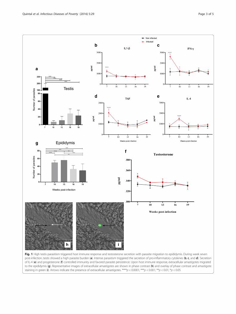
Fig. 1 High testis parasitism triggered host immune response and testosterone secretion with parasite migration to epididymis. During week sevenpost-infection, testis showed a high parasite burden (a). Intense parasitism triggered the secretion of pro-inflammatory cytokines (b, c, and d). Secretionof IL-4 (e) and progesterone (f) controlled immunity and favored parasite persistence. Upon host immune response, extracellular amastigotes migratedto the epididymis (g). Representative images of extracellular amastigotes are shown in phase contrast (h) and overlay of phase contrast and amastigotestaining in green (i). Arrows indicate the presence of extracellular amastigotes. ****p < 0.0001; ***p < 0.001; **p < 0.01; *p < 0.05
Quintal et al. Infectious Diseases of Poverty (2016) 5:29 Page 3 of 5

epididymis, where they might remain adhered to pris-matic cells cilia or be released in sperm [5] (see Fig. 2).
ConclusionThis study revealed the kinetics of L. chagasi migrationthrough the male genital system of hamsters. It also pro-vided evidence that extracellular amastigotes may be amajor factor of venereal transmission of Leishmania inanimals.
Additional file
Additionalfile 1: Multilingual abstracts in the six official workinglanguages of the United Nations. (PDF 356 kb)
AbbreviationsELISA: enzyme-linked immunosorbent assay; PBS: phosphate-bufferedsaline; PCR: polymerase chain reaction; qPCR: real-time PCR; VL: visceralleishmaniasis.
Competing interestsThe authors declare that they have no competing interests.
Authors’ contributionsAPNQ was responsible for developing the project, designed theexperimental approaches, performed the experiments, and drafted thepaper. BCB, PCB, RTS, AFN, and MAS performed the experiments. MAdeSsupervised the experiments and interpreted the data. FGON maintained theparasites. AVM interpreted the data and drafted the paper. GMJC performedthe experimental infections. ABV designed and supervised the experiments,interpreted the data, and drafted the paper. CVS designed and supervised
the experiments, interpreted the data, drafted the paper, and coordinatedthe project. All authors read and approved the final manuscript.
FundingThis study was supported by grants and fellowships from Fundação deAmparo à Pesquisa do Estado de Minas Gerais, Conselho Nacional deDesenvolvimento Científico e Tecnológico, and Coordenação deAperfeiçoamento de Pessoal de Nível Superior.
Author details1Instituto de Ciências Biomédicas, Universidade Federal de Uberlândia,Uberlândia, Minas Gerais, Brasil. 2Hospital Veterinário, Universidade Federal deUberlândia, Uberlândia, Minas Gerais, Brasil. 3Departamento de CiênciasBiomédicas, Universidade Federal de Minas Gerais, Belo Horizonte, Brasil.4Universidade de Uberaba, Uberaba, Minas Gerais, Brasil. 5Departamento deImunologia – Instituto de Ciências Biomédicas, Universidade Federal deUberlândia, Campus Umuarama, Uberlândia, MG, Brasil.
Received: 30 September 2015 Accepted: 23 March 2016
References1. Soto M, Ramirez L, Pineda MA, et al. Searching genes encoding Leishmania
antigens for diagnosis and protection. Schol Res Exchange. 2009;15:1–25.2. Diniz AS, Melo MS, Borges AM, et al. Genital lesions associated with visceral
leishmaniasis and shedding of Leishmania sp. in the semen of naturallyinfected dogs. Vet Pathol. 2005;42:650–8.
3. World Health Organization. Leishmaniasis. Fact sheet N° 375. Available at:http://www.who.int/mediacentre/factsheets/fs375/en/. Accessed 03 Nov 2015.
4. Assis VP, Ribeiro VM, Rachida MA, Castro ACS, Valle GR. Dogs with Leishmaniachagasi infection have semen abnormalities that partially revert during150 days of Allopurinol and Amphotericin B therapy. Animal Reprod Science.2010;117:183–6.
5. Silva FL, Oliveira RG, Silva TMA, Xavier MN, Nascimento EF, Santos RL. Venerealtransmission of canine visceral leishmaniasis. Vet Parasitol. 2009;160:55–9.
Fig. 2 Proposed kinetic of L. chagasi infection in the genital system of male hamsters. Parasites reach testis through blood vessels (a). Upon theonset of a pro-inflammatory immune response, parasitism is controlled (b). The expression of testosterone and IL-4 may have favored the persistenceof a low parasite load (c). Extracellular amastigotes migrate to the epididymis where they might be adhered to prismatic cells cilia or be released withinsperm (d)
Quintal et al. Infectious Diseases of Poverty (2016) 5:29 Page 4 of 5
6. Gonzales JL, Gallego E, Castano M, Rueda A. Testicular amyloidosis inhamsters experimentally infected with Leishmania donovani. British J ExpPathol. 1983;64:518–23.
7. Rieira C, Valladares JE. Viable Leishmania infantum in urine and semen inexperimentally infected dogs. Parasitol Today. 1996;12:412.
8. Amara A, Mrad I, Melki MK, Mrad MB, Rejeb A. Estude histologique dês lésionstesticulaires chez leschiens leishmaniens. Rev Med Vet. 2009;160:54–60.
9. Manna L, Paciello O, Morte RD, Gravino AE. Detection of Leishmaniaparasites in the testis of a dog affected by orchitis: case report. Parasitesand Vectors. 2012;5:1–4.
10. Rodogers MR, Popper SJ, Wirth DF. Amplification of kinetoplast DNA as atool in the detection and diagnosis of Leishmania. Exp Parasitol. 1990;71:267–75.
11. Quintal APN, Ribeiro ES, Rodrigues FP, Rocha FS, Floeter-winter LM, NunesCM. Leishmania spp. in Didelphis albiventris and Micoureus paraguayanus(Didelphimorphia: Didelphidae) of Brasil. Vet Parasitol. 2011;176:112–9.
12. Weinbauer GF, Luetjens CM, Simoni M, Nieschlag E. Physiology of testicularfunction. In: Nieschlag E, Behre HM, Nieschlag S, editors. Andrology: MaleReproductive Health and Dysfunction. 3rd ed. Berlin: Spring Press; 2010. p. 37–8.
13. Naranjo C, Fondevila D, Leiva M, et al. Detection of Leishmania spp. andassociated inflammation in ocular-associated smooth and striated musclesin dogs with patent leishmaniosis. Vet Ophthalmology. 2010;13:139–43.doi:10.1111/j.1463-5224.2010.00768.x.
14. Singh OP, Sundar S. Immunotherapy and targeted therapies in treatment ofvisceral leishmaniasis: current status and future prospects. Front Immunol.2014;5:296.
15. Choi P, Reiser H. IL-4: role in disease and regulation of production. Clin ExpImmunol. 1998;113:317–9.
• We accept pre-submission inquiries
• Our selector tool helps you to find the most relevant journal
• We provide round the clock customer support
• Convenient online submission
• Thorough peer review
• Inclusion in PubMed and all major indexing services
• Maximum visibility for your research
Submit your manuscript atwww.biomedcentral.com/submit
Submit your next manuscript to BioMed Central and we will help you at every step:
Quintal et al. Infectious Diseases of Poverty (2016) 5:29 Page 5 of 5