results, - COnnecting REpositories · were prepared by treatment of polyoma I with pancreatic...
8
THE TWISTED CIRCULAR FORM OF POLYOMA VIRAL DNA* BY J. VINOGRAD, J. LEBOWITZ, R. RADLOFF, R. WATSON, AND P. LAIPIS GATES AND CRELLIN LABORATORIES OF CHEMISTRY AND THE NORMAN W. CHURCH LABORATORY OF CHEMICAL BIOLOGY, CALIFORNIA INSTITUTE OF TECHNOLOGY Communicated by Norman Davidson, March 30, 1965 The major part of the DNA from polyoma virus has been shown to consist of circular base-paired duplex molecules without chain ends.' -' The intertwined cir- cular form accounts for the ease of renaturation4 of this DNA and the failure of the strands to separate in strand-separating solvents. I-' In previous studiesl-3 a minor component, II, observed in variable amounts in sedimentation analyses of preparations of polyoma DNA at neutral pH, was re- garded to be a linear form of the viral DNA. Both the major component I (20S) and II (16S) were infective." I In our further investigations of the minor com- ponent the following results, which are reported below, have been obtained: (1) The minor component is a ring-shaped duplex molecule. (2) It is generated by introducing one single-chain scission in component I by the action of pancreatic DNAase or chemical-reducing agents. (3) The sedimentation coefficient of II is insensitive to several single-strand scissions. (4) The conversion products, when not excessively attacked, are infective. The foregoing results raised a new problem. Why does the viral DNA, an intact duplex ring, sediment 20 per cent faster than a similar duplex ring containing one or more single-strand scissions? Experiments bearing on this problem, presented below, indicate the presence of a twisted circular structure in polyoma DNA I. A mechanism for the formation of this locked-in twisted structure is proposed. Methods.-Isolation and purification of the virus and extraction of the DNA: Two methods6 7 for purification of the virus were used. The DNA was isolated by Weil's method4 except that the phenol was freshly distilled under argon. Ultracentrifugation: Sedimentation analyses were performed in a Spinco model E ultracentri- fuge by band centrifugation.8 Some of the results were recorded with the photoelectric scanning attachment.9, " Sucrose density gradient experiments were performed at 40, 30,000 rpm, and 9 hr. The 3% and 20% sucrose solutions contained SSC (0.15 M NaCl and 0.015 M Na citrate) and 0.05 M Tris chloride pH 8.0. Enzymes: Pancreatic DNAase, 1 X crystallized, was obtained from Worthington Biochemicals Corp. E. coli endonuclease I, 1000 units/ml," and E. coli phosphodiesterase, 2000 units/ml,"2 were gifts from Professor I. R. Lehman. BSA, 30% bovine albumin solution, sterile, was obtained from Armour Pharmaceutical Co. The endonuclease I, 0.12 units/Mg DNA, converted 60% of I into linear molecules in 8 min at 200 in the incubation mixture described by Lehman." Sedimentation velocity-pH titration: Fifteen Al, 40 Mg/nml DNA in SSC10, flowed from the sample well of the type III13 band-forming centerpiece onto an alkaline CsCl bulk-solution. This solution was prepared by titrating 10 ml (Harshaw Chemical Co.) optical grade CsCl, p = 1.35, with 1 M KOH in CsCl, p = 1.35, at 200 under argon, and was transferred to the cell assembly under argon. Usually four samples in a pH series were analyzed simultaneously. A Beckman re- search model pH meter, a general purpose probe glass electrode, and a calomel reference electrode modified with a ground glass junction'4 were used. Plaque assay: Infectivity of polyoma DNA was measured as described by Weil.4 Electron microscopy: Specimens were prepared by the method of Kleinschmidt and Zahn."5 Results.-Preparation of polyoma DNA II: Polyoma II can be prepared from I by treatment with several mild chemical-reducing agents (Table 1). These reagents 1104
Transcript of results, - COnnecting REpositories · were prepared by treatment of polyoma I with pancreatic...

THE TWISTED CIRCULAR FORM OF POLYOMA VIRAL DNA*
BY J. VINOGRAD, J. LEBOWITZ, R. RADLOFF, R. WATSON, AND P. LAIPIS
GATES AND CRELLIN LABORATORIES OF CHEMISTRY AND
THE NORMAN W. CHURCH LABORATORY OF CHEMICAL BIOLOGY,
CALIFORNIA INSTITUTE OF TECHNOLOGY
Communicated by Norman Davidson, March 30, 1965
The major part of the DNA from polyoma virus has been shown to consist ofcircular base-paired duplex molecules without chain ends.' -' The intertwined cir-cular form accounts for the ease of renaturation4 of this DNA and the failure ofthe strands to separate in strand-separating solvents. I-'
In previous studiesl-3 a minor component, II, observed in variable amounts insedimentation analyses of preparations of polyoma DNA at neutral pH, was re-garded to be a linear form of the viral DNA. Both the major component I (20S)and II (16S) were infective." I In our further investigations of the minor com-ponent the following results, which are reported below, have been obtained: (1)The minor component is a ring-shaped duplex molecule. (2) It is generated byintroducing one single-chain scission in component I by the action of pancreaticDNAase or chemical-reducing agents. (3) The sedimentation coefficient of II isinsensitive to several single-strand scissions. (4) The conversion products, whennot excessively attacked, are infective.The foregoing results raised a new problem. Why does the viral DNA, an intact
duplex ring, sediment 20 per cent faster than a similar duplex ring containing oneor more single-strand scissions? Experiments bearing on this problem, presentedbelow, indicate the presence of a twisted circular structure in polyoma DNA I. Amechanism for the formation of this locked-in twisted structure is proposed.
Methods.-Isolation and purification of the virus and extraction of the DNA: Two methods6 7
for purification of the virus were used. The DNA was isolated by Weil's method4 except thatthe phenol was freshly distilled under argon.
Ultracentrifugation: Sedimentation analyses were performed in a Spinco model E ultracentri-fuge by band centrifugation.8 Some of the results were recorded with the photoelectric scanningattachment.9, " Sucrose density gradient experiments were performed at 40, 30,000 rpm, and 9 hr.The 3% and 20% sucrose solutions contained SSC (0.15 M NaCl and 0.015 M Na citrate) and0.05 M Tris chloride pH 8.0.Enzymes: Pancreatic DNAase, 1 X crystallized, was obtained from Worthington Biochemicals
Corp. E. coli endonuclease I, 1000 units/ml," and E. coli phosphodiesterase, 2000 units/ml,"2were gifts from Professor I. R. Lehman. BSA, 30% bovine albumin solution, sterile, was obtainedfrom Armour Pharmaceutical Co. The endonuclease I, 0.12 units/Mg DNA, converted 60% ofI into linear molecules in 8 min at 200 in the incubation mixture described by Lehman."
Sedimentation velocity-pH titration: Fifteen Al, 40 Mg/nml DNA in SSC10, flowed from thesample well of the type III13 band-forming centerpiece onto an alkaline CsCl bulk-solution. Thissolution was prepared by titrating 10 ml (Harshaw Chemical Co.) optical grade CsCl, p = 1.35,with 1 M KOH in CsCl, p = 1.35, at 200 under argon, and was transferred to the cell assemblyunder argon. Usually four samples in a pH series were analyzed simultaneously. A Beckman re-search model pH meter, a general purpose probe glass electrode, and a calomel reference electrodemodified with a ground glass junction'4 were used.
Plaque assay: Infectivity of polyoma DNA was measured as described by Weil.4Electron microscopy: Specimens were prepared by the method of Kleinschmidt and Zahn."5Results.-Preparation of polyoma DNA II: Polyoma II can be prepared from I
by treatment with several mild chemical-reducing agents (Table 1). These reagents1104

VOL. 53, 1965 BIOCHEMISTRY: VINOGRAD ET AL. 1105
TABLE 1ACTION OF REDUCING AGENTS ON POLYOMA DNA
Time Conversion b ofReagent Ma pH (min) I to II (%)
Hydroquinonec 0.0002 8.5 30 100FeCi2 0.001 8.6 30 90Na2SO3 (1 X 10⁻³ M
CU2SO4, 0.1 M 0.01 10.8 60 45NH40H)d
Thiols- 0.01-0.02 3.8-4.2 60 80-90a Final concentration of reducing agents which were diluted fivefold with SSC/10, 0.01 M Tris pH 8.5
containing 40 .ug/ml DNA. The thiols were first dissolved in 0.4 M acetic acid before addition to the DNAsolution.
b Sedimentation analysis at neutral pH.c The reaction product was assayed for infectivity: 0.5 X 10³ pfu/jig DNA compared with 1.5 X 10³
obtained for untreated DNA in the same assay.d Cu + + acts as a catalyst.16 In the absence of Cu + + or NH40H 5% conversion was observed. In the
absence of SOs: no conversion occurred.6 Mercaptoethanol, cysteine, and glutathione. In experiments with acetic acid without thiols, 10%
conversion occurred.
were suggested by the observation that rigorous exclusion of impurities from thephenol, used in the isolation of the DNA, diminished the amount of the minor com-ponent II in the final DNA preparation. A simple conversion of I to II withoutintermediates and without detectable degradation products was observed in sedi-mentation analyses at pH 8.0 (Table 1). Based on the earlier assignment1' 2 of alinear form to component II, the reactions with reducing agents and the infectivenature of the products indicated a specific duplex cleavage. Dulbecco and Vogt1 re-ported a similar conversion of I to II with low concentrations of pancreatic DNAase.These authors postulated that a bond opposite the single-strand scission intro-duced by the enzyme hydrolyzed under the influence of ring strain. The fore-going puzzling results are clarified by the experiments below.
Structure of Polyoma II.-The products from the action of pancreatic DNAaseand of the reducing agents (Table 1) were examined in the electron microscope.They were found to be in the circular form. Figure lb is typical of electron micro-graphs of materials obtained by treatment of I with pancreatic DNAase or with theseveral reducing agents. The possibility that linear molecules were selectively ex-cluded in the preparation of the grids was eliminated by the results of a reconstruc-tion experiment. A synthetic mixture of 10 per cent II and 90 per cent linearpolyoma III (cf. below) showed the expected proportions of linear molecules.The circular form for II is compatible with the proposal that the reducing agents
and pancreatic DNAase introduce single-strand scissions into polyoma DNA I.It may be calculated that the circular form of the molecule should be retained untilon the average about 50 single-strand scissions per molecule have been introduced. 17The material shown in Figure lb contained, on the average, about three breaks permolecule as calculated from the Poisson distribution.A still milder treatment with pancreatic DNAase should give rise to single-
stranded rings and uniform single-stranded linear molecules in strand-separatingsolvents, such as alkaline NaCl or CSCl.2 18 An example of such a result with0.6 breaks per molecule is given in Figure 2b. These two components, s8 20 =18.4S i 0.4, 15.7S + 0.3 for the alkaline Na DNA, have been identified as single-stranded rings (18S) and single-stranded linear molecules (16S), respectively, by thefollowing variation of an experiment originally performed with 4X DNA by Fiersand Sinsheimer.19 An aliquot of the product of the pancreatic DNAase digestion

1106 BIOCHEMISTRY: VINOGRAD ET AL. PROC. N. A. S.
iff en .. /al
~~~~~~~~.~~ ~ ~ ~7.
~~~~~~~~~(N~~~~~~~~~~~~~~~I MW~ .
NO.7-.-.,
FIG. 1. Electron micrographs of polyoma DNA X 21,000. The materials in (a) and (b)were prepared by treatment of polyoma I with pancreatic DNAase, as described under Fig. 3.(a) was withdrawn from the reaction mixture after 5% conversion of I to II; (b) after 95% con-version.
(40%0 conversion) was heat-denatured and treated with E. coli phosphodiesterase.This enzyme attacks single-stranded DNA with a free 3'OH group. It is seen thatthe amount of the 16S component was substantially diminished, while the 18Scomponent was resistant to the exonuclease (Fig. 2c). Thus polyoma DNA IIcan contain a wholly intact circular strand. At this level of digestion the secondstrand in the molecule will contain only one or two breaks if the attack is statistical.We now examine the possibility that one single-strand scission in the duplex is
adequate to convert polyoma ring I to ring II. If only one-chain scission is neces-sary, the rate of conversion of I to II should be the same as the rate of conversionof the 53S component (intact, denatured, double-stranded, cyclic molecules) tothe slower moving single-stranded molecules in alkali. If more than one breakwere necessary to convert I to II, a faster rate of conversion would be seen in alkali.Dulbecco and Vogt1 have already reported that the two rates are alike. In view ofthe importance of the result, we have repeated this experiment with the analysesperformed in the analytical ultracentrifuge.
In Figure 3a it is seen that the alkaline analyses and the neutral analyses give,within the experimental error, the same extent of conversion, a result which con-firms the Dulbecco and Vogt finding. Therefore, the conversion of I to II occurs
VOL. 53, 1965 BIOCHEMISTRY: VINOGRAD ET AL. 1107
53-I1FIG. 2.-Sedimentation velocity patterns of
polyoma DNA in alkaline CsCl. The left and rightpatterns are scans at about 30 min and 90 min aftersedimentation begins. The field is directed towardthe right. CsCl, p = 1.35 gm cm-', pH 12.5,44,000 rpm. (a, al) Control: Component I iso-
16,18 lated in a sucrose gradient experiment treatedi!\ 16 18 identically as in (b) and (c) except for the absenceof enzymes and BSA. Separate experimentsshowed the BSA to be free of DNAase activity
b A tbI under the conditions used. (b, bW) PancreaticDNAase treatment: 106 Ag/ml pure I in 0.048 MNaCl, 0.0075 M MgCl2, 0.01 M Tris pH 8.0, 40
j AI g/ml BSA, and 2.7 X 10⁻⁴ jsg/ml enzyme, 20 min!i+Aft at 200. Reaction stopped by 1/15 volume 1 M
YlVYy4 Ail glycine buffer, pH 9.8. The leading band inAnd~Y - _ w(a), (b), and (c) is the 53S component. The re-
solved slower bands in (al), (bi), and (cl) arethe 16S and 18S components. (c, ci) Effect of heat
t:C 53 C denaturation followed by E. coli phosphodiesterase-16,18 | treatment: Product of (b) heated 5 min 100, cooled
rapidly. 70 ,sg/ml DNA in 0.03 M NaCl, 0.00516'i1s18 M MgC92, 0.007 M Tris, 0.067 M glycine pH 9.8,
yi: \ ^ 0.90 mg/ml BSA, 71 units/ml E. coli phosphodi-weki t [\ ~~~esterase, 90 min at 37°. Reaction stopped by '/lo,r' +| | - 2 , volume 0.1 M EDTA.
whenever the first single-strand scis- AV. BREAKS PER MOLECULEsion is introduced. The conversion
2 4 6 8
appears to be first order. While the 3. T -infectivity (Fig. 3b) declines at a -2__+2slower rate than the conversion of I .7 __ - -1to II, the scatter in the data pre- Z _- _X= _-cludes any conclusions regarding the 8 7- = _kinetics of inactivation. It is clear, 0 *L _ T __ -however, that the first single-strand < 2 1scission in this duplex DNA is not
lethal. 25 50 75 100 200More extensive treatment with a
pancreatic DNAase or the chemical- MINUTESreducing agents so as to completely bconvert I to II (>4 average breaks FIG. 3.-Chemical and biological effects of pan-creatic DNAase treatment. (a) Analyses forper molecule) caused no detectable single-stranded and double-stranded DNA. Ex-change in the sedimentation coeffi- tent of conversion was determined by band-sedi-mentation velocity experiments with photoelectriccient of II. scanner. o, (I)/(I + II) in neutral CsCl bulk
Preparation of Linear Polyoma solutions. *, (53S)/(total) in alkaline CsC1 pH12.3. Areas under bands were corrected for radialDNA with E. coli Endonuclease I.- dilution. Incubation mixture and conditions werePolyoma I was partially converted the same as those given in legend to Fig. 2b, exceptfor enzyme concentration, 2.0 X 10⁻⁴, g/ml. 20-into the linear form with E. coli Xl samples were withdrawn at the indicated timesendonuclease I, which is known to and added to 4 ja! 0.1 MEDTA pH 8.5. The sampleswere frozen prior to analyses. (b) Infectivity ofcleave duplex DNA." The sedli- samples withdrawn from incubation in (a). Thementation velocity of the homo- time for a unit average number of hits was obtainedfrom (a) at 63% conversion. The error bars givegeneous linear molecules was 14.15S the standard deviations from 16 replicate plates.

1108 BIOCHEMISTRY: VINOGRAD ET AL. PROC. N. A. S.
at pH 8.0, the same as previously reported2 for the minor component III, and 16Sin alkali. Polyoma II was not produced in detectable amounts in the above con-version of I to linear molecules. Electron micrographs confirmed the assignmentof a linear form to the enzymatic product and also to the minor component IIIisolated by sucrose gradient sedimentation.
Structure of Component I.-The high sedimentation coefficient of I relative to IIindicates that the viral component is either more compact or larger in mass than thecircular conversion product. In the extreme case of no increase in friction, a 20 percent reduction in mass is required to account for the change in s. An equal amountof mass would have to have been lost as a result of the action of pancreatic DNAaseand the variety of reducing agents used. An excision of viral DNA would havebeen detected by Dulbecco and Vogt,' who were unable to find small fragments oflabeled DNA after preparative band sedimentation of polyoma DNA treated with
pancreatic DNAase. The identical40 I buoyant densities of I and IJJ make it40
unlikely that a nonlabeled, non-DNA35 mass is removed.
Three kinds of experiments suggest30 - that a particular kind of compact struc-
e25 ture-a twisted circularform-is respon-sible for the high sedimentation coeffi-
20 _ cient of polyoma DNA. (1) The elec-0 0 vtron micrographs of the grids prepared
15 0 0 from polyoma I contained' twisted10A---p circles to a variable extent (Fig. la).8.0 11.0 115 12.0 12.5 Grids prepared from polyoma II con-
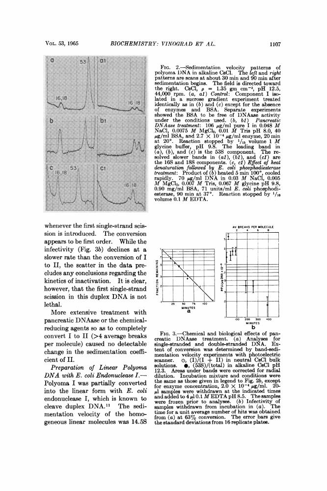
tained extended circles and no twistedFIG. 4.-Sedimentation velocity-pH titration configurations (Fig. lb). The DNA
of the three components in polyoma DNA. I, samples used to prepare the grids for0; II, 0; III, A; I only band present, cf.text; mixture of unresolved single strands in Figures la and b were identical exceptalkali, x; 29,500 rpm. Single linear, -O, and for the time of incubation with the en-single circular, ci, strands in alkali, 44,770rpm. Sedimentation coefficients at 200 in zyme. The spreading forces acting onCsCl, p = 1.35 gm cm-3, are not corrected for the macromolecules in the monolayersolvent viscosity, flr = 0.925, or buoyancyeffects. The values at pH 8.0 and 12.4 are the appear to remove loops and crossoversmeans of 12 and 7 determinations, respectively, that are not locked in the structure of
the DNA.(2) A study of the sedimentation velocity in 3 M CsCl of a mixture of the three
components of polyoma DNA as a function of pH from 8 to 12.5 revealed a com-plicated pH-melting curve for component I (Fig. 4). Component II behavednormally 18, 20 and moved faster as denaturation increased until strand separationoccurred with an attendant sudden drop in sedimentation velocity at pH 11.8.Component I, like II, was at first insensitive to pH. At pH 11.5, however, the sedi-mentation coefficient first dropped, and then in the pH range 11.6 to 11.8 was thesame as for polyoma II. Only one moving band was observed in this pH range.The sedimentation coefficient of I then increased to the very high value characteris-tic of the double-stranded cyclic coil previously reported.2 Essentially the same re-sults were obtained in 1.0 M KCI solutions. The dip in the sedimentation velocity-

VOL. 53, 1965 BIOCHEMISTRY: VINOGRAD ET AL. 1109
pH curve was initially unexplainable. If, however, polyoma DNA I contains left-handed tertiary turns, such a dip in the pH-melting profile would be required. Inthe early stages of denaturation some of the duplex turns, which are known to beright-handed, unwind. The unwinding of the duplex must be accompanied by aright-handed twisting of the remainder of the molecule. If the tertiary turns wereoriginally left-handed (Fig. 5), progressive unwinding would cause the moleculeto pass through configuration I' characterized by the absence of tertiary turns.The extended configuration I' is similar to that in polyoma II (Fig. 5) and bothI' and II would have similar sedimentation velocities. Further unwinding of duplexI' is accompanied by continued right-hand twisting of the whole molecule untilfinally the double-stranded cyclic coil2configuration develops.
(3) The twisted circular structure pro- lqlw.gvides a satisfactory explanation for the 53Sconfigurational change that occurs when 186Sone single-strand scission is introducedinto the molecule. Such a scission gener- -/ " -ates a site for the rotation of the helix in ' Xthe complementary strand opposite the j S 8break. The swivel relieves the topological ', 16 S 16 Srestraint responsible for the twisted con- Ifiguration.Discussion.-A mechanism for the for-
mation of the twisted circular structuressuggested by the above analysis of the pH- Qmelting curve. According to this mecha- 1,20 S ,16 S M,14Snism the last closure of chain ends occursbefore all of the winding of the two DNA xstrands into the Watson-Crick structure FIG. 5.-Diagrammatic representation of
the several forms of polyoma DNA. Theis completed. The closing leaves the du- duplex segments shown contain 12 turns,plex in the configuration I' (Fig. 5) re- about one fortieth of the total number. The
twisted circular duplex shown contains onestrained from converting to I by an as yet left-hand tertiary turn. 8% of the right-unknown factor participating in the DNA hand duplex turns in the model are unwound
to form I'. The dashed circles around thesynthesis. Removal of the restraint then denatured forms indicate the relative hy-allows I' to wind spontaneously into a drodynamic diameters. The sedimentation
coefficients were measured in neutral andcomplete Watson-Crick structure and alkaline NaCl solutions.form the twisted circular structure, I, withno change in winding number.An alternative proposal is that the molecule in form II, which contains a swivel,
is twisted by some organizer, e.g., the virus protein. The last covalent backbonebond is then made while the DNA remains twisted under the constraint of theorganizer. This alternative is unlikely in view of Dulbecco's2' finding that thepolyoma DNA made before virus production begins has the sedimentation velocityof component I.
It is not possible at the present time to estimate reliably the number of tertiaryturns. A turn is defined as a 3600 rotation of the helix. The electron micro-graphs of I usually contain some molecules that are completely extended; these

1110 BIOCHEMISTRY: VINOGRAD ET AL. PROC. N. A. S.
may have suffered a single break during grid preparation or may have been in stateI' due to denaturation induced by the spreading forces. This latter action wouldresult in the unwinding of the twisted circles to form extended circles. The maxi-mum number of crossovers that can be distinguished is 8, which corresponds to 4 turnsor the unwinding of 40 base pairs. This limit may be low because it is difficultto count crossovers in tightly coiled forms. That the number of tertiary turns inthe molecule is not large is suggested by the fact that the transition of I to I' occursbefore substantial melting of II takes place, as indicated by the s versus pH plot.With the new assignment of structure to the three components of polyoma DNA,
it is found that satisfactory agreement obtains between the observed sedimentationcoefficient of the linear form, 14.5S, and the 15.3 4 0.5S predicted by Studier'srelation"8 for a molecular weight2 of 3.0 + 0.3 X 10⁶. The values, 18.4S and15.7S for the alkaline single-stranded circular and linear forms similarly agree withthe predicted values of 17.4S and 15.6S, respectively. The effect of ring closure ofIII to form component II is to increase S by 10 per cent. An effect of similarmagnitude has been reported22 for the cyclization of X DNA.The twisted circular structure observed here for polyoma DNA may be a common
characteristic of covalently closed, circular duplex DNA. A part of the DNA fromrabbit papilloma virus,23 SV40 virus,3 and the replicating form ofOX DNA24-26 haveall been shown to be circular duplex molecules which do not strand-separate inalkali or after heating in formaldehyde. Two sedimentation velocity componentsdiffering by 20-30 per cent have been reported for the above DNA's.27 Crawfordand Black3 observed sedimentation velocity-denaturation curves that are similar toour pH-melting curve upon heating SV40 DNA and polyoma DNA in formaldehydesolutions to various temperatures. No explanation was offered for this behavior,which we interpret as indicating the presence of a left-handed, twisted circular struc-ture.28Burton and Sinsheimer29 have shown that the slow component II in RF-+X
DNA dissociates in alkali to form linear and circular single-stranded moleculesand have concluded that both of the undenatured forms of the DNA are circular.While this communication was in preparation, Jansz and Pouwels30 reported thatthe pancreatic DNAase-induced conversion of I to II in RF-+X DNA representsa conversion between circular duplex molecules. No explanation for the changein the sedimentation coefficient was offered. In view of the results describedhere, it is likely that RF-q5X DNA is in the twisted circular form. A commonmechanism for the incorporation of the tertiary turns during replication is a strongpossibility, and allows us to predict that the tertiary turns in the RF-+X DNA willbe found to be left-handed.Summary.-The results of this study show that circular duplex polyoma DNA
may be converted to a less compact circular duplex by introducing a single-strandscission. The viral form contains tertiary turns which appear to have been lockedin during replication.
It is a pleasure to thank I. R. Lehman for the generous gift of the E. coli enzymes, R. L. Sin-sheimer, J. Petruska, and M. Fried for helpful discussions, R. Dulbecco for allowing us to quotehis unpublished results, and T. Benjamin, L. Wenzel, and A. Drew for advice and assistance inthe culture of the virus and the assay of the DNA. This work was supported in part by grantsHE 03394 and CA 08014 from the U.S. Public Health Service.

VOL. 53, 1965 BIOCHEMISTRY: SETLOW ET AL. 1111
* This work was reported in part at the 9th Annual Meeting of the Biophysical Society, Feb-ruary 24, 1965. Contribution 3227 of Gates and Crellin Laboratories of Chemistry.
I Dulbecco, R., and M. Vogt, these PROCEEDINGS, 50, 236 (1963).2Weil, R., and J. Vinograd, these PROCEEDINGS, 50, 730 (1963).3 Crawford, L. V., and P. H. Black, Virology, 24, 388 (1964).4Weil, R., these PROCEEDINGS, 49, 480 (1963).5 Crawford, L. V., R. Dulbecco, M. Fried, L. Montagnier, and M. Stoker, these PROCEEDINGS,
52, 148 (1964).6 Winocour, E., Virology, 19, 158 (1963).Murikami, W., Science, 142, 56 (1963).
6 Viograd, J., R. Bruner, R. Kent, and J. Weigle, these PROCEEDINGS, 49, 902 (1963).9 Beckman Instruments Co., Spinco Division, Palo Alto, Calif.
l' Hanlon, S., K. Lamers, G. Lauterbach, R. Johnson, and H. K. Schachman, Arch. Biochem.Biophys., 99, 157 (1962).
11 Lehman, I. R., G. G. Roussos, and E. A. Pratt, J. Biol. Chem., 237, 819 (1962).12 Lehman, I. R., J. Biol. Chem., 235, 1479 (1960).13 Vinograd, J., R. Radloff, and R. Bruner, Biopolymers, in press.14 Lebowitz, J., and M. Laskowski, Jr., Biochemistry, 1, 1044 (1962).15 Kleinschmidt, A. K., and R. K. Zahn, Z. Naturforsch., 14b, 770 (1959).16 Swan, J. M., Nature, 180, 643 (1957).17 Thomas, C. A., Jr., J. Am. Chem. Soc., 78, 1861 (1956). The estimate of 50 was obtained for
the case of a 10% lowering of the weight average molecular weight.18 Studier, F. Wm., J. Mol. Biol., 11, 373 (1965)."Fiers, W., and R. L. Sinsheimer, J. Mol. Biol., 5, 408 (1962).20 Davidson, P. F., and D. Freifelder, J. Mol. Biol., 5, 643 (1962).21 Dulbecco, R., private communication.22 Hershey, A. D., E. Burgi, and L. Ingraham, these PROCEEDINGS, 49, 748 (1963).23 Crawford, L. V., J. Mol. Biol., 8, 489 (1964).24 Kleinschmidt, A., A. Burton, and R. L. Sinsheimer, Science, 142, 961 (1963).25 Burton, A., and R. L. Sinsheimer, Science, 142, 962 (1963).26 Burton, A., and R. L. Sinsheimer, Abstracts, 8th Annual Meeting of the Biophysical Society,
1964.27 R. Weil has informed us that the slow component II in rabbit papilloma DNA is circular as
seen in the electron microscope.28 Human papilloma DNA appears to be similar in configuration to SV40 DNA and polyoma
DNA (Crawford, L. V., manuscript submitted for publication).29 Burton, A., and R. L. Sinsheimer, private communication.30 Jansz, H. S., and P. H. Pouwels, Biochem. Biophys. Res. Commun., 18, 589 (1965).
PYRIMIDINE DIMERS IN UV-IRRADIATED POLY dl: dC*
BY R. B. SETLOW, W. L. CARRIER, AND F. J. BOLLUM
BIOLOGY DIVISION, OAK RIDGE NATIONAL LABORATORY, OAK RIDGE, TENNESSEE
Communicated by Alexander Hollaender, March 29, 1965
Ultraviolet irradiation of polynucleotides containing thymine results in the for-mation of dimers between adjacent thymine residues.1' 2 The demonstration thatthese dimers are responsible to a large, but not exclusive, extent for the inactivationof primer DNA3 and of transforming DNA4 made use of a specific photochemicalproperty of the dimers, namely, that they may be monomerized by short-wavelengthirradiation. The fact that many UV effects are photoreactivable and that treat-