Restudying variations of axial skeleton patterning in ... · Key words: Human vertebra morphology...
13
Restudying variations of axial skeleton patterning in Eskimo groups with new data from ancient Chukotka (Ekven archaeological site) ORIGINAL ARTICLE Eur. J. Anat. 23 (3): 187-199 (2019) Marina K. Karapetian 1 , Sergey V. Makarov 2 1 Research Institute and Museum of Anthropology, Lomonosov Moscow State University, Moscow, Russia, Research Centre for Medical Genetics, Moscow, Russia SUMMARY This study continues a series of studies by Stew- art and Merbs on vertebral column variations in Eskimo groups. The focus is on so called cranio- caudal shifts in spine patterning. The study is per- formed on a skeletal sample of ancient Eskimos from Siberia (Ekven site, Chukotka) and compara- tive samples representing population groups of European and African ancestry. In addition to these, literature data are used for comparative analysis to assess the pattern of cranio-caudal border shifts on intra-specific level. The result con- firms the presence of significantly increased pre- disposition of the Eskimos to caudal shifts in spine patterning, expressed both as increased frequen- cies of complete caudal shifts of thoraco-lumbar and lumbo-sacral borders, as well as minor varia- tions in vertebrae morphology, including variation in the type of articular processes (thoracic/lumbar types) and the position of costo-central articulation at T9 level. Hypotheses explaining this specific character of the Eskimo/Inuit groups are proposed and explored, including gene drift, influence of en- vironmental factors and association with morpho- logical characteristics adaptive to survival in the Arctic. One of the explanations may be the associ- ation with characteristic form and size of the tho- racic cage that distinguishes the Arctic groups such as Eskimos and Chukchi from groups leaving in more southern areas. This needs to be tested on other groups, living in similar conditions. Key words: Human vertebra morphology – Cra- nio-caudal shifts – Homeotic transformations – Siberia – Adaptation to Arctic INTRODUCTION Numerical variations of spine formula in humans and in non-human primates have long been a sub- ject of biological and medical interest. First, be- cause of their association with phylogenetic chang- es in locomotion pattern, thus interest in paleoan- thropological studies (Todd, 1922; Schultz, 1930; Williams et al., 2016; Thompson and Almécija, 2017). Second, because of their inheritability (Kühne, 1932; Schapera, 1987) and, thus, interest in populations studies (Shore, 1930; Stewart, 1932; Lanier, 1939; Allbrook, 1955; Bornstein and Peterson, 1966; Tulsi, 1972; de Beer Kaufman, 1974; Merbs, 1974; Jankauskas, 1988). And, third, because of their presumed clinical significance and association with mutations and developmental anomalies (Galis et al., 2006; ten Broek et al., 2012; Nakajima et al., 2014; Tague, 2018). The pattern of the axial skeleton is determined during prenatal development. When a shift in ver- tebra identity is observed at the border between two regions, this represents so-called homeotic transformation. It was shown that homeotic trans- formations along the axial skeleton may be associ- ated with mutations in certain genes responsible for body patterning (e.g. HOX genes) (Mallo et al., 2010). In addition to genetic factors, possible im- pact of external factors, influencing the expression of genes during embryogenesis, is suggested from the experimental studies on animal models (e.g. 187 Submitted: 31 January, 2019. Accepted: 5 April, 2019. Corresponding author: Marina Karapetian. Mokhovaya str. 11/1, Moscow 125009, Russia. E-mail: [email protected]
Transcript of Restudying variations of axial skeleton patterning in ... · Key words: Human vertebra morphology...
Restudying variations of axial skeleton patterning in Eskimo groups with new data from ancient Chukotka
(Ekven archaeological site)
ORIGINAL ARTICLE Eur. J. Anat. 23 (3): 187-199 (2019)
Marina K. Karapetian1, Sergey V. Makarov
2
1Research Institute and Museum of Anthropology, Lomonosov Moscow State University, Moscow, Russia, Research
Centre for Medical Genetics, Moscow, Russia
SUMMARY
This study continues a series of studies by Stew-art and Merbs on vertebral column variations in Eskimo groups. The focus is on so called cranio-caudal shifts in spine patterning. The study is per-formed on a skeletal sample of ancient Eskimos from Siberia (Ekven site, Chukotka) and compara-tive samples representing population groups of European and African ancestry. In addition to these, literature data are used for comparative analysis to assess the pattern of cranio-caudal border shifts on intra-specific level. The result con-firms the presence of significantly increased pre-disposition of the Eskimos to caudal shifts in spine patterning, expressed both as increased frequen-cies of complete caudal shifts of thoraco-lumbar and lumbo-sacral borders, as well as minor varia-tions in vertebrae morphology, including variation in the type of articular processes (thoracic/lumbar types) and the position of costo-central articulation at T9 level. Hypotheses explaining this specific character of the Eskimo/Inuit groups are proposed and explored, including gene drift, influence of en-vironmental factors and association with morpho-logical characteristics adaptive to survival in the Arctic. One of the explanations may be the associ-ation with characteristic form and size of the tho-racic cage that distinguishes the Arctic groups such as Eskimos and Chukchi from groups leaving in more southern areas. This needs to be tested on other groups, living in similar conditions.
Key words: Human vertebra morphology – Cra-nio-caudal shifts – Homeotic transformations – Siberia – Adaptation to Arctic
INTRODUCTION
Numerical variations of spine formula in humans and in non-human primates have long been a sub-ject of biological and medical interest. First, be-cause of their association with phylogenetic chang-es in locomotion pattern, thus interest in paleoan-thropological studies (Todd, 1922; Schultz, 1930; Williams et al., 2016; Thompson and Almécija, 2017). Second, because of their inheritability (Kühne, 1932; Schapera, 1987) and, thus, interest in populations studies (Shore, 1930; Stewart, 1932; Lanier, 1939; Allbrook, 1955; Bornstein and Peterson, 1966; Tulsi, 1972; de Beer Kaufman, 1974; Merbs, 1974; Jankauskas, 1988). And, third, because of their presumed clinical significance and association with mutations and developmental anomalies (Galis et al., 2006; ten Broek et al., 2012; Nakajima et al., 2014; Tague, 2018).
The pattern of the axial skeleton is determined during prenatal development. When a shift in ver-tebra identity is observed at the border between two regions, this represents so-called homeotic transformation. It was shown that homeotic trans-formations along the axial skeleton may be associ-ated with mutations in certain genes responsible for body patterning (e.g. HOX genes) (Mallo et al., 2010). In addition to genetic factors, possible im-pact of external factors, influencing the expression of genes during embryogenesis, is suggested from the experimental studies on animal models (e.g.
187
Submitted: 31 January, 2019. Accepted: 5 April, 2019.
Corresponding author: Marina Karapetian. Mokhovaya str.
11/1, Moscow 125009, Russia.
E-mail: [email protected]
Axial skeleton patterning in Eskimo groups
188
Murakami and Kameyama, 1963; Chernoff and Rogers, 2004). That some homeotic shifts may represent deviations from normal development is suggested from studies, showing existence of sig-nificant prenatal/early postnatal selection of indi-viduals with cervical ribs and association of cervi-cal ribs with malformations in other organ systems (Schumacher et al., 1992; Galis et al., 2006; ten Broek et al., 2012).
Prehistory of the Eskimo/Inuit groups. The first representatives of the Eskimos (Paleo-Eskimos) appeared in Siberia; and about 5-6 thousand years ago they expanded into Alaska, eventually popu-lating Canadian Arctic and Greenland. Later, Pale-Eskimo were replaced by the Neo-Eskimo (or Inuit) who are the direct ancestors of the modern Eski-mo/Inuit groups. The earliest representatives of the Neo-Eskimo are found in Siberia, and in Alaska they first appear as carriers of the early Old Bering Sea culture at the turn of our era. The Thule cul-ture (the direct ancestors of the modern Eskimo/Inuit groups in North America) emerged in Alaska about 1000 AD and expanded throughout the North American Arctic, replacing all Paleo-Eskimo groups living there. The Paleo-Eskimo cultures completely disappear in Alaska at about 800 AD and in Canada and Greenland at about 1300 AD (Raghavan et al., 2014; Friesen and Mason, 2016). Genetic evidence shows continuity in Paleo-Eskimo groups during more than 4000 years of occupation, and discontinuity between Paleo- and Neo-Eskimo groups of North America suggesting incident of population replacement (Raghavan et al., 2014).
Study background. In the early 1930s Stewart presented his detailed observations on the Eskimo spine when he worked with skeletal remains brought by Ales Hrdlička from Alaska (Stewart, 1932). Notably, he pointed to an apparently high prevalence of caudal border shifts at lumbo-sacral border, e.g. elongated pre-sacral spine (25 verte-brae), with no cases of the shortened pre-sacral spines (23 vertebrae). High prevalence of individu-als with 25 pre-sacral vertebrae relative to other population groups was also observed on a sample of Sadlermiut Eskimos from Southampton Island (Merbs, 1974) and on a pooled sample of Native Americans (including Koniag Eskimos, Aleutians and Plain Indians) (Borstein and Peterson, 1966). Lanier, in his detailed study on pre-sacral spine variations in American Whites and Blacks, while performing comparison with other population groups using literature sources, noted that caudal type variations are most frequent in Eskimo group (Lanier, 1939).
Unfortunately, no analogous studies were per-formed on the Asian Eskimos, who are closely re-lated both genetically and by living conditions to the groups from Alaska and North Canada. Thus, it is not known to what extent the morphological specificity observed for the American Eskimo/Inuit groups are characteristic for this circumpolar popu-lation as such, and thus might represent a very ancient morphological complex.
At the Asian part of the Beringia, one of the most outstanding ancient sites is the Ekven burial site. The site was located on the far east point of Chu-kotka near Cape Dezhneva. Most burials belong to the Old Bering Sea (I–III) and Okvik cultures, em-bracing the period from about the end of the 1
st
millennium BC to the beginning of the 2nd
millenni-um AD (Arutyunov and Sergeev, 1975; Friesen and Mason, 2016). Regardless of the wide tem-poral range, during which the burial ground func-tioned, craniological studies showed high level of homogeneity in metric traits, indicating the conser-vation of the anthropological type, well adapted to the extreme environmental conditions (Alexeeva et al., 2008).
The aim of this report was to assess frequencies of numerical variations of spine formula in Eskimo group from the Ekven site, as well as from Alaska and Canada, to perform a comparative analysis with other population groups using original data and data from the literature, and to test that Eski-mos indeed show a strong tendency towards cau-dal shifts in pattern by studying minor variations of vertebrae morphology. Hypotheses explaining this morphological specificity of the Eskimo spine are proposed and explored.
MATERIALS AND METHODS
The Ekven Eskimo collection was studied in the Research Institute and Museum of Anthropology of the Lomonosov Moscow State University. Totally, 59 individuals were studied (30 males and 29 fe-males). All except 7 individuals were adults (≥18 years). The 7 individuals were juveniles older than 14 years of age for whom sex could be estimated. They were studied to increase the sample size, and their inclusion is justified because by the age of 14 all key structures needed to identify the type of a vertebra are formed and synostosed (see Schaefer et al., 2009). The first and the second sacral segments may not fuse completely until the age of 25. However, partial union is usually pre-sent by puberty and, even if the fusion did not begin, the 1
st sacral segment is identifiable based
on its morphology (see Schaefer et al., 2009). Sex was estimated based on pelvic morphology ac-cording to criteria summarized by Buikstra and Ubelaker (1994). Age was estimated as Juvenile and Adult based on the degree of epiphyseal fu-sion (Schaefer et al., 2009).
In addition to the Ekven sample (Ekv), compara-tive samples representing population groups of different ancestry were studied: Russian (2 sam-ples, Moscow region) (Rus); North American resi-dents of European ancestry (samples from the Terry and Grant collections) (NA Eur); North Amer-ican residents of African ancestry (sample from the Terry collection) (NA Afr). Samples studied by the authors are listed and described in Table 1. All individuals in these samples were adults.
Ekven sample and the three comparative sam-ples were reviewed for the following traits:
M.K. Karapetian & S.V. Makarov
189
1) Number of cervical (C), thoracic (T) and lumbar (L) vertebrae and the total number of pre-sacral vertebrae (PCV). Thoracic and sacral vertebrae were identified following classical criteria (Schultz, 1930), used in the overwhelming majority of studies. Following the costal criteria, a vertebra was identified as thoracic if it carried rib facet that indicated presence of articulation with a true rib. If a vertebra carried facet only on one side, it was also considered thoracic. The 24
th vertebra was
considered sacral if it was fused (non-pathologically, unilaterally or bilaterally via lateral masses) to the sacrum. The 25
th vertebra was con-
sidered pre-sacral (lumbar) if it was completely separated from the sacrum and articulated with it only via joint facets (in some cases also via extra facets on the enlarged transverse processes) (Lanier, 1939; Jankauskas, 1988). The number of vertebrae was recorded only when this was cer-tain. For example, 11 thoracic vertebrae were scored only when there was no indication of post-mortem loss of one thoracic vertebra (all present vertebrae were congruent with adjacent verte-brae); and the same was done for the cervical and lumbar spines. If spine formula differed from the modal (7C-12T-5L), a presence of border shift was stated (cranial – towards the cranium; caudal – towards the coccyx).
2) Position of transitional vertebra at T-L bor-
der – lowermost vertebra with superior articular processes of thoracic and inferior articular pro-cesses of lumbar type. Thoracic type superior ar-
ticular processes were scored when they were ori-ented dorsolaterally; lumbar type were scored when they were oriented dorsomedially; and vice versa for the inferior articular processes (after La-nier, 1939). If articular facets were asymmetrical, e.g. left superior facet of T12 was thoracic-type and right was lumbar-type, then 0.5 of this individ-ual had transitional vertebra at T12 level (left side) and another 0.5 – at T11 level (right side).
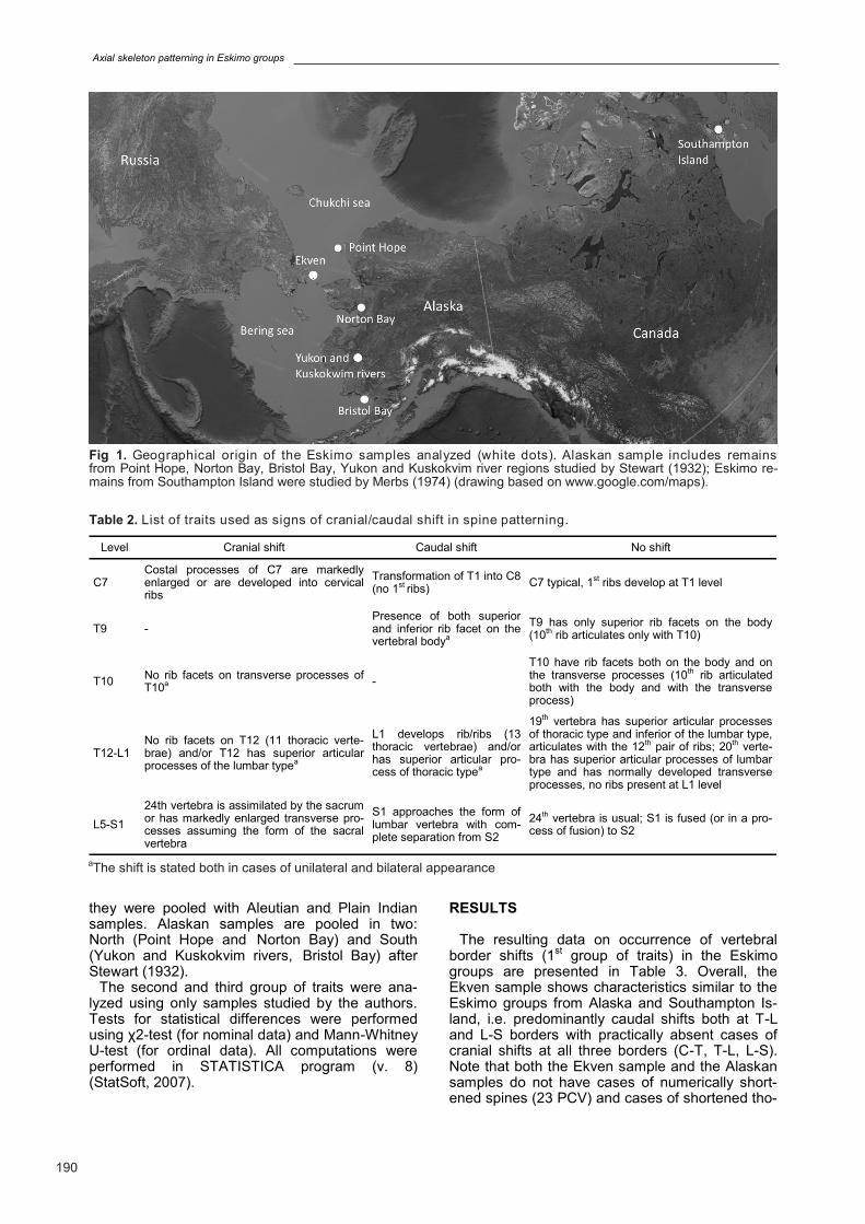
3) Presence of any signs of cranial/caudal shift in
spine patterning at C7, T9 (shift in the position of the last typical thoracic vertebra), T10 (shift in the position of the last vertebra articulating with rib tubercle), T12, L1, L5 and S1 according to criteria, summarized in Table 2. Statement of shift in T9 and T10 morphology proceeded from the custom-ary anatomical description of these vertebrae (Gray, 1858).
The resulting data on the 1st group of traits
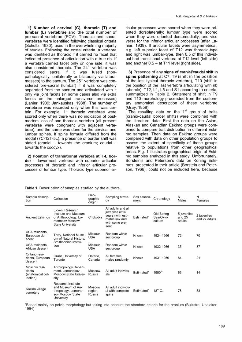
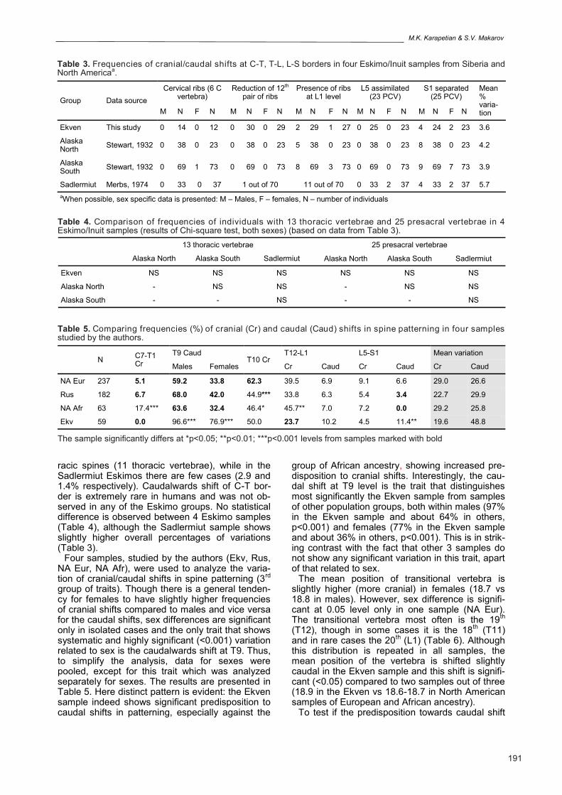
(cranio-caudal border shifts) were combined with the literature data. First the data on the Asian, Alaskan and Canadian Eskimo groups were com-bined to compare trait distribution in different Eski-mo samples. Then data on Eskimo groups were compared with data on other population groups to assess the extent of specificity of these groups relative to populations from other geographical areas. Fig. 1 illustrates geographical origin of Eski-mo samples analyzed in this study. Unfortunately, Borstein’s and Peterson’s data on Koniag Eski-mos, presented in their article (Borstein and Peter-son, 1966), could not be included here, because
Sample descrip-tion
Collection Geo-graphic origin
Sampling strate-gy
Sex assess-ment
Chronology N Males
N Females
Ancient Eskimos
Ekven, Research Institute and Museum of Anthropology, Lo-monosov Moscow State University
Chukotka
All adults and all juveniles (>14 years) with esti-mable sex and with spine pre-sent
Estimateda Old Bering Sea/Okvik culture
5 juveniles and 25 adults
2 juveniles and 27 adults
USA residents, European de-scent
Terry, National Muse-um of Natural History, Smithsonian Institu-tion
Missouri, USA
Random within sex group
Known 1924-1966 72 70
USA residents, African descent
Missouri, USA
Random within sex group
Known 1932-1966 35 37
Ontario resi-dents, European descent
Grant, University of Toronto
Ontario, Canada
All females; males randomly
Known 1931-1950 84 21
Moscow resi-dents (anatomical col-lection)
Anthropology Depart-ment, Lomonosov Moscow State Univer-sity
Moscow, Russia
All adult individu-als
Estimateda 1950th 66 14
Kozino village cemetery
Research Institute and Museum of An-thropology, Lomono-sov Moscow State University
Moscow region, Russia
All adult individu-al with complete spine
Estimateda 18th C. 78 53
Table 1. Description of samples studied by the authors.
aBased mainly on pelvic morphology but taking into account the standard criteria for the cranium (Buikstra, Ubelaker, 1994)
Axial skeleton patterning in Eskimo groups
190
they were pooled with Aleutian and Plain Indian samples. Alaskan samples are pooled in two: North (Point Hope and Norton Bay) and South (Yukon and Kuskokvim rivers, Bristol Bay) after Stewart (1932).
The second and third group of traits were ana-lyzed using only samples studied by the authors. Tests for statistical differences were performed using χ2-test (for nominal data) and Mann-Whitney U-test (for ordinal data). All computations were performed in STATISTICA program (v. 8) (StatSoft, 2007).
RESULTS
The resulting data on occurrence of vertebral border shifts (1
st group of traits) in the Eskimo
groups are presented in Table 3. Overall, the Ekven sample shows characteristics similar to the Eskimo groups from Alaska and Southampton Is-land, i.e. predominantly caudal shifts both at T-L and L-S borders with practically absent cases of cranial shifts at all three borders (C-T, T-L, L-S). Note that both the Ekven sample and the Alaskan samples do not have cases of numerically short-ened spines (23 PCV) and cases of shortened tho-
Fig 1. Geographical origin of the Eskimo samples analyzed (white dots). Alaskan sample includes remains from Point Hope, Norton Bay, Bristol Bay, Yukon and Kuskokvim river regions studied by Stewart (1932); Eskimo re-mains from Southampton Island were studied by Merbs (1974) (drawing based on www.google.com/maps).
Level Cranial shift Caudal shift No shift
C7 Costal processes of C7 are markedly enlarged or are developed into cervical ribs
Transformation of T1 into C8 (no 1st ribs)
C7 typical, 1st ribs develop at T1 level
T9 - Presence of both superior and inferior rib facet on the vertebral bodya
T9 has only superior rib facets on the body (10th rib articulates only with T10)
T10 No rib facets on transverse processes of T10a
-
T10 have rib facets both on the body and on the transverse processes (10th rib articulated both with the body and with the transverse process)
T12-L1 No rib facets on T12 (11 thoracic verte-brae) and/or T12 has superior articular processes of the lumbar typea
L1 develops rib/ribs (13 thoracic vertebrae) and/or has superior articular pro-cess of thoracic typea
19th vertebra has superior articular processes of thoracic type and inferior of the lumbar type, articulates with the 12th pair of ribs; 20th verte-bra has superior articular processes of lumbar type and has normally developed transverse processes, no ribs present at L1 level
L5-S1
24th vertebra is assimilated by the sacrum or has markedly enlarged transverse pro-cesses assuming the form of the sacral vertebra
S1 approaches the form of lumbar vertebra with com-plete separation from S2
24th vertebra is usual; S1 is fused (or in a pro-cess of fusion) to S2
Table 2. List of traits used as signs of cranial/caudal shift in spine patterning.
aThe shift is stated both in cases of unilateral and bilateral appearance
M.K. Karapetian & S.V. Makarov
191
racic spines (11 thoracic vertebrae), while in the Sadlermiut Eskimos there are few cases (2.9 and 1.4% respectively). Caudalwards shift of C-T bor-der is extremely rare in humans and was not ob-served in any of the Eskimo groups. No statistical difference is observed between 4 Eskimo samples (Table 4), although the Sadlermiut sample shows slightly higher overall percentages of variations (Table 3).
Four samples, studied by the authors (Ekv, Rus, NA Eur, NA Afr), were used to analyze the varia-tion of cranial/caudal shifts in spine patterning (3
rd
group of traits). Though there is a general tenden-cy for females to have slightly higher frequencies of cranial shifts compared to males and vice versa for the caudal shifts, sex differences are significant only in isolated cases and the only trait that shows systematic and highly significant (<0.001) variation related to sex is the caudalwards shift at T9. Thus, to simplify the analysis, data for sexes were pooled, except for this trait which was analyzed separately for sexes. The results are presented in Table 5. Here distinct pattern is evident: the Ekven sample indeed shows significant predisposition to caudal shifts in patterning, especially against the
group of African ancestry, showing increased pre-disposition to cranial shifts. Interestingly, the cau-dal shift at T9 level is the trait that distinguishes most significantly the Ekven sample from samples of other population groups, both within males (97% in the Ekven sample and about 64% in others, p<0.001) and females (77% in the Ekven sample and about 36% in others, p<0.001). This is in strik-ing contrast with the fact that other 3 samples do not show any significant variation in this trait, apart of that related to sex.
The mean position of transitional vertebra is slightly higher (more cranial) in females (18.7 vs 18.8 in males). However, sex difference is signifi-cant at 0.05 level only in one sample (NA Eur). The transitional vertebra most often is the 19
th
(T12), though in some cases it is the 18th (T11)
and in rare cases the 20th (L1) (Table 6). Although
this distribution is repeated in all samples, the mean position of the vertebra is shifted slightly caudal in the Ekven sample and this shift is signifi-cant (<0.05) compared to two samples out of three (18.9 in the Ekven vs 18.6-18.7 in North American samples of European and African ancestry).
To test if the predisposition towards caudal shift
Group Data source
Cervical ribs (6 C vertebra)
Reduction of 12th pair of ribs
Presence of ribs at L1 level
L5 assimilated (23 PCV)
S1 separated (25 PCV)
Mean % varia-tion M N F N M N F N M N F N M N F N M N F N
Ekven This study 0 14 0 12 0 30 0 29 2 29 1 27 0 25 0 23 4 24 2 23 3.6
Alaska North
Stewart, 1932 0 38 0 23 0 38 0 23 5 38 0 23 0 38 0 23 8 38 0 23 4.2
Alaska South
Stewart, 1932 0 69 1 73 0 69 0 73 8 69 3 73 0 69 0 73 9 69 7 73 3.9
Sadlermiut Merbs, 1974 0 33 0 37 1 out of 70 11 out of 70 0 33 2 37 4 33 2 37 5.7
aWhen possible, sex specific data is presented: M – Males, F – females, N – number of individuals
Table 3. Frequencies of cranial/caudal shifts at C-T, T-L, L-S borders in four Eskimo/Inuit samples from Siberia and North Americaa.
13 thoracic vertebrae 25 presacral vertebrae
Alaska North Alaska South Sadlermiut Alaska North Alaska South Sadlermiut
Ekven NS NS NS NS NS NS
Alaska North - NS NS - NS NS
Alaska South - - NS - - NS
Table 4. Comparison of frequencies of individuals with 13 thoracic vertebrae and 25 presacral vertebrae in 4 Eskimo/Inuit samples (results of Chi-square test, both sexes) (based on data from Table 3).
N C7-T1 Cr
T9 Caud T10 Cr
T12-L1 L5-S1 Mean variation
Males Females Cr Caud Cr Caud Cr Caud
NA Eur 237 5.1 59.2 33.8 62.3 39.5 6.9 9.1 6.6 29.0 26.6
Rus 182 6.7 68.0 42.0 44.9*** 33.8 6.3 5.4 3.4 22.7 29.9
NA Afr 63 17.4*** 63.6 32.4 46.4* 45.7** 7.0 7.2 0.0 29.2 25.8
Ekv 59 0.0 96.6*** 76.9*** 50.0 23.7 10.2 4.5 11.4** 19.6 48.8
Table 5. Comparing frequencies (%) of cranial (Cr) and caudal (Caud) shifts in spine patterning in four samples studied by the authors.
The sample significantly differs at *p<0.05; **p<0.01; ***p<0.001 levels from samples marked with bold
Axial skeleton patterning in Eskimo groups
192
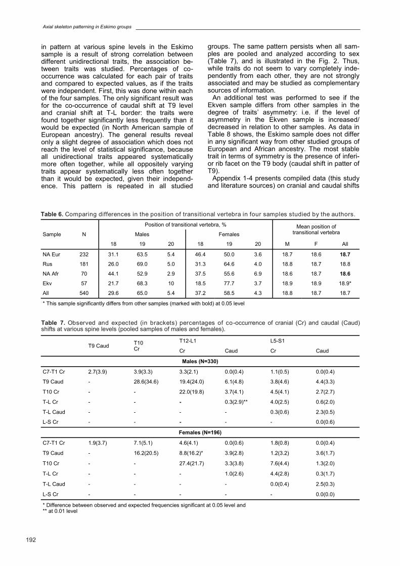
in pattern at various spine levels in the Eskimo sample is a result of strong correlation between different unidirectional traits, the association be-tween traits was studied. Percentages of co-occurrence was calculated for each pair of traits and compared to expected values, as if the traits were independent. First, this was done within each of the four samples. The only significant result was for the co-occurrence of caudal shift at T9 level and cranial shift at T-L border: the traits were found together significantly less frequently than it would be expected (in North American sample of European ancestry). The general results reveal only a slight degree of association which does not reach the level of statistical significance, because all unidirectional traits appeared systematically more often together, while all oppositely varying traits appear systematically less often together than it would be expected, given their independ-ence. This pattern is repeated in all studied
groups. The same pattern persists when all sam-ples are pooled and analyzed according to sex (Table 7), and is illustrated in the Fig. 2. Thus, while traits do not seem to vary completely inde-pendently from each other, they are not strongly associated and may be studied as complementary sources of information.
An additional test was performed to see if the Ekven sample differs from other samples in the degree of traits’ asymmetry: i.e. if the level of asymmetry in the Ekven sample is increased/decreased in relation to other samples. As data in Table 8 shows, the Eskimo sample does not differ in any significant way from other studied groups of European and African ancestry. The most stable trait in terms of symmetry is the presence of inferi-or rib facet on the T9 body (caudal shift in patter of T9).
Appendix 1-4 presents compiled data (this study and literature sources) on cranial and caudal shifts
Sample N
Position of transitional vertebra, % Mean position of transitional vertebra Males Females
18 19 20 18 19 20 M F All
NA Eur 232 31.1 63.5 5.4 46.4 50.0 3.6 18.7 18.6 18.7
Rus 181 26.0 69.0 5.0 31.3 64.6 4.0 18.8 18.7 18.8
NA Afr 70 44.1 52.9 2.9 37.5 55.6 6.9 18.6 18.7 18.6
Ekv 57 21.7 68.3 10 18.5 77.7 3.7 18.9 18.9 18.9*
All 540 29.6 65.0 5.4 37.2 58.5 4.3 18.8 18.7 18.7
* This sample significantly differs from other samples (marked with bold) at 0.05 level
Table 6. Comparing differences in the position of transitional vertebra in four samples studied by the authors.
T9 Caud T10 Cr
T12-L1 L5-S1
Cr Caud Cr Caud
Males (N=330)
C7-T1 Cr 2.7(3.9) 3.9(3.3) 3.3(2.1) 0.0(0.4) 1.1(0.5) 0.0(0.4)
T9 Caud - 28.6(34.6) 19.4(24.0) 6.1(4.8) 3.8(4.6) 4.4(3.3)
T10 Cr - - 22.0(19.8) 3.7(4.1) 4.5(4.1) 2.7(2.7)
T-L Cr - - - 0.3(2.9)** 4.0(2.5) 0.6(2.0)
T-L Caud - - - - 0.3(0.6) 2.3(0.5)
L-S Cr - - - - - 0.0(0.6)
Females (N=196)
C7-T1 Cr 1.9(3.7) 7.1(5.1) 4.6(4.1) 0.0(0.6) 1.8(0.8) 0.0(0.4)
T9 Caud - 16.2(20.5) 8.8(16.2)* 3.9(2.8) 1.2(3.2) 3.6(1.7)
T10 Cr - - 27.4(21.7) 3.3(3.8) 7.6(4.4) 1.3(2.0)
T-L Cr - - - 1.0(2.6) 4.4(2.8) 0.3(1.7)
T-L Caud - - - - 0.0(0.4) 2.5(0.3)
L-S Cr - - - - - 0.0(0.0)
* Difference between observed and expected frequencies significant at 0.05 level and ** at 0.01 level
Table 7. Observed and expected (in brackets) percentages of co -occurrence of cranial (Cr) and caudal (Caud) shifts at various spine levels (pooled samples of males and females).
M.K. Karapetian & S.V. Makarov
193
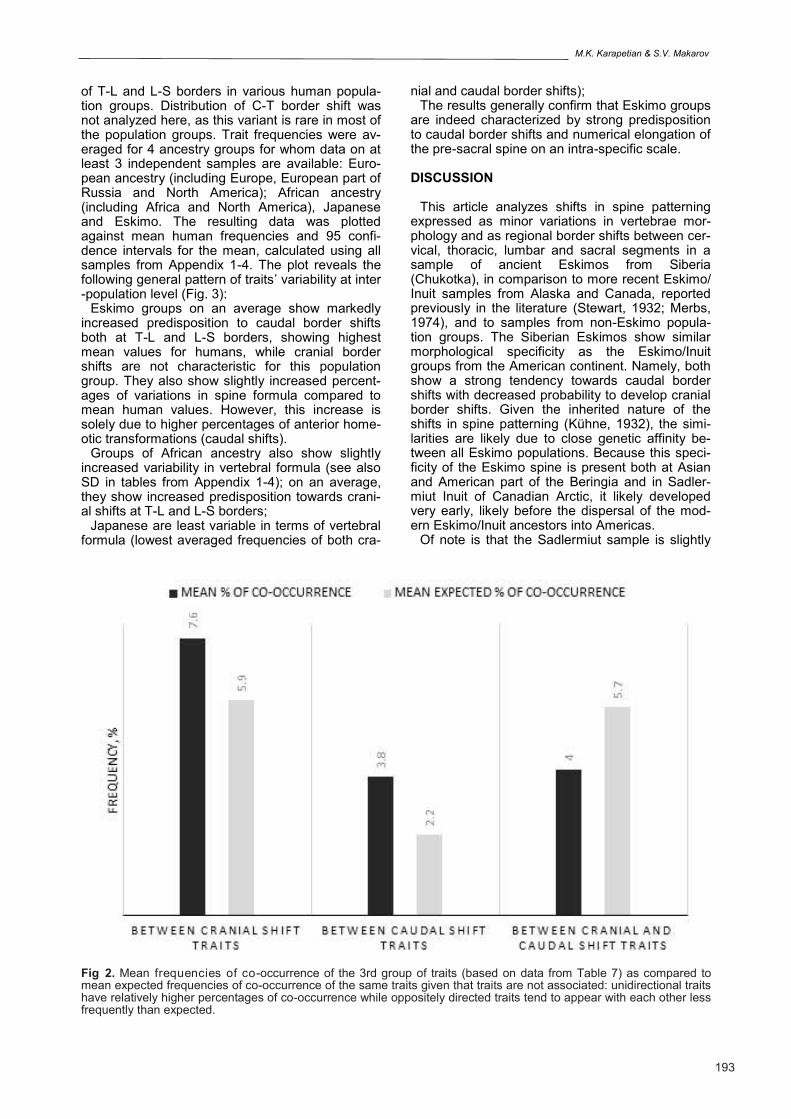
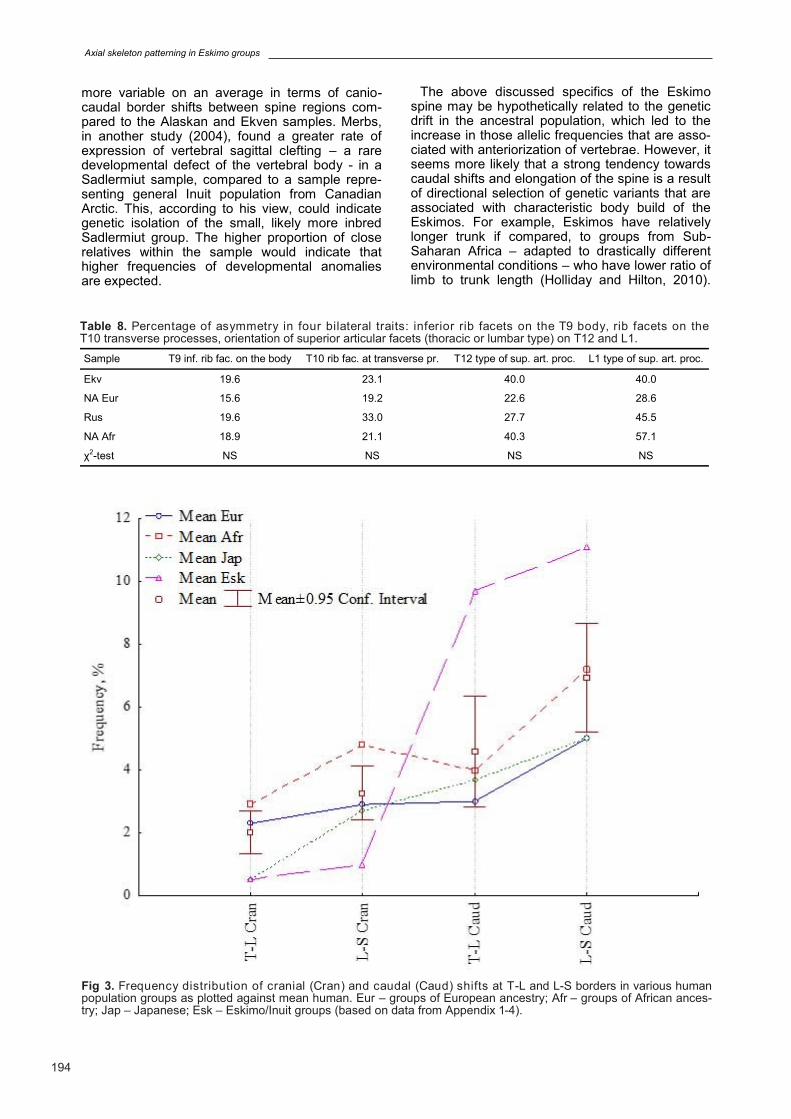
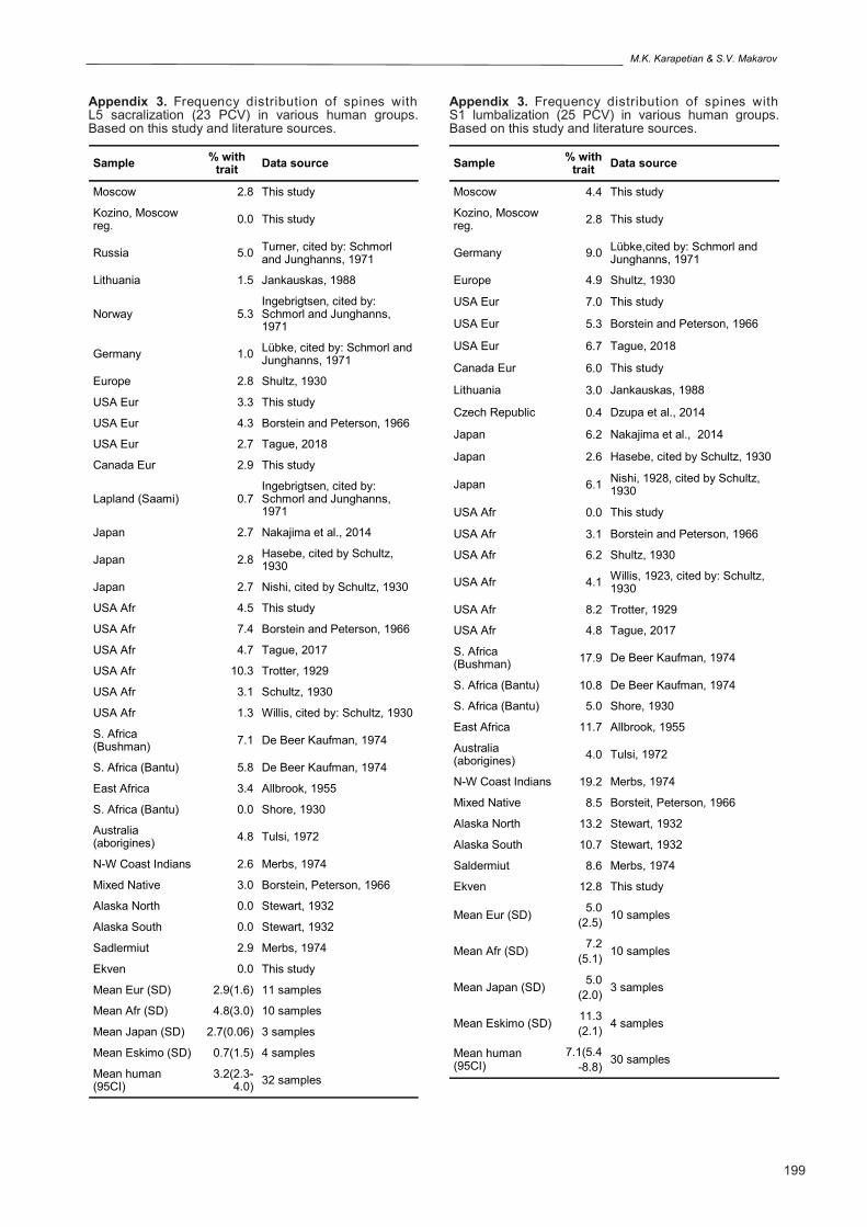
of T-L and L-S borders in various human popula-tion groups. Distribution of C-T border shift was not analyzed here, as this variant is rare in most of the population groups. Trait frequencies were av-eraged for 4 ancestry groups for whom data on at least 3 independent samples are available: Euro-pean ancestry (including Europe, European part of Russia and North America); African ancestry (including Africa and North America), Japanese and Eskimo. The resulting data was plotted against mean human frequencies and 95 confi-dence intervals for the mean, calculated using all samples from Appendix 1-4. The plot reveals the following general pattern of traits’ variability at inter-population level (Fig. 3):
Eskimo groups on an average show markedly increased predisposition to caudal border shifts both at T-L and L-S borders, showing highest mean values for humans, while cranial border shifts are not characteristic for this population group. They also show slightly increased percent-ages of variations in spine formula compared to mean human values. However, this increase is solely due to higher percentages of anterior home-otic transformations (caudal shifts).
Groups of African ancestry also show slightly increased variability in vertebral formula (see also SD in tables from Appendix 1-4); on an average, they show increased predisposition towards crani-al shifts at T-L and L-S borders;
Japanese are least variable in terms of vertebral formula (lowest averaged frequencies of both cra-
nial and caudal border shifts); The results generally confirm that Eskimo groups
are indeed characterized by strong predisposition to caudal border shifts and numerical elongation of the pre-sacral spine on an intra-specific scale.
DISCUSSION
This article analyzes shifts in spine patterning expressed as minor variations in vertebrae mor-phology and as regional border shifts between cer-vical, thoracic, lumbar and sacral segments in a sample of ancient Eskimos from Siberia (Chukotka), in comparison to more recent Eskimo/Inuit samples from Alaska and Canada, reported previously in the literature (Stewart, 1932; Merbs, 1974), and to samples from non-Eskimo popula-tion groups. The Siberian Eskimos show similar morphological specificity as the Eskimo/Inuit groups from the American continent. Namely, both show a strong tendency towards caudal border shifts with decreased probability to develop cranial border shifts. Given the inherited nature of the shifts in spine patterning (Kühne, 1932), the simi-larities are likely due to close genetic affinity be-tween all Eskimo populations. Because this speci-ficity of the Eskimo spine is present both at Asian and American part of the Beringia and in Sadler-miut Inuit of Canadian Arctic, it likely developed very early, likely before the dispersal of the mod-ern Eskimo/Inuit ancestors into Americas.
Of note is that the Sadlermiut sample is slightly
Fig 2. Mean frequencies of co-occurrence of the 3rd group of traits (based on data from Table 7) as compared to mean expected frequencies of co-occurrence of the same traits given that traits are not associated: unidirectional traits have relatively higher percentages of co-occurrence while oppositely directed traits tend to appear with each other less frequently than expected.
Axial skeleton patterning in Eskimo groups
194
more variable on an average in terms of canio-caudal border shifts between spine regions com-pared to the Alaskan and Ekven samples. Merbs, in another study (2004), found a greater rate of expression of vertebral sagittal clefting – a rare developmental defect of the vertebral body - in a Sadlermiut sample, compared to a sample repre-senting general Inuit population from Canadian Arctic. This, according to his view, could indicate genetic isolation of the small, likely more inbred Sadlermiut group. The higher proportion of close relatives within the sample would indicate that higher frequencies of developmental anomalies are expected.
The above discussed specifics of the Eskimo spine may be hypothetically related to the genetic drift in the ancestral population, which led to the increase in those allelic frequencies that are asso-ciated with anteriorization of vertebrae. However, it seems more likely that a strong tendency towards caudal shifts and elongation of the spine is a result of directional selection of genetic variants that are associated with characteristic body build of the Eskimos. For example, Eskimos have relatively longer trunk if compared, to groups from Sub-Saharan Africa – adapted to drastically different environmental conditions – who have lower ratio of limb to trunk length (Holliday and Hilton, 2010).
Sample T9 inf. rib fac. on the body T10 rib fac. at transverse pr. T12 type of sup. art. proc. L1 type of sup. art. proc.
Ekv 19.6 23.1 40.0 40.0
NA Eur 15.6 19.2 22.6 28.6
Rus 19.6 33.0 27.7 45.5
NA Afr 18.9 21.1 40.3 57.1
χ2-test NS NS NS NS
Table 8. Percentage of asymmetry in four bilateral traits: inferior rib facets on the T9 body, rib facets on the T10 transverse processes, orientation of superior articular facets (thoracic or lumbar type) on T12 and L1.
Fig 3. Frequency distribution of cranial (Cran) and caudal (Caud) shifts at T-L and L-S borders in various human population groups as plotted against mean human. Eur – groups of European ancestry; Afr – groups of African ances-try; Jap – Japanese; Esk – Eskimo/Inuit groups (based on data from Appendix 1-4).
M.K. Karapetian & S.V. Makarov
195
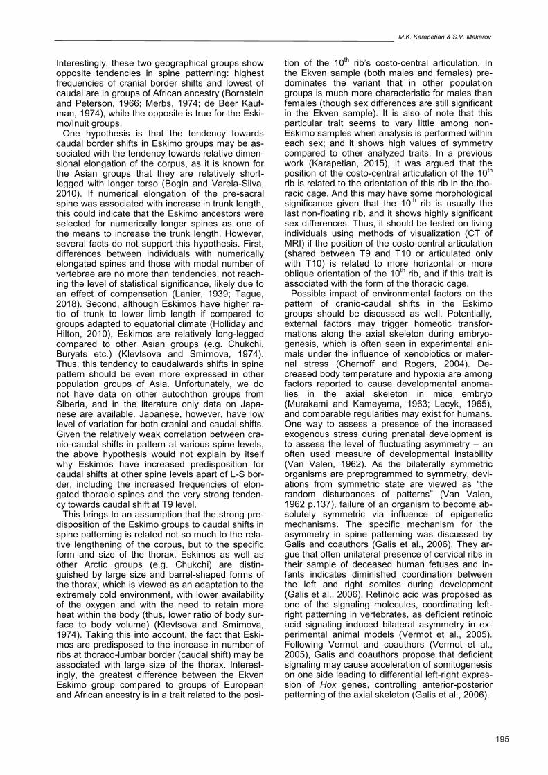
Interestingly, these two geographical groups show opposite tendencies in spine patterning: highest frequencies of cranial border shifts and lowest of caudal are in groups of African ancestry (Bornstein and Peterson, 1966; Merbs, 1974; de Beer Kauf-man, 1974), while the opposite is true for the Eski-mo/Inuit groups.
One hypothesis is that the tendency towards caudal border shifts in Eskimo groups may be as-sociated with the tendency towards relative dimen-sional elongation of the corpus, as it is known for the Asian groups that they are relatively short-legged with longer torso (Bogin and Varela-Silva, 2010). If numerical elongation of the pre-sacral spine was associated with increase in trunk length, this could indicate that the Eskimo ancestors were selected for numerically longer spines as one of the means to increase the trunk length. However, several facts do not support this hypothesis. First, differences between individuals with numerically elongated spines and those with modal number of vertebrae are no more than tendencies, not reach-ing the level of statistical significance, likely due to an effect of compensation (Lanier, 1939; Tague, 2018). Second, although Eskimos have higher ra-tio of trunk to lower limb length if compared to groups adapted to equatorial climate (Holliday and Hilton, 2010), Eskimos are relatively long-legged compared to other Asian groups (e.g. Chukchi, Buryats etc.) (Klevtsova and Smirnova, 1974). Thus, this tendency to caudalwards shifts in spine pattern should be even more expressed in other population groups of Asia. Unfortunately, we do not have data on other autochthon groups from Siberia, and in the literature only data on Japa-nese are available. Japanese, however, have low level of variation for both cranial and caudal shifts. Given the relatively weak correlation between cra-nio-caudal shifts in pattern at various spine levels, the above hypothesis would not explain by itself why Eskimos have increased predisposition for caudal shifts at other spine levels apart of L-S bor-der, including the increased frequencies of elon-gated thoracic spines and the very strong tenden-cy towards caudal shift at T9 level.
This brings to an assumption that the strong pre-disposition of the Eskimo groups to caudal shifts in spine patterning is related not so much to the rela-tive lengthening of the corpus, but to the specific form and size of the thorax. Eskimos as well as other Arctic groups (e.g. Chukchi) are distin-guished by large size and barrel-shaped forms of the thorax, which is viewed as an adaptation to the extremely cold environment, with lower availability of the oxygen and with the need to retain more heat within the body (thus, lower ratio of body sur-face to body volume) (Klevtsova and Smirnova, 1974). Taking this into account, the fact that Eski-mos are predisposed to the increase in number of ribs at thoraco-lumbar border (caudal shift) may be associated with large size of the thorax. Interest-ingly, the greatest difference between the Ekven Eskimo group compared to groups of European and African ancestry is in a trait related to the posi-
tion of the 10th rib’s costo-central articulation. In
the Ekven sample (both males and females) pre-dominates the variant that in other population groups is much more characteristic for males than females (though sex differences are still significant in the Ekven sample). It is also of note that this particular trait seems to vary little among non-Eskimo samples when analysis is performed within each sex; and it shows high values of symmetry compared to other analyzed traits. In a previous work (Karapetian, 2015), it was argued that the position of the costo-central articulation of the 10
th
rib is related to the orientation of this rib in the tho-racic cage. And this may have some morphological significance given that the 10
th rib is usually the
last non-floating rib, and it shows highly significant sex differences. Thus, it should be tested on living individuals using methods of visualization (CT of MRI) if the position of the costo-central articulation (shared between T9 and T10 or articulated only with T10) is related to more horizontal or more oblique orientation of the 10
th rib, and if this trait is
associated with the form of the thoracic cage. Possible impact of environmental factors on the
pattern of cranio-caudal shifts in the Eskimo groups should be discussed as well. Potentially, external factors may trigger homeotic transfor-mations along the axial skeleton during embryo-genesis, which is often seen in experimental ani-mals under the influence of xenobiotics or mater-nal stress (Chernoff and Rogers, 2004). De-creased body temperature and hypoxia are among factors reported to cause developmental anoma-lies in the axial skeleton in mice embryo (Murakami and Kameyama, 1963; Lecyk, 1965), and comparable regularities may exist for humans. One way to assess a presence of the increased exogenous stress during prenatal development is to assess the level of fluctuating asymmetry – an often used measure of developmental instability (Van Valen, 1962). As the bilaterally symmetric organisms are preprogrammed to symmetry, devi-ations from symmetric state are viewed as “the random disturbances of patterns” (Van Valen, 1962 p.137), failure of an organism to become ab-solutely symmetric via influence of epigenetic mechanisms. The specific mechanism for the asymmetry in spine patterning was discussed by Galis and coauthors (Galis et al., 2006). They ar-gue that often unilateral presence of cervical ribs in their sample of deceased human fetuses and in-fants indicates diminished coordination between the left and right somites during development (Galis et al., 2006). Retinoic acid was proposed as one of the signaling molecules, coordinating left-right patterning in vertebrates, as deficient retinoic acid signaling induced bilateral asymmetry in ex-perimental animal models (Vermot et al., 2005). Following Vermot and coauthors (Vermot et al., 2005), Galis and coauthors propose that deficient signaling may cause acceleration of somitogenesis on one side leading to differential left-right expres-sion of Hox genes, controlling anterior-posterior patterning of the axial skeleton (Galis et al., 2006).
Axial skeleton patterning in Eskimo groups
196
One way to assess if the Eskimo groups experi-ence increased level of developmental instability, is to see if these samples show higher levels of homeotic transformations and higher level of asymmetry. The Eskimo samples, especially the Sadlermiut Inuit sample, indeed show on an aver-age slightly higher frequencies of variations com-pared to the mean human values. However, higher frequencies are solely due to higher frequencies of the caudal border shifts, which may be an indicator of selective pressure favoring these variants, ra-ther than increased level of developmental instabil-ity. Another argument would be that there are mini-mal cases of cervical ribs in the Eskimo/Inuit sam-ples. While C-T border forms earlier in embryogen-esis it is theoretically more vulnerable to the influ-ence of factors, altering the development of the fetus, compared to T-L or L-S borders that form later in embryogenesis (see discussion in Galis et al., 2006). Thus if external factors are responsible for the pattern of variations in the vertebral col-umns of the Eskimo groups, C-T border should be more prone to variations compared to groups living in less stressful environmental conditions. Obvi-ously, in the Eskimo samples there might be stronger prenatal/early postnatal selection against individuals with developmental abnormalities, as-sociated with cervical ribs, due to greater con-strains on the organism. When the Ekven sample was tested for differences in the levels of asym-metry in spine traits compared to studied samples of European and African ancestry, all samples showed approximately the same level of asym-metry without any statistically significant devia-tions.
Overall, the hypothesis that the Eskimo groups experienced higher level of instability in the axial skeleton development due to the influence of envi-ronmental factors is possible. However, the pre-sent data are more in agreement with inherent characteristics and existence of selective pressure favoring certain morphological variants. This should be tested by studying other groups adapted to living in the Arctic (primarily Chukchi) and com-pared to other Asian groups living in more south-ern areas.
CONCLUSION
Our study is consistent with increased predispo-sition of the temporally and geographically variable Eskimo/Inuit groups towards caudal shifts in spine patterning. This points to conservation of possibly adaptive traits that formed early in the history of this population.
ACKNOWLEDGMENTS
The research was financially supported by the Russian Science Foundation (grant number 14-50-00029) and the Russian Foundation for Basic Re-search (grant number 17-29-04125). The study of the Terry collection was done at the finantial sup-port of the Smithsonial Institution Fellowship. Au-
thors declare no conflict of interests.
REFERENCES ALEXEEVA TI, BUZHILOVA AP, MEDNIKOVA MB,
DOBROVOLSKAYA MV, editors (2008) Antro-poekologiya severo-vostochnoy Azii: Chukota, Kam-chatka, Komandorskiye ostrova. Taus, Moscow.
ALLBROOK DB (1955) The East African vertebral col-umn: a study in racial variability. Am J Phys Anthropol, 13: 489-513.
ARUTYUNOV SA, SERGEEV DA (1975) Problemy et-nicheskoy istorii Beringomorya: Ekvenskiy mogilnik. Direkt-Media, Moscow.
BOGIN B, VARELA-SILVA MI (2010) Leg length, body proportion, and health: a review with a note on beauty. Int J Environ Res Public Health, 7(3): 1047-1075.
BORNSTEIN PE, PETERSON RR (1966) Numerical variation of the presacral vertebral column in three population groups in North America. Am J Phys An-thropol, 25(2): 139-146.
BUIKSTRA JE, UBELAKER DH (1994) Standards for Data Collecting from Human Skeletal Remains. Arkan-sas Archaeological Survey, Fayetteville.
CHERNOFF N, ROGERS JM (2004) Supernumerary ribs in developmental toxicity bioassays and in human populations: incidence and biological significance. J Toxicol Environ Health, Part B, 7: 437-449.
DE BEER KAUFMAN P (1974) Variation in the number of presacral vertebrae in Bantu-speaking South African Negroes. Am J Phys Anthropol, 40: 369-374.
DZUPA V, SLEPANEK M, STRIZ M, KRBEC M, CHME-LOVA J, KACHLIK D, BACA V (2014) Developmental malformations in the area of the lumbosacral transi-tional vertebrae and sacrum: differences in gender and left/right distribution. Surg Radiol Anat, 36(7): 689-693.
FRIESEN TM, MASON OK, editors (2016) The Oxford handbook of the Prehistoric Arctic. Oxford University Press, New York.
GALIS F, VAN DOOREN TJM, FEUTH JD, METZ JA, WITKAM A, RUINARD S, STEIGENGA MJ, WIJNAENDTS LC (2006) Extreme selection in hu-mans against homeotic transformations of cervical vertebrae. Evolution, 60: 2643-2654.
GRAY H (1858) Anatomy descriptive and surgical. John W. Parker and Son, West strand, London.
HOLLIDAY TW, HILTON CE (2010) Body proportions of circumpolar peoples as evidenced from skeletal data: Ipiutak and Tigara (Point Hope) versus Kodiak Island Inuit. Am J Phys Anthropol, 142: 287-302.
JANKAUSKAS R (1988) Morphologicheskiye osoben-nosti pozvonochnogo stolba i factori ego izmenchivosti [PhD thesis]. Vilnius, Vilnius University.
KARAPETIAN MK (2015) Costal facet variations on the eighth, ninth and tenth thoracic vertebrae: association with sex and shifts in the cranio-caudal pattern of the human axial skeleton. Eur J Anat, 19(2): 179-188.
KLEVTSOVA NI, SMIRNOVA NS (1974) Morphologicheskiye osobennosti tela chukchey i eskimosov. Voprosi antropologii, 48: 18-33.
KÜHNE K (1932) Die Vererbung der variationen der menschlichen Wirbelsäule. Z Morphol Anthropol, Bd. 30(1/2): 1-221.
M.K. Karapetian & S.V. Makarov
197
LANIER RR (1939) The presacral vertebrae of American White and Negro males. Am J Phys Anthropol, 25: 341-420.
LECYK M (1965) The effect of hypothermia applied in the given stages of pregnancy on the number and form of vertebrae in the offspring of white mice. Expe-rientia, 21(8): 452-453.
MALLO M, WELLIK DM, DESCHAMPS J (2010) Hox genes and regional patterning of the vertebrate body plan. Dev Biol, 344: 7-15.
MERBS CF (1974) The effects of cranial and caudal shift in the vertebral columns of northern populations. Arctic Anthropol, 11: 12-19.
MERBS CF (2004) Sagittal clefting of the body and oth-er vertebral developmental errors in Canadian Inuit skeletons. Am J Phys Anthropol, 123(3): 236-249.
MURAKAMI U, KAMEYAMA Y (1963) Vertebral malfor-mations in the mouse foetus caused by maternal hy-poxia during early stages of pregnancy. J Embryol Exp Morph, 11: 107-118.
NAKAJIMA A, USUI A, HOSOKAI Y, KAWASUMI Y, ABIKO K, FUNAYAMA M, SAITO H (2014) The preva-lence of morphological changes in the thoracolumbar spine on whole-spine computed tomographic images. Insights Imaging, 5: 77-83.
RAGHAVAN M, DEGIORGIO M, ALBRECHTSEN A, MOLTKE I, SKOGLUND P, KORNELIUSSEN TS, GRØNNOW B, APPELT M, GULLØV HC, FRIESEN TM, FITZHUGH W, MALMSTRÖM H, RASMUSSEN S, OLSEN J, MELCHIOR L, FULLER BT, FAHRNI SF, STAFORD T, GRIMES V, RENOUF MAP, CYBULSKI J, LYNNERUP N, LAHR MM, BRITTON K, KNECHT R, ARNEBORG J, METSPALU M, CORNEJO OE, MALASPINAS AS, WANG Y, RASMUSSEN M, RAGHAVAN V, HANSEN TVO, KHUSNUTDINOVA E, PIERRE T, DNEPROVSKY K, ANDREASEN C, LANGE H, HAYES MG, COLTRAIN J, SPITSYN VA, GÖTHERSTRÖM A, ORLANDO L, KIVISILD T, VIL-LEMS R, CRAWFORD MH, NIELSEN FC, DISSING J, HEINEMEIER J, MELDGAARD M, BUSTAMANTE C, O’ROURKE DH, JAKOBSSON M, GILBERT MTP, NIELSEN R, WILLERSLEV E (2014) The genetic pre-history of the New World Arctic. Science, 345(6200): 1255832-1-9. doi: 10.1126/science.1255832
SCHAEFER M, BLACK S, SCHEUER L (2009) Juvenile Osteology: a laboratory and field manual. Academic Press, Burlington.
SCHAPERA J (1987) Autosomal dominant inheritance of cervical ribs. Clin Genet, 31: 386-388.
SCHULTZ AH (1930.) The skeleton of the trunk and limbs of higher primates. Hum Biol, 2: 303-438.
SCHUMACHER R, MAI A, GUTJAHR P (1992) Associa-tion of rib anomalies and malignancy in childhood. Eur J Pediatr, 151: 432-434.
SCHMORL G, JUNGHANNS H (1971) The human spine in health and disease. Grune & Stratton, New York.
SHORE LR (1930) Abnormalities of the vertebral column in a series of skeletons of Bantu natives of South Afri-ca. J Anat, 64: 206-238.
SINGER KP, BREIDAHL PD (1990) Accessory ossifica-tion centres at thethoracolumbar junction. Surg Radiol Anat, 12: 53-58.
STATISTICA (data analysis software system) [Computer
program]. Version 8.0. Tulsa (OK): StatSoft Inc., 2007.
STEWART TD (1932) The vertebral column of the Eski-mo. Am J Phys Anthropol, 17: 123-136.
TAGUE RG (2018) Proximate cause, anatomical corre-lates, and obstetrical implication of a supernumerary lumbar vertebra in humans. Am J Phys Anthropol, 165(3): 444-456.
TEN BROEK CMA, BAKKER AJ, VARELA-LASHERAS I, BUGIANI M, VAN DONGEN S, GALIS F (2012) Evo-devo of human vertebral column: on homeotic transfor-mations, pathologies and prenatal selection. Evol Biol, 39: 456-471.
THOMPSON NE, ALMÉCIJA S (2017) The evolution of vertebral formulae in Hominoidea. J Hum Evol, 110: 18-36.
TODD TW (1922) Numerical significance in the thoracol-umbar vertebrae of the mammalian. Anat Rec, 24: 261-286.
TROTTER M (1929) The vertebral column in Whites and in American Negroes. Am J Phys Anthropol, 13: 95-107.
TULSI RS (1972) Vertebral column of the Australian aborigine: selected morphological and metrical fea-tures. Z Morphol Anthropol, 64: 117-144.
VAN VALEN L (1962) A study of fluctuating asymmetry. Evolution, 16(2): 125-142.
VERMOT J, GALLEGO LLAMAS J, FRAULOB V, NIE-DERREITHER K, CHAMBON P, DOLLÉ P (2005) Ret-inoic acid controls the bilateral symmetry of somite formation in the mouse embryo. Science, 308: 563-566.
WILLIAMS SA, MIDDLETON ER, VILLAMIL CI, SHAT-TUCK MR (2016) Vertebral numbers and human evo-lution. Am J Phys Anthropol, 159(Suppl 61): S19-36.
Axial skeleton patterning in Eskimo groups
198
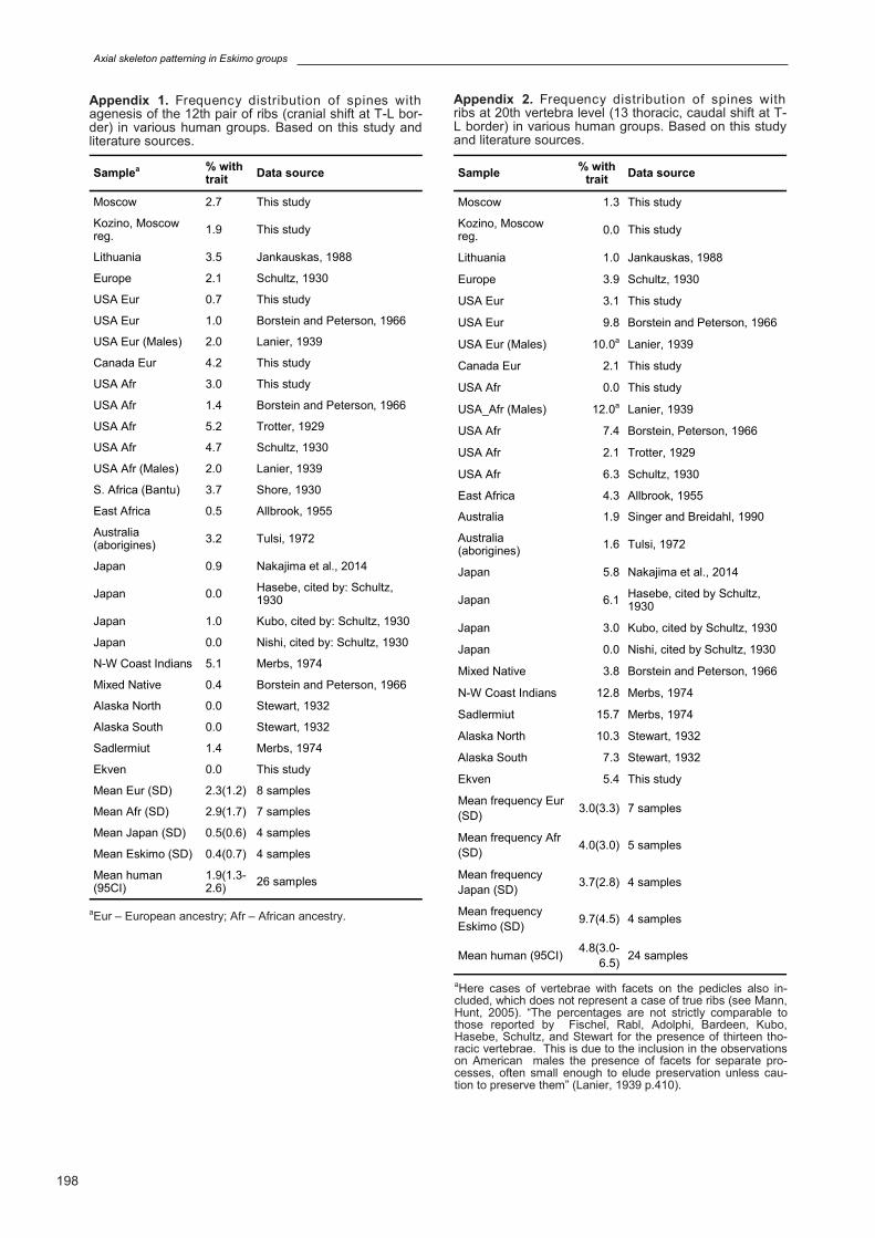
Appendix 1. Frequency distribution of spines with agenesis of the 12th pair of ribs (cranial shift at T-L bor-der) in various human groups. Based on this study and literature sources.
Samplea % with trait
Data source
Moscow 2.7 This study
Kozino, Moscow reg.
1.9 This study
Lithuania 3.5 Jankauskas, 1988
Europe 2.1 Schultz, 1930
USA Eur 0.7 This study
USA Eur 1.0 Borstein and Peterson, 1966
USA Eur (Males) 2.0 Lanier, 1939
Canada Eur 4.2 This study
USA Afr 3.0 This study
USA Afr 1.4 Borstein and Peterson, 1966
USA Afr 5.2 Trotter, 1929
USA Afr 4.7 Schultz, 1930
USA Afr (Males) 2.0 Lanier, 1939
S. Africa (Bantu) 3.7 Shore, 1930
East Africa 0.5 Allbrook, 1955
Australia (aborigines)
3.2 Tulsi, 1972
Japan 0.9 Nakajima et al., 2014
Japan 0.0 Hasebe, cited by: Schultz, 1930
Japan 1.0 Kubo, cited by: Schultz, 1930
Japan 0.0 Nishi, cited by: Schultz, 1930
N-W Coast Indians 5.1 Merbs, 1974
Mixed Native 0.4 Borstein and Peterson, 1966
Alaska North 0.0 Stewart, 1932
Alaska South 0.0 Stewart, 1932
Sadlermiut 1.4 Merbs, 1974
Ekven 0.0 This study
Mean Eur (SD) 2.3(1.2) 8 samples
Mean Afr (SD) 2.9(1.7) 7 samples
Mean Japan (SD) 0.5(0.6) 4 samples
Mean Eskimo (SD) 0.4(0.7) 4 samples
Mean human (95CI)
1.9(1.3-2.6)
26 samples
aEur – European ancestry; Afr – African ancestry.
Sample % with
trait Data source
Moscow 1.3 This study
Kozino, Moscow reg.
0.0 This study
Lithuania 1.0 Jankauskas, 1988
Europe 3.9 Schultz, 1930
USA Eur 3.1 This study
USA Eur 9.8 Borstein and Peterson, 1966
USA Eur (Males) 10.0a Lanier, 1939
Canada Eur 2.1 This study
USA Afr 0.0 This study
USA_Afr (Males) 12.0a Lanier, 1939
USA Afr 7.4 Borstein, Peterson, 1966
USA Afr 2.1 Trotter, 1929
USA Afr 6.3 Schultz, 1930
East Africa 4.3 Allbrook, 1955
Australia 1.9 Singer and Breidahl, 1990
Australia (aborigines)
1.6 Tulsi, 1972
Japan 5.8 Nakajima et al., 2014
Japan 6.1 Hasebe, cited by Schultz, 1930
Japan 3.0 Kubo, cited by Schultz, 1930
Japan 0.0 Nishi, cited by Schultz, 1930
Mixed Native 3.8 Borstein and Peterson, 1966
N-W Coast Indians 12.8 Merbs, 1974
Sadlermiut 15.7 Merbs, 1974
Alaska North 10.3 Stewart, 1932
Alaska South 7.3 Stewart, 1932
Ekven 5.4 This study
Mean frequency Eur
(SD) 3.0(3.3) 7 samples
Mean frequency Afr
(SD) 4.0(3.0) 5 samples
Mean frequency
Japan (SD) 3.7(2.8) 4 samples
Mean frequency
Eskimo (SD) 9.7(4.5) 4 samples
Mean human (95CI) 4.8(3.0-
6.5) 24 samples
Appendix 2. Frequency distribution of spines with ribs at 20th vertebra level (13 thoracic, caudal shift at T-L border) in various human groups. Based on this study and literature sources.
aHere cases of vertebrae with facets on the pedicles also in-cluded, which does not represent a case of true ribs (see Mann, Hunt, 2005). “The percentages are not strictly comparable to those reported by Fischel, Rabl, Adolphi, Bardeen, Kubo, Hasebe, Schultz, and Stewart for the presence of thirteen tho-racic vertebrae. This is due to the inclusion in the observations on American males the presence of facets for separate pro-cesses, often small enough to elude preservation unless cau-tion to preserve them” (Lanier, 1939 p.410).
M.K. Karapetian & S.V. Makarov
199
Sample % with
trait Data source
Moscow 2.8 This study
Kozino, Moscow reg.
0.0 This study
Russia 5.0 Turner, cited by: Schmorl and Junghanns, 1971
Lithuania 1.5 Jankauskas, 1988
Norway 5.3 Ingebrigtsen, cited by: Schmorl and Junghanns, 1971
Germany 1.0 Lübke, cited by: Schmorl and Junghanns, 1971
Europe 2.8 Shultz, 1930
USA Eur 3.3 This study
USA Eur 4.3 Borstein and Peterson, 1966
USA Eur 2.7 Tague, 2018
Canada Eur 2.9 This study
Lapland (Saami) 0.7 Ingebrigtsen, cited by: Schmorl and Junghanns, 1971
Japan 2.7 Nakajima et al., 2014
Japan 2.8 Hasebe, cited by Schultz, 1930
Japan 2.7 Nishi, cited by Schultz, 1930
USA Afr 4.5 This study
USA Afr 7.4 Borstein and Peterson, 1966
USA Afr 4.7 Tague, 2017
USA Afr 10.3 Trotter, 1929
USA Afr 3.1 Schultz, 1930
USA Afr 1.3 Willis, cited by: Schultz, 1930
S. Africa (Bushman)
7.1 De Beer Kaufman, 1974
S. Africa (Bantu) 5.8 De Beer Kaufman, 1974
East Africa 3.4 Allbrook, 1955
S. Africa (Bantu) 0.0 Shore, 1930
Australia (aborigines)
4.8 Tulsi, 1972
N-W Coast Indians 2.6 Merbs, 1974
Mixed Native 3.0 Borstein, Peterson, 1966
Alaska North 0.0 Stewart, 1932
Alaska South 0.0 Stewart, 1932
Sadlermiut 2.9 Merbs, 1974
Ekven 0.0 This study
Mean Eur (SD) 2.9(1.6) 11 samples
Mean Afr (SD) 4.8(3.0) 10 samples
Mean Japan (SD) 2.7(0.06) 3 samples
Mean Eskimo (SD) 0.7(1.5) 4 samples
Mean human (95CI)
3.2(2.3-4.0)
32 samples
Appendix 3. Frequency distribution of spines with L5 sacralization (23 PCV) in various human groups. Based on this study and literature sources.
Sample % with
trait Data source
Moscow 4.4 This study
Kozino, Moscow reg.
2.8 This study
Germany 9.0 Lübke,cited by: Schmorl and Junghanns, 1971
Europe 4.9 Shultz, 1930
USA Eur 7.0 This study
USA Eur 5.3 Borstein and Peterson, 1966
USA Eur 6.7 Tague, 2018
Canada Eur 6.0 This study
Lithuania 3.0 Jankauskas, 1988
Czech Republic 0.4 Dzupa et al., 2014
Japan 6.2 Nakajima et al., 2014
Japan 2.6 Hasebe, cited by Schultz, 1930
Japan 6.1 Nishi, 1928, cited by Schultz, 1930
USA Afr 0.0 This study
USA Afr 3.1 Borstein and Peterson, 1966
USA Afr 6.2 Shultz, 1930
USA Afr 4.1 Willis, 1923, cited by: Schultz, 1930
USA Afr 8.2 Trotter, 1929
USA Afr 4.8 Tague, 2017
S. Africa (Bushman)
17.9 De Beer Kaufman, 1974
S. Africa (Bantu) 10.8 De Beer Kaufman, 1974
S. Africa (Bantu) 5.0 Shore, 1930
East Africa 11.7 Allbrook, 1955
Australia (aborigines)
4.0 Tulsi, 1972
N-W Coast Indians 19.2 Merbs, 1974
Mixed Native 8.5 Borsteit, Peterson, 1966
Alaska North 13.2 Stewart, 1932
Alaska South 10.7 Stewart, 1932
Saldermiut 8.6 Merbs, 1974
Ekven 12.8 This study
Mean Eur (SD) 5.0
(2.5) 10 samples
Mean Afr (SD) 7.2
(5.1) 10 samples
Mean Japan (SD) 5.0
(2.0) 3 samples
Mean Eskimo (SD) 11.3
(2.1) 4 samples
Mean human (95CI)
7.1(5.4
-8.8) 30 samples
Appendix 3. Frequency distribution of spines with S1 lumbalization (25 PCV) in various human groups. Based on this study and literature sources.