Restorative dentistry I., II., III. 3RD Year
134
Transcript of Restorative dentistry I., II., III. 3RD Year

Dental caries
• Etiology and pathogenesis (dental biofilm, remineralization, importance of saliva)

5
tooth
microbs
sugar
time
Dental caries
caries
Factors that are necessary for
origin of dental caries

Dental Caries
Infectious microbiological disease of the teeth
that results in localized dissolution and
destruction of the calcified dental tissues.

Biofilm – Dental Plaque
Complex community
Microbs live in symbiosis
Biofilm is permeable
Microbs have good conditions to survive and are much less sensitive to antimicrobial agents in comparison to planctonic form

Dental Biofilm – Dental Plaque
A gelatinous mass of bacteria adhering to the
tooth surface.

Sugars
Fermentable (mono-, di- tri- sacharides)
Sucrose, glucose, lactose Acids
Demineralization

Dem
inera
lizati
on
Time
Cavitated lesion
Non cavitatated lesion

Importance of saliva
• Plaque formation
• Microbial source
• Mineral source
• Microbial clearence (removes microbs from oral cavity)
• Buffer capacity

Caries danger areas (Habitually unclean places)
• Pits and fissures
• Proximal surfaces
• Cervical area
No self cleaning

Predictable (habitually) clean areas
• Cusps
• Proximal ridge, oblique, transverse ridge
• Incisal edge
• Buccal or oral surface upon the maximal convexity
• Proximal surface upon the contact point
Self cleaning

Caries - depth
• Surface caries (caries superficialis)
• Middle caries (caries media)
• Caries close to pulp (caries pulpae proxima)
• Caries penetrating into the pulp (caries ad pulpam penetrans)
Deep caries

Caries - Topography
• Coronal caries
• Root surface caries
• Enamel caries
• Dentin caries
• Cementum caries

Investigation
• Mirror
• Sharp Probe
• Illimunation
• Magnification
• X- ray, other methods i.e. transillumination, infrared laser fluorescency (Diagnodent, Diagnocam)
Dark spot, white spot, hole, defect

Dental Caries - Treatment
• Non cavitated lesion:
On molecular basis
- Dental hygiene
- Fluorides, Calcium, Phosphates
- Diet
- Antimicrobial agents (ozone, chlorhexidine)

Dental Caries - Treatment
• Cavitated lesion:
Preparation
Filling Drill anf fill

Preparation
Instrumental treatment
Remove caries
Leave the rest of the dental tissues
- to be restored
- to be resistent against the bite forces
- to be prevented against the recurrent caries
(Black 1914)

Class I.
Caries in fissures and pits – occlusal
surfaces of premolars and molars

All pit and fissure restorations.
They are assigned in to three groups. R. on occlusal surface of premolars and molars
R. in foramina coeca – usually on occlusal two thirds
of the facial and lingual surfaces of molars.
R.on lingual surface of maxillary incisors.

Class III.
Proximal surfaces of incisors and canines
without loss of the incisal edge

Class IV
Proximal surfaces of incisors and canines
with the loss of incisal edge

Classification of dental caries Mount and Hume
• Location 1.Occlusal 2. Proximal 3.Cevical • Size 1.Small 2. Medium 3. Big 3.Large

Classification of dental caries Mount and Hume
Examples:
1,2 – caries in fissures or a pit, medium size
3,4 – caries in cervical area, large size

Indication od filling materials Material of the first choice
Material of the second choice
Material of the third choice
Materiális possible to use with
limitations
Material is not indicated

Consideration
Caries
- Size
- Location
Choice of material
Regional circumstances
Intermaxillary relations
Bite forces
Patient
- General health
- Cooperation
LR

Indications of filling materials Class I.
Material Mount
and
Hume
11
12
13
14
Amalgam
Composite
Glassionomer
Indirect
restoration aesth.
Inlay metal

Indications of filling materials class II.
Material 21 22 23 24
Amalgam
Composite
Glassionomer
Indirect
restoration aesth.
Inlay metal

Indications of filling materials class III.
Material 21 22 23 24
Amalgam
Composite
Glassionomer
Indirect
restoration aesth.
Inlay metal
LR

Indications of filling materials class IV.
Material 21 22 23 24
Amalgam
Composite
Glassionomer
Indirect
restoration aesth.
Inlay metal
LR

Indications of filling materials class V. anterior teeth
Material 21 22 23 24
Amalgam
Composite
Glassionomer
Indirect
restoration aesth.
Inlay metal
LR

Indications of filling materials class V. posterior teeth
Material 21 22 23 24
Amalgam
Composite
Glassionomer
Indirect
restoration aesth.
Inlay metal

Indications of filling materials class V. acc. to cavosurface margin
Material Enamel Enamel
cementum
Cement
um
Amalgam
Composite
Glassionomer
Indirect restoration
aesth.
Inlay metal

Amalgam Indication
Moderate to large cavities (heavy occlusal stress, difficut isolation of operating field, subgingival cavities, cavities reaching the root).
13 a 24 p Mounta and Hume
Big reconstruction (core)
Temporary fillings
(intermittent excavation).
Sturdevandt´s Art of Science of Operative Dentistry
45

Amalgam
Highest abrasion resistance
Isolation of operating field is not a critical factor
Preparation must be exact
46
Sturdevandt´s Art of Science…

47
Sturdevandt´s Art of Science…

48

lenka.roubalikovatiscali.cz 49
Sedelmayer J. Amalgám – zapomenuté řemeslo.

The most common mistakes
Preparation - Sharp edges
- Bad configuration of the gingival wall
- Rough margins
- Weakening opf the proximal ridge
Manipulaion
- Trituration – rpm, time.
52

Contemporary trends in treatment of dental caries
• Miniinvasion
• Adhesive techniques
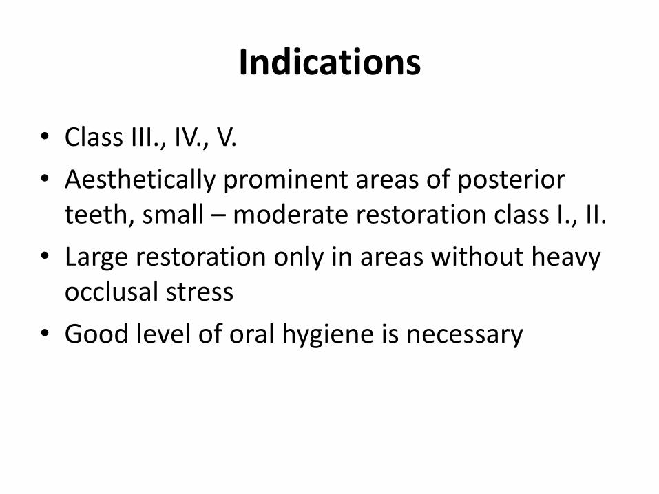
Indications
• Class III., IV., V.
• Aesthetically prominent areas of posterior teeth, small – moderate restoration class I., II.
• Large restoration only in areas without heavy occlusal stress
• Good level of oral hygiene is necessary

Contraindications
• Moderate to large restorations esp. Areas with heavy occlusal stress
• Restorations that are not in highly aesthetics areas
• Restorations that have heavy occlusal contacts
• Restorations that cannot be well isolated
• Restorations that extend onto the root surface
• Abutment teeth for removable partioal dentures
• Temporary or caries control restorations.

Glassionomers - advantages
• Chemical binding to hard dental tissues
• Thermal expansion similar to dentin
• Release fluoride ions (caries control restoration)
• Not sensitive to moisture

Glassionomers-disadvantages
• Long time for setting – sensitive to moisture
• Difficult sculpting - impossible
• Not high aesthetics
• Lower mechanical resistance (wear resistance, flexural strength, hardness)

Glassionomers - indications
• Class V., III. – cavities out of enamel or/and patients with lower level of oral hygiene.
• Class I., II. – caries control filling (inner remoneralization), composite material on the top is strongly recommended (weeks – months later). Tunnel fillings.

Glassionomers contraindications
• Class V., III. – cavities in enamel in patienst with good oral hygiene
• Class IV.
• Class I., II. – permenent filling (esp. larg – moderate restorations)

Composites in posterior teeth

Indications
• Aesthetically prominent areas of posterior teeth
• Small - moderate classes I. that can be well isolated, large cavities only without heavy occlusal stress
• Good level of oral hygiene is necessary

Contraindications
• Moderate to large restorations
• Restorations that are not in highly aesthetics areas
• Restorations that have heavy occlusal contacts
• Restorations that cannot be well isolated
• Restorations that extend onto the root surface
• Abutment teeth for removable partioal dentures
• Temporary or caries control restorations.

Clinical technique
• From the occlusal surface using the diamond burs (roundedn cylinder or ball)

Cavosurface margin
• Outline includes the caries lesion only
• Fissures going into the ceries lesion can be open and sealed (resommended).

Retention principles
• Prepare the box or deep dish – the bottom is in dentin
• Do not prepare any undercuts!
• Do not bevel enamel, finish the border with diamond bur only.

Removal of carious, infected,
dentin and remaining defective enamel.
• Spoon excavator or a slowly revolving ,
round carbid bur of appropriate size.
• Sharp hand instrument

Polymerization shrinkage and polymerization stress

Polymerization shrinkage

Polymerization shrinkage

Surface of adhesion/free surface of the
filling
1/1 and less is optimal
C – factor (Configuration factor)

5
2
1

Forces of polymerization shrinkage depend on
- Composite material (content of filler)
- Geometry of the cavity (C-factor)
- Placement of the composite
- Mode of polymerization

Forces of polymerization shrinkage depend on
- Composite material (content of filler)
Higher content of filler - lower shrinkage, higher polymerization stress.

Forces of polymerization shrinkage depend on
Geometry of the cavity (C-factor)
Higher C-factor – higher stress

Forces of polymerization shrinkage depend on
- Placement of the composite:
- Create the first layer thin, flowable can be used
(Flowables – lower content of filler, higher shrinkage, lower polymerization stress)
- Place the material in increments with respect of the C-factor of each layer (each layer with large free surface). Maximum 1,5 mm

Forces of polymerization shrinkage depend on
- Mode of polymerization
Phases
- Pre-gel (in this phase the material is still soft)
- G-point (material become hard)
- Post –gel (end of shrinkage –postgel shrinkage)

Monomer
Light
Polymerization
Polymer

Pre –gel phase should be prolongated – soft start polymerization
Gel
Post –gel
Now soft start seems not to be so important !!!
Pre gel phase should be long – soft start !!!!

Marginal adaptation depends on
• Placement of composite material
• Dry operating field
• Adhesive systems

Adhesives
• Acid etching technique
• Selfetching adhesive systems

Adhesives
• Acid etching technique
Etching
Washing
Priming Bonding

Adhesives
• Selfetching adhesive systems
Priming
Bonding
Less bonding strength in comparison to acid
teching technique

Adhesives
• Active and passive bonding
Active – rubbing with microbrush (selfetching)
Passive – without any rubbing (acid etching)


WRONG – Higher c- factor


Adhesive preparation in a fissure

Adhesive preparation

Preparation of enamel borders
Preparation 45°

Next to cusp 50-60°, Never cover the cusp
Preparation of enamel borders

Incremental technique
Flowable Building cusp by cusp

Miniinvasive treatment – small cavity, Opening of fissures, preservation of intact areas - ridges

Composite filling – class II.
• Critical factors
- contact area (contact point)
- dry operating field (marginal adaptaion)

Preparation
• Occlusal cavity – class I.
• Proximal cavity

Preparace 30 - 40°
?
Cervical margin
In enamel

No bevel
Cervical margin
Out of enamel

Preparation technique


Interproximal vertical margins
Yes
No

Proximal Preparation with oscillating instrument

Matrices Bands (metal,transparent) Retainers Hawe Neos (0,03 mm) Optra (thin matrices – 10 micrometers)


Sectional matrices With separator and wedge


Optra contact – special instrument OR Contouring of the matrix band using a ball condensor


Miniinvasive techniques
• Adhesive slot
• Tunnel

Sedelmayer
Adhesive slot preparation

Horizontal slot

Horizontal slot – oscillating instrument

Tunnel preparation


1. Low caries risk
2. Good cooperation of the npatient
3. Marginal ridge without any infraction
1. Loupes or microscope
2. Miniinstruments
3. Capsulated GIC or composite
5. BW post op
Success of the tunnel

ART

ART


Bulk fill composites
Placement and curing in one layer 4 mm
1. Flowables – SDR Flow (Dentsply), Venus Bulk Fill (Hereaus Kulzer), X-tra fill (VOCO), Filtek Bulk Fill (3M ESPE).
2. Bulk high density composites (Tetric EvoCeram Bulk Fill (Ivoclar –Vivadent) a QuiXfill (Dentsply).
3. Sonic Fill (KaVo)

Sonic Fill
1 bulk ( 5 mm)
Sonic activation – decreasing of viscosity
Inner scattering of light – good aesthetics
Long term experience necessary


Bulk Fill composites
• Flowables
131 SDR Flow (Dentsply), Venus Bulk Fill (Heraeus Kulzer), X-tra fil (VOCO) nebo Filtek Bulk Fill (3M ESPE).


Bulk Fill composites
• High viscosity
Tetric EvoCeram Bulk Fill (Ivoclar Vivadent) a QuiXfil (Dentsply)

Sonic Fill

Sonic Fill
Možnost plnění kavity v jednom bloku
(do 5 mm)
Sonická „aktivace“ – změna viskozity
Vnitřní rozptyl světla – dobrá estetika
Chybí dlouhodobé zkušenosti
Srovnatelné s jinými materiály

136

Comprehension
• Bulk Fill is a new approach to posterior composite restorations
• The handling must follow instructions
• Maximum layer is 4 mm
• Aesthetics is acceptable bur not so high as composite fillings made by incremental technique

Postoperative sensitivity
marginal
discoloration
gap
cracks in enamel
Versluis 2000
Problems – can we solve them?
Marginal ditching
Recurrent caries Bending of the cusps

![[1][m] minimally invasive restorative dentistry](https://static.fdocuments.us/doc/165x107/587254011a28ab852f8b7e5b/1m-minimally-invasive-restorative-dentistry.jpg)