Resting-StateGlutamateandGABAConcentrationsPredict Task ...
8
Behavioral/Cognitive Resting-State Glutamate and GABA Concentrations Predict Task-Induced Deactivation in the Default Mode Network Yuzheng Hu,* Xi Chen,* Hong Gu,* and Yihong Yang Neuroimaging Research Branch, National Institute on Drug Abuse, National Institutes of Health, Baltimore, Maryland 21224 Deactivation of the human brain’s default mode network (DMN) is regarded as suppression of endogenous activity to support exogenous task-related processes. This phenomenon has important functional relevance and insufficient DMN deactivation has been implicated in several neuropsychiatric disorders. However, the neurochemical mechanism of the DMNs deactivation remains largely unknown. In the present study, we test the hypothesis that the major excitatory and inhibitory neurotransmitters, glutamate and GABA, respectively, are associated with DMN deactivation. We used magnetic resonance spectroscopy to measure neurotransmitter concentrations in the pos- terior cingulate cortex/precuneus (PCC/PCu), a key component of the DMN, and functional magnetic resonance imaging to evaluate DMN deactivation induced by an n-back working memory task. Our results demonstrate significant associations of glutamate and GABA with DMN deactivation. Specifically, high regional GABA concentration in the PCC/PCu area is associated with enhanced deactivation induced by the task in the same region, whereas high glutamate concentration is associated with reduced deactivation. Furthermore, the associ- ation between GABA and DMN deactivation increases with the cognitive loads. These neurochemical characteristics of DMN deactivation may provide novel insights toward better understanding of the DMNs functions under normal physiological conditions and dysfunc- tions in neuropsychiatric disorders. Introduction Neuroimaging studies have demonstrated that, compared with the resting state, activity in a set of brain regions including the posterior cingulate cortex/precuneus (PCC/PCu), medial pre- frontal cortex (mPFC), and hippocampus consistently decrease (termed as deactivation) during a wide range of tasks requiring external orientation (Shulman et al., 1997). These distributed brain regions were defined as the default mode network (DMN) by Raichle et al. (2001). DMN deactivation is thought to be asso- ciated with suppression of spontaneous brain activities and real- location of resources to ongoing, attention-demanding tasks (McKiernan et al., 2003); therefore, the more demanding the task, the stronger the deactivation of the DMN tends to be (Singh and Fawcett, 2008). Furthermore, several studies have found that DMN deactivation is correlated with behavioral performance. For example, stronger deactivation of PCC in a working memory (WM) task predicts better performance (Anticevic et al., 2010). In contrast to DMN function in healthy participants, failure of DMN deactivation has been observed in psychiatric disorders such as schizophrenia (Whitfield-Gabrieli et al., 2009), Alzheimer’s dis- ease (Greicius et al., 2004), and autism (Kennedy et al., 2006). These findings indicate the importance of DMN deactivation contributing to both normal cognitive functions and psychiatric disorders. How- ever, the underlying neurochemical mechanism of DMN deactiva- tion remains largely unknown. DMN deactivation has been well depicted by blood oxygen level– dependent (BOLD) imaging (Ogawa et al., 1990). Al- though influenced by multiple physiological and biophysical fac- tors (Ogawa et al., 1993), BOLD signal has been found to be coupled with neural activity (Logothetis et al., 2001). Particularly, negative BOLD response has been found to associate with de- creased neural activity (Stefanovic et al., 2004; Shmuel et al., 2006). At the cellular level, neuronal activity is regulated by mul- tiple neurochemical processes including cycling of glutamate and GABA, the major excitatory and inhibitory neurotransmitters in the CNS. The coordination between glutamatergic neurons and GABAergic interneurons is believed to have a direct impact on BOLD contrast (Logothetis et al., 2001; Buzsa ´ki et al., 2007). Congruent with this idea, previous studies have shown a negative correlation between GABA concentration and BOLD signals (Northoff et al., 2007; Stagg et al., 2011). A positive correlation between glutamate concentration and BOLD signal change has also been reported (Enzi et al., 2012). In addition, associations among glutamate, GABA, and functional connectivity have been reported (Kapogiannis et al., 2013). However, the coordinative effects of glutamate and GABA on task-induced BOLD change have so far not been well demonstrated. By combining BOLD and magnetic resonance spectroscopy (MRS) techniques, the relationship between neurotransmitters and brain activation/deactivation can be examined at a system level. Based upon the findings mentioned above, we hypothe- Received May 8, 2013; revised Oct. 1, 2013; accepted Oct. 4, 2013. Author contributions: Y.Y. designed research; Y.H., X.C., and H.G. performed research; Y.H., X.C., H.G., and Y.Y. analyzed data; Y.H., X.C., H.G., and Y.Y. wrote the paper. This work was supported by the Intramural Research Program of the National Institute on Drug Abuse, National Institutes of Health. We thank Siemens Medical Solutions USA for providing an MEGA-PRESS sequence and Dr. Vani Pariyadath for help in manuscript preparation. *Y.H., X.C., and H.G. contributed equally to this work. The authors declare no competing financial interests. Correspondence should be addressed to Yihong Yang, PhD, Neuroimaging Research Branch, National Institute on Drug Abuse, National Institutes of Health, Baltimore, MD 21224. E-mail: [email protected]. DOI:10.1523/JNEUROSCI.1973-13.2013 Copyright © 2013 the authors 0270-6474/13/3318566-08$15.00/0 18566 • The Journal of Neuroscience, November 20, 2013 • 33(47):18566 –18573
Transcript of Resting-StateGlutamateandGABAConcentrationsPredict Task ...
Behavioral/Cognitive
Resting-State Glutamate and GABA Concentrations PredictTask-Induced Deactivation in the Default Mode Network
Yuzheng Hu,* Xi Chen,* Hong Gu,* and Yihong YangNeuroimaging Research Branch, National Institute on Drug Abuse, National Institutes of Health, Baltimore, Maryland 21224
Deactivation of the human brain’s default mode network (DMN) is regarded as suppression of endogenous activity to support exogenoustask-related processes. This phenomenon has important functional relevance and insufficient DMN deactivation has been implicated inseveral neuropsychiatric disorders. However, the neurochemical mechanism of the DMN�s deactivation remains largely unknown. In thepresent study, we test the hypothesis that the major excitatory and inhibitory neurotransmitters, glutamate and GABA, respectively, areassociated with DMN deactivation. We used magnetic resonance spectroscopy to measure neurotransmitter concentrations in the pos-terior cingulate cortex/precuneus (PCC/PCu), a key component of the DMN, and functional magnetic resonance imaging to evaluate DMNdeactivation induced by an n-back working memory task. Our results demonstrate significant associations of glutamate and GABA withDMN deactivation. Specifically, high regional GABA concentration in the PCC/PCu area is associated with enhanced deactivation inducedby the task in the same region, whereas high glutamate concentration is associated with reduced deactivation. Furthermore, the associ-ation between GABA and DMN deactivation increases with the cognitive loads. These neurochemical characteristics of DMN deactivationmay provide novel insights toward better understanding of the DMN�s functions under normal physiological conditions and dysfunc-tions in neuropsychiatric disorders.
IntroductionNeuroimaging studies have demonstrated that, compared withthe resting state, activity in a set of brain regions including theposterior cingulate cortex/precuneus (PCC/PCu), medial pre-frontal cortex (mPFC), and hippocampus consistently decrease(termed as deactivation) during a wide range of tasks requiringexternal orientation (Shulman et al., 1997). These distributedbrain regions were defined as the default mode network (DMN)by Raichle et al. (2001). DMN deactivation is thought to be asso-ciated with suppression of spontaneous brain activities and real-location of resources to ongoing, attention-demanding tasks(McKiernan et al., 2003); therefore, the more demanding thetask, the stronger the deactivation of the DMN tends to be (Singhand Fawcett, 2008). Furthermore, several studies have found thatDMN deactivation is correlated with behavioral performance.For example, stronger deactivation of PCC in a working memory(WM) task predicts better performance (Anticevic et al., 2010).In contrast to DMN function in healthy participants, failure ofDMN deactivation has been observed in psychiatric disorders suchas schizophrenia (Whitfield-Gabrieli et al., 2009), Alzheimer’s dis-
ease (Greicius et al., 2004), and autism (Kennedy et al., 2006). Thesefindings indicate the importance of DMN deactivation contributingto both normal cognitive functions and psychiatric disorders. How-ever, the underlying neurochemical mechanism of DMN deactiva-tion remains largely unknown.
DMN deactivation has been well depicted by blood oxygenlevel– dependent (BOLD) imaging (Ogawa et al., 1990). Al-though influenced by multiple physiological and biophysical fac-tors (Ogawa et al., 1993), BOLD signal has been found to becoupled with neural activity (Logothetis et al., 2001). Particularly,negative BOLD response has been found to associate with de-creased neural activity (Stefanovic et al., 2004; Shmuel et al.,2006). At the cellular level, neuronal activity is regulated by mul-tiple neurochemical processes including cycling of glutamate andGABA, the major excitatory and inhibitory neurotransmitters inthe CNS. The coordination between glutamatergic neurons andGABAergic interneurons is believed to have a direct impact onBOLD contrast (Logothetis et al., 2001; Buzsaki et al., 2007).Congruent with this idea, previous studies have shown a negativecorrelation between GABA concentration and BOLD signals(Northoff et al., 2007; Stagg et al., 2011). A positive correlationbetween glutamate concentration and BOLD signal change hasalso been reported (Enzi et al., 2012). In addition, associationsamong glutamate, GABA, and functional connectivity have beenreported (Kapogiannis et al., 2013). However, the coordinativeeffects of glutamate and GABA on task-induced BOLD changehave so far not been well demonstrated.
By combining BOLD and magnetic resonance spectroscopy(MRS) techniques, the relationship between neurotransmittersand brain activation/deactivation can be examined at a systemlevel. Based upon the findings mentioned above, we hypothe-
Received May 8, 2013; revised Oct. 1, 2013; accepted Oct. 4, 2013.Author contributions: Y.Y. designed research; Y.H., X.C., and H.G. performed research; Y.H., X.C., H.G., and Y.Y.
analyzed data; Y.H., X.C., H.G., and Y.Y. wrote the paper.This work was supported by the Intramural Research Program of the National Institute on Drug Abuse, National
Institutes of Health. We thank Siemens Medical Solutions USA for providing an MEGA-PRESS sequence and Dr. VaniPariyadath for help in manuscript preparation.
*Y.H., X.C., and H.G. contributed equally to this work.The authors declare no competing financial interests.Correspondence should be addressed to Yihong Yang, PhD, Neuroimaging Research Branch, National Institute on
Drug Abuse, National Institutes of Health, Baltimore, MD 21224. E-mail: [email protected]:10.1523/JNEUROSCI.1973-13.2013
Copyright © 2013 the authors 0270-6474/13/3318566-08$15.00/0
18566 • The Journal of Neuroscience, November 20, 2013 • 33(47):18566 –18573

sized that DMN deactivation is associated with both excitatoryand inhibitory neurotransmitters and the associations are depen-dent on cognitive load. To test this hypothesis, the present studyexamined the relationship between DMN deactivation inducedby a WM task with different loads and the endogenous concen-trations of glutamate and GABA in the PCC/PCu region. Positiveand negative correlations with BOLD signals were expected forglutamate and GABA, respectively, in a load-dependent fashion.
Materials and MethodsSubjects. Twenty-four healthy subjects (age: 34.4 � 8.6 years; 10 females)participated in the study. Subjects had no histories of neurological orpsychiatric disorders and had no current or past drug abuse. The use ofdrugs that affect neurotransmitter levels or cortical inhibition and exci-tation was exclusionary for participants in this study. Participants werequestioned about prescription medication and illicit drug use and under-
went urine toxicology assessing the presence ofprescription medications and drugs of abuse attheir initial screening visit. They were alsoscreened for drugs of abuse (urine toxicology)including benzodiazepines on the day ofscanning. Before the experiment, all subjectsprovided written informed consent approvedby the Institutional Review Board of the Na-tional Institute on Drug Abuse.
MRS data acquisition and processing. MRSand fMRI scans were performed on a Siemens3T Trio scanner using a 12-channel coil. TheMRS experiments preceded the fMRI task. A2.4 � 3.2 � 3.6 cm 3 voxel of interest (VOI) wasplaced at the PCC/PCu (Fig. 1a) and a controlregion was placed at the primary visual cortex.The 1H spectrum optimized for detectingGABA was acquired by a MEGA-PRESS se-quence (Rothman et al., 1984; Mescher et al.,1998) with the following parameters: TE/TR �68/5000 ms; number of average � 96 (scantime � 8 min). The editing pulses with a durationof 19.9 ms and a bandwidth of 45 Hz were appliedalternatively at frequency of 1.9 or 7.5 ppm. Therewere two loops in the GABA sequence: the inter-leaving of edit on/off in the inner loop and theaveraging in the outer loop. It has been reportedin the literatures that PRESS with TE �30 msachieves reliable glutamate detections (Mullins etal., 2008; Hancu, 2009). Therefore, the glutamateconcentration was obtained by a PRESS sequence(TE/TR�30/3000 ms; number of average�128;scan time � 6 min) from the same VOI. Fieldmap shimming was performed before MRS scansto ensure the line-width of water �14 Hz. A VA-POR (VAriable pulse Power and Optimized Re-laxation delays) module (Tkac et al., 1999) wasused in both sequences to achieve water suppres-sion. Spectra of water were also acquired from thesame VOI using the same MEGA-PRESS andPRESS sequences without the water suppressionmodule, respectively. The number of average ofthe water spectra was 16. All MRS data werequantified using LCModel (version 6.3–0D;Provencher, 1993, 2001; Fig. 1b). The LCModelassumes that the experimental spectrum can befitted in the frequency domain by the linear com-bination of a group of basis sets, each of which isthe frequency spectrum of a certain metabolite/macromolecule (MM), together with a baselineto compensate MM signals that the MM basis setsdo not account for. The reliability of the measure-ment was indicated by the Cramer-Rao lower
bounds (CRLB) and a commonly accepted CRLB criterion of 20% was cho-sen to reject low-quality spectra. Different basis sets from simulation wereused for the MEGA-PRESS and PRESS quantifications. Both GABA andglutamate concentrations were referenced to the unsuppressed water con-centration. The MRS signal of water is subject to different proton densitiesand T1 and T2 relaxation times in gray matter, white matter, and CSF.Therefore, the volume fractions of different tissue types were calculated bysegmenting T1 images in SPM8. The water signal was then corrected by differ-ent tissue fractions and sensitivity factors in gray matter (0.92), white matter(0.81), and CSF (1.0) to account for the partial volume effect (Geramita et al.,2011). T1 and T2 values of GABA (T1 � 1310 ms, T2 � 88 ms) (Edden et al.,2012; Puts et al., 2013) and glutamate (T1 � 1270 ms, T2 � 181 ms) (Mlynariket al., 2001; Ganji et al., 2012) were used for a global relaxation correction forabsolute quantification in millimolar (mM).
Working memory task. Participants performed an n-back WM task(Owen et al., 2005) during a 14 min block design fMRI scan. The n-back
Figure 1. MRS and fMRI experiments. a, A VOI was placed at the PCC/PCu for 1H spectroscopy. b, Regional GABA and glutamateconcentrations were assessed using MRS techniques. c, An n-back working memory task was administered to probe the defaultmode network deactivation, with 0-back as the control condition (time scale is 2 s). d, Behavioral performance (dprime) during theworking memory task demonstrated a significant memory load effect. e, Individual GABA spectra. A weighting function of Lorent-zian–Gaussian transformation (lb � �3 Hz, gb � 8 Hz) was applied, and the displayed peak intensities were normalized toN-acetylaspartic acid.
Hu et al. • Glutamate and GABA Predict PCC/PCu Deactivation J. Neurosci., November 20, 2013 • 33(47):18566 –18573 • 18567

task presented a single letter for 500 ms, followed by an interstimulusinterval of 1500 ms under four conditions: 0-back (0b), 1-back (1b),2-back (2b), and 3-back (3b). In the 0b vigilance condition, participantswere asked to respond with a button press to letter “D”; in the 1b condi-tion, participants responded if the current letter was identical to theprevious one; and in the 2b/3b conditions, participants responded if thecurrent letter was identical to the second/third previous one (Fig. 1c).The task scan included three runs and each run had eight blocks duringwhich each condition (0b, 1b, 2b, or 3b) was presented twice. There were15 consecutive trials in each block (33% targets). The 0b block was placedat the beginning of each run and the order of 1b, 2b, and 3b blocks wascounterbalanced. A 2 s visual instruction preceded each block to indicatethe upcoming condition. The primary behavioral measurement of taskperformance at each task condition was dprime, estimated by the hit ratepenalized by the false alarm rate (Haatveit et al., 2010). All subjects prac-ticed a brief version of the n-back WM task outside the scanner beforeimaging.
fMRI data collection and preprocessing. A single-shot gradient-echoecho-planar imaging sequence was used to acquire BOLD images duringthe WM task scans. The imaging parameters were as follows: TR/TE �2000/27 ms; FA � 78°; slice thickness � 4 mm without gap; 39 slices;FOV � 220 � 220 mm 2 with in-plane resolution of 3.44 � 3.44 mm 2. Tofacilitate spatial normalization, an anatomical scan was acquired using aT1-weighted 3D magnetization prepared rapid gradient echo sequence(256 � 192 � 208 matrix size; 1 � 1�1 mm 3 spatial resolution; TI/TR/TE � 900/1900/3.51 ms; flip angle � 9°).
Preprocessing steps for fMRI data included slice-timing correction,head motion correction, spatial smoothing with a 6 mm Gaussian kernel,and quadratic detrending. General linear models were constructed toobtain the task-evoked activation maps during the 1b, 2b, and 3b WMtask compared with the vigilant 0b condition; the six motion parameterswere included as covariates. The task activation maps were then normal-ized to standard Talairach and Tournoux space with a resampling reso-lution of 3 � 3�3 mm 3 using nonlinear registration (Geng et al., 2009)to facilitate group analysis.
Statistical analyses. WM load effects on behavior performance(dprime) were assessed by repeated-measures ANOVA. The relationshipbetween glutamate and GABA concentrations in the PCC/PCu and rela-tionships of the two neurotransmitters with gender and age were exam-ined by bivariate correlation analyses.
Whole brain voxelwise fMRI activation/deactivation patterns at thegroup level were evaluated by ANOVA. Specifically, contrast maps of 1b,2b, and 3b versus 0b estimated from the first level analysis were modeledas three levels of the WM load. A threshold for significance was set to p �0.01 corrected for whole brain multiple comparisons based on MonteCarlo simulations, which corresponds to uncorrected p � 0.001 and aminimum cluster size of 594 mm 3. To examine brain deactivation inmore detail, a region of interest (ROI) was defined in the PCC/PCu basedon the main effect of WM task revealed from the whole brain voxelwiseANOVA analysis. A post hoc test on BOLD signal changes across differ-ent WM loads was performed. In addition, to explore the functionalrelevance of the PCC/PCu deactivation, correlation between the BOLDsignal change in the PCC/PCu and the task performance (dprime) wasexamined.
To assess how neurotransmitters exert influence on brain response tothe WM task at different cognitive loads, a hierarchical regression proce-dure was performed to regress the PCC/PCu deactivation (BOLD signalchange relative to 0b) on age, fraction of gray matter (GM), and gluta-mate and GABA concentrations, under conditions of 1b, 2b, and 3bseparately. Age was included in the model because of its significant influ-ence on the DMN deactivation (Grady et al., 2006; Spreng and Schacter,2012). GM fraction was included because of its potential influence on therelationship between neurotransmitters and BOLD signal change. At thefirst step of regression (model 1), the PCC/PCu deactivation was pre-dicted by a constant term, GM and age. At the second step (model 2), thePCC/PCu GABA and glutamate concentrations were added in. For anegative control purpose, the same regression procedures were per-formed by replacing GABA and glutamate concentrations from the PCC/PCu with that from the visual primary cortex. Glutamine or Glx (the
combination of glutamate and glutamine) was used to replace gluta-mate in the model 2 to construct two contrast models, which could beused to determine the unique excitatory profile of glutamate.
A previous study (Kapogiannis et al., 2013) suggested that glutamateand GABA measured from posterior medial cortex predict the restingstate functional connectivity within the entire DMN. It would thereforebe interesting to determine whether glutamate and GABA measured fromthe PCC/PCu region associate with the entire DMN deactivation. For thispurpose, we did a complementary analysis. The DMN was defined as deac-tivated regions in the working memory task, mainly including mPFC, PCC/PCu, and bilateral parahippocampus. The same regression models used toexamine the relationship between PCC/PCu deactivation and neurotrans-mitters were performed to examine the relationship between the entireDMN deactivation and neurotransmitters.
ResultsBehaviorWM load was found to modulate task performance as measuredby dprime (Fig. 1d; F(3,69) � 30.78, p � 0.0005). Post hoc t testsusing Bonferroni correction for multiple comparisons deter-mined that dprime was significantly different between each pairof the two conditions (p � 0.04).
Glutamate and GABA levels in the PCC/PCuIndividual GABA spectra are shown in Figure 1e. The full-widthat half-maximum of the MEGA-PRESS and the 30 ms PRESSmetabolite spectra measured by LCModel were 5.3 � 1.5 and5.2 � 1.2 Hz, respectively. The fitting quality, which was quanti-fied as the ratio of the maximum in the spectrum-minus-baselineto twice of the root mean square of the fitting residuals, of theMEGA-PRESS spectra was 45 � 8 and that of 30 ms PRESS was51 � 5. The average CRLB of GABA was 9% (SD 1%, range7–11%), that of glutamate was 5% (SD %1, range 4 – 6%). Nosubject was excluded by the criterion of CRLB. Glutamate andGABA concentrations in the PCC/PCu were 7.46 � 0.56 and1.82 � 0.20 mM, respectively. Age was negatively correlated withglutamate (r � �0.43, p � 0.04), but not GABA (p � 0.18). Nogender effect was found on glutamate or GABA concentration(p 0.65). No correlation was found between glutamate andGABA concentrations (p � 0.84). In the MRS VOI, the fractionaltissue composition was 56 � 5% for gray matter, 33 � 4% forwhite matter, and 11 � 3% for CSF.
Brain activation/deactivation during the WM taskSignificant main effect of the WM task was shown in a number ofbrain regions. To illustrate activation and deactivation inducedby the task, brain activities at the contrast of 3b-0b within theseareas are displayed in Figure 2a. The task-induced positive acti-vation regions included bilateral superior and inferior parietallobules, middle and inferior frontal gyri, anterior insula, anteriorcingulate cortex extending to supplementary motor area, thala-mus, and precuneus. The task-induced deactivation regions werewell overlapped with the DMN, including the PCC/PCu, mPFC,hippocampus, parahippocampal gyrus, superior temporal gyrus,and transverse temporal gyrus.
To elaborate on brain deactivation to the WM task, an ROI inthe PCC/PCu was selected based on the main effect of the WMtask at the significance level of p � 0.01 (corrected for multiplecomparisons), resulting in a volume of 12528 mm 3 (Fig. 2a, ar-rows). A post hoc analysis revealed that the PCC/PCu deactiva-tion was stronger under condition 2b and 3b than undercondition 1b, but did not differ between condition 2b and 3b(Fig. 2b). The PCC/PCu deactivation was significantly correlated
18568 • J. Neurosci., November 20, 2013 • 33(47):18566 –18573 Hu et al. • Glutamate and GABA Predict PCC/PCu Deactivation
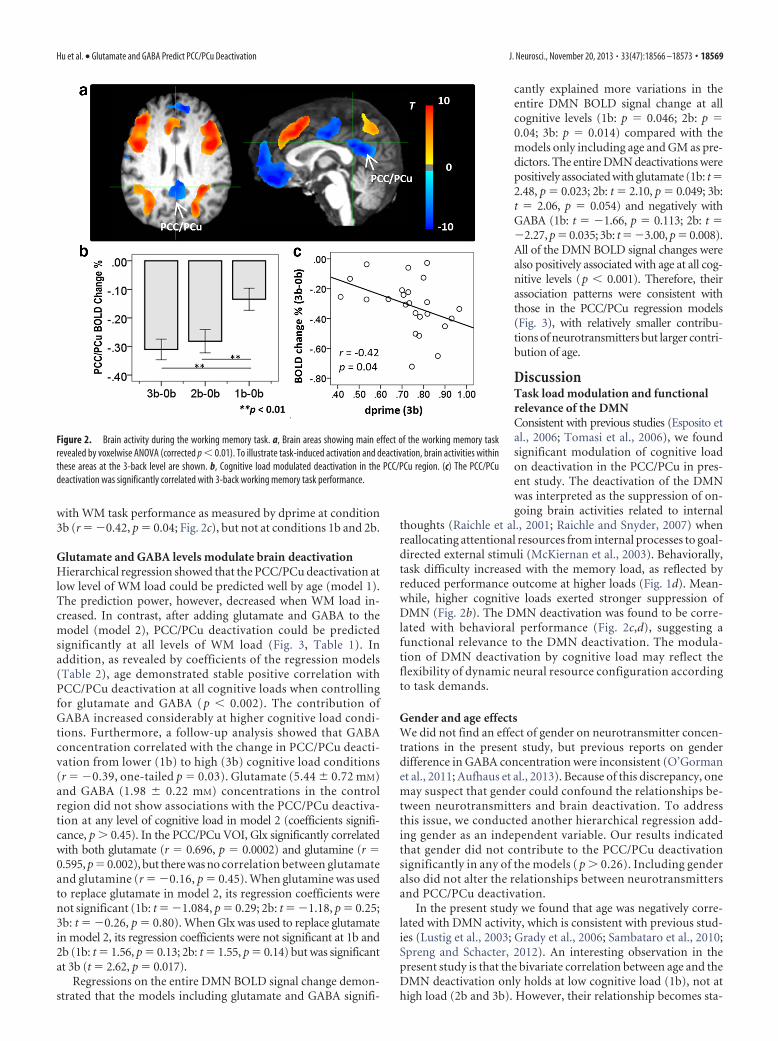
with WM task performance as measured by dprime at condition3b (r � �0.42, p � 0.04; Fig. 2c), but not at conditions 1b and 2b.
Glutamate and GABA levels modulate brain deactivationHierarchical regression showed that the PCC/PCu deactivation atlow level of WM load could be predicted well by age (model 1).The prediction power, however, decreased when WM load in-creased. In contrast, after adding glutamate and GABA to themodel (model 2), PCC/PCu deactivation could be predictedsignificantly at all levels of WM load (Fig. 3, Table 1). Inaddition, as revealed by coefficients of the regression models(Table 2), age demonstrated stable positive correlation withPCC/PCu deactivation at all cognitive loads when controllingfor glutamate and GABA ( p � 0.002). The contribution ofGABA increased considerably at higher cognitive load condi-tions. Furthermore, a follow-up analysis showed that GABAconcentration correlated with the change in PCC/PCu deacti-vation from lower (1b) to high (3b) cognitive load conditions(r � �0.39, one-tailed p � 0.03). Glutamate (5.44 � 0.72 mM)and GABA (1.98 � 0.22 mM) concentrations in the controlregion did not show associations with the PCC/PCu deactiva-tion at any level of cognitive load in model 2 (coefficients signifi-cance, p 0.45). In the PCC/PCu VOI, Glx significantly correlatedwith both glutamate (r � 0.696, p � 0.0002) and glutamine (r �0.595, p � 0.002), but there was no correlation between glutamateand glutamine (r � �0.16, p � 0.45). When glutamine was usedto replace glutamate in model 2, its regression coefficients werenot significant (1b: t � �1.084, p � 0.29; 2b: t � �1.18, p � 0.25;3b: t � �0.26, p � 0.80). When Glx was used to replace glutamatein model 2, its regression coefficients were not significant at 1b and2b (1b: t � 1.56, p � 0.13; 2b: t � 1.55, p � 0.14) but was significantat 3b (t � 2.62, p � 0.017).
Regressions on the entire DMN BOLD signal change demon-strated that the models including glutamate and GABA signifi-
cantly explained more variations in theentire DMN BOLD signal change at allcognitive levels (1b: p � 0.046; 2b: p �0.04; 3b: p � 0.014) compared with themodels only including age and GM as pre-dictors. The entire DMN deactivations werepositively associated with glutamate (1b: t �2.48, p � 0.023; 2b: t � 2.10, p � 0.049; 3b:t � 2.06, p � 0.054) and negatively withGABA (1b: t � �1.66, p � 0.113; 2b: t ��2.27, p � 0.035; 3b: t ��3.00, p � 0.008).All of the DMN BOLD signal changes werealso positively associated with age at all cog-nitive levels (p � 0.001). Therefore, theirassociation patterns were consistent withthose in the PCC/PCu regression models(Fig. 3), with relatively smaller contribu-tions of neurotransmitters but larger contri-bution of age.
DiscussionTask load modulation and functionalrelevance of the DMNConsistent with previous studies (Esposito etal., 2006; Tomasi et al., 2006), we foundsignificant modulation of cognitive loadon deactivation in the PCC/PCu in pres-ent study. The deactivation of the DMNwas interpreted as the suppression of on-going brain activities related to internal
thoughts (Raichle et al., 2001; Raichle and Snyder, 2007) whenreallocating attentional resources from internal processes to goal-directed external stimuli (McKiernan et al., 2003). Behaviorally,task difficulty increased with the memory load, as reflected byreduced performance outcome at higher loads (Fig. 1d). Mean-while, higher cognitive loads exerted stronger suppression ofDMN (Fig. 2b). The DMN deactivation was found to be corre-lated with behavioral performance (Fig. 2c,d), suggesting afunctional relevance to the DMN deactivation. The modula-tion of DMN deactivation by cognitive load may reflect theflexibility of dynamic neural resource configuration accordingto task demands.
Gender and age effectsWe did not find an effect of gender on neurotransmitter concen-trations in the present study, but previous reports on genderdifference in GABA concentration were inconsistent (O’Gormanet al., 2011; Aufhaus et al., 2013). Because of this discrepancy, onemay suspect that gender could confound the relationships be-tween neurotransmitters and brain deactivation. To addressthis issue, we conducted another hierarchical regression add-ing gender as an independent variable. Our results indicatedthat gender did not contribute to the PCC/PCu deactivationsignificantly in any of the models ( p 0.26). Including genderalso did not alter the relationships between neurotransmittersand PCC/PCu deactivation.
In the present study we found that age was negatively corre-lated with DMN activity, which is consistent with previous stud-ies (Lustig et al., 2003; Grady et al., 2006; Sambataro et al., 2010;Spreng and Schacter, 2012). An interesting observation in thepresent study is that the bivariate correlation between age and theDMN deactivation only holds at low cognitive load (1b), not athigh load (2b and 3b). However, their relationship becomes sta-
Figure 2. Brain activity during the working memory task. a, Brain areas showing main effect of the working memory taskrevealed by voxelwise ANOVA (corrected p � 0.01). To illustrate task-induced activation and deactivation, brain activities withinthese areas at the 3-back level are shown. b, Cognitive load modulated deactivation in the PCC/PCu region. (c) The PCC/PCudeactivation was significantly correlated with 3-back working memory task performance.
Hu et al. • Glutamate and GABA Predict PCC/PCu Deactivation J. Neurosci., November 20, 2013 • 33(47):18566 –18573 • 18569

ble when controlling for glutamate and GABA concentrations. Aplausible explanation is that glutamate and GABA may changewith age and these changes may in turn result in changes in DMNactivity. Conversely, the presence of the significant coefficientsfor age and neurotransmitters indicates their independent con-tributions to the variance of the task-induced deactivation. Ide-ally, age-related cognitive changes should be studied in an agerange in which profound changes in cognition are expected.Whether the observed relationships in this study would hold inan aged population and how they link to age-related cognitivechanges are interesting topics that warrant further investigation.
GABA/glutamate levels and neural activityOur results are consistent with previous studies examining theeffects of glutamate or GABA separately on BOLD signal change.The negative correlation between GABA concentration andDMN deactivation agrees with its profile as the major postsynap-
Table 2. Coefficients in regression models
Model Beta (SE) Beta� t p
1b1
Age 0.013 (0.004) 0.574 2.844 0.010GM 0.344 (0.857) 0.081 0.402 0.692
2Age 0.018 (0.004) 0.815 4.612 0.0002GM �0.918 (0.775) �0.216 �1.185 0.251Glutamate 0.261 (0.076) 0.640 3.446 0.003GABA �0.121 (0.060) �0.321 �2.003 0.060
2b1
Age 0.009 (0.005) 0.382 1.704 0.103GM 0.237 (1.004) 0.053 0.236 0.816
2Age 0.016 (0.004) 0.674 3.583 0.002GM �1.262 (0.870) �0.282 �1.451 0.163Glutamate 0.297 (0.085) 0.690 3.489 0.002GABA �0.190 (0.068) �0.480 �2.812 0.011
3b1
Age 0.007 (0.005) 0.337 1.473 0.156GM 0.735 (0.905) 0.186 0.812 0.426
2Age 0.014 (0.004) 0.670 3.947 0.001GM �0.702 (0.694) �0.178 �1.012 0.324Glutamate 0.275 (0.068) 0.724 4.053 0.0007GABA �0.216 (0.054) �0.617 �4.004 0.0008
Dependent variable: PCC/PCu deactivation during WM task; model 1 predictors: (constant), age, GM; model 2predictors: (constant), age, GM, glutamate, GABA.
Beta (SE), unstandardized coefficient (SE); and beta�, standardized coefficient.
Figure 3. Partial regression plots. PCC/PCu deactivation can be predicted by age and regional glutamate and GABA concentrations assessed by MRS at resting state.
Table 1. Models to regress PCC/PCu BOLD signal on age, GM, glutamate, and GABA
Model R 2 Adjusted R 2 R 2 F p
1b1 0.297 0.230 0.297 4.428 0.0252 0.585 0.498 0.289 6.611 0.007
2b1 0.132 0.049 0.132 1.593 0.2272 0.530 0.431 0.398 8.055 0.003
3b1 0.095 0.009 0.095 1.107 0.3492 0.617 0.536 0.521 12.922 0.0003
Model 1 predictors: (constant), age, GM; model 2 predictors: (constant), age, GM, glutamate, GABA; dependentvariable: PCC/PCu BOLD signal change.
18570 • J. Neurosci., November 20, 2013 • 33(47):18566 –18573 Hu et al. • Glutamate and GABA Predict PCC/PCu Deactivation

tic inhibitory neurotransmitter. The relationship between GABAand BOLD signal is highly accordant across different studies(Northoff et al., 2007; Muthukumaraswamy et al., 2009; Sumneret al., 2010). In addition to its association with the magnitude ofBOLD signal, the GABA concentration was found to correlatewith the latency and width of the stimulus-evoked HRF curve(Muthukumaraswamy et al., 2012), as well as electroencephalo-gram measures (Muthukumaraswamy et al., 2009; Rowland et al.,2013). In contrast to the suppression effect of GABA, the gluta-mate concentration has been found to be positively correlatedwith BOLD signal change. For example, when only controllingfor age, regional glutamate concentration in perigenual anteriorcingulate cortex (a part of DMN) was found to be positivelycorrelated with task-induced deactivation (Enzi et al., 2012) andtask-induced activation in supragenual anterior cingulate cortex(Duncan et al., 2011). Similarly, resting-state glutamate concen-tration in the dorsal anterior cingulate cortex (dACC) could pre-dict the strength of the BOLD response to a task requiringcognitive control, not just in the dACC but also in other distinctbrain regions including the retrosplenial cortex and inferior pa-rietal lobule (both are considered as parts of DMN; Falkenberg etal., 2012). These results showed convincing evidence for the di-rect relevance between neurotransmitters and BOLD contrast. Inthe present study, we revealed significant coordinative effects ofglutamate and GABA on task-induced BOLD signal change inPCC/PCu. We further demonstrated significant associations be-tween PCC/PCu neurotransmitters and the entire DMN deacti-vation, suggesting that regional neurotransmitter profile in thehub of a network may significantly affect the activity of the wholenetwork, possibly through functional connectivity (Kapogianniset al., 2013).
The system-level coordinative effects of glutamate and GABAconcentrations on the DMN deactivation may involve complexcellular processes. It might be reasonable to assume that highregional GABA concentrations could facilitate inhibition of localneural activities through GABAergic interneurons, resulting instronger brain deactivation. Similarly, high regional glutamateconcentrations could promote excitation of local glutamatergicneurons. In the resting state, glutamatergic and GABAergic activ-ities in the DMN may reach equilibrium to facilitate endogenousprocesses. When turning from the resting to a task state, theDMN suppression might be achieved by enhancing GABAergicactivities to regulate glutamatergic activities. Therefore, individ-uals with a higher GABA/glutamate ratio tend to suppress ongo-ing neural activities more efficiently. Furthermore, because theinhibition of glutamatergic neurons might also decrease theprobability of excitation of their downstream neurons, the inhib-itory associations with GABA would be enhanced in these indi-viduals.
Implications of abnormal DMN deactivation andneurotransmitters in mental diseaseThe associations between neurotransmitters and the DMN deac-tivation found in the present study may have important implica-tions for neuropsychiatric disease. The DMN is suggested to be afundamental brain network underlying normal and abnormalbrain functions (Buckner et al., 2008; Anticevic et al., 2012). Incontrast to the DMN deactivation patterns seen in healthy sub-jects, insufficient DMN deactivation has been observed in severalpsychiatric disorders. These studies suggest that the DMN abnor-malities observed in fMRI studies may be closely associated withneuropsychiatric disorders. Meanwhile, abnormal neurotrans-mitter function has been reported in disease populations, espe-
cially in schizophrenia (Marsman et al., 2013). Our resultsindicate that coordination of glutamate and GABA is associatedwith task-induced DMN deactivation. Abnormal DMN deactiva-tion in neuropsychiatric disorders may be associated with animbalance of glutamate and GABA neurotransmitters. Furtherassessment of the relationships among clinical observations, ac-tivity, and neurotransmitter function of the DMN in such patientpopulations may provide useful insights into these diseases.
LimitationsWe observed associations of local glutamate and GABA concen-trations at resting state with task-induced BOLD signal change inthe same region. However, as a distributed system, activity of onebrain region is influenced by many other regions. Specifically,release of neurotransmitters could depend on remote afferentprojections, which cannot be determined in the present study.Consideration of both remote afferent projections and local neu-rotransmitters would produce better models to predict BOLDsignal change.
Technically, measurements of GABA and glutamate are chal-lenging. The MM coediting is an important issue for the GABAdetection using editing sequences, and the degree of MM signalcontamination varies with sequences and parameters (Henry etal., 2001; Terpstra et al., 2002; Near et al., 2011). Nevertheless,previous studies (Hofmann et al., 2001; Mader et al., 2002) sug-gested that the MM concentrations in cortical regions of healthyadults are very stable with respect to age and gender. Therefore,individual differences in the contaminated GABA levels may re-flect primarily the differences in the GABA itself (Donahue et al.,2010). In addition, the degree to which glutamate and glutaminecan be separated at 3T is arguable. In the present study, theCRLBs of glutamate and glutamine fitting from LCModel were5 � 1% and 14 � 2%, respectively, suggesting that both arequantifiable. Another limitation of glutamate measurement with1H-MRS is its inability to distinguish signals between metabolicand neurotransmitter pools. However, the relationship betweenneuronal glucose oxidation and glutamate/glutamine cycling issuggested to be linear (de Graaf et al., 2004), which supports theassumption that the total glutamate level measured by 1H-MRScould be a reasonable metric of glutamate involve in glutamater-gic neurotransmission.
In the present study, fMRI and MRS data were collected sep-arately. It would be interesting to assess the association betweenneurotransmitter levels and brain activity at the same functionalstates. In addition, the influence of the menstrual cycle on thecerebral GABA and glutamate levels could be region specific (Ep-person et al., 2002; Batra et al., 2008; Harada et al., 2011). How-ever, as far as we know, there have been no reports about GABAor glutamate level changes in PCC/PCu across the menstrual cy-cle. This is a factor that we did not control for in the present study.
SummaryIn conclusion, the present study demonstrates significant associ-ation between neurotransmitters and the DMN deactivationprobed by a WM task. The major excitatory neurotransmitter,glutamate, prevents BOLD signal from deactivation, whereasGABA, the major inhibitory neurotransmitter, exerts the oppo-site effects. These neurochemical characteristics of DMN deacti-vation may provide novel insights into the function of DMN inhealthy individuals and its dysfunction in brain disorders.
NotesSupplemental material for this article is available at http://irp.drugabuse.gov/PDFs/supp/supHu08282013.pdf. This supplemental material docu-
Hu et al. • Glutamate and GABA Predict PCC/PCu Deactivation J. Neurosci., November 20, 2013 • 33(47):18566 –18573 • 18571

ment includes: (1) individual GABA spectra, (2) an estimation of GABA-macromolecule contamination and glutamate-glutamine separation,and (3) relationships between the entire DMN deactivation and neu-rotransmitters. This material has not been peer reviewed.
ReferencesAnticevic A, Repovs G, Shulman GL, Barch DM (2010) When less is more:
TPJ and default network deactivation during encoding predicts workingmemory performance. Neuroimage 49:2638 –2648. CrossRef Medline
Anticevic A, Cole MW, Murray JD, Corlett PR, Wang XJ, Krystal JH (2012)The role of default network deactivation in cognition and disease. TrendsCogn Sci 16:584 –592. CrossRef Medline
Aufhaus E, Weber-Fahr W, Sack M, Tunc-Skarka N, Oberthuer G, Hoerst M,Meyer-Lindenberg A, Boettcher U, Ende G (2013) Absence of changesin GABA concentrations with age and gender in the human anteriorcingulate cortex: A MEGA-PRESS study with symmetric editing pulsefrequencies for macromolecule suppression. Magn Reson Med 69:317–320. CrossRef Medline
Batra NA, Seres-Mailo J, Hanstock C, Seres P, Khudabux J, Bellavance F,Baker G, Allen P, Tibbo P, Hui E, Le Melledo JM (2008) Proton mag-netic resonance spectroscopy measurement of brain glutamate levels inpremenstrual dysphoric disorder. Biol Psychiatry 63:1178 –1184.CrossRef Medline
Buckner RL, Andrews-Hanna JR, Schacter DL (2008) The brain’s defaultnetwork. Ann N Y Acad Sci 1124:1–38. CrossRef Medline
Buzsaki G, Kaila K, Raichle M (2007) Inhibition and brain work. Neuron56:771–783. CrossRef Medline
de Graaf RA, Mason GF, Patel AB, Rothman DL, Behar KL (2004) Regionalglucose metabolism and glutamatergic neurotransmission in rat brain invivo. Proc Natl Acad Sci U S A 101:12700 –12705. CrossRef Medline
Donahue MJ, Near J, Blicher JU, Jezzard P (2010) Baseline GABA concen-tration and fMRI response. Neuroimage 53:392–398. CrossRef Medline
Duncan NW, Enzi B, Wiebking C, Northoff G (2011) Involvement of glu-tamate in rest-stimulus interaction between perigenual and supragenualanterior cingulate cortex: a combined fMRI-MRS study. Hum BrainMapp 32:2172–2182. CrossRef Medline
Edden RA, Intrapiromkul J, Zhu H, Cheng Y, Barker PB (2012) MeasuringT2 in vivo with J-difference editing: Application to GABA at 3 Tesla.J Magn Reson Imaging 35:229 –234. CrossRef Medline
Enzi B, Duncan NW, Kaufmann J, Tempelmann C, Wiebking C, Northoff G(2012) Glutamate modulates resting state activity in the perigenual ante-rior cingulate cortex–a combined fMRI-MRS study. Neuroscience 227:102–109. CrossRef Medline
Epperson CN, Haga K, Mason GF, Sellers E, Gueorguieva R, Zhang W, WeissE, Rothman DL, Krystal JH (2002) Cortical �-aminobutyric acid levelsacross the menstrual cycle in healthy women and those with premenstrualdysphoric disorder: A proton magnetic resonance spectroscopy study.Arch Gen Psychiatry 59:851– 858. CrossRef Medline
Esposito F, Bertolino A, Scarabino T, Latorre V, Blasi G, Popolizio T, TedeschiG, Cirillo S, Goebel R, Di Salle F (2006) Independent component modelof the default-mode brain function: assessing the impact of active think-ing. Brain Res Bull 70:263–269. CrossRef Medline
Falkenberg LE, Westerhausen R, Specht K, Hugdahl K (2012) Resting-stateglutamate level in the anterior cingulate predicts blood-oxygen level-dependent response to cognitive control. Proc Natl Acad Sci U S A 109:5069 –5073. CrossRef Medline
Ganji SK, Banerjee A, Patel AM, Zhao YD, Dimitrov IE, Browning JD, BrownES, Maher EA, Choi C (2012) T2 measurement of J-coupled metabolitesin the human brain at 3T. NMR Biomed 25:523–529. CrossRef Medline
Geng X, Christensen GE, Gu H, Ross TJ, Yang Y (2009) Implicit reference-based group-wise image registration and its application to structural andfunctional MRI. Neuroimage 47:1341–1351. CrossRef Medline
Geramita M, van der Veen JW, Barnett AS, Savostyanova AA, Shen J,Weinberger DR, Marenco S (2011) Reproducibility of prefrontal�-aminobutyric acid measurements with J-edited spectroscopy. NMRBiomed 24:1089 –1098. CrossRef Medline
Grady CL, Springer MV, Hongwanishkul D, McIntosh AR, Winocur G(2006) Age-related changes in brain activity across the adult lifespan.J Cogn Neurosci 18:227–241. CrossRef Medline
Greicius MD, Srivastava G, Reiss AL, Menon V (2004) Default-mode net-work activity distinguishes Alzheimer’s disease from healthy aging: evi-
dence from functional MRI. Proc Natl Acad Sci U S A 101:4637– 4642.CrossRef Medline
Haatveit BC, Sundet K, Hugdahl K, Ueland T, Melle I, Andreassen OA(2010) The validity of d prime as a working memory index: results fromthe “Bergen n-back task.” J Clin Exp Neuropsychol 32:871– 880. CrossRefMedline
Hancu I (2009) Optimized glutamate detection at 3T. J Magn Reson Imag-ing 30:1155–1162. CrossRef Medline
Harada M, Kubo H, Nose A, Nishitani H, Matsuda T (2011) Measurementof variation in the human cerebral GABA level by in vivo MEGA-editingproton MR spectroscopy using a clinical 3 T instrument and its depen-dence on brain region and the female menstrual cycle. Hum Brain Mapp32:828 – 833. CrossRef Medline
Henry PG, Dautry C, Hantraye P, Bloch G (2001) Brain GABA editing with-out macromolecule contamination. Magn Reson Med 45:517–520.CrossRef Medline
Hofmann L, Slotboom J, Boesch C, Kreis R (2001) Characterization of themacromolecule baseline in localized 1H-MR spectra of human brain.Magn Reson Med 46:855– 863. CrossRef Medline
Kapogiannis D, Reiter DA, Willette AA, Mattson MP (2013) Posteromedialcortex glutamate and GABA predict intrinsic functional connectivity ofthe default mode network. Neuroimage 64:112–119. CrossRef Medline
Kennedy DP, Redcay E, Courchesne E (2006) Failing to deactivate: Restingfunctional abnormalities in autism. Proc Natl Acad Sci U S A 103:8275–8280. CrossRef Medline
Logothetis NK, Pauls J, Augath M, Trinath T, Oeltermann A (2001) Neuro-physiological investigation of the basis of the fMRI signal. Nature 412:150 –157. CrossRef Medline
Lustig C, Snyder AZ, Bhakta M, O’Brien KC, McAvoy M, Raichle ME, MorrisJC, Buckner RL (2003) Functional deactivations: change with age anddementia of the Alzheimer type. Proc Natl Acad Sci U S A 100:14504 –14509. CrossRef Medline
Mader I, Seeger U, Karitzky J, Erb M, Schick F, Klose U (2002) Protonmagnetic resonance spectroscopy with metabolite nulling reveals regionaldifferences of macromolecules in normal human brain. J Magn ResonImaging 16:538 –546. CrossRef Medline
Marsman A, van den Heuvel MP, Klomp DW, Kahn RS, Luijten PR, HulshoffPol HE (2013) Glutamate in schizophrenia: a focused review and meta-analysis of 1H-MRS studies. Schizophr Bull 39:120 –129. CrossRefMedline
McKiernan KA, Kaufman JN, Kucera-Thompson J, Binder JR (2003) Aparametric manipulation of factors affecting task-induced deactivation infunctional neuroimaging. J Cogn Neurosci 15:394 – 408. CrossRefMedline
Mescher M, Merkle H, Kirsch J, Garwood M, Gruetter R (1998) Simultane-ous in vivo spectral editing and water suppression. NMR Biomed 11:266 –272. CrossRef Medline
Mlynarik V, Gruber S, Moser E (2001) Proton T1 and T2 relaxation times ofhuman brain metabolites at 3 Tesla. NMR Biomed 14:325–331. CrossRefMedline
Mullins PG, Chen H, Xu J, Caprihan A, Gasparovic C (2008) Comparativereliability of proton spectroscopy techniques designed to improve detec-tion of J-coupled metabolites. Magn Reson Med 60:964 –969. CrossRefMedline
Muthukumaraswamy SD, Edden RA, Jones DK, Swettenham JB, Singh KD(2009) Resting GABA concentration predicts peak gamma frequencyand fMRI amplitude in response to visual stimulation in humans. ProcNatl Acad Sci U S A 106:8356 – 8361. CrossRef Medline
Muthukumaraswamy SD, Evans CJ, Edden RA, Wise RG, Singh KD (2012)Individual variability in the shape and amplitude of the BOLD-HRF cor-relates with endogenous GABAergic inhibition. Hum Brain Mapp 33:455– 465. CrossRef Medline
Near J, Simpson R, Cowen P, Jezzard P (2011) Efficient �-aminobutyricacid editing at 3T without macromolecule contamination: MEGA-SPECIAL. NMR Biomed 24:1277–1285. CrossRef Medline
Northoff G, Walter M, Schulte RF, Beck J, Dydak U, Henning A, Boeker H,Grimm S, Boesiger P (2007) GABA concentrations in the human ante-rior cingulate cortex predict negative BOLD responses in fMRI. Nat Neu-rosci 10:1515–1517. CrossRef Medline
Ogawa S, Lee TM, Kay AR, Tank DW (1990) Brain magnetic resonanceimaging with contrast dependent on blood oxygenation. Proc Natl AcadSci U S A 87:9868 –9872. CrossRef Medline
18572 • J. Neurosci., November 20, 2013 • 33(47):18566 –18573 Hu et al. • Glutamate and GABA Predict PCC/PCu Deactivation
Ogawa S, Menon RS, Tank DW, Kim SG, Merkle H, Ellermann JM, Ugurbil K(1993) Functional brain mapping by blood oxygenation level-dependentcontrast magnetic resonance imaging–a comparison of signal character-istics with a biophysical model. Biophys J 64:803– 812. CrossRef Medline
O’Gorman RL, Michels L, Edden RA, Murdoch JB, Martin E (2011) In vivodetection of GABA and glutamate with MEGA-PRESS: reproducibility andgender effects. J Magn Reson Imaging 33:1262–1267. CrossRef Medline
Owen AM, McMillan KM, Laird AR, Bullmore E (2005) N-back workingmemory paradigm: a meta-analysis of normative functional neuroimag-ing studies. Hum Brain Mapp 25:46 –59. CrossRef Medline
Provencher SW (1993) Estimation of metabolite concentrations from local-ized in vivo proton NMR spectra. Magn Reson Med 30:672– 679.CrossRef Medline
Provencher SW (2001) Automatic quantitation of localized in vivo 1H spec-tra with LCModel. NMR Biomed 14:260 –264. CrossRef Medline
Puts NA, Barker PB, Edden RA (2013) Measuring the longitudinal relax-ation time of GABA in vivo at 3 tesla. J Magn Reson Imaging 37:999 –1003.CrossRef Medline
Raichle ME, Snyder AZ (2007) A default mode of brain function: a briefhistory of an evolving idea. Neuroimage 37:1083–1090; discussion1097–9. CrossRef Medline
Raichle ME, MacLeod AM, Snyder AZ, Powers WJ, Gusnard DA, ShulmanGL (2001) A default mode of brain function. Proc Natl Acad Sci U S A98:676 – 682. CrossRef Medline
Rothman DL, Behar KL, Hetherington HP, Shulman RG (1984) Homo-nuclear 1H double-resonance difference spectroscopy of the rat brain invivo. Proc Natl Acad Sci U S A 81:6330 – 6334. CrossRef Medline
Rowland LM, Edden RA, Kontson K, Zhu H, Barker PB, Hong LE (2013)GABA predicts inhibition of frequency-specific oscillations in schizo-phrenia. J Neuropsychiatry Clin Neurosci 25:83– 87. CrossRef Medline
Sambataro F, Murty VP, Callicott JH, Tan HY, Das S, Weinberger DR, MattayVS (2010) Age-related alterations in default mode network: impact onworking memory performance. Neurobiol Aging 31:839 – 852. CrossRefMedline
Shmuel A, Augath M, Oeltermann A, Logothetis NK (2006) Negative func-
tional MRI response correlates with decreases in neuronal activity inmonkey visual area V1. Nat Neurosci 9:569 –577. CrossRef Medline
Shulman GL, Fiez JA, Corbetta M, Buckner RL, Miezin FM, Raichle ME,Petersen SE (1997) Common blood flow changes across visual tasks: II.decreases in cerebral cortex. J Cogn Neurosci 9:648 – 663. CrossRefMedline
Singh KD, Fawcett IP (2008) Transient and linearly graded deactivation ofthe human default-mode network by a visual detection task. Neuroimage41:100 –112. CrossRef Medline
Spreng RN, Schacter DL (2012) Default network modulation and large-scale network interactivity in healthy young and old adults. Cereb Cortex22:2610 –2621. CrossRef Medline
Stagg CJ, Bachtiar V, Johansen-Berg H (2011) The role of GABA in humanmotor learning. Curr Biol 21:480 – 484. CrossRef Medline
Stefanovic B, Warnking JM, Pike GB (2004) Hemodynamic and metabolicresponses to neuronal inhibition. Neuroimage 22:771–778. CrossRefMedline
Sumner P, Edden RA, Bompas A, Evans CJ, Singh KD (2010) More GABA,less distraction: a neurochemical predictor of motor decision speed. NatNeurosci 13:825– 827. CrossRef Medline
Terpstra M, Ugurbil K, Gruetter R (2002) Direct in vivo measurement ofhuman cerebral GABA concentration using MEGA-editing at 7 Tesla.Magn Reson Med 47:1009 –1012. CrossRef Medline
Tkac I, Starcuk Z, Choi IY, Gruetter R (1999) In vivo 1H NMR spectroscopyof rat brain at 1 ms echo time. Magn Reson Med 41:649 – 656. CrossRefMedline
Tomasi D, Ernst T, Caparelli EC, Chang L (2006) Common deactivationpatterns during working memory and visual attention tasks: an intra-subject fMRI study at 4 Tesla. Hum Brain Mapp 27:694 –705. CrossRefMedline
Whitfield-Gabrieli S, Thermenos HW, Milanovic S, Tsuang MT, Faraone SV,McCarley RW, Shenton ME, Green AI, Nieto-Castanon A, LaViolette P, Wo-jcik J, Gabrieli JD, Seidman LJ (2009) Hyperactivity and hyperconnectivityof the default network in schizophrenia and in first-degree relatives of personswith schizophrenia. Proc Natl Acad Sci U S A 106:1279–1284. CrossRefMedline
Hu et al. • Glutamate and GABA Predict PCC/PCu Deactivation J. Neurosci., November 20, 2013 • 33(47):18566 –18573 • 18573