RESPIRATORY SYSTEMRESPIRATORY SYSTEMfacultymembers.sbu.ac.ir/rajabi/ppt...
53
RESPIRATORY SYSTEM RESPIRATORY SYSTEM
Transcript of RESPIRATORY SYSTEMRESPIRATORY SYSTEMfacultymembers.sbu.ac.ir/rajabi/ppt...
RESPIRATORY SYSTEMRESPIRATORY SYSTEM
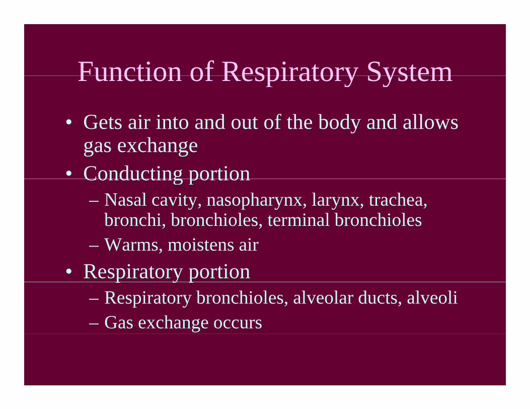
Function of Respiratory SystemFunction of Respiratory System• Gets air into and out of the body and allowsGets air into and out of the body and allows
gas exchange• Conducting portionConducting portion
– Nasal cavity, nasopharynx, larynx, trachea, bronchi, bronchioles, terminal bronchioles
– Warms, moistens air• Respiratory portionp y p
– Respiratory bronchioles, alveolar ducts, alveoli– Gas exchange occurs

Main Divisions ofDivisions of Respiratory
TTract

Layers of Wall
• Mucosa– Epithelium– Lamina propria (loose CT)– Smooth muscle
• Submucosa– Dense irregular CT– Glands often present
• Adventitia

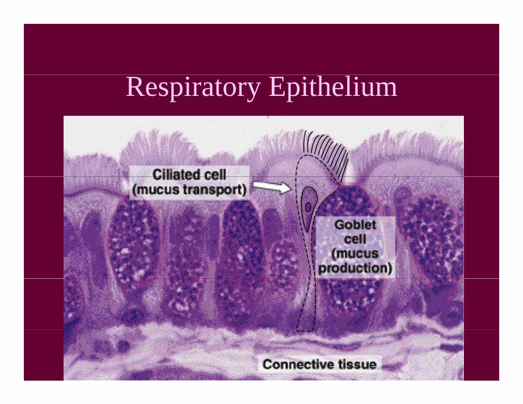
Respiratory EpitheliumRespiratory Epithelium• In the conducting portion:g p
– Ciliated columnar cells– Mucous goblet cellsg– Brush cells (microvilli)
• Sensory receptor cellsy p
– Basal cells• Generative stem cells that replace other cellsp
– Small granule cell (DNES)• Produce biogenic amines (NE, Ep, 5-HT); paracrine
cells
Respiratory Epithelium

Ciliated C lColumnar
Cells

S f fSurface of Respiratory esp ato y
Mucosa
Ciliated cell
Goblet cell
Brush cell
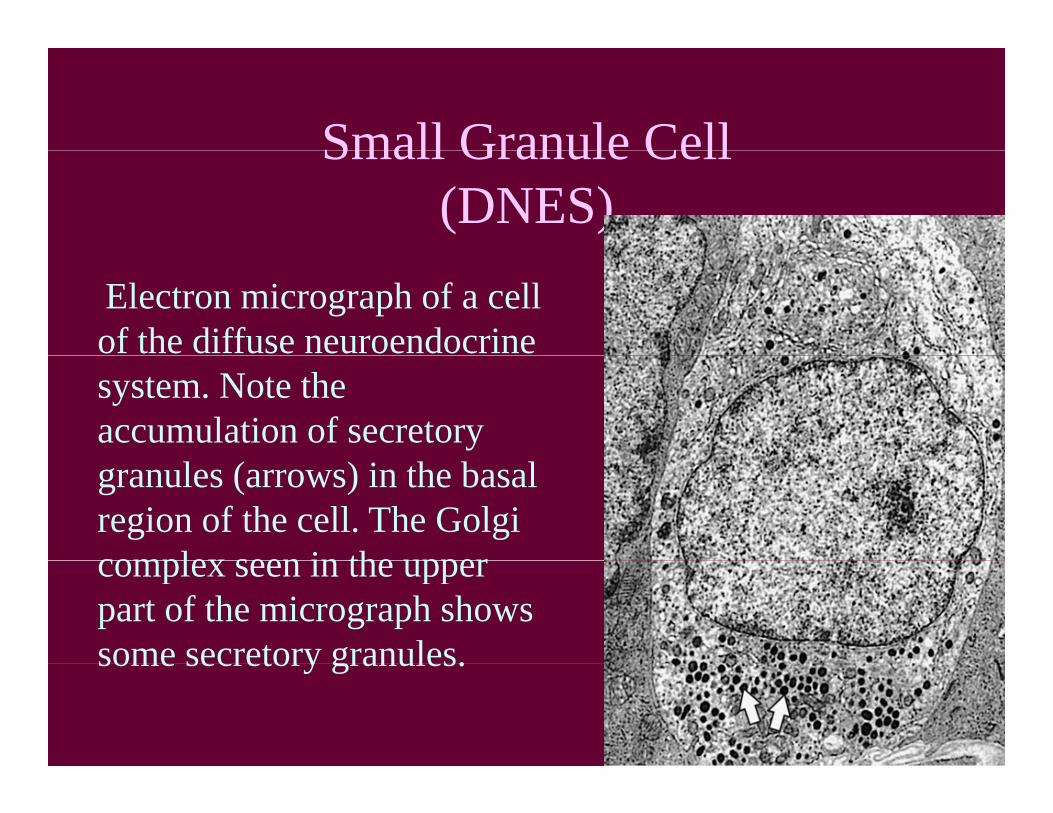
Small Granule CellSmall Granule Cell(DNES)
Electron micrograph of a cell of the diffuse neuroendocrine system. Note the accumulation of secretory granules (arrows) in the basal region of the cell. The Golgi complex seen in the uppercomplex seen in the upper part of the micrograph shows some secretory granules.some secretory granules.

Nasal Passage
TurbinatesTurbinates

Nasal CavityNasal Cavity
• Vestibule (outer nasal area)Vestibule (outer nasal area)– Keratinized epithelium transitions to respiratory– Short hairs filterShort hairs filter
• Nasal FossaeCh h– Chonchae
• Respiratory epith (pseudostratified squamous)• Swell bodies; extensive venous system for• Swell bodies; extensive venous system for
countercurrent flow to warm air.• Olfactory epithelium of superior choncha
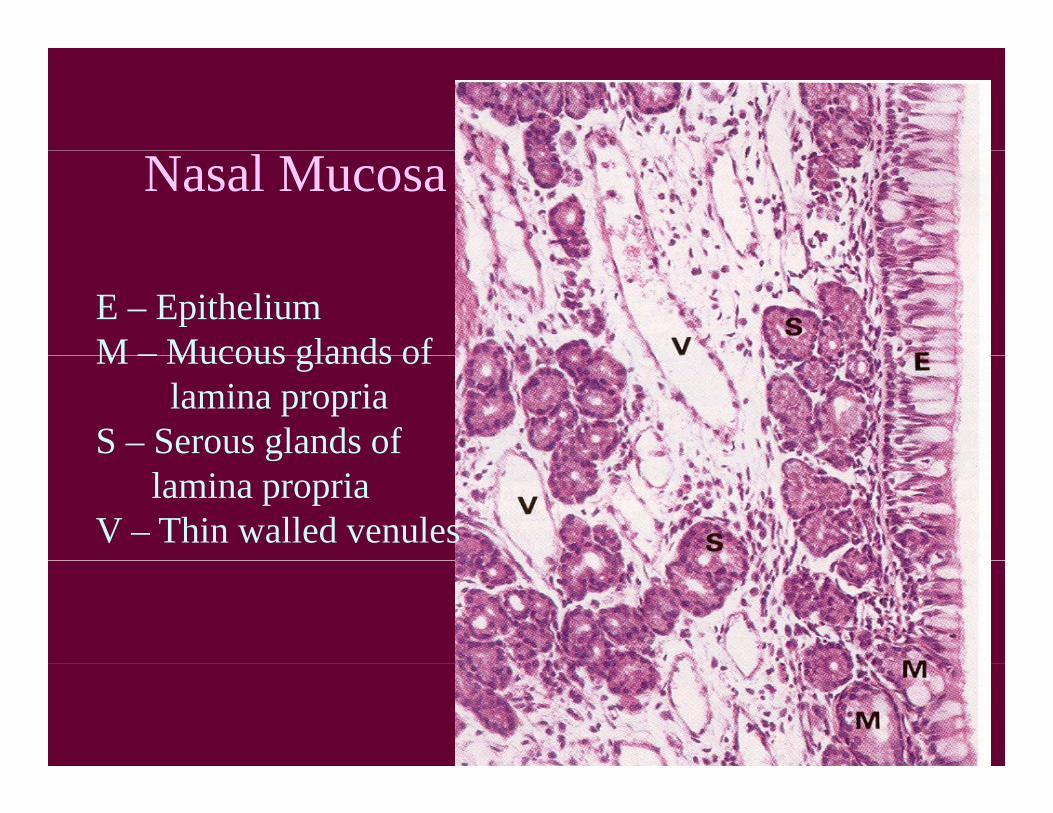
Nasal Mucosa
E – EpitheliumM – Mucous glands ofM – Mucous glands of
lamina propriaS – Serous glands ofg
lamina propriaV – Thin walled venules

Olfactory Mucosa

Sinuses
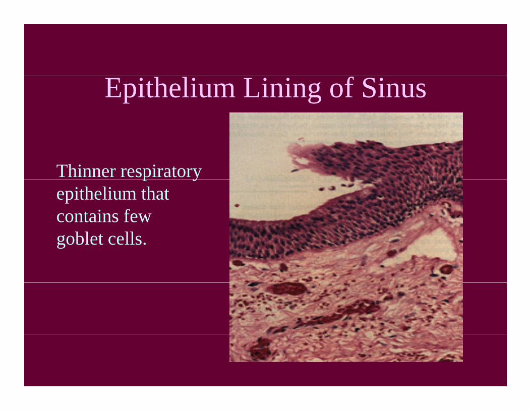
Epithelium Lining of Sinus
Thinner respiratoryp yepithelium thatcontains fewgoblet cells.
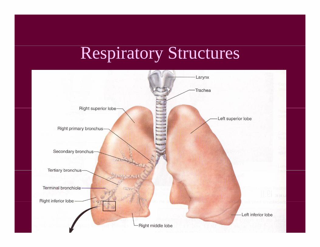
Respiratory Structures
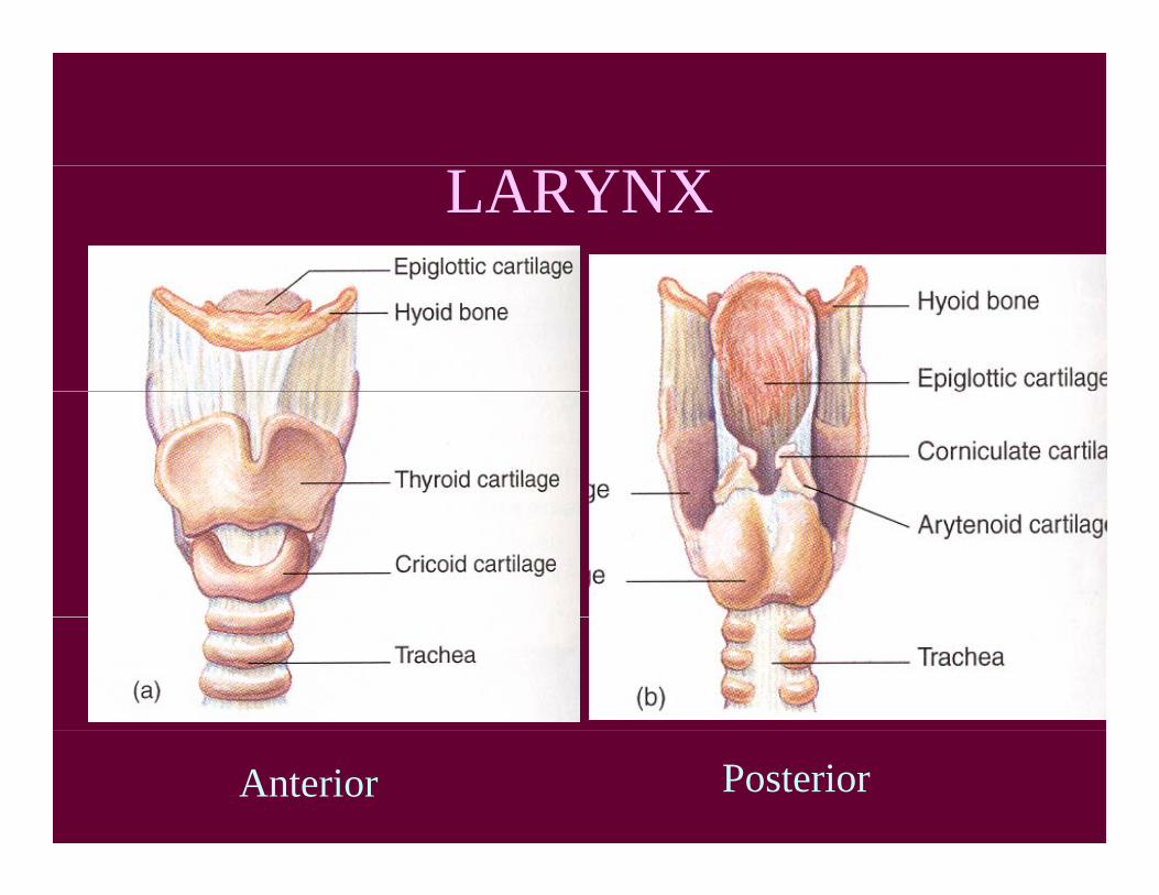
LARYNX
Anterior Posterior
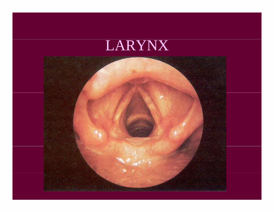
LARYNX

TRACHEA
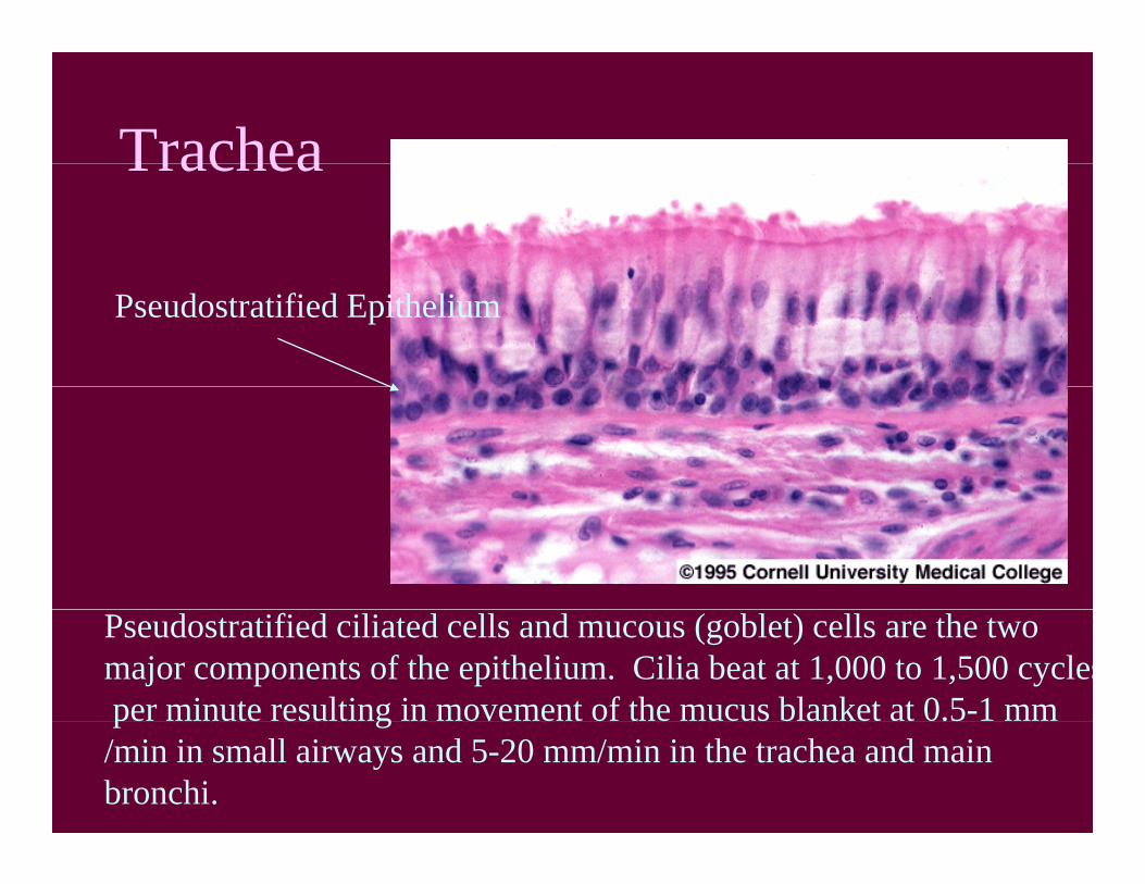
TracheaTrachea
Pseudostratified Epithelium
Pseudostratified ciliated cells and mucous (goblet) cells are the two major components of the epithelium. Cilia beat at 1,000 to 1,500 cyclesper minute resulting in movement of the mucus blanket at 0.5-1 mmpe u e esu g ove e o e ucus b e 0.5/min in small airways and 5-20 mm/min in the trachea and main bronchi.

Trachea x40
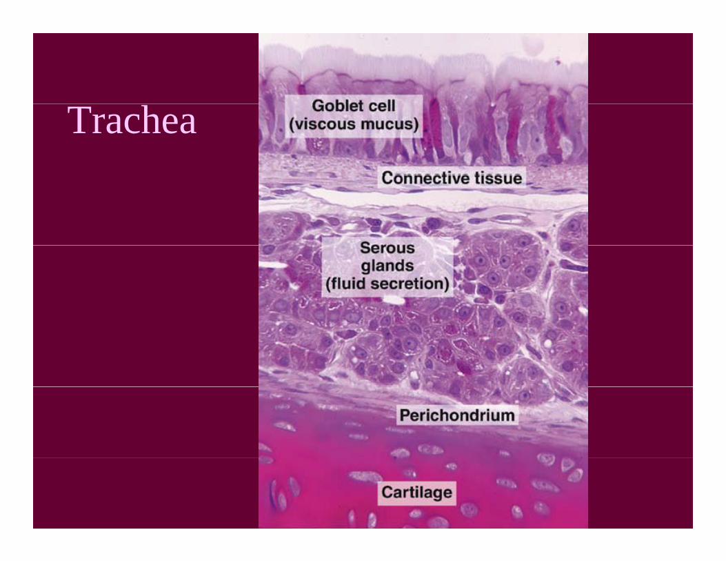
Trachea
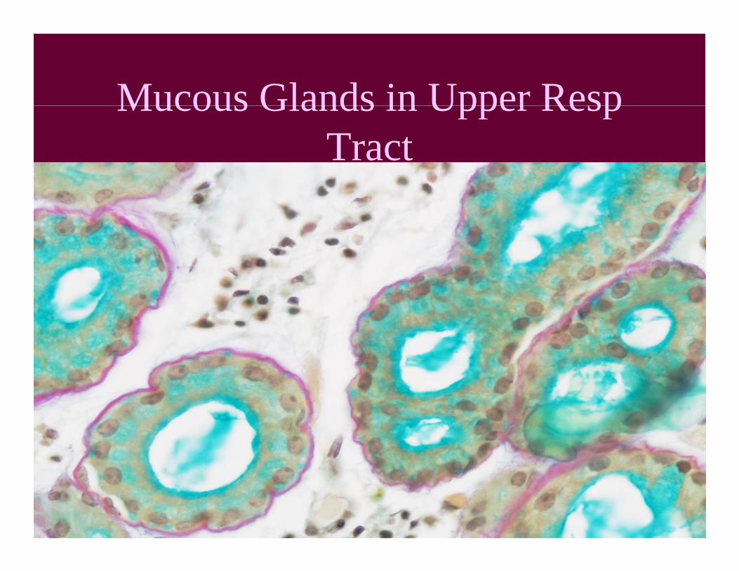
Mucous Glands in Upper RespMucous Glands in Upper Resp Tract

Trachea x10

Trachea x10

Glands of Trachea x40

BRONCHIAL TREE
Primary
SecondaryTertiary

BRONCHIAL TREE
• 1, 2, 3 Bronchi– Cartilage plates, glands present, smallest is
5mm, many lymphocytes present• Bronchioles
– No cartilage, no glands• Alveolar Duct• Alveolar Sac
– Gas exchange• Alveolus

HISTOLOGY OF BRONCHIALHISTOLOGY OF BRONCHIAL TREE
• Cartilage– Rings, plates, disappears
Smooth Muscle LayerBegins in bronchi, more prominent in eg s b o c , o e p o ebronchioles, then disappears
• Epitheliump– Pseudostratified, Simple columnar, cuboidal,
squamous
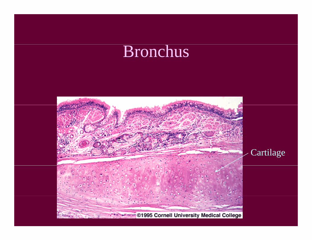
Bronchus
Cartilage

BronchusLamina propria

Bronchus

Bronchioles
• > 5mm diameter• No cartilage or glands in mucosa• Epithelium changes from pseudostratified to
cuboidal epith., shortening along the way.O l tt d bl t ll i iti ll• Only scattered goblet cells initially.
• Clara cells secrete protective proteins.Lamina propria contains onl smooth m scle and• Lamina propria contains only smooth muscle and elastic fibers.
• Vagus nerve sympathetic neuronsVagus nerve, sympathetic neurons

Bronchiole x10

Bronchiole x40

Alveolus, Bronchiole

Location of Gas Exchange

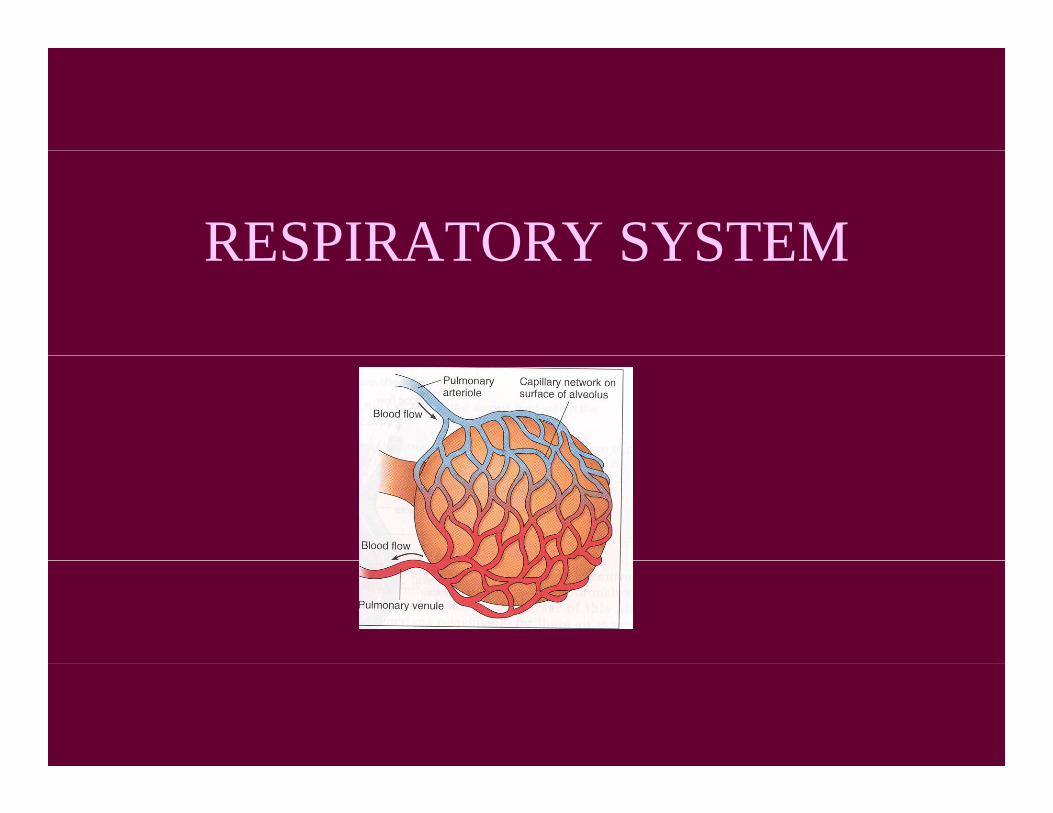
ALVEOLUS AND CAPILLARY
GAS EXCHANGE
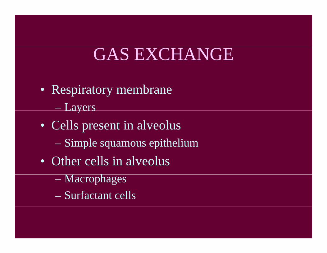
• Respiratory membrane– LayersLayers
• Cells present in alveolusSimple squamous epithelium– Simple squamous epithelium
• Other cells in alveolush– Macrophages
– Surfactant cells
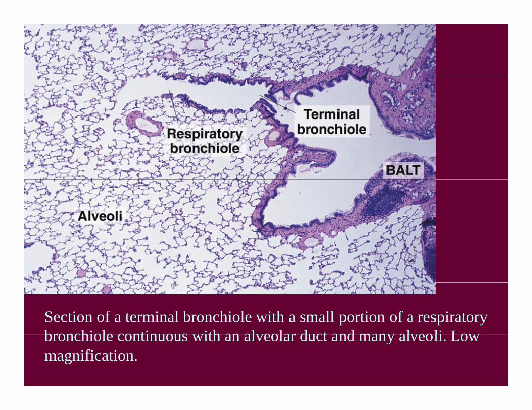
Section of a terminal bronchiole with a small portion of a respiratory bronchiole continuous with an alveolar duct and many alveoli Lowbronchiole continuous with an alveolar duct and many alveoli. Low magnification.
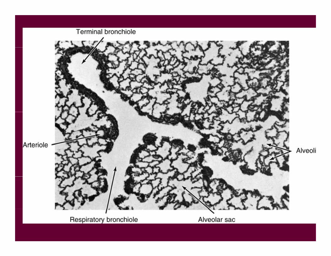

Diagram of aDiagram of a portion of the bronchial tree. Note that the smooth muscle in h l l dthe alveolar duct
disappears in the alveoli (Redrawnalveoli. (Redrawn from Baltisberger.)
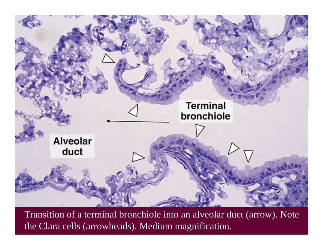
Transition of a terminal bronchiole into an alveolar duct (arrow). Note the Clara cells (arrowheads). Medium magnification.
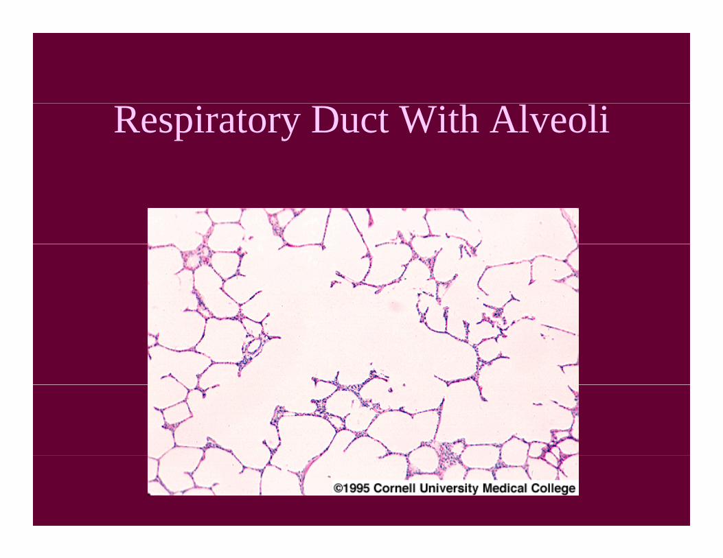
Respiratory Duct With Alveoli
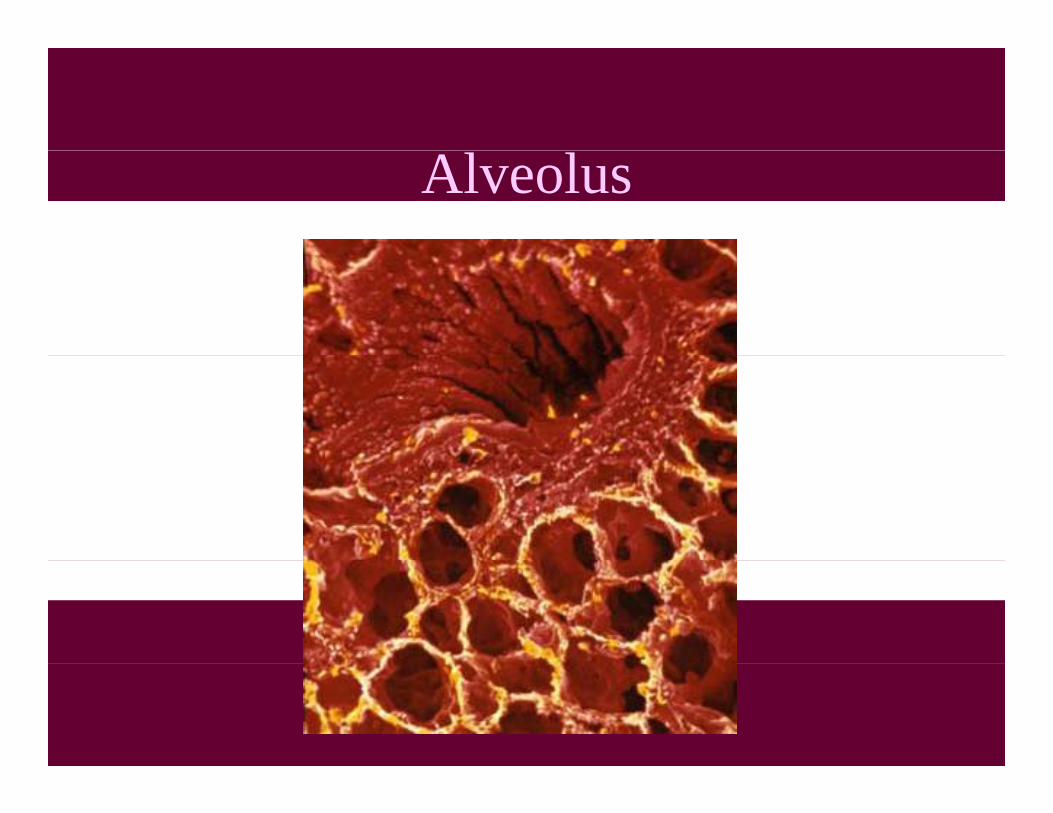
Alveolus
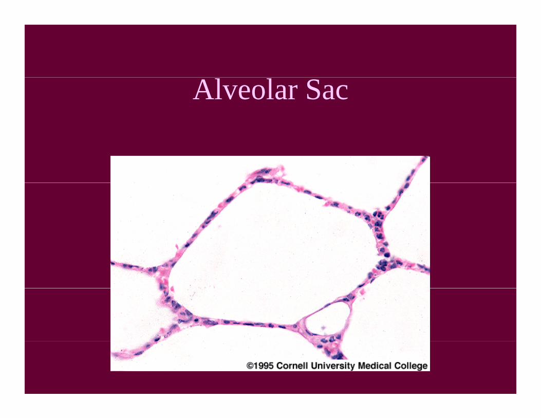
Alveolar Sac

Blood Vessel in Lung

Alveolus with Capillary
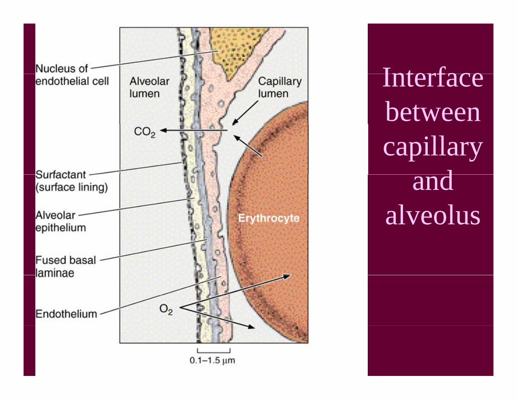
InterfaceInterfacebetweencapillary
dand alveolus

Interface Between Capillary, Alveolus
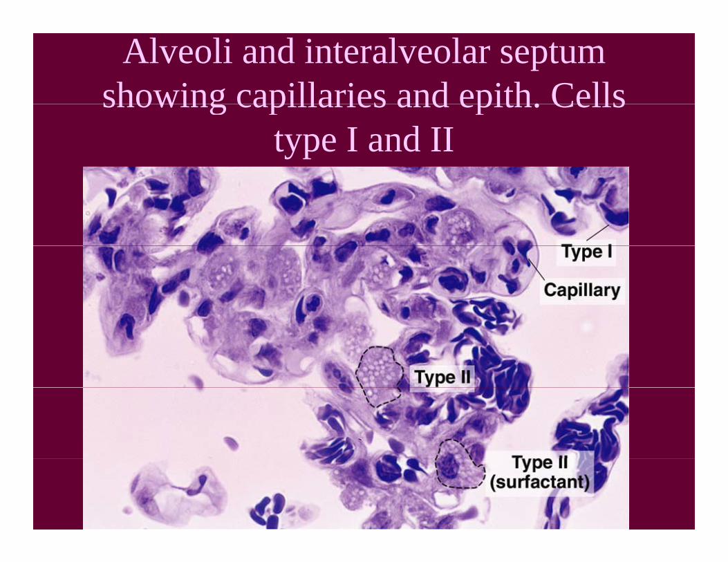
Alveoli and interalveolar septum showing capillaries and epith. Cellsshowing capillaries and epith. Cells
type I and II
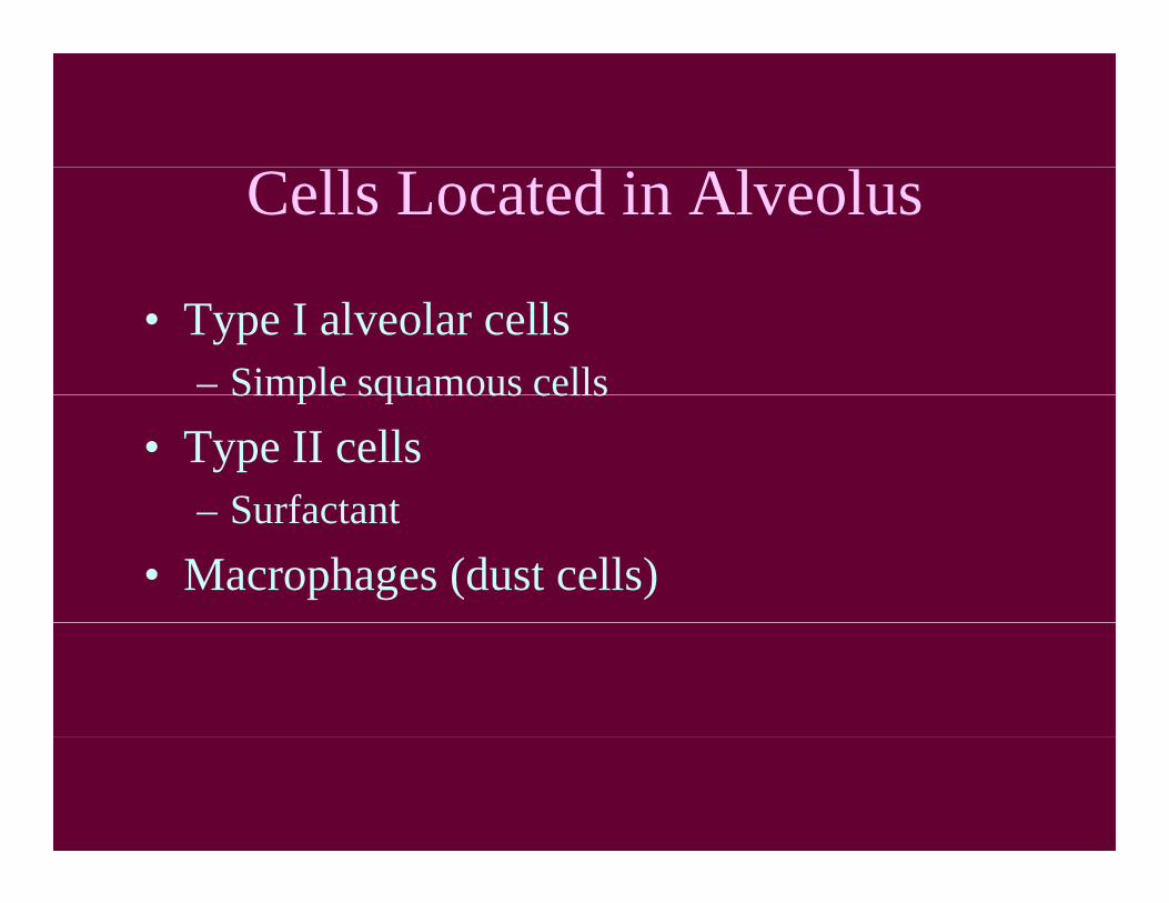
Cells Located in Alveolus
• Type I alveolar cells– Simple squamous cellsSimple squamous cells
• Type II cellsSurfactant– Surfactant
• Macrophages (dust cells)

http://www lab anhb uwa edu au/http://www.lab.anhb.uwa.edu.au/mb140/Big/Big.htm