Respiratory Physiology - Regulation
58
RESPIRATORY PHYSIOLOGY RESPIRATORY PHYSIOLOGY Agaton T. Panopio, Jr., MD, Agaton T. Panopio, Jr., MD, MHPEd MHPEd
-
Upload
roy-acosta-gumban -
Category
Documents
-
view
128 -
download
12
description
Respiratory Physiology - Regulation
Transcript of Respiratory Physiology - Regulation

RESPIRATORY RESPIRATORY PHYSIOLOGYPHYSIOLOGY
Agaton T. Panopio, Jr., MD, MHPEdAgaton T. Panopio, Jr., MD, MHPEd

Respiratory Patterns
EupneaNormal breathing
ApneaTemporary cessation of breathing
SighA larger than normal breath occurring automatically at regular intervals to counteract collapse of alveoli
Respiratory Patterns
Yawning - Pandiculation
An exaggerated sigh
May be due to:
A reflex occurring if there is O2 need
Boredom
A primitive behavior to warn others
Contagious ?

Yawning
Increase in dopamine, serotonin, acetylcholine, nitric oxide, ACTH-related peptides and oxytocin
Decrease in endorphins
Yawning
Expansion of the chest and descent of diaphragm and larynx
Elevation of ala nasi and soft palate
Downward and backward movement of the tongue
Abduction of vocal cords
Wide opening of the mouth

Yawning
Contraction of tensor veli palatini muscle
Closure of the eyes and lacrimation
Pulling of head backwards
Stretching of arms sideward, and forearms upward
Vasoconstriction esp in digits
Cardiac acceleration

Respiratory Patterns
Tachypnea (Polypnea)
An increase in respiratory rate
May be reflex in origin
Hyperpnea
An increase in rate and depth of respiration, which matches an increase in metabolic demand as in exercise
Respiratory Patterns
HyperventilationAn increase in rate and depth of respiration, which exceeds metabolic demand as in high altitudes
HypoventilationA decrease in rate and depth of respiration, which may occur as compensation to metabolic alkalosis

Respiratory Patterns
DyspneaConsciousness of the necessity for increased respiratory efforts, seen in pulmonary congestion of left heart failure
Kussmaul respirationExtremely deep, rapid breathing due to intense stimulation of the respiratory center, seen in metabolic acidosis
Respiratory Patterns
OrthopneaDyspnea in the recumbent position
Cheyne-Stokes respirationCycles of gradual increase followed by gradual decrease in tidal volumes, followed by equal periods of apnea, seen in bilateral cortical disease, congestive heart failure, or during sleep at high altitudes

Respiratory Patterns
Biot’s respirationBreaths of equal volume separated by unequal periods of apnea, seen in medullary or pontine lesions
GaspingMaximal, brief inspiratory efforts separated by long periods of expiration, seen in severe anoxia or terminal brain stem lesions
Respiratory Patterns
Apneusis
Prolonged inspiration separated by brief expirations
Rarely seen in humans
Due to lesion in pneumotaxic center

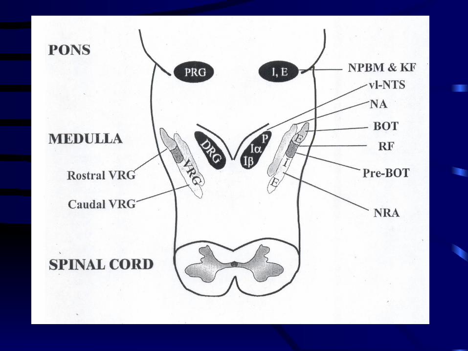
Respiratory Centers
Centers at the Pons
The Pons modulates – but is not essential for – respiratory output
Pontine Respiratory Group
Apneustic Center

Centers at the Pons
Pontine Respiratory GroupPneumotaxic Center (Rostral Pons)
Nucleus parabrachialis medialisKolliker-Fuse nucleus
Promotes coordinated respirations by modulating activity of Apneustic Center
Not necessary for eupnea
Centers at the Pons
Apneustic Center (Caudal Pons)
Stimulation causes apneuses
Presently, the role of the Apneustic Center on respiration is still not clearly understood
Centers at the Medulla
Dorsal Respiratory Group (DRG)
Primarily contains inspiratory neurons
Located at Nucleus Tractus Solitarius
Responsible for processing of sensory inputs
Inputs come from CN IX and X, as well as from peripheral chemoreceptors
Centers at the Medulla
Ventral Respiratory Group (VRG)Contains both inspiratory and expiratory
neuronsContains motor neurons that innervate
the muscles of the larynx, pharynx, viscera of thorax and abdomen
Receives sensory information from DRG
Centers at the Medulla
Ventral Respiratory Group
Rostral VRG
Botzinger complex or Nucleus Retrofacialis
Contain interneurons that drive expiratory activity of the caudal region
Centers at the Medulla
Ventral Respiratory Group
Intermediate VRG
Nucleus Ambiguus
Nucleus Para-ambigualis
Contain somatic motor neurons which supply the upper airways

Centers at the Medulla
Ventral Respiratory Group
Intermediate VRG
Also contains premotor neurons that project to inspiratory motor neurons in the spinal cord and medulla

Centers at the Medulla
Ventral Respiratory GroupIntermediate VRG
At the rostral pole of the intermediate VRG is a group of inspiratory neurons
PreBotzinger complexGenerates respiratory
rhythmicity

Centers at the Medulla
Ventral Respiratory Group
Caudal VRG (Nucleus Retroambigualis)
Contains premotor neurons that travel down the spinal cord to
synapse on motor neurons that innervate accessory muscles of expiration

Centers at the Medulla
Respiratory RhythmicityRestricted Site Models
Nucleus Tractus Solitarius (Cajal)Reported that neurons in the NTS receive afferents from pulmonary stretch receptors and project directly to the phrenic motor nucleus

Centers at the Medulla
Respiratory RhythmicityRestricted Site Models
PreBotzinger Complex (Suzue)Neurons at the complex generate rhythmic motor
output in the phrenic and hypoglossal nerves; destruction of these neurons causes respiratory outputs to stop
Centers at the Medulla
Respiratory RhythmicityDistributed Oscillator Model
There is more than one Central Pattern Generator (CPG)
Parafacial respiratory groupTRH
Only one CPG exists for eupnea, the rest only augment the rhythm

Centers at the Medulla
Respiratory Rhythmicity
Emergent Property Model (Lumsden)
No individual region of DRG or VRG is sufficient to generate a rhythm, but that many of them are necessary

Center at the Spinal Cord
Serves as site of integration of descending impulses from higher centers
Nerve fibers:Alpha motor neurons
Innervate extrafusal fibersGamma efferent neurons
Innervate intrafusal fibers

Normal Ventilation
Spontaneous depolarization of Central Pattern Generator (CPG)
Dorsal Respiratory Group
Ventral Respiratory Group
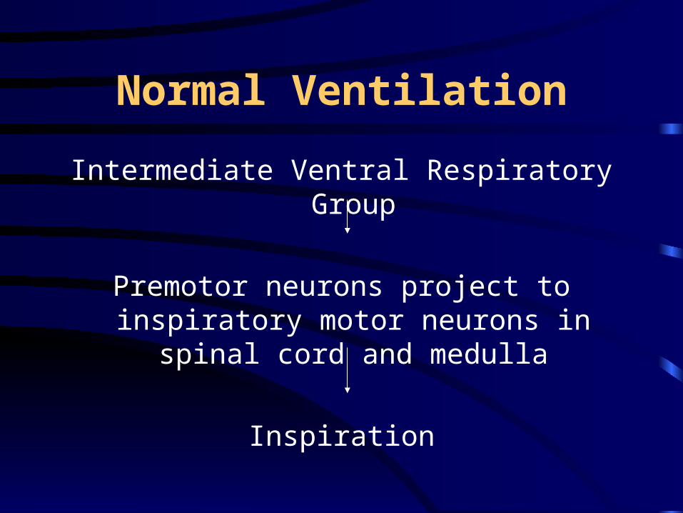
Normal Ventilation
Intermediate Ventral Respiratory Group
Premotor neurons project to inspiratory motor neurons in spinal cord and
medulla
Inspiration

Normal Ventilation
Intermediate Ventral Respiratory Group
Somatic motor neurons whose axons leave the medulla through CN IX and X
Maximize caliber of the upper airways during inspiration

Modulation of Respiration
Receptors In Airways and Lung Parenchyma
Provide feedback about lung volume and the presence of irritants
Slowly adapting pulmonary stretch receptors
Rapidly adapting pulmonary stretch receptors
C fiber receptors (J receptors)

Higher Brain Centers
Cerebral Cortex
Modulates the respiratory system by:
Sending axons to respiratory centers at the medulla
Sending axons to motor neurons that control muscles of respiration

Higher Brain Centers
Cerebral Cortex
Coordinates voluntary behaviors that use respiratory muscles
Voluntary hyperventilating
Breath-holding Speaking
Singing Whistling
Playing musical wind instruments

Higher Brain Centers
Cerebral Cortex
Coordinates complex non-ventilatory behaviors
Yawning Chewing
Swallowing Sucking
Defecating Grunting
Vomiting
Higher Brain Centers
Cerebral CortexLesions in specific areas of cerebral cortex
Abolish voluntary breath-holding(Respiratory apraxia)
Lesions in reticulospinal tractRespiratory failure while asleep (Ondine’s curse)
Other Centers
Limbic System and Hypothalamus
Modify respiration in affective states
Fear
Horror
Rage
Passion
Other Centers
Reticular Activating System
At brain stem
One of the sources of tonic drive to the respiratory CPG
Increase in drive occurs during arousal from sleep

Reflex MechanismsReflex Mechanisms
Receptors – stretch receptors in airwaysReceptors – stretch receptors in airways
ResponseResponse
Apnea/Decreased respirationApnea/Decreased respiration
BronchodilationBronchodilation
Hering-Breuer Inflation ReflexHering-Breuer Inflation Reflex
Stimulus – lung inflationStimulus – lung inflation
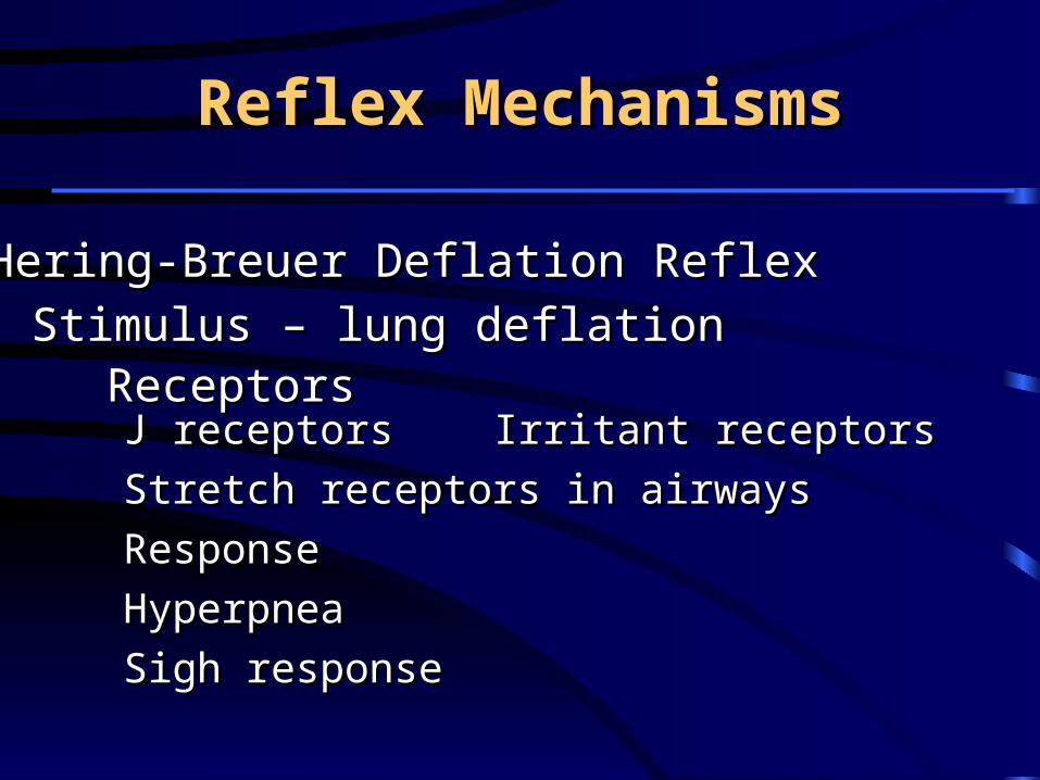
J receptors J receptors Irritant receptorsIrritant receptors
Stretch receptors in airwaysStretch receptors in airways
ResponseResponse
HyperpneaHyperpnea
Sigh responseSigh response
Reflex MechanismsReflex Mechanisms
Hering-Breuer Deflation ReflexHering-Breuer Deflation Reflex
Stimulus – lung deflationStimulus – lung deflationReceptorsReceptors
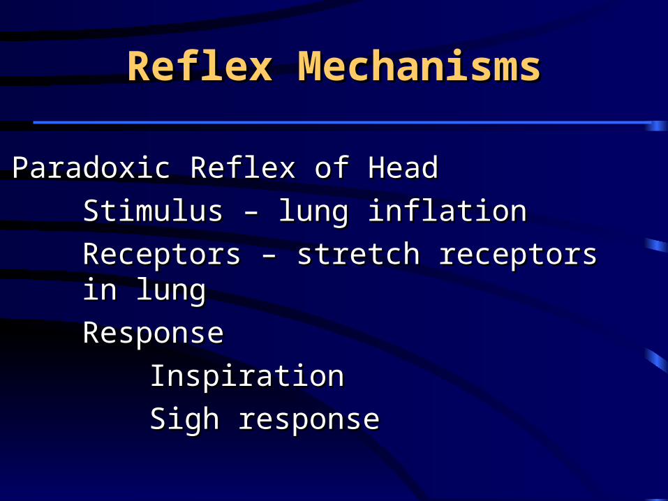
Stimulus – lung inflationStimulus – lung inflation
Receptors – stretch receptors in lungReceptors – stretch receptors in lung
Response Response
InspirationInspiration
Sigh responseSigh response
Reflex MechanismsReflex Mechanisms
Paradoxic Reflex of HeadParadoxic Reflex of Head

Stimulus – irritation at the noseStimulus – irritation at the nose
Receptors – nasal mucosa receptorsReceptors – nasal mucosa receptors
ResponseResponse
SneezeSneeze
BronchoconstrictionBronchoconstriction
Rise in BPRise in BP
Reflex MechanismsReflex Mechanisms
Sneeze ReflexSneeze Reflex
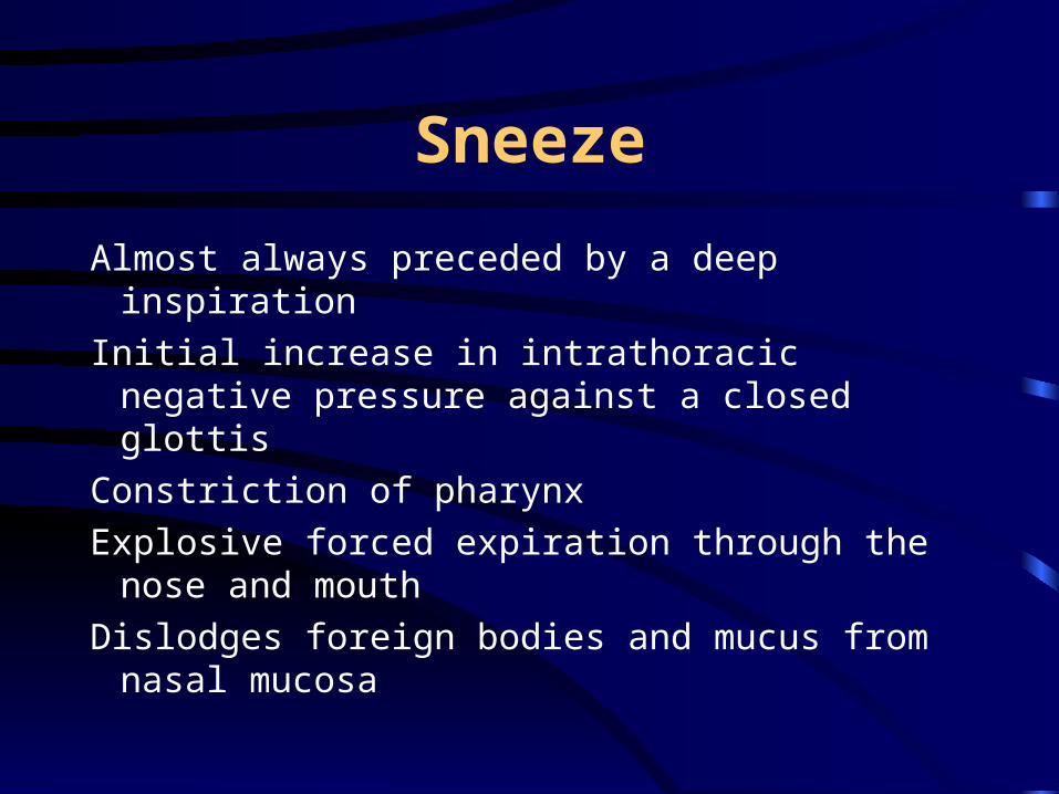
Sneeze
Almost always preceded by a deep inspiration
Initial increase in intrathoracic negative pressure against a closed glottis
Constriction of pharynx
Explosive forced expiration through the nose and mouth
Dislodges foreign bodies and mucus from nasal mucosa
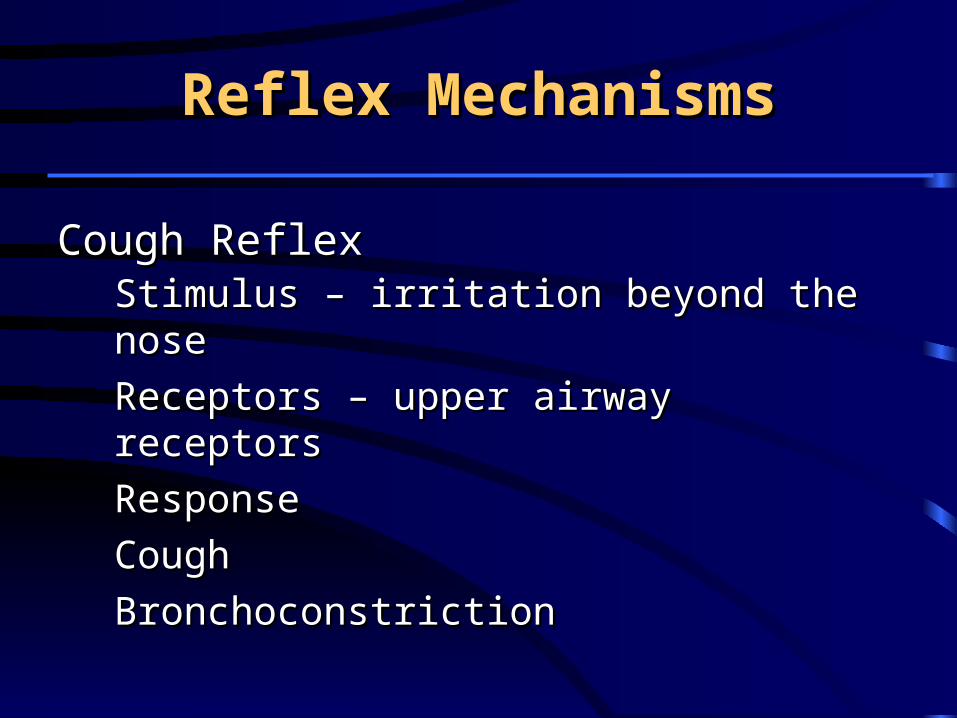
Stimulus – irritation beyond the noseStimulus – irritation beyond the nose
Receptors – upper airway receptorsReceptors – upper airway receptors
ResponseResponse
CoughCough
BronchoconstrictionBronchoconstriction
Reflex MechanismsReflex Mechanisms
Cough ReflexCough Reflex
Cough
Upper airway causes
Inspiration is absent
Lower airway causes
Small inspiration
Cough
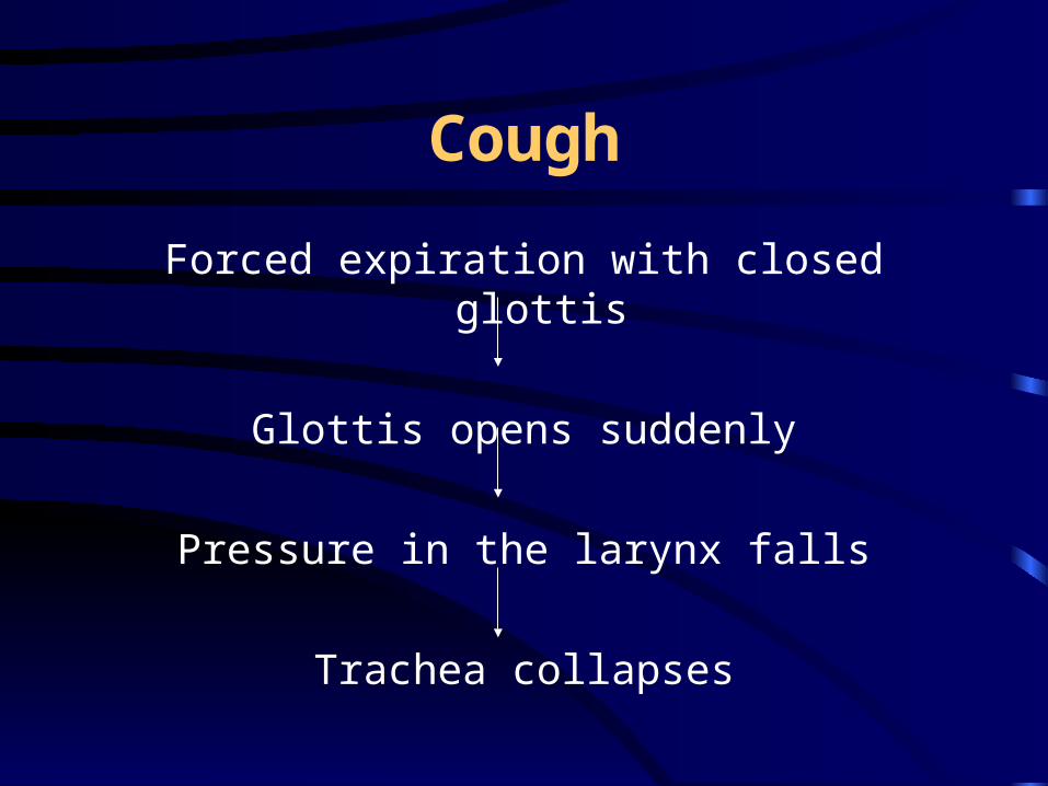
Forced expiration with closed glottis
Glottis opens suddenly
Pressure in the larynx falls
Trachea collapses

Cough
Brief but violent rush of air out of the trachea
Loosens mucus or foreign bodies and moves them upward
Stimulus – immersion of the face in waterStimulus – immersion of the face in water
Receptors – receptors in nasal mucosa Receptors – receptors in nasal mucosa and and faceface
ResponseResponse
ApneaApnea
Decreased heart rateDecreased heart rate
VasoconstrictionVasoconstriction
Reflex MechanismsReflex Mechanisms
Diving ReflexDiving Reflex

Stimulus – drugs in pulmonary Stimulus – drugs in pulmonary circulationcirculation
Receptors – J receptorsReceptors – J receptors
ResponseResponse
Apnea/TachypneaApnea/Tachypnea
BronchoconstrictionBronchoconstriction
Reflex MechanismsReflex Mechanisms
Pulmonary ChemoreflexPulmonary Chemoreflex
Stimulus – low Pa O2, low pHStimulus – low Pa O2, low pH
Receptors – chemoreceptorsReceptors – chemoreceptors
ResponseResponse
HyperpneaHyperpnea BronchoconstrictionBronchoconstriction
BradycardiaBradycardia VasoconstrictionVasoconstriction
Reflex MechanismsReflex Mechanisms
Arterial Chemoreceptor ReflexArterial Chemoreceptor Reflex

Stimulus – increased systemic BPStimulus – increased systemic BP
Receptors – baroreceptorsReceptors – baroreceptors
ResponseResponse
ApneaApnea BronchodilationBronchodilation
BradycardiaBradycardia VasodilationVasodilation
Reflex MechanismsReflex Mechanisms
Arterial Baroreceptor ReflexArterial Baroreceptor Reflex
StimulusStimulus
Stretch of muscle/tendonStretch of muscle/tendon
Movement of jointsMovement of joints
Receptors – muscle spindle, Golgi Receptors – muscle spindle, Golgi tendon organ, proprioceptorstendon organ, proprioceptors
Response - hyperpneaResponse - hyperpnea
Reflex MechanismsReflex Mechanisms
Reflexes From Muscles, Tendons, JointsReflexes From Muscles, Tendons, Joints

Stimulus – somatic painStimulus – somatic pain
Receptors – nociceptorsReceptors – nociceptors
ResponseResponse
HyperpneaHyperpnea
TachycardiaTachycardia
VasoconstrictionVasoconstriction
Reflex MechanismsReflex Mechanisms
Reflexes Due to PainReflexes Due to Pain
Central Chemoreceptors
Ventrolateral medullaStimulation leads to an increase in ventilation
Medullary rapheNeurons stimulated by acidosis contain
serotonin as transmitterNeurons inhibited by acidosis contain
GABA as transmitter

Central Chemoreceptors
Nucleus ambiguusNucleus tractus solitariusLocus ceruleusHypothalamus
Neurons are primarily sensitive to arterial hypercapnia, but actual parameter appears to be a low pH in or around the chemoreceptors

T H A N K Y O U !