Respiratory dickson cv presentation
59
THE PARTS AND FUNCTIONS OF HUMAN RESPIRATORY SYTEM
-
Upload
akankwatsa-cv-dickson -
Category
Health & Medicine
-
view
102 -
download
2
Transcript of Respiratory dickson cv presentation

THE PARTS AND FUNCTIONS OF
HUMAN RESPIRATORY SYTEM

* It is the system, consisting of tubes and is responsible for the exchange of gases in
Humans by filtering incoming air and transporting it into the microscopic
alveoli where gases are exchanged
THE HUMAN RESPIRATORY SYSTEM

THE HUMAN RESPIRATORY SYSTEM

The organs of the “Respiratory Tract”
can be divided into two groups“STRUCTURALLY”
** The Upper Respiratory Tract ** The Lower Respiratory Tract
* Nose
* Nasal cavity
* Sinuses
* Pharynx
* Larynx
* Trachea
* Bronchial Tree
* Lungs

THE HUMAN RESPIRATORY SYSTEM
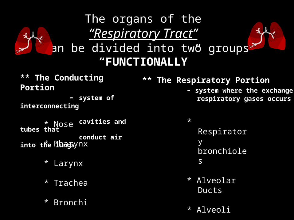
The organs of the “Respiratory Tract”
can be divided into two groups“FUNCTIONALLY”
** The Conducting Portion - system of interconnecting cavities and tubes that conduct air into the lungs
** The Respiratory Portion - system where the exchange of respiratory gases occurs
* Nose
* Pharynx
* Larynx
* Trachea
* Bronchi
* Respiratory bronchioles
* Alveolar Ducts
* Alveoli

THE HUMAN RESPIRATORY SYSTEMI. N O S E
A. N a s a l C a v i t y B. P a r a n a s a l S i n u s e s
II. P H A R Y N X
III. L A R Y N X A. E p I g i o t t i s B. V o c a l C o r d s
IV. T R A C H E A
v. B R O N C H I A. B r o n c h i a l T r e e
VI. L U N G S A. L o b e s o f t h e L u n g s B. P l e u r a l C a v i t i e s C. A l v e o l i

THE NOSE

* It provides an entrance for air in which air is
filtered by coarse hairs inside the nostrils.
* It has 2 portions : the external and internal
* The external portion is supported by a framework
of bone and cartilage covered with skin and lined with mucous membrane.
* The internal portion is a large cavity in the skull,
merging with the extrenal nose anteriorly and communicating with the throat posteriorly.
THE NOSE

The Nasal Cavity

* Interior area of the nose; lined with a sticky mucous membrane and contains tiny, surface hairs, cilia. divided medially by the nasal septum.
* Nasal conchae divide the cavity into passageways that are lined with mucous membrane, and help increase the surface area available to warm and filter incoming air.
•Particles trapped in the mucus are carried to the pharynx by ciliary action, swallowed, and carried to the stomach where gastric juice destroys any microorganisms in the mucus.
The Nasal Cavity

Paranasal Sinuses
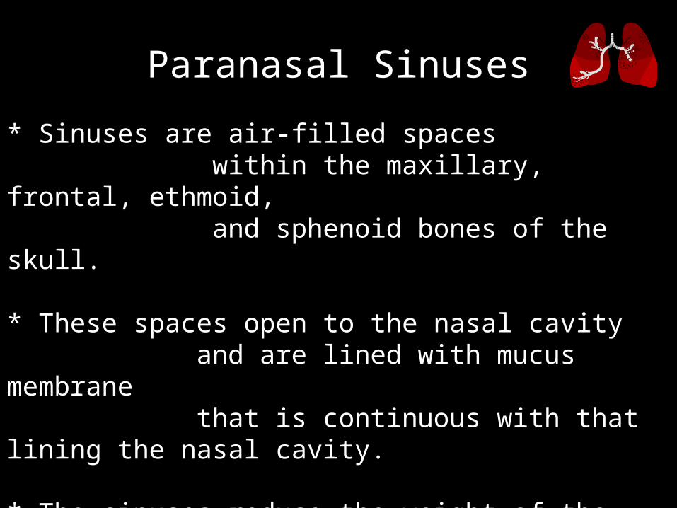
* Sinuses are air-filled spaces within the maxillary, frontal, ethmoid, and sphenoid bones of the skull.
* These spaces open to the nasal cavity and are lined with mucus membrane that is continuous with that lining the nasal cavity.
* The sinuses reduce the weight of the skull and serve as a resonant chamber to affect the quality of the voice.
Paranasal Sinuses
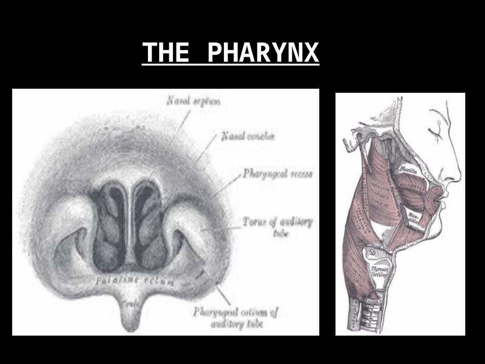
THE PHARYNX

* The “throat” is a funnel shaped tube that lies posterior to the nasal cavity, oral cavity and larynx; and anteriorly to the cervical vertebra.
* It is composed of: Nasopharynx – uppermost portion Oropharynx – middle portion Laryngopharynx – lowermost portion
* It is a common passageway for air and food and it provides a resonating chamber for speech sounds
THE PHARYNX

* It is an enlargement in the airway superior to the trachea and inferior to the pharynx.
* It helps keep particles from entering the trachea and also houses the vocal cords.
* It is composed of a framework of muscles and cartilage bound by elastic tissue
THE LARYNX

* It is a large leaf-shaped piece of cartilage.
* A flap of cartilage that prevents food from entering the trachea (or windpipe).
* During swallowing, there is elevation of the larynx
The Epiglottis

* Inside the larynx, 2 pairs of folds of muscle and connective tissues covered with mucous membrane make up the vocal cords.
a. The upper pair is the false vocal cords.
b. The lower pair is the true vocal cords.
c. Changing tension on the vocal cords controls pitch, while increasing the loudness depends upon increasing the force of air vibrating the vocal cords.
The Vocal Cords

* During normal breathing,
the vocal cords are relaxed and the glottis is a triangular slit. * During swallowing,
the false vocal cords and epiglottis close off the glottis.
The Vocal Cords

THE TRACHEA
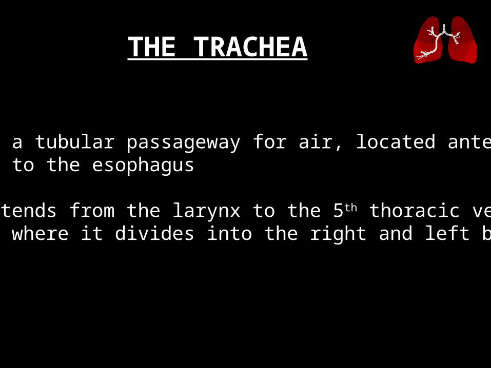
* It is a tubular passageway for air, located anterior to the esophagus
* It extends from the larynx to the 5th thoracic vertebra where it divides into the right and left bronchi.
THE TRACHEA

THE TRACHEA

* The inner wall of the trachea is lined with ciliated mucous membrane with many goblet cells that serve to trap incoming particles.
* The tracheal wall is supported by 20 incomplete cartilaginous rings.
THE TRACHEA
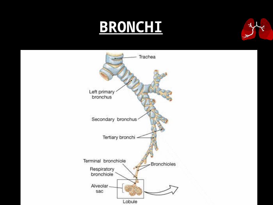
BRONCHI

* The Bronchi are the two main air passages into the lungs.
* They are composed of the:
** “Right Primary Bronchus” - leading to the right lung.
** “Left Primary Bronchus” - leading to the left lung.
BRONCHI

The Bronchial Tree

* The bronchial tree consists of branched tubes leading from the trachea to the alveoli.
* The bronchial tree begins with the two primary bronchi, each leading to a lung.
* The branches of the bronchial tree from the trachea are right and left primary bronchi; these further subdivide until bronchioles give rise to alveolar ducts which terminate in alveoli.
* It is through the thin epithelial cells of the alveoli that gas exchange between the blood and air occurs.
The Bronchial Tree
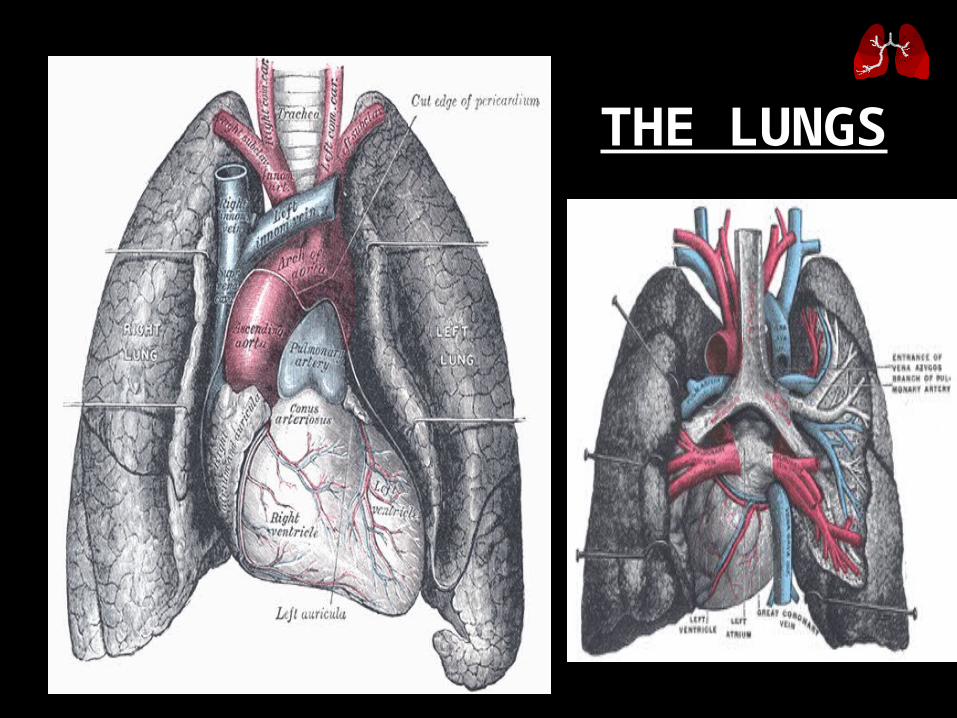
THE LUNGS

•The paired soft, spongy, cone-shaped lungs, separated medially by the mediastinum and are enclosed by the diaphragm and thoracic cage.
•2 layers of serous membrane, collectively known as pleural membrane, enclose and protect each lung.
** Parietal Pleura - outer layer attached to the thoracic cavity ** Visceral Pleura - inner layer covering the lung itself
THE LUNGS
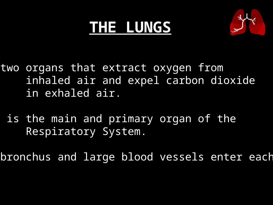
* The two organs that extract oxygen from inhaled air and expel carbon dioxide in exhaled air.
* This is the main and primary organ of the Respiratory System.
* The bronchus and large blood vessels enter each lung.
THE LUNGS

Lobes of the Lungs

* The right lung has three lobes. * The left lung has two lobes.
* Each lobe is composed of lobules
that contain air passages, alveoli, nerves,
blood vessels, lymphatic vessels,
and connective tissues.
Lobes of the Lungs
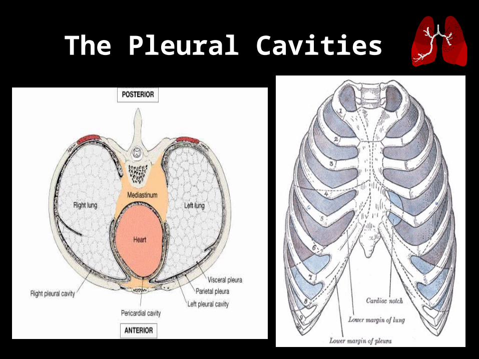
The Pleural Cavities

* A layer of serous membrane, between the visceral pleura and the parietal pleura.
* It contains a lubricating fluid secreted by the membranes that prevents friction between the membranes and allows their easy movement on one another during breathing.
The Pleural Cavities

The Alveoli

* They are cup-shaped out pouching lined by epithelium and supported by a thin elastic basement membrane.
•With that you can imagine having bunch of grapes with each grape indicating and alveolus.
* Alveolar sacs are 2 or more alveoli that share a common opening.
* This is where the primary exchange of gases occur.
The Alveoli

Physiology of respiratory system
The noseThe major functions of nasal breathing are:
to heat and moisten the air to remove particulate matter.
About 10 000 L of air are inhaled daily. The relatively low flow rates and turbulence of inspired air are ideal for
particle deposition, and few particles greater than 10 microns pass through the nose. Deposited particles are
removed from the nasal mucosa within 15 minutes, compared with 60-120 days from the alveolus.

Cont…
• Nasal secretion contains many protective proteins in the form of IgA antibodies, lysozyme and interferon. In addition, the cilia of the nasal epithelium move the mucous gel layer rapidly back to the oropharynx where it is swallowed.

Breathing• Lung ventilation can be considered in two
parts:the mechanical process of inspiration and
expirationthe control of respiration to a level
appropriate for themetabolic needs.
Mechanical process
• Inspiration is an active process and results from the descent of the diaphragm and movement of the ribs upwards and outwards under the influence of the inter costal muscles.
• In healthy individuals at rest, inspiration is almost entirely due to contraction of the diaphragm. Respiratory muscles are similar to other skeletal muscles but are less prone to fatigue

Cont…
• Expiration follows passively as a result of gradual relaxation of the intercostal muscles, allowing the lungs to collapse under the influence of their own elastic forces.
• Inspiration against increased resistance may require the use of the accessory muscles of ventilation, such as the sternomastoid and scalene muscles.

Forced expiration is also accomplished with the aid of accessory muscles, chiefly those of the abdominal wall, which
help to push up the diaphragm.
The lungs have an inherent elastic property that causes them to tend to collapse away from the thoracic wall, generating a
negative pressure within the pleural space.
The strength of this retractive force relates to the volume of the lung; thus, at higher lung volumes the lung is stretched more,
and a greater negative intrapleural press ure is generated.

• Lung compliance is a measure of the relationship between this retractive force and lung volume.
• It is defined as the change in lung volume brought about by unit change in transpulmonary (intrapleural) pressure and is measured in litres per kilopascal (L/kPa). At the end of a quiet expiration, the retractive force exerted by the lungs is balanced by the tendency of the thoracic wall to spring outwards
Cont…
• At this point, respiratory muscles are resting and the volume of air in the lung is known as the functional residual capacity (FRC).

The control of respiration
• Coordinated respiratory movements result from rhyth mical discharges arising in an anatomically ill-defined group of interconnected neurones in the reticular sub stance of the brainstem, known as the respiratory centre.
• Motor discharges from the respiratory centre travel via the phrenic and intercostal nerves to the respiratory musculature.

Cont…• The pressures of oxygen and carbon dioxide in arterial
blood are closely controlled. In a typical normal adult at rest:
• The pulmonary blood flow of 5 L/min carries11 mmol/min (250 mL/min) of oxygen from the lungsto the tissues.
• Ventilation at about 6 L/min carries 9 mmol/min(200 mL/min) of carbon dioxide out of the body.

Cont…
• The normal pressure of oxygen in arterial blood (Pao2) is between 11 and 13 kPa (83 and 98 mmHg).
• ■ The normal pressure of carbon dioxide in arterialblood (Paco2) is 4.8-6.0 kPa (36-45 mmHg).

Airflow• Movement of air through the airways
results from a difference between the pressure in the alveoli and the atmospheric pressure; alveolar pressure is positive in expiration and negative in inspiration.
• During quiet breathing the pleural pressure is sub-atmospheric throughout the breathing cycle.

Cont…
• With vigorous expiratory efforts (e.g. cough), the central airways are compressed by positive pleural pressures exceeding 10 kPa, but the airways do not close completely because the driving pressure for expiratory flow (alveolar pressure) is also increased.
• Alveolar pressure (PALV) is equal to the pleural pressure (Ppi) plus the elastic recoil pressure (PEi) of the lung.

Cont…
• When there is no airflow (i.e. during a pause in breathing) the tendency of the lungs to collapse (the positive recoil pressure) is exactly balanced by an equivalent negative pleural pressure.
• As air flows from the alveoli towards the mouth there is a gradual loss of pressure owing to flow resistance

The airways of the lungs
•• From the trachea to the periphery, the
airways become smaller in size (although greater in number). The cross-sectional area available for airflow increases as the total number of airways increases.
Cont…
• The flow of air is greatest in the trachea and slows progressively towards the periphery (as the velocity of airflow depends on the ratio of flow to cross-sectional area). In the terminal airways, gas flow occurs solely by diffusion. The resistance to airflow is very low (0.1-0.2 kPa/L in a normal tracheobronchial tree), steadily increasing from the small to the large airways.

Cont…
• Airways expand as lung volume is increased, and at full inspiration (total lung capacity, TLC) they are 30-40% larger in calibre than at full expiration (residual volume, RV). In chronic obstructive pulmonary disease (COPD) the small airways are narrowed and this can be partially compensated by breathing at a larger lung volume.

• In forced expiration, the driving pressure raises both the alveolar pressure and the intrapleural pressure. Between the alveolus and the mouth, there is a point where the airway pressure equals the intrapleural pressure, and the airway collapses.
• However, this collapse is temporary, as the transient occlu sion of the airway results in an increase in pressure behind it (i.e. upstream) and this raises the intra-airway pressure so that the airways open and flow is restored.
• The airways thus tend to vibrate at this point of 'dynamic collapse'.
Cont…
• The elastic recoil pressure of the lungs decreases with decreasing lung volume and the 'collapse point' moves upstream (i.e. towards the smaller airways .
• Where there is pathological loss of recoil pressure (as in chronic obstructive pulmonary disease, COPD), the 'collapse point' starts even further upstream and causes expiratory flow limitation.
• The measurement of the forced expiratory volume in 1 second (FEVj) is a useful clinical index of this phenomenon.

Cont…
• To compensate, these patients often 'purse their lips' in order to increase airway pressure so that their peripheral airways do not collapse.
• On inspiration, the intrapleural pressure is always less than the intraluminal pressure within the intrathoracic airways, so there is no limitation to airflow with increasing effort. Inspiratory flow is limited only by the power of the inspiratory muscles.
Cont…• The measure of the volume that can be forced in from
the residual volume in 1 second (FIVj) will always be greater than that which can be forced out from TLC in 1 second (FEV,). Thus, the ratio of FEVj to FIV] is below 1. The only exception to this occurs when there is significant obstruction to the airways outside the thorax, such as with a tumour mass in the upper part of the trachea.
• Under these circumstances expiratory airway narrowing is prevented by the tracheal resistance (a situation similar to pursing the lips) and expiratory airflow becomes more effort-dependen
Cont…
• During forced inspiration this same resistance causes such negative intraluminal pressure that the trachea is compressed by the surrounding atmospheric pressure. Inspiratory flow thus becomes less effort-dependent, and the ratio of FEVj to FIVj becomes greater than 1. This phenomenon, and the characteristic flow-volume loop, is used to diagnose extrathoracic airways obstruction.

THANK YOU