Resolvin E1 attenuates atherosclerosis in absence of ...mice. Briefly, pharmacokinetic analysis of...
8
Resolvin E1 attenuates atherosclerosis in absence of cholesterol- lowering effects and on top of atorvastatin Kanita Salic a, * , Martine C. Morrison a , Lars Verschuren b , Peter Y. Wielinga a , Lijun Wu c, 1 , Robert Kleemann a , Per Gjorstrup c, ** , Teake Kooistra a a Department of Metabolic Health Research, Netherlands Organisation for Applied Scientific Research (TNO), Zernikedreef 9, 2333 CK, Leiden, The Netherlands b Department of Microbiology and Systems Biology, Netherlands Organisation for Applied Scientific Research (TNO), Utrechtseweg 48, 3704 HE, Zeist, The Netherlands c Resolvyx Pharmaceuticals, Inc., 222 Third Street, Cambridge, MA, 02142, United States article info Article history: Received 25 November 2015 Received in revised form 26 April 2016 Accepted 1 May 2016 Available online 2 May 2016 Keywords: Resolvin E1 Atherosclerosis Inflammation Resolution Statin abstract Background and aims: Besides LDL-cholesterol, local vascular inflammation plays a key role in athero- genesis. Efficient therapies to treat the inflammatory component of the disease have not been estab- lished. The discovery of specialized inflammation-resolving mediators, such as resolvins may provide new opportunities for treatment. This study examines whether the u-3 fatty acid eicosapentaenoic acid- derived resolvin E1 (RvE1), can reduce atherosclerosis, when administered alone or in combination with a cholesterol-lowering statin. Methods: ApoE*3Leiden mice were fed a hypercholesterolemic diet for 9 weeks and subsequently treated with RvE1-low (1 mg/kg/day), RvE1-high (5 mg/kg/day), atorvastatin (1.5 mg/kg/day) or the combination of atorvastatin and RvE1-low for the following 16 weeks. Results: RvE1-low and RvE1-high reduced atherosclerotic lesion size to the same extent (35%; p < 0.05), attenuated the formation of severe lesions, also seen as a proportional increase in the presence of mild lesions, but did not alter plasma cholesterol levels. Cholesterol-lowering atorvastatin reduced athero- sclerosis (27%, p < 0.05), and the combination of RvE1 and atorvastatin further attenuated lesion size (51%, p < 0.01) and increased the content of mild lesions. RvE1 did not affect plasma SAA, E-selectin, VCAM-1 or MCP-1 but did reduce plasma EPHX4 and down-regulated the local expression of pro- atherogenic genes in the aortae, (e.g. Cd74, Cd44, Ccl2, Ccr5 and Adam17) and significantly inactivated IFN-g (p < 0.001) and TNF-a (p < 0.001) signalling pathways. Conclusions: RvE1 attenuates atherogenesis both alone and on top of a statin. The local effects of RvE1 are demonstrated by the modulated aortic expression of genes involved in inflammatory and immune responses, without altering plasma cholesterol or circulating SAA. © 2016 The Authors. Published by Elsevier Ireland Ltd. This is an open access article under the CC BY-NC- ND license (http://creativecommons.org/licenses/by-nc-nd/4.0/). 1. Introduction Atherosclerotic plaque buildup is closely linked to an increased exposure to low-density lipoprotein (LDL) cholesterol, which has been the rationale for treatment with LDL-lowering statins over the last several decades. While the outcome of statin treatment un- deniably is a reduction in cardiovascular disease (CVD) mortality, this still remains a major cause of death worldwide [1]. However, elevated LDL levels may be more important in the induction of disease, while plaque progression and later potential rupture is caused by vascular inflammation rather than high LDL levels per se. Intimal retention of LDL and its subsequent modification and oxidation provide a chronic trigger of vascular inflammation. This initially involves the innate immune system, with monocyte recruitment and macrophage and dendritic cell activation, which initiates later adaptive immune responses [2,3]. Due to the * Corresponding author. TNO Metabolic Health Research, Zernikedreef 9, 2333CK Leiden, The Netherlands. ** Corresponding author. Present address: Anida Pharma Inc., 155 Brookline Street, Cambridge, MA 02139, United States. E-mail addresses: [email protected] (K. Salic), [email protected] (P. Gjorstrup). 1 Present address: Concert Pharmaceuticals Inc., 99 Hayden Avenue Lexington, MA 02421 United States. Contents lists available at ScienceDirect Atherosclerosis journal homepage: www.elsevier.com/locate/atherosclerosis http://dx.doi.org/10.1016/j.atherosclerosis.2016.05.001 0021-9150/© 2016 The Authors. Published by Elsevier IrelandLtd. This is an open access article under the CC BY-NC-ND license (http://creativecommons.org/licenses/by-nc- nd/4.0/). Atherosclerosis 250 (2016) 158e165
Transcript of Resolvin E1 attenuates atherosclerosis in absence of ...mice. Briefly, pharmacokinetic analysis of...

lable at ScienceDirect
Atherosclerosis 250 (2016) 158e165
Contents lists avai
Atherosclerosis
journal homepage: www.elsevier .com/locate/atherosclerosis
Resolvin E1 attenuates atherosclerosis in absence of cholesterol-lowering effects and on top of atorvastatin
Kanita Salic a, *, Martine C. Morrison a, Lars Verschuren b, Peter Y. Wielinga a, Lijun Wu c, 1,Robert Kleemann a, Per Gjorstrup c, **, Teake Kooistra a
a Department of Metabolic Health Research, Netherlands Organisation for Applied Scientific Research (TNO), Zernikedreef 9, 2333 CK, Leiden, TheNetherlandsb Department of Microbiology and Systems Biology, Netherlands Organisation for Applied Scientific Research (TNO), Utrechtseweg 48, 3704 HE, Zeist, TheNetherlandsc Resolvyx Pharmaceuticals, Inc., 222 Third Street, Cambridge, MA, 02142, United States
a r t i c l e i n f o
Article history:Received 25 November 2015Received in revised form26 April 2016Accepted 1 May 2016Available online 2 May 2016
Keywords:Resolvin E1AtherosclerosisInflammationResolutionStatin
* Corresponding author. TNO Metabolic Health ReseLeiden, The Netherlands.** Corresponding author. Present address: AnidaStreet, Cambridge, MA 02139, United States.
E-mail addresses: [email protected] (K. Salic), p(P. Gjorstrup).
1 Present address: Concert Pharmaceuticals Inc., 9MA 02421 United States.
http://dx.doi.org/10.1016/j.atherosclerosis.2016.05.0010021-9150/© 2016 The Authors. Published by Elseviernd/4.0/).
a b s t r a c t
Background and aims: Besides LDL-cholesterol, local vascular inflammation plays a key role in athero-genesis. Efficient therapies to treat the inflammatory component of the disease have not been estab-lished. The discovery of specialized inflammation-resolving mediators, such as resolvins may providenew opportunities for treatment. This study examines whether the u-3 fatty acid eicosapentaenoic acid-derived resolvin E1 (RvE1), can reduce atherosclerosis, when administered alone or in combination witha cholesterol-lowering statin.Methods: ApoE*3Leiden mice were fed a hypercholesterolemic diet for 9 weeks and subsequently treatedwith RvE1-low (1 mg/kg/day), RvE1-high (5 mg/kg/day), atorvastatin (1.5 mg/kg/day) or the combinationof atorvastatin and RvE1-low for the following 16 weeks.Results: RvE1-low and RvE1-high reduced atherosclerotic lesion size to the same extent (�35%; p < 0.05),attenuated the formation of severe lesions, also seen as a proportional increase in the presence of mildlesions, but did not alter plasma cholesterol levels. Cholesterol-lowering atorvastatin reduced athero-sclerosis (�27%, p < 0.05), and the combination of RvE1 and atorvastatin further attenuated lesion size(�51%, p < 0.01) and increased the content of mild lesions. RvE1 did not affect plasma SAA, E-selectin,VCAM-1 or MCP-1 but did reduce plasma EPHX4 and down-regulated the local expression of pro-atherogenic genes in the aortae, (e.g. Cd74, Cd44, Ccl2, Ccr5 and Adam17) and significantly inactivatedIFN-g (p < 0.001) and TNF-a (p < 0.001) signalling pathways.Conclusions: RvE1 attenuates atherogenesis both alone and on top of a statin. The local effects of RvE1are demonstrated by the modulated aortic expression of genes involved in inflammatory and immuneresponses, without altering plasma cholesterol or circulating SAA.© 2016 The Authors. Published by Elsevier Ireland Ltd. This is an open access article under the CC BY-NC-
ND license (http://creativecommons.org/licenses/by-nc-nd/4.0/).
1. Introduction
Atherosclerotic plaque buildup is closely linked to an increasedexposure to low-density lipoprotein (LDL) cholesterol, which has
arch, Zernikedreef 9, 2333CK
Pharma Inc., 155 Brookline
9 Hayden Avenue Lexington,
Ireland Ltd. This is an open access
been the rationale for treatment with LDL-lowering statins over thelast several decades. While the outcome of statin treatment un-deniably is a reduction in cardiovascular disease (CVD) mortality,this still remains a major cause of death worldwide [1]. However,elevated LDL levels may be more important in the induction ofdisease, while plaque progression and later potential rupture iscaused by vascular inflammation rather than high LDL levels per se.Intimal retention of LDL and its subsequent modification andoxidation provide a chronic trigger of vascular inflammation. Thisinitially involves the innate immune system, with monocyterecruitment and macrophage and dendritic cell activation, whichinitiates later adaptive immune responses [2,3]. Due to the
article under the CC BY-NC-ND license (http://creativecommons.org/licenses/by-nc-
K. Salic et al. / Atherosclerosis 250 (2016) 158e165 159
complexity of these local inflammatory events in CVD develop-ment, it is challenging to single out one pathway as a therapeutictarget, when considering an immune-based approach. Statins, inaddition to their LDL-lowering effects, have specific anti-inflammatory properties in the vasculature [4] but may not offersufficient control [5], nor have other attempts at treating athero-sclerosis with anti-inflammatory drugs been successful [6]. A majorcontributing factor to plaque build-up and instability is thought tobe compromised clearance mechanisms at the plaque level,possibly suggesting a local imbalance between pro-inflammatoryevents and counter-acting resolution mechanisms of the immunesystem [7] suggesting that resolution may be dysfunctional.
The discovery of specialized pro-resolving mediators (SPMs)may represent a therapeutic alternative to classic anti-inflammatory drugs by harnessing the body’s own systems toregulate inflammation and promote homeostasis, including acti-vation of endogenous clearance mechanisms [8]. Originally iden-tified as oxygenation products of omega-3 fatty acids in resolvingexudates of acute inflammation [9], these lipid-derived mediatorswith agonistic properties, such as resolvins, protectins, and mar-esins, are now established regulators of both acute and chronicinflammatory responses [10]. In a dual action these mediatorsdampen active pro-inflammatory pathways and concurrently acti-vate pro-resolution functions such as phagocytic clearance andtissue repair mechanisms. Uniquely, they appear to do so withoutcompromising host immune defense [10].
The ApoE*3Leiden transgenic mouse has a humanized lipopro-tein profile with elevated plasma cholesterol levels confinedmainlyto the VLDL/LDL-sized lipoprotein fractions [11]. The model is well-established to quantify the build-up of lesions and their potentialamelioration by drug intervention, and is well-characterized forstatins [12]. Using this model, we herein investigated the efficacy ofresolvin E1 (RvE1), an endogenous SPM derived from eicosa-pentaenoic acid, in attenuating atherosclerotic lesion development.A putative anti-atherogenic effect of RvE1was studied at two doses,and in combination with atorvastatin. A reference group wastreated with atorvastatin alone.
2. Materials and methods
2.1. Ethics statement
Experiments were approved by an independent Ethical Com-mittee on Animal Care and Experimentation (Zeist, theNetherlands; approval number DEC2680) and were performed incompliance with the European Commission Directive on the use ofanimals for scientific purposes.
2.2. Animals and treatments
Eighty female ApoE*3Leiden transgenic mice were fed anatherogenic Western-type diet (HC) for a nine-week run-in period,starting at 12e16 weeks of age. This diet contains 40.5% sucrose,20% acid casein, 15% cocoa butter, 10% corn starch, 5.45% cellulose,5.1% mineral mixture, 1% choline chloride, 1% corn oil, 0.2%methionine and 0.4% cholesterol (all w/w) (AB-diets, Woerden, theNetherlands). After the run-in period, in which plasma cholesterolincreased from 2.8 ± 0.4 mM to 17.5 ± 2.5 mM as expected forApoE*3Leiden transgenic mice, mice were divided into 5 treatmentgroups that were matched for total plasma cholesterol (n ¼ 16/group) continuing on HC with 1) vehicle control, 2) 1.5 mg/kg/dayatorvastatin, 3) 1 mg/kg/day RvE1 (RvE1-low), 4) 5 mg/kg/day RvE1(RvE1-high), 5) or a combination of atorvastatin 1.5 mg/kg/day andRvE11mg/kg/day (combination group), for the following 16 weeks.Atorvastatin (Lipitor, Pfizer, Capelle a/d IJssel, the Netherlands) was
supplemented to the diet (0.0015% w/w), and RvE1 and vehiclewere administered daily by oral gavage between 9 and 10 a.m.Resolvin E1 (RvE1; RX-10001) was provided by Resolvyx Pharma-ceuticals Inc. (Cambridge, MA, USA). The dose RvE1 used in thisstudy was defined based on previous experiments in humans andmice. Briefly, pharmacokinetic analysis of a single dose, 10 mg, 14C-labeled RvE1 (approximately 0.15 mg/kg) in a Phase 1 clinical studyrevealed a half-life of about 7 h (Resolvyx unpublished; trialnumber NCT00941018). Furthermore, administration of repeateddoses of 100 mg and higher in the same study were well-toleratedand without safety-related issues. In mice, 14C-labeled RvE1 at1 mg/kg resulted in plasma concentrations greater than 3 nM,which is the EC50 defined in in vitro experiments. These levels(>3 nM) were maintained for a period of 8 h post-administration.(Resolvyx unpublished).
All animals had free access to water and food. Body weight andfood intake were monitored throughout the study. Animals weresacrificed by CO2 asphyxiation after 16 weeks of treatment (at37e41 weeks of age) to collect hearts including the aortic rootswhich were fixed in formalin and embedded in paraffin foratherosclerosis analysis.
Prior to the main study described above, a pilot experiment wasperformed using the same experimental conditions but with 9weeks of RvE1 intervention. This experiment served to investigatewhether a dose of 1mg/kg is efficacious in a setting of experimentaldiet-induced atherosclerosis and to establish a microarray geneexpression dataset of aortae.
2.3. Biochemical, histological and microarray gene expressionanalyses
A detailed description of biochemical, histological and aorticmicroarray analyses is provided in Supplemental methods. In brief,plasma parameters (total cholesterol, serum amyloid A, E-selectinand alanine aminotransferase) were quantified using commerciallyavailable assays as described previously [13,14]. Atherosclerosiswas scored blindly in 4 serial cross-sections (at 50 mm intervals) ofthe aortic root. Morphometric analysis of lesion number and areawas performed using cell̂ D software (version 2.7; Olympus SoftImaging Solutions, Hamburg, Germany), and lesion severity wasscored according to the established classification of the AmericanHeart Association [15,16]. Aortic genome-wide gene expressionanalyses were performed using Illumina microarray analysis fol-lowed by established normalisation and quality control protocolsand pathway analysis as described [17,18].
2.4. Statistical analysis
Significance of differences was tested using one-way ANOVAwith LSD post-hoc test. Statistical tests were performed using SPSSsoftware (version 20, IBM, Armonk USA). A p-value <0.05 wasconsidered statistically significant. All data are presented asmean ± SEM.
3. Results
3.1. RvE1 treatment reduces atherosclerotic lesion area
ApoE*3Leiden transgenic mice were fed the HC diet for 9 weeksto induce atherosclerotic lesions, after which treatment with RvE1was started and continued for another 16weeks until the end of thestudy. Analysis of atherosclerotic lesions in the valve area of theaortic root (Fig. 1AeE) revealed a clear treatment effect of RvE1relative to vehicle in which the total lesion area was163,200 ± 17,410 mm2. Both RvE1-low (105,300 ± 12,200 mm2) and

Fig. 1. Treatment with RvE1 reduces atherosclerotic lesion area. (A) Representative images of haematoxylin-phloxine-saffron stained cross-sections of the aortic root after 16weeks of treatment with vehicle, (B) RvE1-low 1 mg/kg/day, (C) RvE1-high 5 mg/kg/day, (D) atorvastatin 1.5 mg/kg/day, (E) combination of atorvastatin 1.5 mg/kg/day and RvE11 mg/kg/day. (F)Quantification of total atherosclerotic lesion area in serial cross-sections of the aortic root after 16 weeks of treatment. Results are shown as mean ± SEM(n ¼ 15e16/group). *p < 0.05, **p < 0.01 vs. the vehicle group.
K. Salic et al. / Atherosclerosis 250 (2016) 158e165160
RvE1-high (103,700 ± 17,640 mm2) significantly attenuated lesionarea by around 35% compared with vehicle control (both p < 0.05vs. vehicle) (Fig. 1F). The reference compound atorvastatin(119,400 ± 16,410 mm2) reduced atherosclerosis to a comparableextent as RvE1 (27% reduction; p < 0.05 vs. vehicle). Notably, thecombination treatment had the strongest effect on atherosclerosis(82,300 ± 1522 mm2) equaling a 51% reduction (p < 0.001 vs.vehicle). The lesion area of this group tended to be 31% smaller thanwith atorvastatin (p ¼ 0.11 vs. atorvastatin).
In ApoE*3Leiden transgenic mice, the reduction in lesion areaupon statin treatment is closely linked to the cholesterol-loweringeffects of these drugs [11,12]. In the current study, atorvastatinreduced plasma cholesterol levels from 17.5 ± 0.3 mM before thestart of treatment to 12.0 ± 0.4 mM at endpoint (18% reduction vs.vehicle; p < 0.001) (Fig. 2A). RvE1 had no effect on plasmacholesterol levels at either dose, nor did RvE1 affect the lipoproteinprofile or HDL cholesterol levels (data not shown). Combinedtreatment with RvE1 and atorvastatin reduced cholesterol levels to
the same degree as atorvastatin alone (18% reduction at endpoint;p < 0.01 vs. vehicle) (Fig. 2A). Atorvastatin and the combinationtreatment reduced the total cholesterol exposure, calculated overthe entire treatment period, by 21% and 17% (both p < 0.001 vs.vehicle) respectively (Fig. 2B), while cholesterol exposure was notaffected by RvE1.
3.2. RvE1 treatment improves lesion severity
To further elucidate how RvE1 interferes with the progression ofatherosclerosis, aortic segments were analyzed for presence orabsence of lesions and the number of lesion-free segments wasdetermined (Fig. 3A). When lesions were present, each individuallesion was graded to determine its severity according to the clas-sification system developed by the American Heart Association[15,16] (Fig. 3B).
In the vehicle group, 8 ± 3% of all aortic segments were free oflesions (Fig. 3A). A higher content of lesion-free segments was
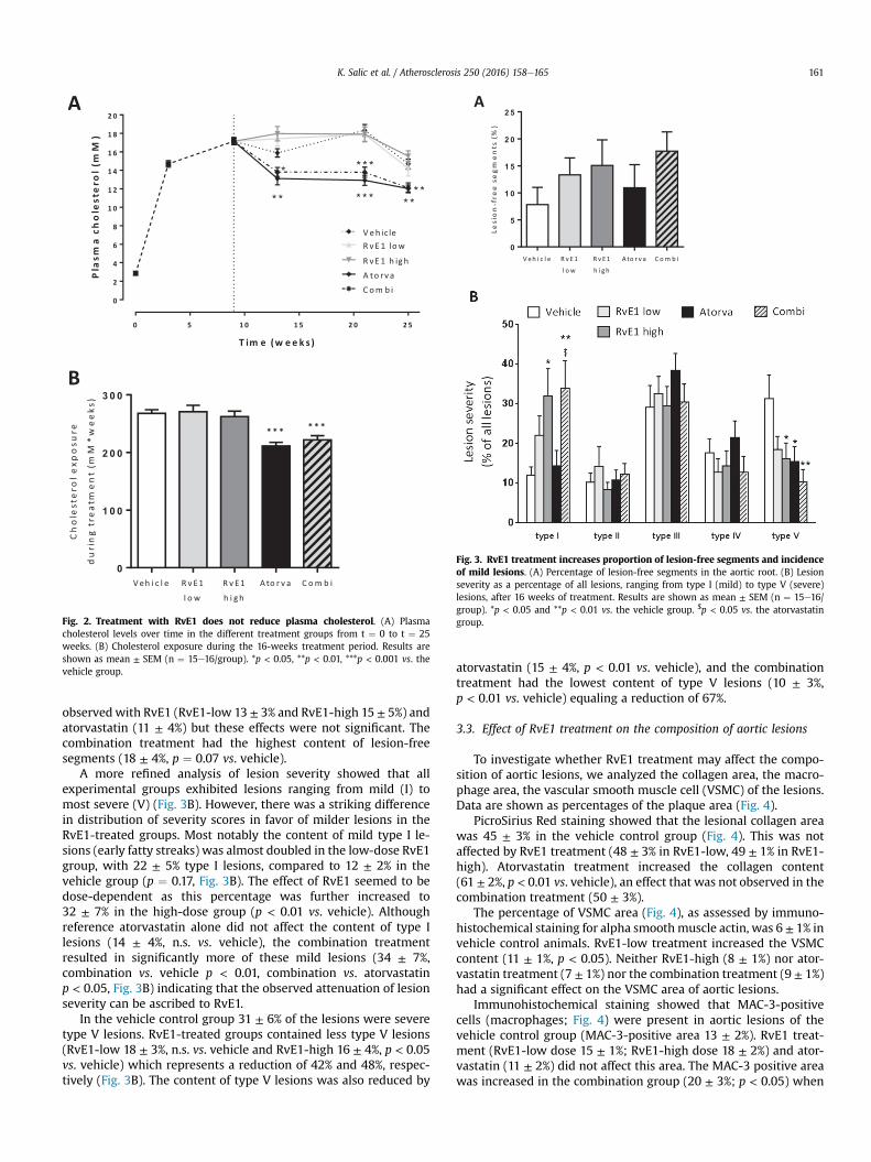
Fig. 2. Treatment with RvE1 does not reduce plasma cholesterol. (A) Plasmacholesterol levels over time in the different treatment groups from t ¼ 0 to t ¼ 25weeks. (B) Cholesterol exposure during the 16-weeks treatment period. Results areshown as mean ± SEM (n ¼ 15e16/group). *p < 0.05, **p < 0.01, ***p < 0.001 vs. thevehicle group.
Fig. 3. RvE1 treatment increases proportion of lesion-free segments and incidenceof mild lesions. (A) Percentage of lesion-free segments in the aortic root. (B) Lesionseverity as a percentage of all lesions, ranging from type I (mild) to type V (severe)lesions, after 16 weeks of treatment. Results are shown as mean ± SEM (n ¼ 15e16/group). *p < 0.05 and **p < 0.01 vs. the vehicle group. $p < 0.05 vs. the atorvastatingroup.
K. Salic et al. / Atherosclerosis 250 (2016) 158e165 161
observedwith RvE1 (RvE1-low 13 ± 3% and RvE1-high 15 ± 5%) andatorvastatin (11 ± 4%) but these effects were not significant. Thecombination treatment had the highest content of lesion-freesegments (18 ± 4%, p ¼ 0.07 vs. vehicle).
A more refined analysis of lesion severity showed that allexperimental groups exhibited lesions ranging from mild (I) tomost severe (V) (Fig. 3B). However, there was a striking differencein distribution of severity scores in favor of milder lesions in theRvE1-treated groups. Most notably the content of mild type I le-sions (early fatty streaks) was almost doubled in the low-dose RvE1group, with 22 ± 5% type I lesions, compared to 12 ± 2% in thevehicle group (p ¼ 0.17, Fig. 3B). The effect of RvE1 seemed to bedose-dependent as this percentage was further increased to32 ± 7% in the high-dose group (p < 0.01 vs. vehicle). Althoughreference atorvastatin alone did not affect the content of type Ilesions (14 ± 4%, n.s. vs. vehicle), the combination treatmentresulted in significantly more of these mild lesions (34 ± 7%,combination vs. vehicle p < 0.01, combination vs. atorvastatinp < 0.05, Fig. 3B) indicating that the observed attenuation of lesionseverity can be ascribed to RvE1.
In the vehicle control group 31 ± 6% of the lesions were severetype V lesions. RvE1-treated groups contained less type V lesions(RvE1-low 18 ± 3%, n.s. vs. vehicle and RvE1-high 16 ± 4%, p < 0.05vs. vehicle) which represents a reduction of 42% and 48%, respec-tively (Fig. 3B). The content of type V lesions was also reduced by
atorvastatin (15 ± 4%, p < 0.01 vs. vehicle), and the combinationtreatment had the lowest content of type V lesions (10 ± 3%,p < 0.01 vs. vehicle) equaling a reduction of 67%.
3.3. Effect of RvE1 treatment on the composition of aortic lesions
To investigate whether RvE1 treatment may affect the compo-sition of aortic lesions, we analyzed the collagen area, the macro-phage area, the vascular smooth muscle cell (VSMC) of the lesions.Data are shown as percentages of the plaque area (Fig. 4).
PicroSirius Red staining showed that the lesional collagen areawas 45 ± 3% in the vehicle control group (Fig. 4). This was notaffected by RvE1 treatment (48 ± 3% in RvE1-low, 49 ± 1% in RvE1-high). Atorvastatin treatment increased the collagen content(61 ± 2%, p < 0.01 vs. vehicle), an effect that was not observed in thecombination treatment (50 ± 3%).
The percentage of VSMC area (Fig. 4), as assessed by immuno-histochemical staining for alpha smoothmuscle actin, was 6 ± 1% invehicle control animals. RvE1-low treatment increased the VSMCcontent (11 ± 1%, p < 0.05). Neither RvE1-high (8 ± 1%) nor ator-vastatin treatment (7 ± 1%) nor the combination treatment (9 ± 1%)had a significant effect on the VSMC area of aortic lesions.
Immunohistochemical staining showed that MAC-3-positivecells (macrophages; Fig. 4) were present in aortic lesions of thevehicle control group (MAC-3-positive area 13 ± 2%). RvE1 treat-ment (RvE1-low dose 15 ± 1%; RvE1-high dose 18 ± 2%) and ator-vastatin (11 ± 2%) did not affect this area. The MAC-3 positive areawas increased in the combination group (20 ± 3%; p < 0.05) when
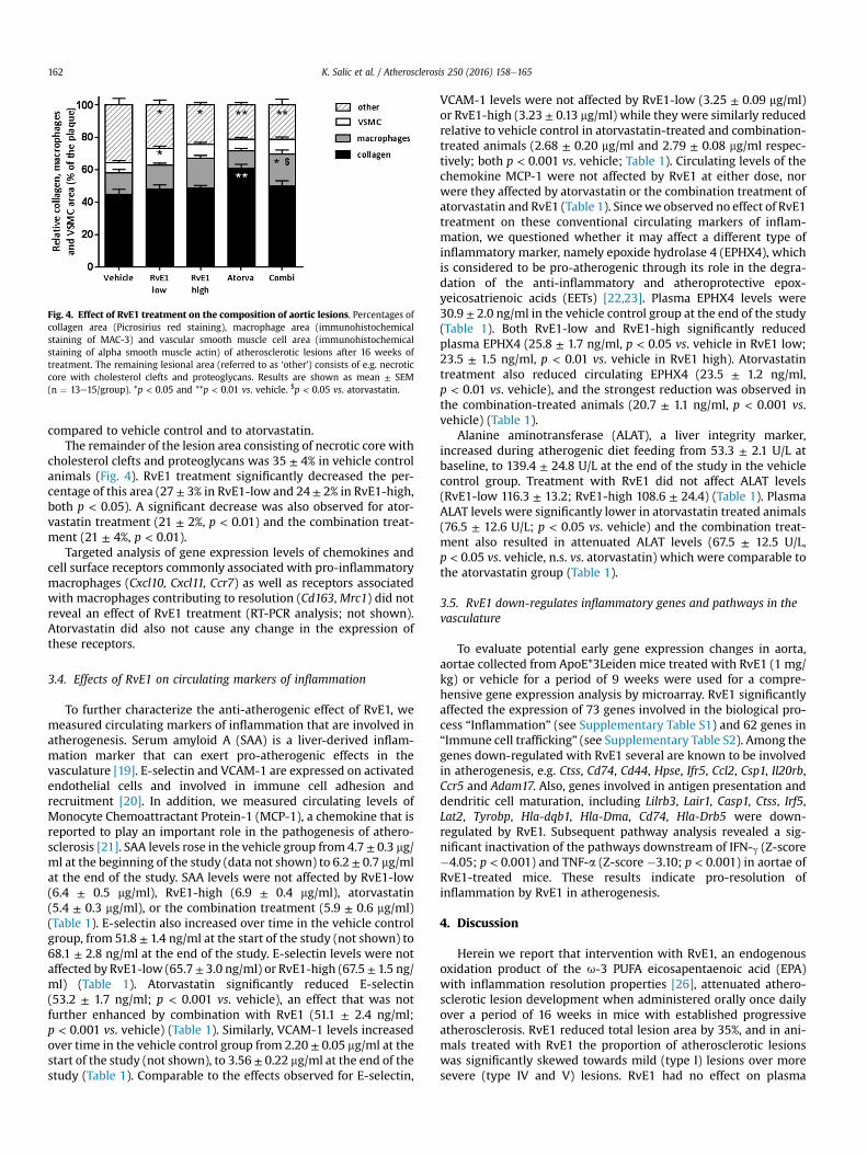
Fig. 4. Effect of RvE1 treatment on the composition of aortic lesions. Percentages ofcollagen area (Picrosirius red staining), macrophage area (immunohistochemicalstaining of MAC-3) and vascular smooth muscle cell area (immunohistochemicalstaining of alpha smooth muscle actin) of atherosclerotic lesions after 16 weeks oftreatment. The remaining lesional area (referred to as ‘other’) consists of e.g. necroticcore with cholesterol clefts and proteoglycans. Results are shown as mean ± SEM(n ¼ 13e15/group). *p < 0.05 and **p < 0.01 vs. vehicle. $p < 0.05 vs. atorvastatin.
K. Salic et al. / Atherosclerosis 250 (2016) 158e165162
compared to vehicle control and to atorvastatin.The remainder of the lesion area consisting of necrotic core with
cholesterol clefts and proteoglycans was 35 ± 4% in vehicle controlanimals (Fig. 4). RvE1 treatment significantly decreased the per-centage of this area (27 ± 3% in RvE1-low and 24 ± 2% in RvE1-high,both p < 0.05). A significant decrease was also observed for ator-vastatin treatment (21 ± 2%, p < 0.01) and the combination treat-ment (21 ± 4%, p < 0.01).
Targeted analysis of gene expression levels of chemokines andcell surface receptors commonly associated with pro-inflammatorymacrophages (Cxcl10, Cxcl11, Ccr7) as well as receptors associatedwith macrophages contributing to resolution (Cd163,Mrc1) did notreveal an effect of RvE1 treatment (RT-PCR analysis; not shown).Atorvastatin did also not cause any change in the expression ofthese receptors.
3.4. Effects of RvE1 on circulating markers of inflammation
To further characterize the anti-atherogenic effect of RvE1, wemeasured circulating markers of inflammation that are involved inatherogenesis. Serum amyloid A (SAA) is a liver-derived inflam-mation marker that can exert pro-atherogenic effects in thevasculature [19]. E-selectin and VCAM-1 are expressed on activatedendothelial cells and involved in immune cell adhesion andrecruitment [20]. In addition, we measured circulating levels ofMonocyte Chemoattractant Protein-1 (MCP-1), a chemokine that isreported to play an important role in the pathogenesis of athero-sclerosis [21]. SAA levels rose in the vehicle group from 4.7 ± 0.3 mg/ml at the beginning of the study (data not shown) to 6.2 ± 0.7 mg/mlat the end of the study. SAA levels were not affected by RvE1-low(6.4 ± 0.5 mg/ml), RvE1-high (6.9 ± 0.4 mg/ml), atorvastatin(5.4 ± 0.3 mg/ml), or the combination treatment (5.9 ± 0.6 mg/ml)(Table 1). E-selectin also increased over time in the vehicle controlgroup, from 51.8 ± 1.4 ng/ml at the start of the study (not shown) to68.1 ± 2.8 ng/ml at the end of the study. E-selectin levels were notaffected by RvE1-low (65.7± 3.0 ng/ml) or RvE1-high (67.5 ± 1.5 ng/ml) (Table 1). Atorvastatin significantly reduced E-selectin(53.2 ± 1.7 ng/ml; p < 0.001 vs. vehicle), an effect that was notfurther enhanced by combination with RvE1 (51.1 ± 2.4 ng/ml;p < 0.001 vs. vehicle) (Table 1). Similarly, VCAM-1 levels increasedover time in the vehicle control group from 2.20 ± 0.05 mg/ml at thestart of the study (not shown), to 3.56 ± 0.22 mg/ml at the end of thestudy (Table 1). Comparable to the effects observed for E-selectin,
VCAM-1 levels were not affected by RvE1-low (3.25 ± 0.09 mg/ml)or RvE1-high (3.23 ± 0.13 mg/ml) while they were similarly reducedrelative to vehicle control in atorvastatin-treated and combination-treated animals (2.68 ± 0.20 mg/ml and 2.79 ± 0.08 mg/ml respec-tively; both p < 0.001 vs. vehicle; Table 1). Circulating levels of thechemokine MCP-1 were not affected by RvE1 at either dose, norwere they affected by atorvastatin or the combination treatment ofatorvastatin and RvE1 (Table 1). Sincewe observed no effect of RvE1treatment on these conventional circulating markers of inflam-mation, we questioned whether it may affect a different type ofinflammatory marker, namely epoxide hydrolase 4 (EPHX4), whichis considered to be pro-atherogenic through its role in the degra-dation of the anti-inflammatory and atheroprotective epox-yeicosatrienoic acids (EETs) [22,23]. Plasma EPHX4 levels were30.9 ± 2.0 ng/ml in the vehicle control group at the end of the study(Table 1). Both RvE1-low and RvE1-high significantly reducedplasma EPHX4 (25.8 ± 1.7 ng/ml, p < 0.05 vs. vehicle in RvE1 low;23.5 ± 1.5 ng/ml, p < 0.01 vs. vehicle in RvE1 high). Atorvastatintreatment also reduced circulating EPHX4 (23.5 ± 1.2 ng/ml,p < 0.01 vs. vehicle), and the strongest reduction was observed inthe combination-treated animals (20.7 ± 1.1 ng/ml, p < 0.001 vs.vehicle) (Table 1).
Alanine aminotransferase (ALAT), a liver integrity marker,increased during atherogenic diet feeding from 53.3 ± 2.1 U/L atbaseline, to 139.4 ± 24.8 U/L at the end of the study in the vehiclecontrol group. Treatment with RvE1 did not affect ALAT levels(RvE1-low 116.3 ± 13.2; RvE1-high 108.6 ± 24.4) (Table 1). PlasmaALAT levels were significantly lower in atorvastatin treated animals(76.5 ± 12.6 U/L; p < 0.05 vs. vehicle) and the combination treat-ment also resulted in attenuated ALAT levels (67.5 ± 12.5 U/L,p < 0.05 vs. vehicle, n.s. vs. atorvastatin) which were comparable tothe atorvastatin group (Table 1).
3.5. RvE1 down-regulates inflammatory genes and pathways in thevasculature
To evaluate potential early gene expression changes in aorta,aortae collected from ApoE*3Leiden mice treated with RvE1 (1 mg/kg) or vehicle for a period of 9 weeks were used for a compre-hensive gene expression analysis by microarray. RvE1 significantlyaffected the expression of 73 genes involved in the biological pro-cess “Inflammation” (see Supplementary Table S1) and 62 genes in“Immune cell trafficking” (see Supplementary Table S2). Among thegenes down-regulated with RvE1 several are known to be involvedin atherogenesis, e.g. Ctss, Cd74, Cd44, Hpse, Ifr5, Ccl2, Csp1, Il20rb,Ccr5 and Adam17. Also, genes involved in antigen presentation anddendritic cell maturation, including Lilrb3, Lair1, Casp1, Ctss, Irf5,Lat2, Tyrobp, Hla-dqb1, Hla-Dma, Cd74, Hla-Drb5 were down-regulated by RvE1. Subsequent pathway analysis revealed a sig-nificant inactivation of the pathways downstream of IFN-g (Z-score�4.05; p < 0.001) and TNF-a (Z-score �3.10; p < 0.001) in aortae ofRvE1-treated mice. These results indicate pro-resolution ofinflammation by RvE1 in atherogenesis.
4. Discussion
Herein we report that intervention with RvE1, an endogenousoxidation product of the u-3 PUFA eicosapentaenoic acid (EPA)with inflammation resolution properties [26], attenuated athero-sclerotic lesion development when administered orally once dailyover a period of 16 weeks in mice with established progressiveatherosclerosis. RvE1 reduced total lesion area by 35%, and in ani-mals treated with RvE1 the proportion of atherosclerotic lesionswas significantly skewed towards mild (type I) lesions over moresevere (type IV and V) lesions. RvE1 had no effect on plasma
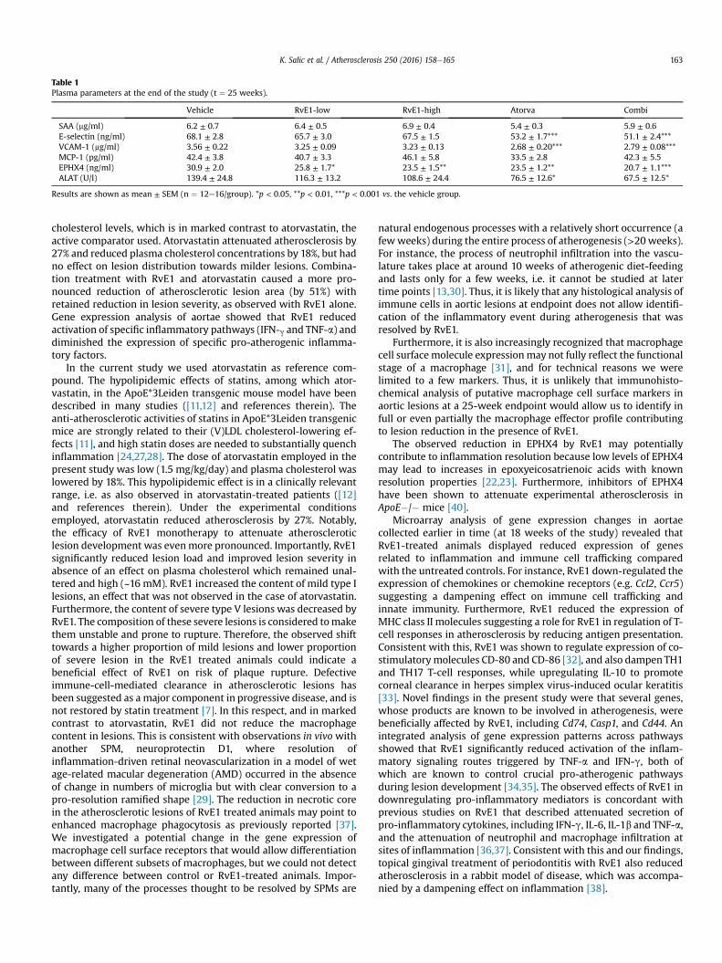
Table 1Plasma parameters at the end of the study (t ¼ 25 weeks).
Vehicle RvE1-low RvE1-high Atorva Combi
SAA (mg/ml) 6.2 ± 0.7 6.4 ± 0.5 6.9 ± 0.4 5.4 ± 0.3 5.9 ± 0.6E-selectin (ng/ml) 68.1 ± 2.8 65.7 ± 3.0 67.5 ± 1.5 53.2 ± 1.7*** 51.1 ± 2.4***VCAM-1 (mg/ml) 3.56 ± 0.22 3.25 ± 0.09 3.23 ± 0.13 2.68 ± 0.20*** 2.79 ± 0.08***MCP-1 (pg/ml) 42.4 ± 3.8 40.7 ± 3.3 46.1 ± 5.8 33.5 ± 2.8 42.3 ± 5.5EPHX4 (ng/ml) 30.9 ± 2.0 25.8 ± 1.7* 23.5 ± 1.5** 23.5 ± 1.2** 20.7 ± 1.1***ALAT (U/l) 139.4 ± 24.8 116.3 ± 13.2 108.6 ± 24.4 76.5 ± 12.6* 67.5 ± 12.5*
Results are shown as mean ± SEM (n ¼ 12e16/group). *p < 0.05, **p < 0.01, ***p < 0.001 vs. the vehicle group.
K. Salic et al. / Atherosclerosis 250 (2016) 158e165 163
cholesterol levels, which is in marked contrast to atorvastatin, theactive comparator used. Atorvastatin attenuated atherosclerosis by27% and reduced plasma cholesterol concentrations by 18%, but hadno effect on lesion distribution towards milder lesions. Combina-tion treatment with RvE1 and atorvastatin caused a more pro-nounced reduction of atherosclerotic lesion area (by 51%) withretained reduction in lesion severity, as observed with RvE1 alone.Gene expression analysis of aortae showed that RvE1 reducedactivation of specific inflammatory pathways (IFN-g and TNF-a) anddiminished the expression of specific pro-atherogenic inflamma-tory factors.
In the current study we used atorvastatin as reference com-pound. The hypolipidemic effects of statins, among which ator-vastatin, in the ApoE*3Leiden transgenic mouse model have beendescribed in many studies ([11,12] and references therein). Theanti-atherosclerotic activities of statins in ApoE*3Leiden transgenicmice are strongly related to their (V)LDL cholesterol-lowering ef-fects [11], and high statin doses are needed to substantially quenchinflammation [24,27,28]. The dose of atorvastatin employed in thepresent study was low (1.5 mg/kg/day) and plasma cholesterol waslowered by 18%. This hypolipidemic effect is in a clinically relevantrange, i.e. as also observed in atorvastatin-treated patients ([12]and references therein). Under the experimental conditionsemployed, atorvastatin reduced atherosclerosis by 27%. Notably,the efficacy of RvE1 monotherapy to attenuate atheroscleroticlesion development was evenmore pronounced. Importantly, RvE1significantly reduced lesion load and improved lesion severity inabsence of an effect on plasma cholesterol which remained unal-tered and high (~16 mM). RvE1 increased the content of mild type Ilesions, an effect that was not observed in the case of atorvastatin.Furthermore, the content of severe type V lesions was decreased byRvE1. The composition of these severe lesions is considered tomakethem unstable and prone to rupture. Therefore, the observed shifttowards a higher proportion of mild lesions and lower proportionof severe lesion in the RvE1 treated animals could indicate abeneficial effect of RvE1 on risk of plaque rupture. Defectiveimmune-cell-mediated clearance in atherosclerotic lesions hasbeen suggested as a major component in progressive disease, and isnot restored by statin treatment [7]. In this respect, and in markedcontrast to atorvastatin, RvE1 did not reduce the macrophagecontent in lesions. This is consistent with observations in vivowithanother SPM, neuroprotectin D1, where resolution ofinflammation-driven retinal neovascularization in a model of wetage-related macular degeneration (AMD) occurred in the absenceof change in numbers of microglia but with clear conversion to apro-resolution ramified shape [29]. The reduction in necrotic corein the atherosclerotic lesions of RvE1 treated animals may point toenhanced macrophage phagocytosis as previously reported [37].We investigated a potential change in the gene expression ofmacrophage cell surface receptors that would allow differentiationbetween different subsets of macrophages, but we could not detectany difference between control or RvE1-treated animals. Impor-tantly, many of the processes thought to be resolved by SPMs are
natural endogenous processes with a relatively short occurrence (afewweeks) during the entire process of atherogenesis (>20weeks).For instance, the process of neutrophil infiltration into the vascu-lature takes place at around 10 weeks of atherogenic diet-feedingand lasts only for a few weeks, i.e. it cannot be studied at latertime points [13,30]. Thus, it is likely that any histological analysis ofimmune cells in aortic lesions at endpoint does not allow identifi-cation of the inflammatory event during atherogenesis that wasresolved by RvE1.
Furthermore, it is also increasingly recognized that macrophagecell surface molecule expressionmay not fully reflect the functionalstage of a macrophage [31], and for technical reasons we werelimited to a few markers. Thus, it is unlikely that immunohisto-chemical analysis of putative macrophage cell surface markers inaortic lesions at a 25-week endpoint would allow us to identify infull or even partially the macrophage effector profile contributingto lesion reduction in the presence of RvE1.
The observed reduction in EPHX4 by RvE1 may potentiallycontribute to inflammation resolution because low levels of EPHX4may lead to increases in epoxyeicosatrienoic acids with knownresolution properties [22,23]. Furthermore, inhibitors of EPHX4have been shown to attenuate experimental atherosclerosis inApoE�/� mice [40].
Microarray analysis of gene expression changes in aortaecollected earlier in time (at 18 weeks of the study) revealed thatRvE1-treated animals displayed reduced expression of genesrelated to inflammation and immune cell trafficking comparedwith the untreated controls. For instance, RvE1 down-regulated theexpression of chemokines or chemokine receptors (e.g. Ccl2, Ccr5)suggesting a dampening effect on immune cell trafficking andinnate immunity. Furthermore, RvE1 reduced the expression ofMHC class II molecules suggesting a role for RvE1 in regulation of T-cell responses in atherosclerosis by reducing antigen presentation.Consistent with this, RvE1 was shown to regulate expression of co-stimulatorymolecules CD-80 and CD-86 [32], and also dampen TH1and TH17 T-cell responses, while upregulating IL-10 to promotecorneal clearance in herpes simplex virus-induced ocular keratitis[33]. Novel findings in the present study were that several genes,whose products are known to be involved in atherogenesis, werebeneficially affected by RvE1, including Cd74, Casp1, and Cd44. Anintegrated analysis of gene expression patterns across pathwaysshowed that RvE1 significantly reduced activation of the inflam-matory signaling routes triggered by TNF-a and IFN-g, both ofwhich are known to control crucial pro-atherogenic pathwaysduring lesion development [34,35]. The observed effects of RvE1 indownregulating pro-inflammatory mediators is concordant withprevious studies on RvE1 that described attenuated secretion ofpro-inflammatory cytokines, including IFN-g, IL-6, IL-1b and TNF-a,and the attenuation of neutrophil and macrophage infiltration atsites of inflammation [36,37]. Consistent with this and our findings,topical gingival treatment of periodontitis with RvE1 also reducedatherosclerosis in a rabbit model of disease, which was accompa-nied by a dampening effect on inflammation [38].

K. Salic et al. / Atherosclerosis 250 (2016) 158e165164
In the present study we studied for the first time the efficacy ofresolvin E1 in a diet-inducible model of atherosclerosis. While wedemonstrate the principle of atherosclerosis treatment withresolvin E1, the study was not designed to study or unravel theunderlying mechanisms such as phenotypic changes of inflamma-tory cells. Future studies should include a refined characterizationof circulating as well as tissue-resident inflammatory cells to pro-vide insight into the mechanisms underlying these effects.
In the current study, en face Oil Red O lipid staining of the aorta,which is a commonly used method for quantification of athero-sclerosis extension, was not performed. The method employedherein is an established method for analysis of atherosclerosis[11,13,24,25] that not only allows quantification of lesion load (withresults comparable to those obtained from en face staining [25,39]),but also provides information on the severity of atheroscleroticlesions.
Taken together, our results demonstrate for the first time theefficacy of an SPM, RvE1, in an experimental model of diet-inducedatherosclerosis, the ApoE*3Leiden transgenic mouse, and furthersupport the concept that defective resolution of inflammation maycontribute to progression of atherosclerotic disease. While RvE1showed comparable efficacy to that of an established statin inattenuating atherosclerosis, the more pronounced effect of theircombined actions is indicative of a therapeutic potential of SPM inatherosclerosis, and the added benefit of activating pro-resolutionpathways on top of existing first line treatment.
Conflict of interest
PG and LW were employees of Resolvyx Pharmaceuticals Inc.
Financial support
This work was supported by Resolvyx Pharmaceuticals Inc.MCM received funding from Top Institute Food and Nutrition (grantno: CH001), a public-private partnership on pre-competitiveresearch in food and nutrition.
Acknowledgements
We would like to thank Wim van Duyvenvoorde and Karin Toetfor their excellent technical assistance.
Appendix A. Supplementary data
Supplementary data related to this article can be found at http://dx.doi.org/10.1016/j.atherosclerosis.2016.05.001.
References
[1] J.C. Fruchart, J. Davignon, M.P. Hermans, et al., Residual macrovascular risk in2013: what have we learned? Cardiovasc. Diabetol. 13 (2014), 26e2840-2813-2826.
[2] G.K. Hansson, A. Hermansson, The immune system in atherosclerosis, Nat.Immunol. 12 (2011) 204e212.
[3] C.G. Santos-Gallego, B. Picatoste, J.J. Badimon, Pathophysiology of acute cor-onary syndrome, Curr. Atheroscler. Rep. 16 (2014) 401.
[4] E. van der Meij, G.G. Koning, P.W. Vriens, et al., A clinical evaluation of statinpleiotropy: statins selectively and dose-dependently reduce vascular inflam-mation, PLoS One 8 (2013) e53882.
[5] D. Tousoulis, C. Psarros, M. Demosthenous, et al., Innate and adaptiveinflammation as a therapeutic target in vascular disease: the emerging role ofstatins, J. Am. Coll. Cardiol. 63 (2014) 2491e2502.
[6] I.F. Charo, R. Taub, Anti-inflammatory therapeutics for the treatment ofatherosclerosis, Nat. Rev. Drug Discov. 10 (2011) 365e376.
[7] I. Tabas, Macrophage death and defective inflammation resolution inatherosclerosis, Nat. Rev. Immunol. 10 (2010) 36e46.
[8] C.N. Serhan, N. Chiang, T.E. Van Dyke, Resolving inflammation: dual anti-inflammatory and pro-resolution lipid mediators, Nat. Rev. Immunol. 8
(2008) 349e361.[9] C.N. Serhan, C.B. Clish, J. Brannon, et al., Novel functional sets of lipid-derived
mediators with antiinflammatory actions generated from omega-3 fatty acidsvia cyclooxygenase 2-nonsteroidal antiinflammatory drugs and transcellularprocessing, J. Exp. Med. 192 (2000) 1197e1204.
[10] C.N. Serhan, Pro-resolving lipid mediators are leads for resolution physiology,Nature 510 (2014) 92e101.
[11] S. Zadelaar, R. Kleemann, L. Verschuren, et al., Mouse models for atheroscle-rosis and pharmaceutical modifiers, Arterioscler. Thromb. Vasc. Biol. 27 (2007)1706e1721.
[12] E. van de Steeg, R. Kleemann, H.T. Jansen, et al., Combined analysis of phar-macokinetic and efficacy data of preclinical studies with statins markedlyimproves translation of drug efficacy to human trials, J. Pharmacol. Exp. Ther.347 (2013) 635e644.
[13] M. Morrison, R. van der Heijden, P. Heeringa, et al., Epicatechin attenuatesatherosclerosis and exerts anti-inflammatory effects on diet-induced human-CRP and NFkappaB in vivo, Atherosclerosis 233 (2014) 149e156.
[14] M.C. Morrison, W. Liang, P. Mulder, et al., Mirtoselect, an anthocyanin-richbilberry extract, attenuates non-alcoholic steatohepatitis and associatedfibrosis in ApoE *3Leiden mice, J. Hepatol. 62 (2015) 1180e1186.
[15] H.C. Stary, A.B. Chandler, R.E. Dinsmore, et al., A definition of advanced typesof atherosclerotic lesions and a histological classification of atherosclerosis. Areport from the committee on vascular lesions of the council on arterioscle-rosis, american heart association, Arterioscler. Thromb. Vasc. Biol. 15 (1995)1512e1531.
[16] H.C. Stary, A.B. Chandler, S. Glagov, et al., A definition of initial, fatty streak,and intermediate lesions of atherosclerosis. A report from the committee onvascular lesions of the council on arteriosclerosis, american heart association,Arterioscler. Thromb. Vasc. Biol. 14 (1994) 840e856.
[17] L. Verschuren, M. Radonjic, P.Y. Wielinga, et al., Systems biology analysisunravels the complementary action of combined rosuvastatin and ezetimibetherapy, Pharmacogen. Genom. 22 (2012) 837e845.
[18] R. Kleemann, L. Verschuren, M.J. van Erk, et al., Atherosclerosis and liverinflammation induced by increased dietary cholesterol intake: a combinedtranscriptomics and metabolomics analysis, Genome Biol. 8 (2007) R200.
[19] V.L. King, J. Thompson, L.R. Tannock, Serum amyloid A in atherosclerosis, Curr.Opin. Lipidol. 22 (2011) 302e307.
[20] R.P. McEver, Selectins: initiators of leucocyte adhesion and signalling at thevascular wall, Cardiovasc. Res. 107 (2015) 331e339.
[21] L.C. Freitas Lima, VdA. Barga, M. do Socorro de França Silva, et al., Adipokines,diabetes and atherosclerosis: an inflammatory association, Front. Physiol.(2015) 6.
[22] J. He, C. Wang, Y. Zhu, et al., Soluble epoxide hydrolase: a potential target formetabolic diseases, J. Diabetes 8 (2016) 305e313.
[23] Y.-X.J. Wang, A. Ulu, L.-N. Zhang, et al., Soluble epoxide hydrolase in athero-sclerosis, Curr. Atheroscler. Rep. 12 (2010) 174e183.
[24] R. Kleemann, H.M. Princen, J.J. Emeis, et al., Rosuvastatin reduces athero-sclerosis development beyond and independent of its plasma cholesterol-lowering effect in APOE*3-Leiden transgenic mice: evidence for antiin-flammatory effects of rosuvastatin, Circulation 108 (2003) 1368e1374.
[25] L. Verschuren, R. Kleemann, E.H. Offerman, et al., Effect of low dose atorvas-tatin versus diet-induced cholesterol lowering on atherosclerotic lesion pro-gression and inflammation in apolipoprotein E*3-Leiden transgenic mice,Arterioscler. Thromb. Vasc. Biol. 25 (2005) 161e167.
[26] H. Seki, Y. Tani, M. Arita, Omega-3 PUFA derived anti-inflammatory lipidmediator resolvin E1, Prostagl. Other Lipid Mediat. 89 (2009) 126e130.
[27] R. Kleemann, T. Kooistra, HMG-CoA reductase inhibitors: effects on chronicsubacute inflammation and onset of atherosclerosis induced by dietarycholesterol, Curr. Drug Targets Cardiovasc. Haematol. Disord. 5 (2005)441e453.
[28] R. Kleemann, L. Verschuren, B.J. de Rooij, et al., Evidence for anti-inflammatoryactivity of statins and PPARalpha activators in human C-reactive proteintransgenic mice in vivo and in cultured human hepatocytes in vitro, Blood 103(2004) 4188e4194.
[29] K.G. Sheets, B. Jun, Y. Zhou, et al., Microglial ramification and redistributionconcomitant with the attenuation of choroidal neovascularization by neuro-protectin D1, Mol. Vis. 19 (2013) 1747e1759.
[30] M. van Leeuwen, M.J. Gijbels, A. Duijvestijn, et al., Accumulation ofmyeloperoxidase-positive neutrophils in atherosclerotic lesions in LDLR-/-mice, Arterioscler. Thromb. Vasc. Biol. 28 (2008) 84e89.
[31] T. R}oszer, Understanding the mysterious M2 macrophage through activationmarkers and effector mechanisms, Mediat. Inflamm. (2015) 2015.
[32] E.K. Vassiliou, O.M. Kesler, J.H. Tadros, et al., Bone marrow-derived dendriticcells generated in the presence of resolvin E1 induce apoptosis of activatedCD4þ T cells, J. Immunol. 181 (2008) 4534e4544.
[33] N.K. Rajasagi, P.B. Reddy, A. Suryawanshi, et al., Controlling herpes simplexvirus-induced ocular inflammatory lesions with the lipid-derived mediatorresolvin E1, J. Immunol. 186 (2011) 1735e1746.
[34] P. Kleinbongard, G. Heusch, R. Schulz, TNFalpha in atherosclerosis, myocardialischemia/reperfusion and heart failure, Pharmacol. Ther. 127 (2010) 295e314.
[35] I. Voloshyna, M.J. Littlefield, A.B. Reiss, Atherosclerosis and interferon-gamma:new insights and therapeutic targets, Trends Cardiovasc. Med. 24 (2014)45e51.
[36] Y. Jin, M. Arita, Q. Zhang, et al., Anti-angiogenesis effect of the novel anti-inflammatory and pro-resolving lipid mediators, Invest. Ophthalmol. Vis.

K. Salic et al. / Atherosclerosis 250 (2016) 158e165 165
Sci. 50 (2009) 4743e4752.[37] J.M. Schwab, N. Chiang, M. Arita, et al., Resolvin E1 and protectin D1 activate
inflammation-resolution programmes, Nature 447 (2007) 869e874.[38] H. Hasturk, R. Abdallah, A. Kantarci, et al., Resolvin E1 (RvE1) attenuates
atherosclerotic plaque formation in diet and inflammation-induced athero-genesis, Arterioscler. Thromb. Vasc. Biol. 35 (2015) 1123e1133.
[39] S. Kühnast, J.W. van der Hoorn, E.J. Pieterman, et al., Alirocumab inhibitsatherosclerosis, improves the plaque morphology, and enhances the effects ofa statin, J. Lipid Res. 55 (10) (2014) 2103e2112.
[40] A. Ulu, B.B. Davis, H.-J. Tsai, et al., Soluble epoxide hydrolase inhibitors reducethe development of atherosclerosis in apolipoprotein E-knockout mousemodel, J. Cardiovasc. Pharmacol. 52 (4) (2008) 314e323.