Residual insulin positivity and pancreatic atrophy in ... · insulin positivity) four to five...
6
Diabetologia (1987) 30:757-762 Diabetologia Springer-Verlag1987 Originals Residual insulin positivity and pancreatic atrophy in relation to duration of chronic Type 1 (insulin-dependent) diabetes mellitus and microangiopathy* M. Lrhr and G. Klrppel** Institute of Pathology, University of Hamburg, Hamburg, FRG Summary. The relationship of residual insulin positivity in chronic Type I (insulin-dependent) diabetes and atrophy of the exocrine pancreas to duration of diabetes, age at onset and microangiopathy was studied in 26 patients (disease du- ration 2 to 54 years, mean 26 years). Islets containing insulin cells were found in 13/26 pancreata. In 5/13 pancreata insu- lin positive cells were detected in only one lobule, while in 8/13 insulin positivity was multifocal. All patients with dia- betes duration less than 11 years had residual insulin cells; whereas, the rate of insulin positivity was near 40% with dia- betes duration of more than 11 and 21 years, respectively. Survival of insulin cells was not clearly related to age at on- set. HLA-DR expression on insulin cells was seen in one case. lnsulitis was lacking. Pancreatic volume determined in 18 patients ranged from 14-110ml (age adjusted mean 56.3 ml) and was significantly less than that of control sub- jects (age adjusted, mean 89.9 ml, p < 0.0001). Computerized morphometry of the exocrine pancreas revealed severe aci- nat atrophy due to a reduction in size of acinar cells. Acinar atrophy correlated neither with the degree of insulin positivi- ty, disease duration nor severity of microangiopathy. The findings suggest that in about 40% of patients with Type 1 diabetes a small population of insulin cells may escape auto- immune destruction, irrespective of disease duration or age at onset. Though exocrine atrophy and insulin deficiency are associated, the variable extent of pancreatic atrophy could not to be related to such factors as amount of surviving insu- lin cells, duration of diabetes or microangiopathy. Key words: Chronic Type 1 (insulin-dependent) diabetes, re- sidual insulin cells, exocrine atrophy, HLA-DR expression, diabetic microangiopathy. Loss of insulin cells characterises the islet lesions in patients with Type 1 (insulin-dependent) diabetes mel- litus [1-4]. This loss, however, need not to be complete, since B cells, though severely reduced, have been dem- onstrated even in patients with a diabetes duration up to 37 years [1, 3], The mechanisms which may protect some insulin cells from destruction by the autoimmune process underlying Type I diabetes are largely un- krlown. Another finding that characterises the pancreas in chronic Type 1 diabetes is a reduction in weight and volume [3, 5-8]. The pancreatic atrophy is attributed to the lacking trophic effect of insulin on the acinar cells [2, 3, 9-13]. The extent of the exocrine atrophy, how- ever, appears to vary considerably from patient to pat- ient and has not yet been related to the remaining insu- lin positivity in the pancreas, to diabetes duration or to the degree of microangiopathy. * Presented in part at the 22 nd EASD meeting in Rome, 1986 ** Present address: Department of Pathology, Free University of Brussels, Belgium The aim of the present study was to investigate the degree and distribution of residual insulin positivity in the pancreas of patients with long-standing Type 1 dia- betes by immunocytochemistry and to determine the degree of pancreatic atrophy by morphometry. The re- sults from these two determinations were then related to diabetes duration, age at onset and extent of micro- angiopathy to see whether these parameters have any impact on the survival of insulin cells and a decrease of pancreatic volume. Subjects and methods In 26 patients (14 males, 12 females) with Type1 diabetes of long duration (2-54 years, mean 26 years), the pancreas and the kidneys were studied at autopsy. Autopsies were performed at varying inter- vals post mortem, with a range from 14 to 26 h. Clinical data regard- ing age at onset and duration of diabetes were collected. Death was related to complications of diabetes, i. e. nepbropathy, myocardial in- farction and coma. The pancreas was carefully dissected and in 18 cases its volume was determined by Archimedes' principle. The same was done in
Transcript of Residual insulin positivity and pancreatic atrophy in ... · insulin positivity) four to five...

Diabetologia (1987) 30:757-762 Diabetologia �9 Springer-Verlag 1987
Originals
Residual insulin positivity and pancreatic atrophy in relation to duration of chronic Type 1 (insulin-dependent) diabetes mellitus and microangiopathy*
M. L r h r and G. Klrppel**
Institute of Pathology, University of Hamburg, Hamburg, FRG
Summary. The relationship of residual insulin positivity in chronic Type I (insulin-dependent) diabetes and atrophy of the exocrine pancreas to duration of diabetes, age at onset and microangiopathy was studied in 26 patients (disease du- ration 2 to 54 years, mean 26 years). Islets containing insulin cells were found in 13/26 pancreata. In 5/13 pancreata insu- lin positive cells were detected in only one lobule, while in 8/13 insulin positivity was multifocal. All patients with dia- betes duration less than 11 years had residual insulin cells; whereas, the rate of insulin positivity was near 40% with dia- betes duration of more than 11 and 21 years, respectively. Survival of insulin cells was not clearly related to age at on- set. HLA-DR expression on insulin cells was seen in one case. lnsulitis was lacking. Pancreatic volume determined in 18 patients ranged from 14-110ml (age adjusted mean 56.3 ml) and was significantly less than that of control sub-
jects (age adjusted, mean 89.9 ml, p < 0.0001). Computerized morphometry of the exocrine pancreas revealed severe aci- nat atrophy due to a reduction in size of acinar cells. Acinar atrophy correlated neither with the degree of insulin positivi- ty, disease duration nor severity of microangiopathy. The findings suggest that in about 40% of patients with Type 1 diabetes a small population of insulin cells may escape auto- immune destruction, irrespective of disease duration or age at onset. Though exocrine atrophy and insulin deficiency are associated, the variable extent of pancreatic atrophy could not to be related to such factors as amount of surviving insu- lin cells, duration of diabetes or microangiopathy.
Key words: Chronic Type 1 (insulin-dependent) diabetes, re- sidual insulin cells, exocrine atrophy, HLA-DR expression, diabetic microangiopathy.
Loss of insulin cells characterises the islet lesions in patients with Type 1 (insulin-dependent) diabetes mel- litus [1-4]. This loss, however, need not to be complete, since B cells, though severely reduced, have been dem- onstrated even in patients with a diabetes duration up to 37 years [1, 3], The mechanisms which may protect some insulin cells f rom destruction by the autoimmune process underlying Type I diabetes are largely un- krlown.
Another finding that characterises the pancreas in chronic Type 1 diabetes is a reduction in weight and volume [3, 5-8]. The pancreatic a t rophy is attributed to the lacking trophic effect of insulin on the acinar cells [2, 3, 9-13]. The extent of the exocrine atrophy, how- ever, appears to vary considerably from patient to pat- ient and has not yet been related to the remaining insu- lin positivity in the pancreas, to diabetes duration or to the degree o f microangiopathy.
* Presented in part at the 22 nd EASD meeting in Rome, 1986 ** Present address: Department of Pathology, Free University of
Brussels, Belgium
The aim of the present study was to investigate the degree and distribution of residual insulin positivity in the pancreas of patients with long-standing Type 1 dia- betes by immunocytochemistry and to determine the degree of pancreatic a t rophy by morphometry. The re- sults from these two determinations were then related to diabetes duration, age at onset and extent of micro- angiopathy to see whether these parameters have any impact on the survival of insulin cells and a decrease of pancreatic volume.
Subjects and methods
In 26 patients (14 males, 12 females) with Type 1 diabetes of long duration (2-54 years, mean 26 years), the pancreas and the kidneys were studied at autopsy. Autopsies were performed at varying inter- vals post mortem, with a range from 14 to 26 h. Clinical data regard- ing age at onset and duration of diabetes were collected. Death was related to complications of diabetes, i. e. nepbropathy, myocardial in- farction and coma.
The pancreas was carefully dissected and in 18 cases its volume was determined by Archimedes' principle. The same was done in
758
Table 1. Clinical characteristics of patients with Type 1 (insulin-de- pendent) diabetes with and without residual insulin cells
M. Lthr and G. K1Oppel: Residual insulin cells in chronic Type 1 (insulin-dependent) diabetes
Insulin- Number Age Diabetes Age at score (years) duration onset
(years) (years) Mean+ Mean+ Mean_+ SD SD SD
Chronic Type 1 0 13 52+15 30+ 9 23+11 diabetes 1 + 5 49 + 27 27 + 19 22___ 24 mellitus 2+ 8 41+21 20+12 21___14
All 26 48+19 26+12 22_+15
Control subjects 3+ 19 40+18
45 nondiabetic control subjects who died of diseases unrelated to the pancreas. In 19 of these 45 patients, the pancreas was used for mor- phometric evaluation of the exocrine compartment. The interval be- tween death and examination in the control subjects averaged 24 h. The pancreas was cut into slices and fixed in Bouin's solution. Mate- rial was obtained from eight regions coded by anatomic location ac- cording to the protocol proposed by Malaisse-Lagae et al. [14]. From each block, serial sections (3 ~tm) were cut after routine paraffin pro- cessing. The first three sections were stained with H&E, periodic ac- id Schiff (PAS) and Masson-Goldner.
Immunocytochemistry was performed on the subsequent depar- affinised sections, as described elsewhere [15]. A monoclonal anti- body was used against insulin (Biogenex via Camon, Wiesbaden, FRG) and polyclonal antibodies against glucagon (INC, Interna- tional Nuclear Corporation, Stillwater, Minn, USA), somatostatin (INC) and human pancreatic polypeptide (HPP, gift of Dr. R. E. Chance, Indianapolis, Ind, USA). The primary dilutions were 1:20, 1:5000, 1:5000 and 1:10000 respectively. In addition to the hormonal antibodies, we used two antibodies against the non-poly- morphous region of the HLA-DR antigen, EPl13 (gift of Dr. H. G. Thiele, Hamburg, FRG [16[) and TAL-B 5 (gift of C. Dixon, London, UK [17]) known to react on paraffin embedded tissue at di- lutions of I : 10 000 and 1 : 20 respectively.
The sections were evaluated regarding insulitis, insulin cell con- tent of the islets and localisation and frequency of insulin positive is- lets in the pancreas. The presence of insulin positive islets in one lob- ule was graded 1 + , while insulin positive islets in more than one lobule were graded 2+ . The islets in control subjects were graded 3 + . In five pancreata (two with t + insulin positivity, three with 2 + insulin positivity) four to five randomly selected insulin positive is- lets were evaluated by point counting to determine the percentage of total islet surface area occupied by each of the four islet cell types. The percentages were then averaged and a group mean was calculat- ed.
To evaluate the volume density of the acinar cell compartment in the patients with diabetes and 19 normogiycaemic normal weight control subjects, the areas occupied by the nuclei and the cytoplasm of the acinar ceils were separately measured on a Masson-Goldner stained cross-sectional slide of the pancreas. The morphometric equipment included a standard light microscope (Zeiss, Oberkochen, FRG) with a semi-automatic image analyser (Kontron, Mtinchen, FRG) [18]. The image of the pancreatic parenchyma, as seen in the microscope at a total magnification of x 63, appeared on a color monitor and was then transformed by color-tone and grey-tone level discrimination into a black and white picture. Four different areas from the body and tail of the pancreas were arbitrarily chosen for evaluation. From the two parameters measured, the nucleus/cell (cytoplasm plus nucleus)-ratio (N/C ratio) was calculated. In addi- tion, the number of nuclei per 0.5 mm 2 (monitor screen area at • 63 magnification) was determined.
To estimate the degree of microvascular changes, thickening of the basement membrane of capillaries and arterioles, as visualized
5 5 -
5 0 -
4 . 5 -
4 -0 -
3 5 -
~ 30
~ 25 ,,l.-
P 2o
~ 1 5
21o
0
~b �9 o ~
�9 �9
*-' 5 �9 �9
0 I 1 '1 2+ 1," 0 lnsu t in -pos i t i v i t y
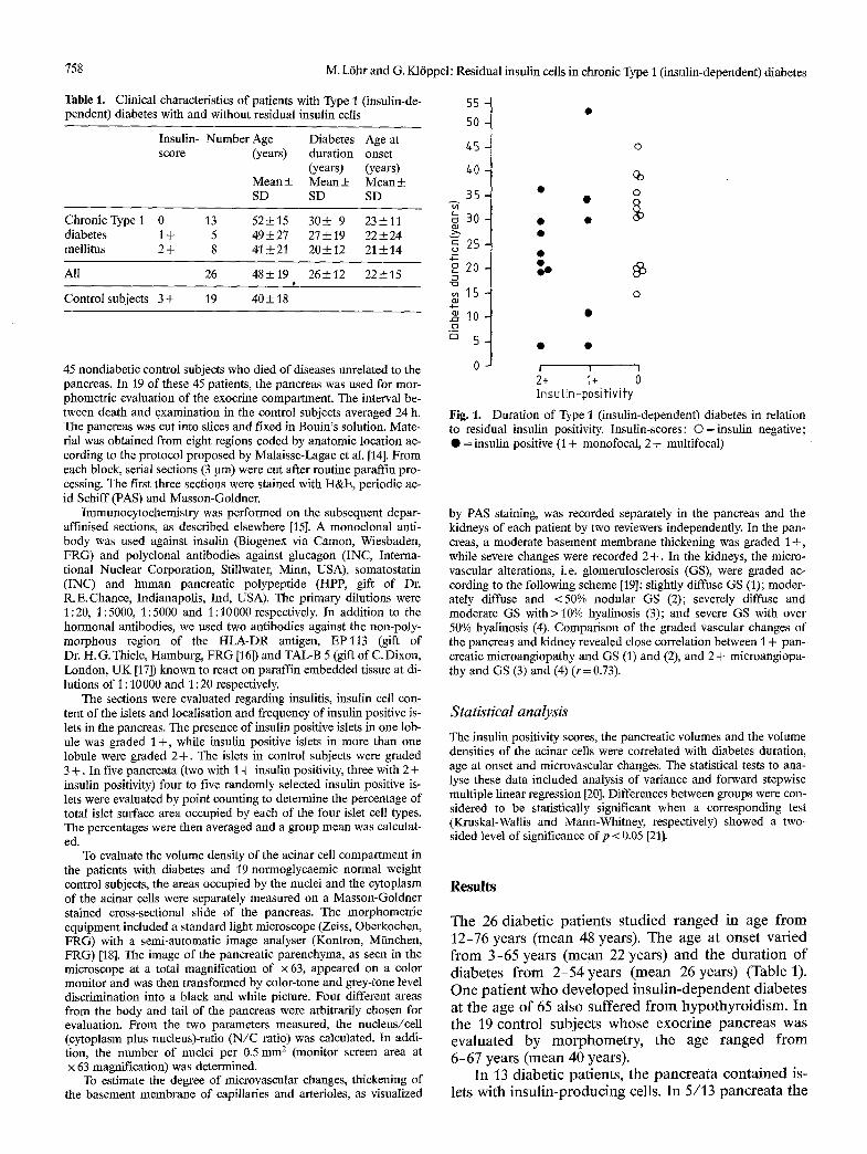
Fig, 1. Duration of Type 1 (insulin-dependent) diabetes in relation to residual insulin positivity. Insulin-scores: O =insulin negative; �9 =insulin positive (1 + monofocal, 2+ multifocal)
by PAS staining, was recorded separately in the pancreas and the kidneys of each patient by two reviewers independently. In the pan- creas, a moderate basement membrane thickening was graded 1 + , while severe changes were recorded 2 + . In the kidneys, the micro- vascular alterations, i.e. glomerulosclerosis (GS), were graded ac- cording to the following scheme [19]: slightly diffuse GS (1); moder- ately diffuse and <50% nodular GS (2); severely diffuse and moderate GS with>10% hyalinosis (3); and severe GS with over 50% hyalinosis (4). Comparison of the graded vascular changes of the pancreas and kidney revealed close correlation between 1 + pan- creatic microangiopathy and GS (1) and (2), and 2 + microangiopa- thy and GS (3) and (4) (r = 0.73).
Statistical analysis
The insulin positivity scores, the pancreatic volumes and the volume densities of the acinar cells were correlated with diabetes duration, age at onset and microvascular changes. The statistical tests to ana- lyse these data included analysis of variance and forward stepwise multiple linear regression [20]. Differences between groups were con- sidered to be statistically significant when a corresponding test (Kruskal-Wallis and Mann-Whitney, respectively) showed a two- sided level of significance of p < 0.05 [21].
Results
The 26 diabetic patients studied ranged in age from 12-76 years (mean 48 years). The age at onset varied from 3-65 years (mean 22 years) and the duration of diabetes from 2-54years (mean 26 years) (Table 1). One patient who developed insulin-dependent diabetes at the age of 65 also suffered from hypothyroidism. In the 19 control subjects whose exocrine pancreas was evaluated by morphometry, the age ranged from 6-67 years (mean 40 years).
In 13 diabetic patients, the pancreata contained is- lets with insulin-producing cells. In 5/13 pancreata the
M. L6hr and G. K15ppel: Residual insulin cells in chronic Type I (insulin-dependent) diabetes
Table 2. Residual insulin positivity in long standing Type 1 (insulin-dependent) diabetes related to parameters of pancreatic atrophy
759
Insulin- Number Pancreatic % Parenchyma Parenchyma Number of score volume a (% Volome (ml) nuclei per
(ml) density) 0.5 mm 2
N / C ratio Pancreatic volume a (age adjusted) (ml)
M e a n _ S D Mean+SD Mean+SD Mean+SD M e a n • Mean+SD
Chronic Type l 0 13 47 +25 63.7+11 30.25+ 3.2 627+305 7.01• diabetes meUitus 1+ 5 40 _+11 59.1+10 25.64+ 2.6 456+108 3.1 +1.8
2+ 8 35 +_22 63.4+10 22.23+- 2.3 545+-267 5.6 +2.7
All 26 43 +-20 56.3+16.7 60.5+16 27.28+13,9 569+266 5.84+-3.43
Control subjects 3+ 19 86.5• 89.9• 85 + 6.7 71.87• 1011+342 9.4 _+2.9
p-values 0.0001 0.0001 0.0001 0.0001 0.0001 0.01
a These values apply only to 18 patients whose pancreatic volume could be determined (Insulin-score 0: 9/18; t + : 4/18; 2 + : 5/18)
insulin positive islets were restricted to one lobule of the pancreas (1 +), whereas insulin-positive islets in more than one lobule (2+) were found in 8/13 pan- creata. All 1 + pancreata had a greater loss of B cells than the 2 + pancreata. Insulin positive islets showed no preferred localisation in the pancreas. In the PP- lobe insulin cells were always absent. The vast majority of insulin positive islets were strongly depleted of B cells. In five pancreata where the percentage of each endocrine cell type was determined, the proportion of insulin cells ranged from 3 to 28% (mean 14%), while the mean percentage of glucagon, somatostatin and PP cells was 63%, 17% and 6% respectively.
Insulitis could not be detected, even after serial sec- tioning of blocks with insulin positivity, which was done in two cases. Coexpression of insulin and HLA- D R was observed on 3 ~tm serial sections in one case of a 13-year-old boy with diabetes duration of 4 years. In this case, 4 blocks showed insulin-positive cells but HLA-DR expression on B cells was only present in a few islets from the head region. HLA-DR was consis- tently found to be expressed on endothelial cells. Patchy interstitial lymphocytic infiltration of the exo- crine pancreas was observed in 3/26 patients.
Incidence and degree of insulin positivity did not correlate significantly with a diabetes duration of less or more than 21 years. Insulin positive cells were found in all 3 patients with a diabetes duration of less than 11 years (Fig.l). Between 11 and 21 years diabe- tes duration, the positivity rate was 37%, and over 21 years 46%. No significant relationship was found between the patients' age at onset of diabetes and re- sidual insulin positivity.
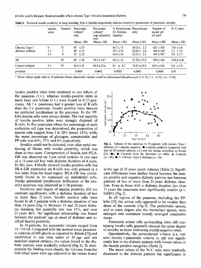
In 18 patients, the pancreatic volume ranged from 14-110 ml. Compared with the normal mean pancreat- ic volumes of 600 glands as reported by RSssle [22] and established in our own series of 45 age and sex matched control subjects, the values found in the dia- betic patients were markedly reduced (Fig. 1). To dem- onstrate the finding more clearly, the volumes from the individual cases were age adjusted to the values found
120-
E
(D ~_ 4o
O-
�9 A~ OA
f . 0 20 40 60 80
Age (years)
Fig. 2. Volume of the pancreas in 18 patients with chronic Type 1 diabetes (O = insulin negative; �9 = insulin positive) compared with that of 45 control subjects (A) and the means according to RSssle [21] ( I ) . �9 Controls from the literature (n=600); �9 Controls (n =45); �9 O Chronic Type 1 diabetes (n =18)
at the age of 35 years (peak volume) (Table 2). Signifi- cant differences were neither found between the insu- lin positive and negative diabetic patients nor between patients of less or more than 21 years diabetes dura- tion. Even in those with a diabetes duration less than 11 years the pancreata were significantly smaller (p < 0.0001) (Fig. 2).
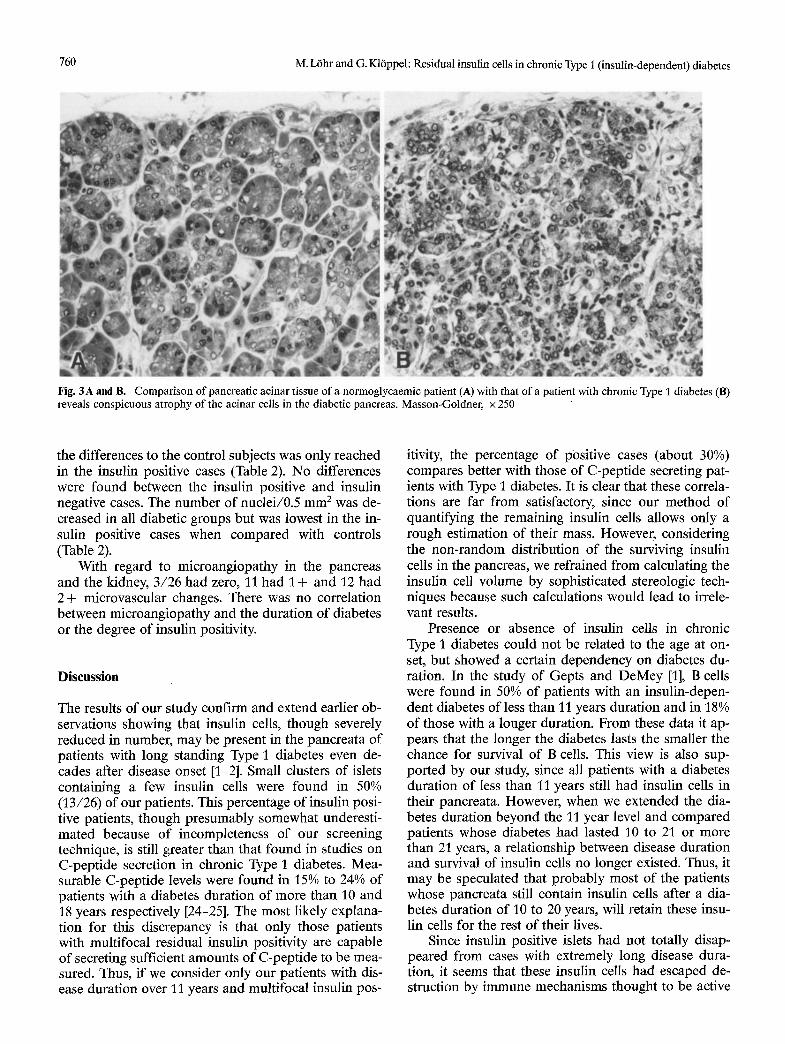
In all regions of the pancreas, including the PP- lobe [23], the acinar cells appeared to be smaller than those of the controls (Fig.3). The perilobular spaces, and to some degree also the interacinar spaces, were enlarged and contained loosely arranged connective tissue.
Pancreatic acinar cells surrounding islets still con- taining insulin cells generally showed the same degree of atrophy as those embracing insulin negative islets.
Quantitatively, the parenchymal volume (i.e. vol- ume density • pancreatic volume in ml) was signifi- cantly less in the diabetic patients with lowest values in the insulin positive categories (Table 2).
The mean values of the N / C ratio were markedly decreased in the diabetic patients but significance in
760 M. Lrhr and G. K16ppel: Residual insulin cells in chronic Type 1 (insulin-dependent) diabetes
Fig. 3 A and B. Comparison of pancreatic acinar tissue of a normoglycaemic patient (A) with that of a patient with chronic Type 1 diabetes (B) reveals conspicuous atrophy of the acinar cells in the diabetic pancreas. Masson-Goldner, x 250
the differences to the control subjects was only reached in the insulin positive cases (Table 2). No differences were found between the insulin positive and insulin negative cases. The number of nuclei/0.5 mm 2 was de- creased in all diabetic groups but was lowest in the in- sulin positive cases when compared with controls (Table 2).
With regard to microangiopathy in the pancreas and the kidney, 3/26 had zero, 11 had 1 + and 12 had 2 + microvascular changes. There was no correlation between micr0angiopathy and the duration of diabetes or the degree of insulin positivity.
Discussion
The results of our study confirm and extend earlier ob- servations showing that insulin cells, though severely reduced in number, may be present in the pancreata of patients with long standing Type 1 diabetes even de- cades after disease onset [1-2]. Small clusters of islets containing a few insulin cells were found in 50% (13/26) of our patients. This percentage of insulin posi- tive patients, though presumably somewhat underesti- mated because of incompleteness of our screening technique, is still greater than that found in studies on C-peptide secretion in chronic Type 1 diabetes. Mea- surable C-peptide levels were found in 15% to 24% of patients with a diabetes duration of more than 10 and 18 years respectively [24-25]. The most likely explana- tion for this discrepancy is that only those patients with multifocal residual insulin positivity are capable of secreting sufficient amounts of C-peptide to be mea- sured. Thus, if we consider only our patients with dis- ease duration over 11 years and multifocal insulin pos-
itivity, the percentage of positive cases (about 30%) compares better with those of C-peptide secreting pat- ients with Type 1 diabetes. It is clear that these correla- tions are far from satisfactory, since our method of quantifying the remaining insulin cells allows only a rough estimation of their mass. However, considering the non-random distribution of the surviving insulin cells in the pancreas, we refrained from calculating the insulin cell volume by sophisticated stereologic tech- niques because such calculations would lead to irrele- vant results.
Presence or absence of insulin cells in chronic Type 1 diabetes could not be related to the age at on- set, but showed a certain dependency on diabetes du- ration. In the study of Gepts and DeMey [1], B cells were found in 50% of patients with an insulin-depen- dent diabetes of less than 11 years duration and in 18% of those with a longer duration. From these data it ap- pears that the longer the diabetes lasts the smaller the chance for survival of B cells. This view is also sup- ported by our study, since all patients with a diabetes duration of less than 11 years still had insulin ceils in their pancreata. However, when we extended the dia- betes duration beyond the 11 year level and compared patients whose diabetes had lasted 10 to 21 or more than 21 years, a relationship between disease duration and survival of insulin cells no longer existed. Thus, it may be speculated that probably most of the patients whose pancreata still contain insulin cells after a dia- betes duration of 10 to 20 years, will retain these insu- lin cells for the rest of their lives.
Since insulin positive islets had not totally disap- peared from cases with extremely long disease dura- tion, it seems that these insulin cells had escaped de- struction by immune mechanisms thought to be active

M. L6hr and G. K16ppel: Residual insulin cells in chronic Type 1 (insulin-dependent) diabetes 761
in Type I diabetes [26]. One of the factors under recent investigation is an aberrant HLA-DR expression on B cells which is thought to be involved in the induction of the autoimmune process and possibly precedes in- sulitis [26-28]. In our series of 13 insulin positive pat- ients, we found no insulitis and in only one patient co- expression of insulin and HLA-DR in very few islet cells. There are two hypotheses which might explain the phenomenon of long surviving insulin cells. Either these residual insulin cells had never expressed HLA- DR and have to be considered as remnants of an origi- nally HLA-DR free B-cell population or they represent regenerated B cells which no longer express HLA-DR [26]. Though there is as yet no ready explanation for the longtime survival of insulin cells, this observation is of interest with respect to the theoretical possibility to reactivate the growth of this resting pool of B cells.
Patients with chronic Type 1 diabetes display a considerable reduction in weight and volume of the pancreas [1, 6-8]. These findings are confirmed by our study, since all but one pancreas of our series showed volume values far below those found in nondiabetic subjects [22]. This volume reduction of the pancreas was found to be due to a severe atrophy of all acinar cells (including those of the PP-lobe), since the reduc- tion in parenchymal volume was paralleled by a de- cline in cell numbers per given area (expressed as nu- clei per 0.5 mm2). The fact that in the diabetic pancrea- ta the acinar cells, despite their smaller size, did not increase in number per area can be explained by the development of a diffuse interstitial fibrosis conspicu- ously enlarging the interstitial spaces. A comparison of the ratio of the nuclear area to the acinar cell area (N/C ratio) of control subjects with that in diabetic patients showed that the decrease in cell size was par- ticularly due to a decline of the nuclear volume.
Lack of endogenous insulin has been suggested as causing the atrophy of the pancreas in chronic Type 1 diabetes [12, 29-31], as insulin has been shown to pro- mote growth and protein synthesis of the acinar cells [11-13, 30]. This conclusion is substantiated by clinical studies on the exocrine secretory capacity of the pan- creas in chronic Type I diabetes demonstrating de- creased secretion of enzymes [32-36]. Frier et al. found that the preservation of pancreatic function correlates with the duration of diabetes [32] and the persistence of B-cell secretory activity measured by C-peptide lev- els [33]. Domschke et al. [34] and Lankisch et al. [35], however, were not able to confirm these correlations. Our results are more in keeping with the latter observa- tions, since we failed to demonstrate clear relationships between the extent of exocrine atrophy and residual insulin positivity, the age at onset or duration of diabe- tes. This suggests that insulin secretion from a few re- maining insulin cells is not sufficient to exert a signifi- cant trophic effect on the acinar cells.
As a further factor which may influence pancreatic exocrine atrophy, microangiopathy has to be dis-
cussed. In our study, however, microangiopathy, as judged by the degree of renal glomerulosclerosis and pancreatic arteriosclerosis, was found to be unrelated to the extent of exocrine atrophy and thus seems to play no significant role in the pathogenesis of pan- creatic atrophy in chronic Type 1 diabetes.
In conclusion, we demonstrated persisting residual insulin positivity in about 40% of patients with Type 1 diabetes after a disease duration of more than 11 years. Exocrine atrophy, though closely linked to a state of insulin deficiency, appeared not to be further influ- enced by such variables as surviving insulin cells, dia- betes duration or microangiopathy.
Acknowledgments. We would like to express our sincere thanks to Ms. S. Peters, Ms. R. Malik and Ms. L. Blaudzun for their excellent technical assistance. We are thankful to Pl'of. G. Delling, Institute of Pathology, Univ. of Hamburg, for the collaboration regarding the morphometric evaluation. We thank Ms. U.Roggenbuck and Dr. W. Rehpenning for their help regarding statistical analysis of the data. Sincere thanks are also due to Dr. A. Foulis, Glasgow, for his help in the analysis of the HLA-DR stained sections.
References
1. Gepts W, Demey J (1978) Islet cell survival determined by mor- phology. An immunocytochemical study of the islets of Langer- hans in juvenile diabetes mellitus. Diabetes 27 [Suppl 1]: 251-261
2. Foulis AK, Liddle CN, Farquharson MA, Richmond JA, Weir RS (1986) The histopathology of the pancreas in Type 1 (insulin- dependent) diabetes mellitus: a 25-year review of deaths in pat- ients under 20 years of age in the United Kingdom. Diabetologia 29:267-274
3. Ki6ppel G (1984) Islet histopathology in diabetes mellitus. In: K16ppel G, Heitz PhU (eds) Pancreatic pathology. Churchill Liv- ingstone, London, pp 154-192
4. K16ppel G, L6hr M, Habich K, Oberholzer M, Heitz PhU (1985) Islet pathology and the pathogenesis of type I and 2 diabetes re- visited. Surv Synth Path Res 4:110-125
5. Gepts W (1965) Pathologic anatomy of the pancreas in juvenile diabetes mellitus. Diabetes 14:61%633
6. Doniach I (1974) Pathology of the islets of Langerhans in diabe- tes mellitus. In: Bastenie PA, Gepts W, Addison GM (eds) Im- munity and autoimmunity in diabetes mellitus. Excerpta Medica, Amsterdam New York, pp 175-182
7. MacLean N, Ogilvie RF (1955) Quantitative estimation of the pancreatic islet tissue in diabetic subjects. Diabetes 4:367-376
8. MacLean N, Ogilvie RF (1959) Observations on the pancreatic islet tissue of young diabetic subjects. Diabetes 8:83-91
9. Foulis AK, Stewart JA (1984) The pancreas in recent onset Type 1 (insulin-dependent) diabetes mellitus: insulin content of islets, insulitis and associated changes in the exocrine acinar tis- sue. Diabetologia 26:456-461
10. Gepts W (1981) Islet changes in human diabetes. In: Cooperstein S J, Watkins D (eds) The islets of Langerhans. Academic Press, London, pp 321-356
11. Adler G, Kern HF (1975) Regulation of exocrine pancreatic se- cretory process by insulin. Horm Metab Res 7:290-296
12. Henderson JR, Daniel PM, Fraser PA (1981) The pancreas as a single organ: the influence of the endocrine upon the exocrine part of the gland. Gut 22:158-167
13. Korc M, Owerbach D, Quinto C, Rutter WJ (1981) Pancreatic is- let-acinar cell interaction: amylase messanger RNA levels are de- termined by insulin. Science 213:351-353
14. Malaisse-Lagae F, Stefan Y, Cox J, Pen'elet A, Orci L (1979)
762 M. L6hr and G. K16ppel: Residual insulin cells in chronic Type 1 (insulin-dependent) diabetes
Identification of a lobe rich in pancreatic polypeptide. Diabeto- logia 17:361-365
15. Kltppel G, Drenck CR, Oberholzer M, Heitz PhU (1984) Mor- phometric evidence for a striking B-cell reduction at the clinical onset of type 1 diabetes. Virchows Arch [A] 403: 441-452
16. Becker WM, Grtning G, Arndt tL Thiele HG (1981) Monoclonal antibody EP 113 raised against Reh ceils recognizes a subspe- cies of la like antigens shared by cALL + and B-cell lines. In: Knapp W (ed) Leucemia Markers. Academic Press, London
17. Epenetos AA, Bobrow LG, Adams TE, Collins CM, Isaacson PG, Bodmer WF (1985) A monoclonal antibody that detects HLA-D region antigen in routinely fixed, wax embedded sec- tions of normal and neoplastic lymphoid tissues. J Clin Pathol 38:12-17
18. Aherne WA, Dunnill MS (1982) Morphometry. Edward Arnold, London
19. Bloodworth JMB jr (1978) A re-evaluation of diabetic glomerulo- sclerosis 50 years after the discovery of insulin. Hum Pathol 9: 439-453
20. Dixon WJ (Chief Editor) (1983) BMDP Statistical software. Univ. of California Press, Berkeley, California
21. Sachs L (1984) Angewandte Statistik: Statistische Methoden und ihre Anwendungen. Springer Verlag, Berlin Heidelberg New York
22. Rdssle R (1921) Beitr~ge zur Kenntnis der gesunden und kranken Bauchspeicheldrfise. Beitr Pathol Anat Allg Patho169:163-184
23. Rahier J, Wallon J, Loozen S, Lefevre A, Gepts W, Haot J (1983) The pancreatic polypeptide cells in the human pancreas: the ef- fect of age and diabetes. J Clin Endocrinol Metab 56:441-444
24. Madsbad S (1983) Prevalence of residual B-cell function and its metabolic consequences in Type I (insulin-dependent) diabetes. Diabetologia 24:141-147
25. Eft C, Faber O, Deckert T (1978) Persistent insulin secretion, as- sessed by plasma C-peptide estimation in long-term juvenile dia- betics with a low insulin requirement. Diabetologia 15:169-172
26. Bottazzo GF, Dean BM, McNally JM, Mackay EH, Swift PGF, Gamble DR (1985) In situ characterization of autoimmune phe- nomena and expression of HLA molecules in the pancreas in diabetic insulitis. N Engl J Med 313:353-360
27. Foulis AK, Farquharson MA (1986) Aberrant expression of HLA-DR antigens by insulin-containing t-cells in recent-onset type 1 diabetes mellitus. Diabetes 35:1215-1224
28. Foulis AK (1986) Class II major histocompatibility complex and organ specific autoimmunity in man. J Pathol 150:5-11
29. Malaisse-Lagae F, Ravazzola M, Robberecht P, Vandermeers A, Malaisse WJ, Orci L (1975) Exocrine pancreas: evidence for topographic partition of secretory function. Science 190:795-797
30. M6ssner J, Logsdon CD, Williams JA, Goldfine ID (1985) Insu- lin via its receptors regulates growth and amylase synthesis in pancreatic acinar cells. Diabetes 34:891-897
31. Williams JA, Goldfine ID (1985) The insulin pancreatic acinar axis: a review. Diabetes 34:980-986
32. Frier BM, Saunders JHB, Wormsley K, Bouchier IAD (1976) Ex- ocrine pancreatic function in juvenile-onset diabetes mellitus. Gut 17:695-691
33. Frier BM, Faber OK, Binder C, Elliot HL (1978) The effect of re- sidual insulin secretion on exocrine pancreatic function in juve- nile onset diabetes mellitus. Diabetologia 14:301-304
34. Domschke W, Tymper F, Domschke S, Demling L (1975) Exo- crine pancreatic function in juvenile diabetics. J Dig Dis 20: 309-312
35. Lankisch PG, Manthey G, Otto J, Koop H, Talaulicar M, Willms B, Creutzfeldt W (1982) Exocrine pancreatic function in insulin-dependent diabetes mellitus. Digestion 25:211-216
36. Hitanant S, Vanasaeng S, Tan-Ngarm-Trong D, Plengvanit U, Viranuvatti V (1986) Exocrine pancreatic function among diabet- ic patients in Thailand. Am J Gastroenterol 81:559-561
Received: 10 June 1987 and in revised form: 21 August 1987
Professor Gfinter K16ppel Department of Pathology Free University of Brussels Laarbeeklaan 101 B-1090 Brussels Belgium