research papers IUCr channels imaged by cryo-EM receptors and a ribbon of dimers are shaded in pink...
10
research papers IUCrJ (2017). 4 https://doi.org/10.1107/S2052252517005243 1 of 7 IUCrJ ISSN 2052-2525 BIOLOGY j MEDICINE Received 13 February 2017 Accepted 6 April 2017 Edited by S. S. Hasnain, University of Liverpool, England Keywords: nicotinic acetylcholine receptor; cholesterol; lipid microdomain; cryo-EM; helical image reconstruction. Supporting information: this article has supporting information at www.iucrj.org Segregation of lipids near acetylcholine-receptor channels imaged by cryo-EM Nigel Unwin* MRC Laboratory of Molecular Biology, Francis Crick Avenue, Cambridge Biomedical Campus, Cambridge CB2 0QH, England. *Correspondence e-mail: [email protected] Rapid communication at the chemical synapse depends on the action of ion channels residing in the postsynaptic membrane. The channels open transiently upon the binding of a neurotransmitter released from the presynaptic nerve terminal, eliciting an electrical response. Membrane lipids also play a vital but poorly understood role in this process of synaptic transmission. The present study examines the lipid distribution around nicotinic acetylcholine (ACh) receptors in tubular vesicles made from postsynaptic membranes of the Torpedo ray, taking advantage of the recent advances in cryo-EM. A segregated distribution of lipid molecules is found in the outer leaflet of the bilayer. Apparent cholesterol-rich patches are located in specific annular regions next to the transmembrane helices and also in a more extended ‘microdomain’ between the apposed subunits of neighbouring receptors. The particular lipid distribution can be interpreted straightforwardly in relation to the gating movements revealed by an earlier time-resolved cryo-EM study, in which the membranes were exposed briefly to ACh. The results suggest that in addition to stabilizing the protein, cholesterol may play a mechanical role by conferring local rigidity to the membrane so that there is productive coupling between the extracellular and membrane domains, leading to opening of the channel. 1. Introduction Rapid communication in the nervous system takes place by synaptic transmission, a process in which a neurotransmitter released from a nerve terminal binds transiently to ion chan- nels located in the oppositely facing postsynaptic membrane of the target cell, stimulating the channels to open and effecting a change in membrane potential. Models for the conformational change underlying the postsynaptic response have been derived from both X-ray and cryo-EM studies of several kinds of transmitter-gated ion channel, using recom- binant detergent-solubilized protein [for channels in the acetylcholine (ACh) receptor family, see Hibbs & Gouaux, 2011; Miller & Aricescu, 2014; Hassaine et al. , 2014; Du et al., 2015; Morales-Perez et al., 2016]. These studies have provided a wealth of structural insight at near-atomic resolution on the channels themselves. However, membrane lipids also play a vital role (daCosta & Baenziger, 2013), and the actual physiological mechanism is therefore best understood by analyzing both the channel and the lipid components together in their natural membrane setting. Postsynaptic membranes isolated from the Torpedo electric organ provide such a setting. They form tubular vesicles having the same lipid composition as, and near-equivalent
Transcript of research papers IUCr channels imaged by cryo-EM receptors and a ribbon of dimers are shaded in pink...

research papers
IUCrJ (2017). 4 https://doi.org/10.1107/S2052252517005243 1 of 7
IUCrJISSN 2052-2525
BIOLOGYjMEDICINE
Received 13 February 2017
Accepted 6 April 2017
Edited by S. S. Hasnain, University of Liverpool,
England
Keywords: nicotinic acetylcholine receptor;
cholesterol; lipid microdomain; cryo-EM; helical
image reconstruction.
Supporting information: this article has
supporting information at www.iucrj.org
Segregation of lipids near acetylcholine-receptorchannels imaged by cryo-EM
Nigel Unwin*
MRC Laboratory of Molecular Biology, Francis Crick Avenue, Cambridge Biomedical Campus, Cambridge CB2 0QH,
England. *Correspondence e-mail: [email protected]
Rapid communication at the chemical synapse depends on the action of ion
channels residing in the postsynaptic membrane. The channels open transiently
upon the binding of a neurotransmitter released from the presynaptic nerve
terminal, eliciting an electrical response. Membrane lipids also play a vital but
poorly understood role in this process of synaptic transmission. The present
study examines the lipid distribution around nicotinic acetylcholine (ACh)
receptors in tubular vesicles made from postsynaptic membranes of the Torpedo
ray, taking advantage of the recent advances in cryo-EM. A segregated
distribution of lipid molecules is found in the outer leaflet of the bilayer.
Apparent cholesterol-rich patches are located in specific annular regions next to
the transmembrane helices and also in a more extended ‘microdomain’ between
the apposed � subunits of neighbouring receptors. The particular lipid
distribution can be interpreted straightforwardly in relation to the gating
movements revealed by an earlier time-resolved cryo-EM study, in which the
membranes were exposed briefly to ACh. The results suggest that in addition to
stabilizing the protein, cholesterol may play a mechanical role by conferring
local rigidity to the membrane so that there is productive coupling between the
extracellular and membrane domains, leading to opening of the channel.
1. Introduction
Rapid communication in the nervous system takes place by
synaptic transmission, a process in which a neurotransmitter
released from a nerve terminal binds transiently to ion chan-
nels located in the oppositely facing postsynaptic membrane
of the target cell, stimulating the channels to open and
effecting a change in membrane potential. Models for the
conformational change underlying the postsynaptic response
have been derived from both X-ray and cryo-EM studies of
several kinds of transmitter-gated ion channel, using recom-
binant detergent-solubilized protein [for channels in the
acetylcholine (ACh) receptor family, see Hibbs & Gouaux,
2011; Miller & Aricescu, 2014; Hassaine et al., 2014; Du et al.,
2015; Morales-Perez et al., 2016]. These studies have provided
a wealth of structural insight at near-atomic resolution on the
channels themselves. However, membrane lipids also play a
vital role (daCosta & Baenziger, 2013), and the actual
physiological mechanism is therefore best understood by
analyzing both the channel and the lipid components together
in their natural membrane setting.
Postsynaptic membranes isolated from the Torpedo electric
organ provide such a setting. They form tubular vesicles
having the same lipid composition as, and near-equivalent

architecture to, the postsynaptic membrane in situ (Heuser &
Salpeter, 1979; Unwin, 2013). Furthermore, the tubes are
amenable to crystallographic analysis, since their constituent
ion channels, ACh receptors, arrange on a helical surface
lattice. As a result, it has been possible to obtain an atomic
model of the membrane-bound ACh receptor, giving mole-
cular details of the five subunits (��, ��, �, � and �) encircling
the central ion path (Miyazawa et al., 2003; Unwin, 2005).
Most recently, a time-resolved cryo-EM study of ACh-reacted
tubes allowed a simple mechanical description of how ACh
opens the channel (Unwin & Fujiyoshi, 2012). In brief, ACh
binding to �� and �� triggers small concerted displacements
in the extracellular domain, and these communicate to the
membrane domain through the action of �� pushing against
the � subunit, which tilts outwards in the membrane to open
the pore. Although other changes, such as straightening of the
adjacent �� and � pore-lining helices, also occur, the
displacement outwards of � near the outer membrane surface
is the motion that most directly affects the lipids.
Here, we investigate the likely role played by the lipids
in facilitating this transient conformational change, taking
advantage of the recent advances in direct-electron detector
technology and computer software, which enable improved
definition of low-contrast features in the image. We find that
the outer leaflet of the bilayer has a segregated distribution of
lipids, indicated by patches of differing density in the region of
the phospholipid headgroups. The low-density patches, which
we identify with cholesterol-rich areas, are at equivalent
annular sites (involving helices M1 and M4) around the
receptor and in a more extended space between the apposed �subunits of neighbouring receptors. This distribution suggests
that cholesterol may play a dual role in (i) stabilizing the
transmembrane �-helical organization and (ii) conferring local
rigidity to the membrane so that there is productive coupling
between the extracellular and membrane domains, leading to
opening of the channel. Similar facilitating roles for the lipids
have been proposed for other membrane proteins that
undergo large-scale conformational changes (see, for example,
Cornelius et al., 2015; Landreh et al., 2016).
2. Methods
2.1. Specimen preparation
Tubular postsynaptic membrane vesicles were prepared
from fresh Torpedo electric organ as described by Kubalek et
al. (1987) and suspended in 100 mM sodium cacodylate, 1 mM
calcium chloride pH 7.0. Aliquots (3.8 ml) of this solution were
applied to holey carbon support grids and blotted to retain the
specimens in a thin aqueous film before plunging into liquid
nitrogen-cooled ethane. Possible heterogeneity in lipid
composition owing to individual or seasonal variation was
minimized in these experiments by using tubes from only one
fish.
2.2. Cryo-EM data collection and image processing
We chose �750 A diameter tubes belonging to the (�17, 5)
helical family (Miyazawa et al., 1999) for the structure analysis,
which applies the single-particle method of helical image
reconstruction (Egelman, 2000; Sachse et al., 2007; He &
Scheres, 2017) rather than the Fourier–Bessel method
(Toyoshima & Unwin, 1990; Beroukhim & Unwin, 1997)
research papers
2 of 7 Nigel Unwin � Segregation of lipids near acetylcholine-receptor channels IUCrJ (2017). 4
Figure 1Two-dimensional classification and image selection. (a) Micrograph of a (�17, 5) tube recorded at 2.4 mm underfocus with a Falcon 2 direct-electrondetector. (b) Examples of good class averages. (c) Fourier transform of a good class average, showing ‘pairing layer lines’ (arrows) reflecting the dimericarrangement of receptors on the tube surface lattice (Brisson & Unwin, 1984). The figures in parentheses denote the (h, k; n) indices for major low-resolution layer lines (Toyoshima & Unwin, 1990). Data from class averages where the pairing layer lines [(0, 5; �25) and (1, 3; 2)] were weak or non-existent were omitted from the subsequent image-processing steps.

applied in earlier studies. Both methods correct for long-range
variations in the surface lattice and for distortions, such as
bending and out-of-plane tilt, which are inevitably present in
ACh-receptor tubes because of their shell-like architecture
and the fluid nature of the lipid matrix in which the protein is
embedded. The (�17, 5) tubes have dihedral (D1) symmetry
and form a single-start helix with average twist and rise values
of 147.0� and 5.9 A, respectively.
The specimens were imaged with an FEI Titan Krios
transmission electron microscope incorporating a 70 mm
diameter objective aperture and operating in nanoprobe mode
at an accelerating voltage of 300 kV. Micrographs were
recorded on a Falcon 2 4096 � 4096 pixel direct-electron
detector after searching for straight (�17, 5) tubes spanning
holes in the carbon support film (Fig. 1a). The calibrated pixel
size was 1.34 A and the total dose on the specimen was
35 e A�2 fractionated over 22 frames.
We used 295 micrographs of the tubes, recorded with an
underfocus range of 1–3 mm and selected by inspection of their
Fourier transforms. Each tube image was divided into over-
lapping segments using a box size of 1024� 1024 pixels and an
inter-box spacing of 100 pixels. Micrograph frame stacks were
drift-corrected using UCSF MotionCorr (Li et al., 2013), and
contrast-transfer function parameters were determined locally
along the tube axis using Gctf (Zhang, 2016). For three-
dimensional classification, an initial 20 A resolution model
was generated by Fourier–Bessel synthesis from structure-
factor terms along the layer lines (Miyazawa et al., 1999). All
subsequent image-processing steps were performed in
RELION (Scheres, 2012; He & Scheres, 2017) after binning
the images times two.
Reference-free two-dimensional classification was applied
to the extracted segments, yielding 34 class averages (Fig. 1b),
of which 80% were considered good on the basis of the quality
of their Fourier transforms (Fig. 1c). Rejected segments
included disordered or defective regions of tubes and regions
where the projected p2 symmetry (indicated the presence of
‘pairing layer lines’; arrows in Fig. 1c) was weak or absent.
Only segments associated with the good classes were retained
for subsequent three-dimensional classification.
Three-dimensional classification was performed on 7196
extracted segments using a cylindrical mask of inner and outer
radii sufficient to include all densities comprising the tube with
a margin of �40 A on either side. The ‘central Z-length’, used
for searching and applying helical symmetry in real space, was
10% of the box size. In an initial step, three class averages
were generated. One of these class averages (class 1; 2563
segments) showed better alignment errors and resolution than
research papers
IUCrJ (2017). 4 Nigel Unwin � Segregation of lipids near acetylcholine-receptor channels 3 of 7
Figure 2Single-particle helical reconstruction from (�17, 5) tubes. (a) Masked-out volume showing the helical arrangement of ACh receptors; a �–� linked dimerof receptors and a ribbon of dimers are shaded in pink and grey, respectively. (b) Central cross-section (top) and radial section (bottom) from the three-dimensional density map cutting through receptors and the lipid-bilayer matrix in which they are embedded. (c) Enlargements of regions in (b) withatomic models of the ACh receptor (PDB entry 2bg9) superimposed. Circles identify areas next to the � subunit where densities corresponding to theouter phospholipid headgroups are weak or missing; the adjacent helices are M1 (lower circle), and M4 and M1 (upper circle). Subunit colours: ��, red;��, orange; �, green; �, cyan; �, blue. Inverted contrast.

the other two. Moreover, the reconstructed tube from this
class had the expected diameter, whereas the reconstructed
tubes from the other two classes, while displaying the same
features, were slightly wider. Based on these criteria, the class
1 segments appeared to be the least affected by flattening
owing to insufficient thickness of the ice and/or other possible
sources of heterogeneity, such as variation in lipid content.
This class was therefore refined subsequently, yielding a
resolution for the final three-dimensional density map of 8.4 A
(FSC = 0.143 threshold, two independently refined half-data
sets).
An atomic model of the ACh receptor (PDB entry 2bg9;
Unwin, 2005) was fitted to the densities using UCSF Chimera
(Pettersen et al., 2004) and was incorporated in Fig. 2 using
PyMOL (DeLano, 2002).
3. Results
3.1. Architecture of a tubular vesicle
The single-particle helical reconstruction of the (�17, 5)
tubes (Fig. 2a) shows very similar molecular details as estab-
lished using the alternative Fourier–Bessel approach. The
receptors form dimers linked by a disulfide bridge between the
� subunits of neighbouring molecules (Chang & Bock, 1977;
Brisson & Unwin, 1984), as indicated by the pink shading in
Fig. 2(a). The helical surface lattice is built from ribbons of
dimers (grey shading), which associate side by side.
Cross-sectional slices through the
three-dimensional map normal to the
membrane plane (Fig. 2b) are domi-
nated by paired tracks of density
corresponding to the phospholipid
headgroups of the lipid bilayer and by
irregular blocks of density corre-
sponding to individual receptors packed
tightly in the lipid matrix. The extra-
cellular domain of the receptor, built
around a �-sandwich core, extends
about 60 A from the outer membrane
surface. The intracellular domain, which
is shaped largely by a conical arrange-
ment of �-helices, extends about 40 A
from the inner membrane surface. The
receptor has an all-�-helical transmem-
brane domain, with four helices (M1,
M2, M3 and M4) per subunit. The
helices are sufficiently long to span the
outer phospholipid headgroups as well
as the central low-density hydrophobic
portion of the bilayer (see x3.2).
To evaluate the protein structure, we
fitted an atomic model of the ACh
receptor (PDB entry 2bg9) to the
densities (see x2). Fig. 2(c) shows slices
through the atomic model super-
imposed on several views of the
channel, with each subunit identified in a different colour. As
can be seen, most of the protein densities are accounted for by
the atomic model. An exception is the density arising from a
short transverse helix, which has been modelled in the related
�4�2 nicotinic and 5HT3 receptor structures (Hassaine et al.,
2014; Morales-Perez et al., 2016) and lies on the inner
membrane surface. The clustering protein rapsyn may be
responsible for some of the density at the base of the receptor,
but is otherwise invisible because it forms variable networks
that do not match the helical symmetry of a tube (Zuber &
Unwin, 2013).
3.2. The lipid bilayer
The single-particle helical method applied in the present
study, unlike the Fourier–Bessel helical method, uses classifi-
cation to ensure radial uniformity of the tubes and does not
rely on the averaging of equatorial Fourier terms affected by
the edges of the boxed-out areas, which are prone to error. As
a result, the radial density distribution is now more accurate
and the membrane appears with better definition than could
be achieved previously. The outer leaflet of the bilayer is of
particular interest because it is the portion that is most
significantly implicated in the conformational change to open
the channel.
The outer leaflet presents a fairly uniform�10 A thick band
of density, arising from the phospholipid headgroups, that
occupies almost the entire space between the embedded
research papers
4 of 7 Nigel Unwin � Segregation of lipids near acetylcholine-receptor channels IUCrJ (2017). 4
Figure 3Sections tangential to the tube axis spanning the outer phospholipid headgroup region of the lipidbilayer. Low-density patches are present in the space between the � subunits of neighbouringreceptors (see, for example, the box in the third panel) and at the M1/M4 lipid–helix interface of theremaining four subunits (arrows). Inset: enlargement of the boxed dimer of receptors and thecentral low-density microdomain, identifying individual subunits and the M1 (orange) and M4(green) helices of �. The panels are at 2.7 A intervals from the outermost side (left) to the innermostside of the headgroup region. Inverted contrast.
helices of the receptors. However, in several locations imme-
diately adjacent to a protein surface, the densities comprising
the band are diminished or missing altogether (circles in
Fig. 2c).
Sections tangential to the tube axis, encompassing the outer
phospholipid headgroups, provide a more complete picture of
these ‘low-density patches’ next to the protein surfaces. Fig. 3
shows four such sections extending from one side of the
headgroup region to the other. The helices of the embedded
receptors are now displayed in cross-section, arranged
pentagonally around the water-filled pore, with densities from
the lipids occupying the space surrounding them. Two kinds of
low-density patch are apparent within the otherwise relatively
uniform densities associated with the phospholipid head-
groups. The largest occupies the area between the � subunits
of neighbouring receptors (box in Fig. 3) and is at right angles
to the slices through the same region circled in Fig. 2(c). This
patch forms a ‘microdomain’: an elongated (�26 � 11 A)
parallelogram framed on its sides by the polar phospholipid
headgroups and on its ends by the helices M1 and M4 (inset in
Fig. 3). In addition, there are smaller patches located next to
the M1 and M4 helices of the �, ��, � and �� subunits (arrows
in Fig. 3). Both kinds of patch appear to extend across the
entire headgroup region, although the details of the smaller
patches are less clear.
4. Discussion
Most structural studies of the roles played by lipids in the
function of membrane proteins are conducted on the protein
in detergent or in an artificial membrane-like environment,
viewing lipid molecules that are immobilized on the protein
surface. In this regard, single-particle cryo-EM of detergent-
solubilized protein combined with lipid-nanodisc technology
offers a promising approach (Bayburt et al., 2002; Gao et al.,
2016). Structure determination based on reconstituted
proteoliposomes (see, for example, Gonen et al., 2005; Wang &
Sigworth, 2009; Kudryashev et al., 2016) provides a potential
alternative route. However, none of these methods are likely
to recapitulate precisely the lipid environment as it exists in
situ. Here, by imaging and reconstructing the unperturbed
natural membrane, we obtain the most complete physiological
perspective, showing how native lipids are distributed near the
protein in the outer leaflet of the native bilayer. Low-density
patches are visible among the phospholipid headgroups
(Fig. 3), indicating that the lipid composition is modified not
only at specific annular sites around the protein but also in a
more extended microdomain bordering the protein surfaces.
The resolution achieved from the limited number of images
has not been sufficient to define any of the lipids creating these
low-density patches as discrete molecular entities. However,
we can confidently interpret the patches to reflect areas that
are enriched in (or composed entirely of) cholesterol, which
has a much smaller headgroup than the other lipids. For
example, the microdomain within the phospholipid headgroup
region (inset in Fig. 3) gives rise to a density equal to that of
water in the pore at the centre of the receptor (Supplementary
Fig. S1). Cholesterol in the underlying membrane accounts for
this density, since it exposes only a hydroxyl and contributes
no mass this far from the hydrophobic core of the membrane.
Furthermore, the fraction of the outer leaflet of the bilayer
occupied by the microdomain (�6%) is readily furnished by
available cholesterol molecules, which have a concentration
of at least 35 mol% in Torpedo postsynaptic membrane
(Rotstein et al., 1987). Phospholipids with relatively small
headgroups could not be responsible for the low density
because they are present in only very small amounts (for
example, phosphatidic acid at <0.5 mol%; Rotstein et al.,
1987).
An association of cholesterol with the ACh receptor has
been well documented (for a recent review, see Barrantes,
2010), despite its absence from the structures of all ACh-
receptor family members solved to date. Cholesterol plays an
essential part in enabling the classical physiological transitions
associated with rapid switching of the protein between closed
(or resting), open and desensitized states (Criado et al., 1982;
Ochoa et al., 1983; Sunshine & McNamee, 1992; Ryan et al.,
1996; Rankin et al., 1997; Hamouda et al., 2006). The protein
adopts an uncoupled conformation, in which the pore cannot
open in response to ACh binding, in reconstituted membranes
lacking anionic lipids and this neutral lipid (daCosta &
Baenziger, 2009). Evidently, the high concentration of
cholesterol in the Torpedo postsynaptic membrane is needed
for normal synaptic activity, since the functionality of the
receptor diminishes progressively as the concentration is
research papers
IUCrJ (2017). 4 Nigel Unwin � Segregation of lipids near acetylcholine-receptor channels 5 of 7
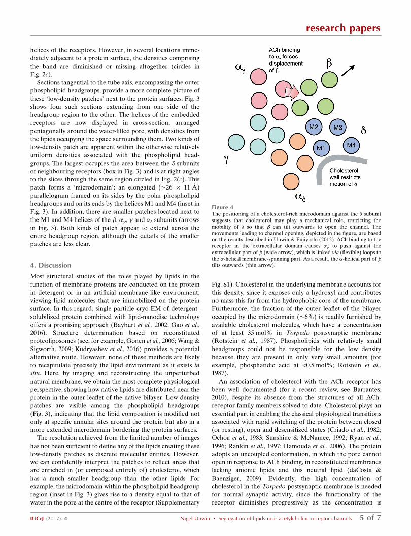
Figure 4The positioning of a cholesterol-rich microdomain against the � subunitsuggests that cholesterol may play a mechanical role, restricting themobility of � so that � can tilt outwards to open the channel. Themovements leading to channel opening, depicted in the figure, are basedon the results described in Unwin & Fujiyoshi (2012). ACh binding to thereceptor in the extracellular domain causes �� to push against theextracellular part of � (wide arrow), which is linked via (flexible) loops tothe �-helical membrane-spanning part. As a result, the �-helical part of �tilts outwards (thin arrow).

lowered from the natural amount (see, for example, Rankin et
al., 1997; Hamouda et al., 2006).
How does the inferred segregation of cholesterol into areas
where it is enriched (arrows and box in Fig. 3) influence the
ACh-induced conformational change to open the channel?
Clearly, specific annular lipid–protein interactions may be
required to stabilize the �-helical organization in the
membrane: for example, to restrict relative motion between
the implicated helices M1 and M4 (which does not occur in
any of the subunits; Unwin & Fujiyoshi, 2012). Indeed,
molecular-dynamics simulations predict that cholesterol binds
in the groove between M1 and M4 (Brannigan et al., 2008).
However, the presence of a microdomain next to the � subunit,
which is large enough to incorporate several cholesterol
molecules (Supplementary Fig. S2), hints that a less direct,
physical involvement may also be important (Fig. 4).
As sketched in Fig. 4, the ACh-binding subunit, ��, drives
the conformational change by pushing against the extra-
cellular part of the � subunit (wide arrow), causing the
membrane part of this subunit to tilt outwards (thin arrow).
To achieve the required outward tilt, and hence productive
coupling between the extracellular and membrane parts, the �subunit must resist the force transmitted through � that would
push against it. We suggest that the cholesterol-rich micro-
domain helps to impose this resistance by reducing the free
volume available for molecular motion in the hydrophobic
portion of the bilayer (Song et al., 2014), hence conferring
rigidity next to �. Without a supporting wall of rigid sterol
groups, the required shift-to-tilt coupling of the � subunit
might not be favoured, leading to an unproductive confor-
mational change when ACh binds to the receptor, rather than
conversion to an open channel.
To corroborate this suggestion and illuminate further how
the lipids assist in channel opening, more detailed structural
information on the lipids in the intact membrane will be
required. This should be achievable by the helical recon-
struction approach used in the present study when applied to
more images of tubes. Of particular interest are the organi-
zation and extent of mobility of the microdomain lipids. Since
they span both channels of the dimer completely, it seems
possible that in addition to their anchoring effect (Fig. 4) they
are able to mediate cooperative interactions between the
paired channels, thereby accounting for the synchronous
gating activity shown by single-channel conductance experi-
ments (Schindler et al., 1984).
5. Conclusion
This paper extends a time-resolved cryo-EM study of gating of
membrane-embedded ACh-receptor channels by examining
the distribution of lipids surrounding the channels in the outer
leaflet of the bilayer, where the displacements are greatest.
The results suggest that cholesterol, in addition to stabilizing
the protein, may be needed to achieve local rigidity of the
membrane so that a productive conformational change takes
place when ACh binds, triggering the channel to open.
6. Related literature
The following reference is cited in the Supporting Information
for this article: Wennberg et al. (2012).
Acknowledgements
I thank the staff of the Station Biologique de Roscoff for the
supply of T. marmorata electric rays, the staff of the British
Antarctic Survey for providing facilities for handling and
dissection of the fish, and Theresa Langford, who set up and
coordinated these aspects of the project. I also wish to thank
Shaoda He, Jude Short and Sjors Scheres for their help and
advice with the single-particle approach to helical image
reconstruction.
Funding information
Funding for this research was provided by: National Institutes
of Health (award No. GM61941); Medical Research Council
(award No. MC-A023-5PD1D).
References
Barrantes, F. J. (2010). Subcell. Biochem. 51, 467–487.Bayburt, T. H., Grinkova, Y. V. & Sligar, S. G. (2002). Nano Lett. 2,
853–856.Beroukhim, R. & Unwin, N. (1997). Ultramicroscopy, 70, 57–81.Brannigan, G., Henin, J., Law, R., Eckenhoff, R. & Klein, M. L.
(2008). Proc. Natl Acad. Sci. USA, 105, 14418–14423.Brisson, A. & Unwin, N. (1984). J. Cell Biol. 99, 1202–1211.Chang, H. W. & Bock, E. (1977). Biochemistry, 16, 4513–4520.Cornelius, F., Habeck, M., Kanai, R., Toyoshima, C. & Karlish, S. J. D.
(2015). Biochim. Biophys. Acta, 1848, 1729–1743.Criado, M., Eibl, H. & Barrantes, F. J. (1982). Biochemistry, 21, 3622–
3629.daCosta, C. J. & Baenziger, J. E. (2009). J. Biol. Chem. 284, 17819–
17825.daCosta, C. J. & Baenziger, J. E. (2013). Structure, 21, 1271–1283.DeLano, W. L. (2002). PyMOL. http://www.pymol.org.Du, J., Lu, W., Wu, S., Cheng, Y. & Gouaux, E. (2015). Nature
(London), 526, 224–229.Egelman, E. H. (2000). Ultramicroscopy, 85, 225–234.Gao, Y., Cao, E., Julius, D. & Cheng, Y. (2016). Nature (London), 534,
347–351.Gonen, T., Cheng, Y., Sliz, P., Hiroaki, Y., Fujiyoshi, Y., Harrison, S. C.
& Walz, T. (2005). Nature (London), 438, 633–638.Hamouda, A. K., Sanghvi, M., Sauls, D., Machu, T. K. & Blanton,
M. P. (2006). Biochemistry, 45, 4327–4337.Hassaine, G., Deluz, C., Grasso, L., Wyss, R., Tol, M. B., Hovius, R.,
Graff, A., Stahlberg, H., Tomizaki, T., Desmyter, A., Moreau, C., Li,X.-D., Poitevin, F., Vogel, H. & Nury, H. (2014). Nature (London),512, 276–281.
He, S. & Scheres, S. H. W. (2017). J. Struct. Biol., https://doi.org/10.1016/j.jsb.2017.02.003.
Heuser, J. E. & Salpeter, M. (1979). J. Cell Biol. 82, 150–173.Hibbs, R. E. & Gouaux, E. (2011). Nature (London), 474, 54–60.Kubalek, E., Ralston, S., Lindstrom, J. & Unwin, N. (1987). J. Cell
Biol. 105, 9–18.Kudryashev, M., Castano-Dıez, D., Deluz, C., Hassaine, G., Grasso,
L., Graf-Meyer, A., Vogel, H. & Stahlberg, H. (2016). Structure, 24,165–170.
Landreh, M., Marklund, E. G., Uzdavinys, P., Degiacomi, M. T.,Coincon, M., Gault, J., Gupta, K., Liko, I., Benesch, J. L. P., Drew,D. & Robinson, C. V. (2016). Nat. Commun. 8, 13993.
research papers
6 of 7 Nigel Unwin � Segregation of lipids near acetylcholine-receptor channels IUCrJ (2017). 4
Li, X., Mooney, P., Zheng, S., Booth, C. R., Braunfeld, M. B.,Gubbens, S., Agard, D. A. & Cheng, Y. (2013). Nat. Methods, 10,584–590.
Miller, P. S. & Aricescu, A. R. (2014). Nature (London), 512, 270–275.
Miyazawa, A., Fujiyoshi, Y., Stowell, M. & Unwin, N. (1999). J. Mol.Biol. 288, 765–786.
Miyazawa, A., Fujiyoshi, Y. & Unwin, N. (2003). Nature (London),423, 949–955.
Morales-Perez, C. L., Noviello, C. M. & Hibbs, R. E. (2016). Nature(London), 538, 411–415.
Ochoa, E. L., Dalziel, A. W. & McNamee, M. G. (1983). Biochim.Biophys. Acta, 727, 151–162.
Pettersen, E. F., Goddard, T. D., Huang, C. C., Couch, G. S.,Greenblatt, D. M., Meng, E. C. & Ferrin, T. E. (2004). J. Comput.Chem. 25, 1605–1612.
Rankin, S. E., Addona, G. H., Kloczewiak, M. A., Bugge, B. & Miller,K. W. (1997). Biophys. J. 73, 2446–2455.
Rotstein, N. P., Arias, H. R., Barrantes, F. J. & Aveldano, M. I. (1987).J. Neurochem. 49, 1333–1340.
Ryan, S. E., Demers, C. N., Chew, J. P. & Baenziger, J. E. (1996). J.Biol. Chem. 271, 24590–24597.
Sachse, C., Chen, J. Z., Coureux, P.-D., Stroupe, M. E., Fandrich, M. &Grigorieff, N. (2007). J. Mol. Biol. 371, 812–835.
Scheres, S. H. W. (2012). J. Struct. Biol. 180, 519–530.Schindler, H., Spillecke, F. & Neumann, E. (1984). Proc. Natl Acad.
Sci. USA, 81, 6222–6226.Song, Y., Kenworthy, A. K. & Sanders, C. R. (2014). Protein Sci. 23, 1–22.Sunshine, C. & McNamee, M. G. (1992). Biochim. Biophys. Acta,
1108, 240–246.Toyoshima, C. & Unwin, N. (1990). J. Cell Biol. 111, 2623–2635.Unwin, N. (2005). J. Mol. Biol. 346, 967–989.Unwin, N. (2013). Q. Rev. Biophys. 46, 283–322.Unwin, N. & Fujiyoshi, Y. (2012). J. Mol. Biol. 422, 617–634.Wang, L. & Sigworth, F. L. (2009). Nature (London), 461, 292–295.Wennberg, C. L., van der Spoel, D. & Hub, J. S. (2012). J. Am. Chem.
Soc. 134, 5351–5361.Zhang, K. (2016). J. Struct. Biol. 193, 1–12.Zuber, B. & Unwin, N. (2013). Proc. Natl Acad. Sci. USA, 110, 10622–
10627.
research papers
IUCrJ (2017). 4 Nigel Unwin � Segregation of lipids near acetylcholine-receptor channels 7 of 7

IUCrJ (2017). 4, doi:10.1107/S2052252517005243 Supporting information
IUCrJ Volume 4 (2017)
Supporting information for article:
Segregation of lipids near acetylcholine-receptor channels imaged by cryo-EM
Nigel Unwin

IUCrJ (2017). 4, doi:10.1107/S2052252517005243 Supporting information, sup-1
Figure S1 Contour map of the densities in the middle of the outer headgroup region,
corresponding to the boxed area in Fig. 3. The phospholipid headgroups (PL) give rise to a
fairly uniform positive density in the space between individual protein molecules, because of
their (presumably) random disposition. The space framed by the M1 and M4 helices of the δ
subunits, and associated with the microdomain (M), is an exception. The weaker density here
is equal to that of the lumen of the pore, indicating a depletion of the large phospholipid
headgroups and their substitution by water. The presence of cholesterol in the underlying
membrane explains this anomaly, since cholesterol exposes only a hydroxyl and contributes
no mass this far from the hydrophobic core of the membrane (for examples of simulated
cholesterol-containing membranes, see: http://cmb.bio.uni-goettingen.de/cholmembranes.html).
Contours are at 0.6σ, with the relevant microdomain and pore regions in red.

IUCrJ (2017). 4, doi:10.1107/S2052252517005243 Supporting information, sup-2
Figure S2 Hypothetical depiction of a cluster of five cholesterol molecules located in the
microdomain space between the δ subunits of neighbouring receptors. The figure suggests that
the microdomain, fully occupied, could harbour as many as eight cholesterols. The co-
ordinates for the cluster were taken from a molecular dynamics simulation of a DOPC bilayer
containing 30 mol % cholesterol (Wennberg et al., 2012). The co-ordinates for the receptors
were obtained by fitting the atomic model (PDB entry 2bg9) to the densities corresponding to
a dimer in the 3D map; subunit colours as in Fig. 2 (αδ, orange; β, green; δ, blue).