
RESEARCH Open Access Decreased expression of LATS1 is … · RESEARCH Open Access Decreased...
9
RESEARCH Open Access Decreased expression of LATS1 is correlated with the progression and prognosis of glioma Tianhai Ji 1,2† , Dan Liu 2† , Wei Shao 2† , Wensheng Yang 2 , Haiqiao Wu 2 and Xiuwu Bian 1* Abstract Background: LATS1 is a tumor suppressor genes implicated in the pathogenesis of certain types of tumors, but its role is not known in human glioma. Methods: Using real-time PCR and immunohistochemistry, we detected the mRNA and protein expression of LATS1 in glioma. The effect of LATS1 on cell growth and invasion were investigated. Results: We found that mRNA and protein of LATS1 expression is significantly downregulated in glioma compared with normal control brain tissues. Furthermore, reduced LATS1 expression was markedly negatively correlated with WHO grade and KPS (p<0.001 and p<0.001) in glioma patients. Patients with lower LATS1 expression had a significantly shorter overall survival time than did patients with higher LATS1 expression. Multivariate analysis suggested that the level of LATS1 expression was an independent prognostic indicator (p<0.001) for the survival of patients with glioma. Forced expression of LATS1 in glioma U251 cells not only significantly suppressed cell growth, migration and invasion, but retarded cell cycle progression from G2/M to G1 in vitro. Finally, we found that overexpressed LATS1 markedly inhibited the expression level of cell cycle factor CCNA1. Conclusion: These results indicate that LATS1 is an important candidate tumor suppressor and its downregulated expression may contribute to glioma progression. Keywords: LATS1, Tumor suppressor, Prognosis, CCNA1 Background Gliomas are neuroectodermal tumors contributing to 30–45% of all human intracranial tumors that commonly arise in the white matter of cerebral hemisphere [1]. Due to its highly invasive ability, angiogenesis and the pres- ence of necrosis surrounding brain [2,3], malignant gli- omas are often incurable by surgery alone. The molecular pathogenesis of malignant gliomas is still un- clear, thus a major research effort has been directed at identifying novel specific glioma-associated genes which might play significant roles in glioma carcinogenesis. The LATS1 gene, a mammalian homolog of fly LATS originally isolated in Drosophila as a cell proliferation in- hibitor [4,5], is a speculative serine/threonine kinase that localizes to the mitotic apparatus. In mammalian cells, LATS1 is phosphorylated in a cell-cycle-dependent manner and complexes with CDC2 in early mitosis. The N-terminal region of the LATS1 protein binds CDC2 to form a complex showing decreased H1 histone kinase activity, indicating a role as a negative regulator of CDC2/cyclin A [6]. Lats1- knockout mice spontaneously developed large soft tissue sarcomas and ovarian stromal cell tumors and a high sensitivity to carcinogenic treat- ments, suggesting that Lats1 is a tumor suppressor at least in mice [7]. The human LATS1 gene has been mapped to chromosome 6q24-25 where loss of heterozy- gosity has been observed in ovarian [8], cervical [9], and breast cancers [10]. Overexpressed LATS1 not only causes G2-M arrest through the inhibition of CDC2 kin- ase activity in breast cancer cell line in vitro [11], but also significantly inhibited the tumorigenicity in vivo by inducing apoptosis [12]. Furthermore, recent investiga- tions demonstrated that hypermethylation of LATS1 gene promoter which caused downregulated expression of LATS1 is frequently observed in a few human tumors, such as breast cancer and astrocytoma [13,14]. * Correspondence: [email protected] † Equal contributors 1 Institute of Pathology and Southwest Cancer Center, Southwest Hospital, Third Military Medical University, Chongqing 400038, China Full list of author information is available at the end of the article © 2012 Ji et al.; licensee BioMed Central Ltd. This is an Open Access article distributed under the terms of the Creative Commons Attribution License (http://creativecommons.org/licenses/by/2.0), which permits unrestricted use, distribution, and reproduction in any medium, provided the original work is properly cited. Ji et al. Journal of Experimental & Clinical Cancer Research 2012, 31:67 http://www.jeccr.com/content/31/1/67
Transcript of RESEARCH Open Access Decreased expression of LATS1 is … · RESEARCH Open Access Decreased...

Ji et al. Journal of Experimental & Clinical Cancer Research 2012, 31:67http://www.jeccr.com/content/31/1/67
RESEARCH Open Access
Decreased expression of LATS1 is correlated withthe progression and prognosis of gliomaTianhai Ji1,2†, Dan Liu2†, Wei Shao2†, Wensheng Yang2, Haiqiao Wu2 and Xiuwu Bian1*
Abstract
Background: LATS1 is a tumor suppressor genes implicated in the pathogenesis of certain types of tumors, but itsrole is not known in human glioma.
Methods: Using real-time PCR and immunohistochemistry, we detected the mRNA and protein expression ofLATS1 in glioma. The effect of LATS1 on cell growth and invasion were investigated.
Results: We found that mRNA and protein of LATS1 expression is significantly downregulated in glioma comparedwith normal control brain tissues. Furthermore, reduced LATS1 expression was markedly negatively correlated withWHO grade and KPS (p<0.001 and p<0.001) in glioma patients. Patients with lower LATS1 expression had asignificantly shorter overall survival time than did patients with higher LATS1 expression. Multivariate analysissuggested that the level of LATS1 expression was an independent prognostic indicator (p<0.001) for the survival ofpatients with glioma. Forced expression of LATS1 in glioma U251 cells not only significantly suppressed cell growth,migration and invasion, but retarded cell cycle progression from G2/M to G1 in vitro. Finally, we found thatoverexpressed LATS1 markedly inhibited the expression level of cell cycle factor CCNA1.
Conclusion: These results indicate that LATS1 is an important candidate tumor suppressor and its downregulatedexpression may contribute to glioma progression.
Keywords: LATS1, Tumor suppressor, Prognosis, CCNA1
BackgroundGliomas are neuroectodermal tumors contributing to30–45% of all human intracranial tumors that commonlyarise in the white matter of cerebral hemisphere [1]. Dueto its highly invasive ability, angiogenesis and the pres-ence of necrosis surrounding brain [2,3], malignant gli-omas are often incurable by surgery alone. Themolecular pathogenesis of malignant gliomas is still un-clear, thus a major research effort has been directed atidentifying novel specific glioma-associated genes whichmight play significant roles in glioma carcinogenesis.The LATS1 gene, a mammalian homolog of fly LATS
originally isolated in Drosophila as a cell proliferation in-hibitor [4,5], is a speculative serine/threonine kinase thatlocalizes to the mitotic apparatus. In mammalian cells,LATS1 is phosphorylated in a cell-cycle-dependent
* Correspondence: [email protected]†Equal contributors1Institute of Pathology and Southwest Cancer Center, Southwest Hospital,Third Military Medical University, Chongqing 400038, ChinaFull list of author information is available at the end of the article
© 2012 Ji et al.; licensee BioMed Central Ltd. TCommons Attribution License (http://creativecreproduction in any medium, provided the or
manner and complexes with CDC2 in early mitosis. TheN-terminal region of the LATS1 protein binds CDC2 toform a complex showing decreased H1 histone kinaseactivity, indicating a role as a negative regulator ofCDC2/cyclin A [6]. Lats1- knockout mice spontaneouslydeveloped large soft tissue sarcomas and ovarian stromalcell tumors and a high sensitivity to carcinogenic treat-ments, suggesting that Lats1 is a tumor suppressor atleast in mice [7]. The human LATS1 gene has beenmapped to chromosome 6q24-25 where loss of heterozy-gosity has been observed in ovarian [8], cervical [9], andbreast cancers [10]. Overexpressed LATS1 not onlycauses G2-M arrest through the inhibition of CDC2 kin-ase activity in breast cancer cell line in vitro [11], butalso significantly inhibited the tumorigenicity in vivo byinducing apoptosis [12]. Furthermore, recent investiga-tions demonstrated that hypermethylation of LATS1gene promoter which caused downregulated expressionof LATS1 is frequently observed in a few human tumors,such as breast cancer and astrocytoma [13,14].
his is an Open Access article distributed under the terms of the Creativeommons.org/licenses/by/2.0), which permits unrestricted use, distribution, andiginal work is properly cited.
Ji et al. Journal of Experimental & Clinical Cancer Research 2012, 31:67 Page 2 of 9http://www.jeccr.com/content/31/1/67
Based on Takahashi et al’s report that the LATS1 genepromoter is hypermethylated in the glioma U251 cellline [13], we hypothesized that expression of LATS1gene is decreased in glioma pathogenesis. In the presentstudy, we examined the expression of LATS1 in gliomasand explored its role as a tumor-suppressor gene in gli-oma cells in vitro. We provided a preliminary molecularmechanism of LATS1-mediated cell growth suppressionin glioma.
Materials and methodsCell cultureHuman glioma cells U251 were cultured in RPMI1640medium (HyClone Inc, USA) supplemented with 12%new calf bovine serum (NCBS) (PAA Laboratories, Inc,Austria) in a 37°C, 5% CO2 incubator.
Clinical sample collectionSamples with confirmed pathological diagnosis were col-lected from Chenggong Hospital, Xiamen University,China, at the time of first resections before any therapywith informed consent of all patients and approval ofthe ethics committee for the use of these clinical materi-als for research purposes. This included 17 fresh pairedgliomas and adjacent normal brain tissues, 32 archivedparaffin-embedded normal brain tissues and 103
archived paraffin-embedded gliomas. For the use ofthese clinical materials for research purposes, prior writ-ten consents from the patients and approval from theEthics Committees of our hospitals were obtained. Allarchived paraffin-embedded glioma samples were stagedaccording to the 2000 glioma staging system of WHO.
ImmunohistochemistryParaffin sections (3 μm) from 103 gliomas were deparaf-finized in 100% xylene and re-hydrated in descendingdilutions of ethanol and water washes. Heat-inducedantigen retrieval was performed followed by blocking en-dogenous peroxidase activity and non-specific antigenwith peroxidase blocking reagent containing 3% hydro-gen peroxide and serum, respectively. Subsequently sam-ples were incubated with goat anti-human LATS1antibody (1:100) (Abcam, MA, USA) overnight. The sec-tions were incubated with biotin-labeled rabbit anti-goatantibody, and subsequently incubated with streptavidin-conjugated horseradish peroxidase (HRP) (Maixin Inc,China). Sections were visualized with DAB and counter-stained with hematoxylin, mounted in neutral gum, andanalyzed using a bright field microscope.
Evaluation of stainingThe immunohistochemically stained tissue sections werereviewed and scored separately by two pathologistsblinded to the clinical parameters. The staining intensity
was scored as previously described [15]. For statisticalanalysis, a final staining scores of f 0–1, 2–3, 4–5, and6–7 were respectively considered to be Negative, weak,positive and strong expression.
Quantitative real-time PCR (qPCR)The expression of LATS1 mRNA was measured byqPCR using SYBR Premix Ex Taq (Takara, Japan) withan Mx3000P real-time PCR system (Stratagene, La Jolla,CA, USA). For LATS1 analysis, the sequence for senseprimer was 5’- GTTAAGGGGAGAGCCAGGTCCTT-3’,and antisense primer was 5’- TCAAGGAAGTCCCCAG-GACTGT-3’. Parallel reactions were performed usingprimers (the sense primer 5’- TCATGGGTGTGAAC-CATGAGAA -3’ and antisense primer 5’- GGCATG-GACTGTGGTCATGAG -3’) for GAPDH as an internalcontrol. Comparative quantification was determinedusing the 2-ΔΔCt method [16].
Establishment of glioma U251 cell line stably expressingLATS1A LATS1 cDNA clone was purchased from GeneCopoeiaIncorporation. The preparation of pCDF-GFP lentiviralvectors (SBI Corporation,USA) expressing human LATS1was performed using the following method: 1) LATS1open reading frame(ORF) was amplified using the forwardprimer 5’- CTACAGATCTATGAAGAGGAGTGAAAAGC-CAGA-3’ and the reverse primer 5’-CAGTAGATCTT-TAAACATATACTAGATCGCGATTT -3’ and a BglIIrestriction endonuclease site was introduced; 2) LATS1ORF digested with BglII was cloned into a BglII-digestedpCDF-GFP lentivirus expression vector; 3) The LATS1sequence was confirmed by sequence analysis. Further,the resulting lentivirus vector together with two pack-aging plasmids including pFIV-34 N and pVSV-G werecotransfected into 293FT cells using lipofectamine 2000(Invitrogen, Carlsbad, CA). An “empty” vector pCDF-GFP was utilized as a negative control. After the titerswere determined, the lentiviral particles were used toinfect LAST-negative U251 glioma cells. Colonies withGFP expression were selected to expand culture andtotal RNA of all single cell clones were isolated andquantitative real-time PCR was performed to detectthe mRNA level of LATS1. Each sample was measuredat least three times.
Western blot analysisApproximately 5 × 10⁶ U251 cells were lysed in RIPABuffer and total protein concentration determined withBCA assay (Beyotime Inc, China) and 30 μg of total pro-tein was loaded onto a 8% SDS-PAGE gel. Antibodiesused for Western blot analysis included: CCNA1(Abcam, MA, USA, 1:500), anti-ACTB antibody (SantaCruz, USA, 1:400), and HRP-conjugated anti-rabbit

Ji et al. Journal of Experimental & Clinical Cancer Research 2012, 31:67 Page 3 of 9http://www.jeccr.com/content/31/1/67
secondary antibody (Zhongshan Inc, 1:2000). Each ex-periment was performed in triplicate.
Cell proliferation analysisCell growth was determined by MTT assay (Sigma,USA). Briefly, 1 × 10³ cells were seeded into 96-well platewith quadruplicate for each condition. MTT reagent wasadded to each well at 5 mg/mL in 20 μL 72 h later andincubated for another 4 h. The formazan crystals formedby viable cells were then solubilized in DMSO and mea-sured at 490 nm for the absorbance (A) values. Each ex-periment was performed in triplicate.
Plate colony formation assayApproximately 100 cells were added to each well of asix-well culture plate. After incubation at 37 °C for15 days, cells were washed twice with PBS and stainedwith Giemsa solution. The number of colonies contain-ing ≥ 50 cells was counted under a microscope [plateclone formation efficiency = (number of colonies / num-ber of cells inoculated) × 100%]. Each experiment wasperformed in triplicate.
Cell cycle analysisThe cells grown in the regular growth or the serum-freemedia for 36 h were collected, fixed in methanol andstained with PBS containing 10 μg/mL propidium iodideand 0.5 mg/mL RNase A for 15 min at 37 °C. The DNAcontent of labeled cells was acquired using FACS Calibercytometry (BD Biosciences). Each experiment was per-formed in triplicate.
Migration and invasion assayCells growing in the log phase were treated with trypsinand re-suspended as single-cell solution. A total of1 × 105 cells were seeded on a fibronectin-coated poly-carbonate membrane insert in a transwell apparatus(Corning Inc., Corning, NY). In the lower chamber,600 μl of RPMI 1640 with 10% NBCS was added aschemoattractant. After the cells were incubated for 18 h,the insert was washed with PBS, and cells on the topsurface of the insert were removed by a cotton swab.Cells adhering to the lower surface were fixed withmethanol, stained with Giemsa and counted under amicroscope in five predetermined fields (×100). Allassays were independently repeated at least three times.For the matrigel invasion assay, the procedure was simi-lar to the cell migration assay, except transwell mem-branes were precoated with 25 μg/μl Matrigel (R&DSystems, USA). The cells were incubated for 18 hours at37 °C and 5% CO2 incubator. Cells adhering to the lowersurface were fixed by methanol, stained by Giemsa andcounted under a microscope in five predetermined fields
(×200). All assays were independently repeated at leastthree times.
Statistical analysesAll statistical analyses were performed using SPSS 13.0software. The χ2 test was used to analyze the correlationbetween the levels of LATS1 expression and clinico-pathologic characteristics. Survival curves were plottedusing the Kaplan-Meier method and compared using thelog-rank test. The significances of various variables insurvival were analyzed using Multivariate Cox Propor-tional Hazards Model. One-way ANOVA was used todetermine the differences between groups for all in vitroanalyses. A P value of less than 0.05 was considered sta-tistically significant.
ResultsDownregulated mRNA expression of LATS1 in GliomaIn order to assess the role of LATS1 in glioma, we per-formed real-time PCR to measure the expression ofLATS1 mRNA transcripts in 17 paired gliomas and theiradjacent brain tissues. As shown in Figure 1A, 13 gliomatissues showed the markedly decreased expression (>2-fold change) of LATS1 compared to their matched nor-mal tissues (Figure 1A).
Reduced LATS1 protein expression in gliomaWe measured the expression levels and subcellularlocalization of LATS1 protein in archived paraffin-embedded normal brain and glioma samples usingimmunohistochemical staining (Figure 1B1-B5). LATS1protein is primarily localized within the cytoplasm. Fur-thermore, we observed expression of LATS1 was mark-edly decreased in glioma samples compared to normalbrain tissues (p<0.001) (Table 1).
Relationship between clinicopathologic features andLATS1 expression in glioma patientsThe relationships between clinicopathologic featuresand LATS1 expression levels in individuals with gliomawere analyzed. We did not find a significant associationof LATS1 expression levels with patient’s age and sex in103 glioma cases. However, we observed that the expres-sion level of LATS1 was negatively correlated withWHO grade (P<0.016) and KPS in glioma patients(Table 2).
Survival analysisTo investigate the prognostic value of LATS1 expressionfor glioma, we assessed the association between levels ofLATS1 expression and patients’ survival using Kaplan–Meier analysis with the log-rank test. In 103 glioma caseswith prognosis information, we observed that the levelof LATS1 protein expression was significantly correlated

Figure 1 The reduced expression levels of LATS1 mRNA and protein in glioma and Kaplan–Meier plots of overall survival duration inpatients with glioma. A. LATS1 mRNA level was markedly downregulated in glioma samples compared to their matched normal brain tissues. B.Reduced protein expression of LATS1 in glioma. 1: Strong expression of LATS1 in normal brain; 2: Strong expression of LATS1 in glioma WHOgrade-1; 3: Strong expression of LATS1 in glioma WHO grade-2; 4: Weak expression of LATS1 in glioma WHO grade-3. 5. Negative expression ofLATS1 in glioma WHO grade-4; C. Kaplan–Meier survival analysis of overall survival duration in 103 glioma patients according to LATS1 proteinexpression. The log-rank test was used to calculate p values.
Ji et al. Journal of Experimental & Clinical Cancer Research 2012, 31:67 Page 4 of 9http://www.jeccr.com/content/31/1/67
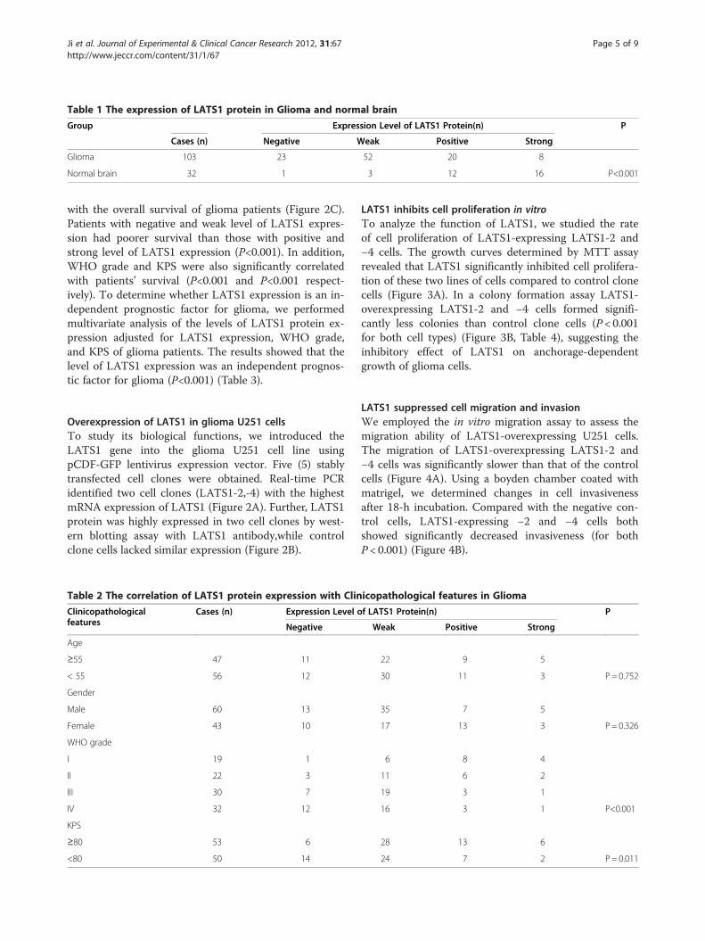
Table 1 The expression of LATS1 protein in Glioma and normal brain
Group Expression Level of LATS1 Protein(n) P
Cases (n) Negative Weak Positive Strong
Glioma 103 23 52 20 8
Normal brain 32 1 3 12 16 P<0.001
Ji et al. Journal of Experimental & Clinical Cancer Research 2012, 31:67 Page 5 of 9http://www.jeccr.com/content/31/1/67
with the overall survival of glioma patients (Figure 2C).Patients with negative and weak level of LATS1 expres-sion had poorer survival than those with positive andstrong level of LATS1 expression (P<0.001). In addition,WHO grade and KPS were also significantly correlatedwith patients’ survival (P<0.001 and P<0.001 respect-ively). To determine whether LATS1 expression is an in-dependent prognostic factor for glioma, we performedmultivariate analysis of the levels of LATS1 protein ex-pression adjusted for LATS1 expression, WHO grade,and KPS of glioma patients. The results showed that thelevel of LATS1 expression was an independent prognos-tic factor for glioma (P<0.001) (Table 3).
Overexpression of LATS1 in glioma U251 cellsTo study its biological functions, we introduced theLATS1 gene into the glioma U251 cell line usingpCDF-GFP lentivirus expression vector. Five (5) stablytransfected cell clones were obtained. Real-time PCRidentified two cell clones (LATS1-2,-4) with the highestmRNA expression of LATS1 (Figure 2A). Further, LATS1protein was highly expressed in two cell clones by west-ern blotting assay with LATS1 antibody,while controlclone cells lacked similar expression (Figure 2B).
Table 2 The correlation of LATS1 protein expression with Clin
Clinicopathologicalfeatures
Cases (n) Expression Level o
Negative
Age
≥55 47 11
< 55 56 12
Gender
Male 60 13
Female 43 10
WHO grade
I 19 1
II 22 3
III 30 7
IV 32 12
KPS
≥80 53 6
<80 50 14
LATS1 inhibits cell proliferation in vitroTo analyze the function of LATS1, we studied the rateof cell proliferation of LATS1-expressing LATS1-2 and−4 cells. The growth curves determined by MTT assayrevealed that LATS1 significantly inhibited cell prolifera-tion of these two lines of cells compared to control clonecells (Figure 3A). In a colony formation assay LATS1-overexpressing LATS1-2 and −4 cells formed signifi-cantly less colonies than control clone cells (P < 0.001for both cell types) (Figure 3B, Table 4), suggesting theinhibitory effect of LATS1 on anchorage-dependentgrowth of glioma cells.
LATS1 suppressed cell migration and invasionWe employed the in vitro migration assay to assess themigration ability of LATS1-overexpressing U251 cells.The migration of LATS1-overexpressing LATS1-2 and−4 cells was significantly slower than that of the controlcells (Figure 4A). Using a boyden chamber coated withmatrigel, we determined changes in cell invasivenessafter 18-h incubation. Compared with the negative con-trol cells, LATS1-expressing −2 and −4 cells bothshowed significantly decreased invasiveness (for bothP < 0.001) (Figure 4B).
icopathological features in Glioma
f LATS1 Protein(n) P
Weak Positive Strong
22 9 5
30 11 3 P = 0.752
35 7 5
17 13 3 P = 0.326
6 8 4
11 6 2
19 3 1
16 3 1 P<0.001
28 13 6
24 7 2 P = 0.011

Figure 2 Reexpression of LATS1 in glioma U251 cells. A. Real-time PCR analysis indicated the highest mRNA expression of LATS1in two cell clones pLATS1-2 and −4. B. Western blotting assay showssignificantly increased protein expression of LATS1 in pLATS1-2 and−4 suppressed the expression of cell cycle factor CCNA1 proteincompared to Control-vector cells. β-actin was used as the internalcontrol.
Ji et al. Journal of Experimental & Clinical Cancer Research 2012, 31:67 Page 6 of 9http://www.jeccr.com/content/31/1/67
Inhibition of cell cycle progression by LATS1To detect the effect of LATS1 on cell cycle, we mea-sured cell cycle distribution in LATS1-expressing −2and −4 cells. The G2 phase population was markedlyincreased and G1 phase population significantlydecreased in both cell lines compared to the Ctr-vector cells and U251 cells (P < 0.001). However, inboth two lines the change in S phase population wasnot significant (Figure 4C)(Additional file 1: Figure S1)(Additional file 2: Table S1).
Table 3 Summary of univariate and multivariate Cox regressi
Parameter Univariate an
P HR
Age
≥55vs. <55 years 0.069 0.777
Gender
Male vs. female 0.160 0.820
WHO grade
Ivs.II vs.III vs.IV 0.000 1.715
KPS
≥80 vs. < 80 0.000 2.033
LAST1 expression
Strong vs.Positive vs.Weak vs.Negative* 0.000 0.437
LATS1 inhibits the expression of CCNA1In exploring the molecular mechanism of LATS1 tumor-suppressing function in glioma, we found that restor-ation of LATS1 expression significantly inhibitedexpression of cell cycle factor CCNA1 in glioma U251cells (Figure 4D). This suggested that LATS1 may beinvolved in G2/M cell cycle pathway in glioma.
DiscussionMalignant gliomas occur more frequently than othertypes of primary CNS tumors, having a combined inci-dence of 5–8/100,000 population. Due to its highly inva-sive nature, median reported survival is less than 1 yeareven with aggressive treatment using surgery, radiation,and chemotherapy [17]. Thus, there is a need for a bet-ter understanding of the molecular basis of gliomapathogenesis to improve prognosis prediction and de-velop targeted, molecular-based therapies.Accumulating evidence suggests that the LATS (Large
Tumor Suppressor) family of human tumor suppressors(LATS1 and LATS2) as regulators of cellular homeosta-sis. Loss of function of either LATS1 or LATS2 leads toa variety of tumor types including soft tissue sarcomas,leukemia, as well as breast, prostate, lung and esopha-geal cancers [18], which suggests they function as tumorsuppressors in tumor pathogenesis. LATS1 gene islocated at chromosome 6q25.1 and its open readingframe is 3393 bp encoding a 1130-amino acid polypep-tide with molecular weight of 126.87 kDa. LATS1 ex-pression is significantly decreased in some tumorsincluding breast cancer and astrocytoma [13,14], andthis downregulation has been attributed to its promoterhypermethylation. Negative regulator of oncoproteinYAP1 in the Hippo signaling pathway plays a pivotal rolein organ size control and tumor suppression by restrict-ing proliferation and promoting apoptosis. LATS1 phos-phorylates YAP1 protein and inhibits its translocation
on analysis of overall survival duration
alysis Multivariate analysis
95%CI P HR 95%CI
0.593-1.019
0.621-1.082
1.454-2.023 0.000 1.463 1.233-1.735
1.540-2.684 0.000 2.437 1.810-3.283
0.362-0.528 0.000 0.389 0.316-0.478

Figure 3 Overexpression of LATS1 inhibted cell proliferationin vitro. A. The cell growth of Control-vector cells and pLATS1-2and −4 cells, were examined by MTT assay over a seven-day period.*P < 0.05, as compared to control-vector cells. B. The cell growth ofcontrol-vector cells and pLATS1-2 and −4 cells, were examined byplate colony formation assay. *P < 0.05, as compared tocontrol-vector cells.
Ji et al. Journal of Experimental & Clinical Cancer Research 2012, 31:67 Page 7 of 9http://www.jeccr.com/content/31/1/67
into the nucleus to regulate cellular genes important forcell proliferation, cell death, and cell migration [19]. Fur-thermore, in previous studies LATS1 overexpressioninduced cell apoptosis by increasing pro-apoptotic pro-teins p53 and Bax [11] and suppressed cell proliferationthrough p53 upregulation to ensure genomic integrity[20]. Conversely, knockdown of LATS1 induced cell mi-gration in HeLa cells [21]. These results together sup-ported that LATS1 played a suppressive role in tumorpathogenesis.In order to assess the role of LATS1 in glioma, we first
performed real-time PCR to measure the expression of
Table 4 Plate clone formation assay among pLATS1-2,pLATS1-4, and Ctr-vector cells
Cells Number P value
pLATS1-2 45.33 ± 4.16
pLATS1-4 34.67 ± 6.25
Ctr-vector 77.33 ± 7.12 p<0.001
LATS1 mRNA transcripts in 17 paired glioma samplesand their adjacent brain tissues. Similar to reports ofother tumor types [13,14], we observed that LATS1 ex-pression was significantly decreased in 13 glioma tissuescompared to their matched normal tissues. This sug-gested LATS1 functions as a tumor suppressor in gli-oma. We validated this downregulation of LATS1protein by immunohistochemistry. In addition, we foundthat LATS1 expression levels were inversely associatedwith WHO grade of glioma and KPS. Further, we pre-sented the evidence that LATS1 protein expression inglioma was positively correlated with patient’s overallsurvival. The patients with lower expression of LATS1protein had shorter survival time. According to multi-variate analyses, decreased expression of LATS1 proteinwas a significant predictor of poor prognosis for gliomapatients. These results were analogous to Takahashi etal’s report in the study of breast cancer [13] and stronglysuggested a suppressive role of LATS1 in gliomatumorigenesis.Next, we used a gain-of-function approach by introdu-
cing the LATS1 gene into LATS1-negative U251 gliomacells, to investigate its biological functions. We observedthat overexpression of LATS1 caused significant reducedin vitro cell growth and G(2)/M arrest. These are con-sistent with the findings by Yang et al. [11] and Xia et al.[12] that upregulation of LATS1 suppresses cell growthand cell cycle progression, which further demonstratesthat the suppressive biological functions of LATS1 arecommon to multiple cancers. Additionally, our studyalso revealed a novel function of LATS1 in glioma insuppression of cell migration and invasion. This suggestsLATS1 may be involved in invasion and metastasis ofcancer, a concept which would need to be confirmed byin vivo animal model. The observations that LATS1 reg-ulates multiple cellular processes such as cell prolifera-tion, cell cycle progression, migration, invasionemphasizes its importance as a therapeutic target fortreating glioma.In a previous investigation, increased LATS1 expres-
sion inhibited cell proliferation by blocking the G2/Mtransition, mainly through inhibition of the kinase activ-ity of Cdc2/ Cyclin A/B complex [18]. We also observedthat overexpressed LATS1 caused the G2/M phaseblockade in glioma U251 cells. Therefore, we investi-gated the expression change of CCNA1, a cell cycle fac-tor in the Cdc2/ Cyclin A/B complex. This gene bindsboth CDK2 and CDC2 kinases and thus regulates thecell cycle transition at G2/M [22-25]. We speculatedCCNA1 might be involved in the cell cycle regulationpathway of LATS1 in glioma. Consistent with this pre-sumption, we found that overexpression of LATS1 sig-nificantly reduced the expression of CCNA1 by westernblot assay in glioma U251 cells. Further investigation is

Figure 4 Increased LATS1 expression inhibited cell migration, invasion and cell cycle progression. (A) Cell migration and (B) invasioncapabilities of pLATS1-2, -4 cells and Control-vector cells, were examined using transwell and boyden chamber assay. Data were presented asmean± SD for three independent experiments. *P< 0.05, as compared to control-vector cells. C. Cell cycle in pLATS1-2 and −4 cells andcontrol-vector cells, was determined by FACS Caliber Cytometry. *P< 0.05, as compared to control-vector cells.
Ji et al. Journal of Experimental & Clinical Cancer Research 2012, 31:67 Page 8 of 9http://www.jeccr.com/content/31/1/67
necessary to determine the exact role LATS1 plays incell cycle pathway in glioma.
ConclusionsOur results indicate that the decreased expression ofLATS1 appears to favor the development of glioma andmight serve a suppressive role in glioma. Further, we ap-plied a gain-of-function approach and to examine thebiological processes regulated by LATS1 in glioma cells.We demonstrated the functional importance of LATS1in suppressing glioma cell growth, migration, invasion
and cell cycle transition from G2 to M phase. Finally, weobserved that overexpression of LATS1 could inhibit theexpression of cell cycle factor CCNA1, which mightpartly explain the mechanism by which LATS1 in con-trols cell proliferation.
Additional files
Additional file 1: Figure S1. Cell cycle map of pLATS1-2, -4 cellsand Control-vector cells.

Ji et al. Journal of Experimental & Clinical Cancer Research 2012, 31:67 Page 9 of 9http://www.jeccr.com/content/31/1/67
Additional file 2: Table S1. Overexpression of LATS1 reduced DNAcontent of G2 phase and increased DNA content of G1 phase.
Competing interestsThe authors declare that they have no competing interests.
Authors’ contributionsXB designed and directed the study. TJ, DL and WS performed experiments,conducted the analysis and drafted the manuscript. WY and HW assisted inthe analysis and interpretation of results. All authors read and approved thefinal manuscript.
AcknowledgementsThis study was supported by National Natural Science Foundation of P.R.China (30900559, 81101904) and Science and Technology Project of Xiamen(3502Z20104015;3502Z20124019).
Author details1Institute of Pathology and Southwest Cancer Center, Southwest Hospital,Third Military Medical University, Chongqing 400038, China. 2Department ofPathology, Chenggong Hospital, Xiamen University, Xiamen, Fujian 361003,China.
Received: 17 January 2012 Accepted: 7 August 2012Published: 21 August 2012
References1. Kleihues P, Cavenee WK: World Health Organization Classification of Tumours-
Pathology and Genetics -Tumors of the Nervous System. Lyon. France: IARCPress; 2000:9–52.
2. Wu M, Chen Q, Li D, Li X, Li X, Huang C, Tang Y, Zhou Y, Wang D, Tang K,Cao L, Shen S, Li G: LRRC4 inhibits human glioblastoma cells proliferation,invasion, and proMMP-2 activation by reducing SDF-1 alpha/CXCR4-mediated ERK1/2 and Akt signaling pathways. J Cell Biochem 2008,103:245–255.
3. Louis DN, Ohgaki H, Wiestler OD, Cavenee WK, Burger PC, Jouvet A,Scheithauer BW, Kleihues P: The 2007 WHO classification of tumours ofthe central nervous system. Acta Neuropathol 2007, 114:97–109.
4. Justice RW, Zilian O, Woods DF, Noll M, Bryant PJ: The Drosophila tumorsuppressor gene warts encodes a homolog of human myotonicdystrophy kinase and is required for the control of cell shape andproliferation. Genes Dev 1995, 9:534–546.
5. Xu T, Wang W, Zhang S, Stewart RA, Yu W: Identifying tumor suppressorsin genetic mosaics: the Drosophila lats gene encodes a putative proteinkinase. Development 1995, 121:1053–1063.
6. Tao W, Zhang S, Turenchalk GS, Stewart RA, St John MA, Chen W, Xu T:Human homologue of the Drosophila melanogaster lats tumoursuppressor modulates CDC2 activity. Nat Genet 1999, 21:177–181.
7. St John MA, Tao W, Fei X, Fukumoto R, Carcangiu ML, Brownstein DG,Parlow AF, McGrath J, Xu T: Mice deficient of Lats1 develop soft-tissuesarcomas, ovarian tumours and pituitary dysfunction. Nat Genet 1999,21:182–186.
8. Cooke IE, Shelling AN, Le Meuth VG, Charnock ML, Ganesan TS: Allele losson chromosome arm 6q and fine mapping of the region at 6q27 inepithelial ovarian cancer. Genes Chromosomes Cancer 1996, 15:223–233.
9. Mazurenko N, Attaleb M, Gritsko T, Semjonova L, Pavlova L, Sakharova O,Kisseljov F: High resolution mapping of chromosome 6 deletions incervical cancer. Oncol Rep 1999, 6:859–863.
10. Fujii H, Zhou W, Gabrielson E: Detection of frequent allelic loss of 6q23–q25.2 in microdissected human breast cancer tissues. GenesChromosomes Cancer 1996, 16:35–39.
11. Yang X, Li D, Chen W, Xu T: Human homologue of the Drosophila lats,LATS1, negatively regulate growth by inducing G2/M arrest or apoptosis.Oncogene 2001, 20:6516–6523.
12. Xia H, Qi H, Li Y, Pei J, Barton J, Blackstad M, Xu T, Tao W: LATS1 tumorsuppressor regulates G2/M transition and apoptosis. Oncogene 2002,21:1233–1241.
13. Takahashi Y, Miyoshi Y, Takahata C, Irahara N, Taguchi T, Tamaki Y, NoguchiS: Down-regulation of LATS1 and LATS2 mRNA expression by promoter
hypermethylation and its association with biologically aggressivephenotype in human breast cancers. Clin Cancer Res 2005, 11:1380–1385.
14. Jiang Z, Li X, Hu J, Zhou W, Jiang Y, Li G, Lu D: Promoterhypermethylation-mediated down-regulation of LATS1 and LATS2 inhuman astrocytoma. Neurosci Res 2006, 56:450–458.
15. Liu Z, Li X, He X, Jiang Q, Xie S, Yu X, Zhen Y, Xiao G, Yao K, Fang W:Decreased expression of updated NESG1 in nasopharyngeal carcinoma:its potential role and preliminarily functional mechanism. Int J Cancer. IntJ Cancer 2011, 128:2562–2571.
16. Livak KJ, Schmittgen TD: Analysis of relative gene expression data usingreal-time quantitative PCR and the 2(−Delta Delta C(T)) Method. Methods2001, 25:402–408.
17. Avgeropoulos NG, Batchelor TT: New treatment strategies for malignantgliomas. Oncologist 1999, 4:209–224.
18. Visser S, Yang X: LATS tumor suppressor: a new governor of cellularhomeostasis. Cell Cycle 2010, 9:3892–3903.
19. Zhang J, Smolen GA, Haber DA: Negative regulation of YAP by LATS1underscores evolutionary conservation of the Drosophila Hippopathway. Cancer Res 2008, 68:2789–2794.
20. Iida S, Hirota T, Morisaki T, et al: Tumor suppressor WARTS ensuresgenomic integrity by regulating both mitotic progression and G1tetraploidy checkpoint function. Oncogene 2004, 23:5266–5274.
21. Visser S, Yang X: Identification of LATS transcriptional targets in HeLacells using whole human genome oligonucleotide microarray. Gene 2010,449:22–29.
22. Liu D, Liao C, Wolgemuth DJ: A role for cyclin A1 in the activation of MPFand G2-M transition during meiosis of male germ cells in mice. Dev Biol2000, 224:388–400.
23. Diederichs S, Bäumer N, Ji P, Metzelder SK, Idos GE, Cauvet T, Wang W,Möller M, Pierschalski S, Gromoll J, Schrader MG, Koeffler HP, Berdel WE,Serve H, Müller-Tidow C: Identification of interaction partners andsubstrates of the cyclin A1-CDK2 complex. J Biol Chem 2004,279:33727–33741.
24. Cho NH, Choi YP, Moon DS, Kim H, Kang S, Ding O, Rha SY, Yang YJ, ChoSH: Induction of cell apoptosis in non-small cell lung cancer cells bycyclin A1 small interfering RNA. Cancer Sci 2006, 97:1082–1092.
25. Bois C, Delalande C, Bouraïma-Lelong H, Durand P, Carreau S: 17β-Estradiolregulates cyclin A1 and cyclin B1 gene expression in adult ratseminiferous tubules. J Mol Endocrinol 2012, 48:89–97.
doi:10.1186/1756-9966-31-67Cite this article as: Ji et al.: Decreased expression of LATS1 is correlatedwith the progression and prognosis of glioma. Journal of Experimental &Clinical Cancer Research 2012 31:67.
Submit your next manuscript to BioMed Centraland take full advantage of:
• Convenient online submission
• Thorough peer review
• No space constraints or color figure charges
• Immediate publication on acceptance
• Inclusion in PubMed, CAS, Scopus and Google Scholar
• Research which is freely available for redistribution
Submit your manuscript at www.biomedcentral.com/submit


![Lecture – 3 Dr. Zahoor Ali Shaikh 1. What is Anemia? Anemia means - Decreased hemoglobin - Decreased RBC count - Decreased Hematocrit [PCV] Therefore,](https://static.fdocuments.us/doc/165x107/56649c9e5503460f9495e870/lecture-3-dr-zahoor-ali-shaikh-1-what-is-anemia-anemia-means-decreased.jpg)
