Immunohistochemistry Antibody Validation Report for Anti-Phospho-Akt (T308) Antibody (STJ90750)

Experimental Hematology & Oncology
Islam et al. Experimental Hematology & Oncology 2014, 3:25http://www.ehoonline.org/content/3/1/25
RESEARCH Open Access
Activation of Akt at T308 and S473 in alcohol,tobacco and HPV-induced HNSCC: is thereevidence to support a prognostic or diagnosticrole?Mohammad R Islam1, Ian R Ellis1, Michaelina Macluskey1, Lynda Cochrane2 and Sarah J Jones1*
Abstract
Background: Tobacco, alcohol and HPV infection are associated with increased risk of HNSCC. However, little isknown about the underlying signaling events influencing risk. We aimed to investigate the relationship betweenthese risk factors and Akt phosphorylation, to determine prognostic value.
Method: VEGF-positive HNSCC biopsies, with known HPV status, were analyzed by immunohistochemistry (IHC)for Akt, phosphorylated at residues S473 and T308. Comparisons between the tissues were carried out using aMann–Whitney U test. Associations between the variables and continuous immunohistochemical parameterswere evaluated with general linear models. Patient characteristics and pAkt IHC score were analyzed for possibleassociation with overall survival by Cox proportional hazard models.
Results: Immunohistochemistry revealed that cancer patients had significantly higher levels of pAkt T308 thanS473 (P < 0.001). Smoking and alcohol were found to be independent risk factors for Akt phosphorylation at T308(P = 0.022 and 0.027, respectively). Patients with tumors positive for HPV or pAkt S473 had a poorer prognosis(P = 0.005, and 0.004, respectively). Patients who were heavy drinkers were 49 times more likely to die thannon-drinkers (P = 0.003). Patients with low pAkt T308 were more likely to be HPV positive (P = 0.028). Non-drinkerswere also found to have lower levels of pAkt T308 and were more likely to have tumors positive for HPV thanheavy drinkers (P = 0.044 and 0.007, respectively).
Conclusion: This study suggests different mechanisms of carcinogenesis are initiated by smoking, alcohol andHPV. Our data propose higher phosphorylation of Akt at T308 as a reliable biomarker for smoking and alcoholinduced HNSCC progression and higher phosphorylation of Akt at S473 as a prognostic factor for HNSCC.
Keywords: pAkt T308, pAkt S473, HNSCC, Risk factors, Prognosis, Biomarker
BackgroundHNSCC (Head and Neck Squamous Cell Carcinoma) in-cludes cancers that involve the oral cavity, pharynx andlarynx. Each year there are approximately 400 000 casesof cancer of the oral cavity and pharynx, with 160 000cancers of the larynx, resulting in approximately 300 000deaths [1]. It is the sixth most common type of cancerworldwide with a five year survival rate of 40-50%, which
* Correspondence: [email protected] of Oral and Maxillofacial Clinical Sciences, The Dental School,University of Dundee, Dundee DD1 4HR, UKFull list of author information is available at the end of the article
© 2014 Islam et al.; licensee BioMed Central LCommons Attribution License (http://creativecreproduction in any medium, provided the orDedication waiver (http://creativecommons.orunless otherwise stated.
has shown only moderate improvement over the last twodecades [2]. Two modifiable risk factors, tobacco use and al-cohol consumption, are thought to explain approximately75% of the incidence of HNSCC (up to 100 times higher forboth) [3]; another important risk factor is HPV infection,which has been detected in around 20% of all cases [4].Recent studies have focused on the genetic and epigen-
etic alterations of HNSCC, providing a better understand-ing of the molecular events underlying the pathogenesis ofHNSCC [5,6]. One of the most frequently altered signalingpathways in HNSCC is the PI3K/Akt cascade [6]. Akt, alsoknown as PKB, is a serine-threonine protein kinase and is
td. This is an Open Access article distributed under the terms of the Creativeommons.org/licenses/by/4.0), which permits unrestricted use, distribution, andiginal work is properly credited. The Creative Commons Public Domaing/publicdomain/zero/1.0/) applies to the data made available in this article,
Islam et al. Experimental Hematology & Oncology 2014, 3:25 Page 2 of 9http://www.ehoonline.org/content/3/1/25
central to the phosphatidylinositol 3-kinase (PI3K) signal-ing pathway [7-9]. PI3K is activated by tyrosine-kinasetransmembrane receptors and other signaling intermedi-ates, such as Ras oncogenes and G proteins [10]. PI3Kthen phosphorylates PtdIns (4,5) P2 (PIP2) yielding PtdIns(3,4,5) P3 (PIP3), which serves as an anchor for intracellularproteins (primarily mediated by pleckstrin homology do-mains), including Akt amongst others. Membrane-boundAkt is phosphorylated at T308 in the catalytic domain bythe kinase PDK1 and at S473 in the regulatory domain bymTORC2 [11,12]. Full Akt kinase activity is dependent uponphosphorylation at both T308 and S473 residues and this isgreatly increased by growth factor receptor signaling [13].PIP3 is converted back into PIP2 through the action of thelipid phosphatase PTEN, thus terminating the PI3K-initiatedsignal and avoiding further Akt activation [14]. Growth fac-tor receptor over-expression [15,16], mutation and down-regulation of PTEN protein [17] and amplification of thePIK3CA gene (the gene coding for the catalytic unit ofPI3K) [18] can lead to increased Akt activity. EnhancedAkt activity has indeed been found in 20 to 60% of tumorsamples and in the majority of HNSCC-derived cell lines[18-21]. Once Akt is phosphorylated and activated, it iscapable of phosphorylating multiple substrates generatingdiverse cellular processes, such as metabolism, prolifera-tion, survival and protein synthesis [22].An increasing number of mucosal changes and cellular
atypia occur over large areas of the carcinogen-exposedupper aero-digestive tract epithelium, which initiate thestepwise carcinogenesis process in HNSCC. Acquisitionof a transformed phenotype and accumulation of specificmolecular genetic events are associated with this process[23-28]; yet histopathological evaluation remains thetime honored method in risk assessment of carcinomalesions. In a search for better biological models of risk,Akt activation was recently identified as an early cellularresponse to carcinogen exposure and may be a signifi-cant step in environmental carcinogenesis [29]. Akt acti-vation has also been found to correlate with squamouscell carcinoma progression from normal epithelium toinvasive cancer [30].These observations encouraged us to investigate the
role of demographic, pathological and major risk factors(smoking, alcohol and HPV) of HNSCC patients on theactivation of Akt (phosphorylation of Akt at Threonine308 and Serine 473) and to determine their prognosticrole.
ResultsAnalysis of patient details58 HNSCC patient details were analyzed in this study. 33(57%) were male and 25 (43%) female with ages rangingfrom 36 to 97 years (median age 64 years). 42 (72%) weresmokers and 16 (28%) non-smokers. 7 patients (12%) were
non-drinkers, 21 (36%) medium or moderate drinkers and30 (52%) were heavy drinkers. 31 (53%) patients hadnegative and 27 (47%) had positive nodal metastasis.Half of the cohort was HPV positive. 43 (74%) patientshad T1/T2 and 15 (26%) had T3/T4 tumor size with 5(9%) of them grade I, 39 (67%) of them grade II and 14(24%) of them grade III. These data are summarized inTable 1.
Immunohistochemistry for Akt phosphorylationBoth normal and VEGF-positive carcinoma patient sam-ples were stained with pAkt S473 and pAkt T308 anti-bodies. Some samples which were highly stained forpAkt S473 and pAkt T308 were then selected and testedwith the blocking peptide for the respective antibodyand were used as negative controls (Figure 1A). Nostaining was observed in the blocking peptide treated tis-sues and this confirmed the specific binding of theantibodies.Normal tissue samples were also regarded as negative
controls and the level of Akt phosphorylation at T308was higher in the HNSCC group than the control (me-dian 5.8 vs 2.0, P < 0.001) (Figure 1B). Normal tissue ad-jacent to the tumors was also tested and the resultantdata indicated that there was very low or no pAkt T308/pAkt S473 present (Figure 1C). There is also some evidenceto suggest higher levels of pAkt S473 in the cancer groupthan the controls (P = 0.054) (Figure 1B). There is a statisti-cally significant difference between pAkt T308 and pAktS473 levels in the cancer patients (pAkt T308, median 5.8vs pAkt S473, median 0.3, P < 0.001) (Figure 1B). VEGFA isnot correlated with pAkt T308 (r = 0.062, P = 0.644) andpAkt S473 (r = 0.181, P = 0.175). Table 1 compares thecharacteristics between pAkt expression groups. All thesamples were found to be phosphorylated at Akt T308so there is no ‘no phosphorylation’ group for this resi-due. On the other hand, there is no ‘high phosphoryl-ation’ group for pAkt S473.
Association of patient characteristics with AktphosphorylationSmoking and alcohol were found to be independent riskfactors for phosphorylation of Akt at T308 (P = 0.022and 0.027, respectively) but not for pAkt S473 (P = 0.449and 0.968, respectively). HNSCC with nodal metastasiswas associated with a higher level of pAkt T308 thanHNSCC without nodal metastasis (P = 0.018). Smokersand HNSCC with nodal metastasis were found to havehigher levels of Akt phosphorylated at T308 than non-smokers (P = 0.022) and HNSCC without modal metas-tasis (P = 0.018) patients, respectively. HPV-negative patientsexhibited higher levels of pAkt T308 compared to those whowere HPV positive (P = 0.028). There is some evidence to
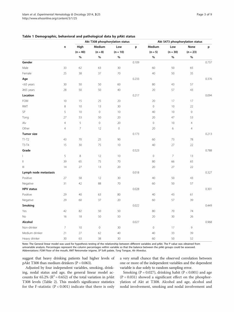
Table 1 Demographic, behavioral and pathological data by pAkt status
Akt T308 phosphorylation status Akt S473 phosphorylation status
n High Medium Low p Medium Low None p
(n = 40) (n = 8) (n = 10) (n = 5) (n = 30) (n = 23)
% % % % % %
Gender 0.109 0.737
Male 33 62 63 30 60 50 65
Female 25 38 37 70 40 50 35
Age 0.233 0.376
<65 years 30 50 50 60 80 43 57
≥65 years 28 50 50 40 20 57 43
Location 0.217 0.094
FOM 10 15 25 20 20 17 17
RMT 8 10 13 30 0 10 22
SP 5 10 0 10 40 10 0
Tong 27 53 50 20 20 47 53
Alv 4 5 0 20 0 10 4
Other 4 7 12 0 20 6 4
Tumor size 0.173 0.213
T1-T2 43 70 25 90 60 73 78
T3-T4 15 30 75 10 40 27 22
Grade 0.523 0.788
I 5 8 12 10 0 7 13
II 39 65 75 70 80 66 65
III 14 27 13 20 20 27 22
Lymph node metastasis 0.018 0.327
Positive 27 58 12 30 40 50 43
Negative 31 42 88 70 60 50 57
HPV status 0.028 0.301
Positive 29 40 63 80 40 43 61
Negative 29 60 37 20 60 57 39
Smoking 0.022 0.449
Yes 42 82 50 50 80 70 74
No 16 18 50 50 20 30 26
Alcohol 0.027 0.968
Non-drinker 7 10 0 30 0 17 9
Medium drinker 21 27 62 40 40 33 39
Heavy drinker 30 63 38 30 60 50 52
Note: The General linear model was used for hypothesis testing of the relationship between different variables and pAkt. The P value was obtained fromunivariable analysis. Percentages represent the column percentages within variable so that the balance between the pAkt groups could be assessed.Abbreviations: FOM Floor of the mouth, RMT Retromolar trigone, SP Soft palate, Tong Tongue, Alv Alveolus.
Islam et al. Experimental Hematology & Oncology 2014, 3:25 Page 3 of 9http://www.ehoonline.org/content/3/1/25
suggest that heavy drinking patients had higher levels ofpAkt T308 than medium drinkers (P = 0.063).Adjusted by four independent variables, smoking, drink-
ing, nodal status and age, the general linear model ac-counts for 65.2% (R2 = 0.652) of the total variation in pAktT308 levels (Table 2). This model’s significance statisticsfor the F-statistic (P < 0.001) indicate that there is only
a very small chance that the observed correlation betweenone or more of the independent variables and the dependentvariable is due solely to random sampling error.Smoking (P = 0.027), drinking habit (P < 0.001) and age
(P = 0.031) showed a significant effect on the phosphor-ylation of Akt at T308. Alcohol and age, alcohol andnodal involvement, smoking and nodal involvement and
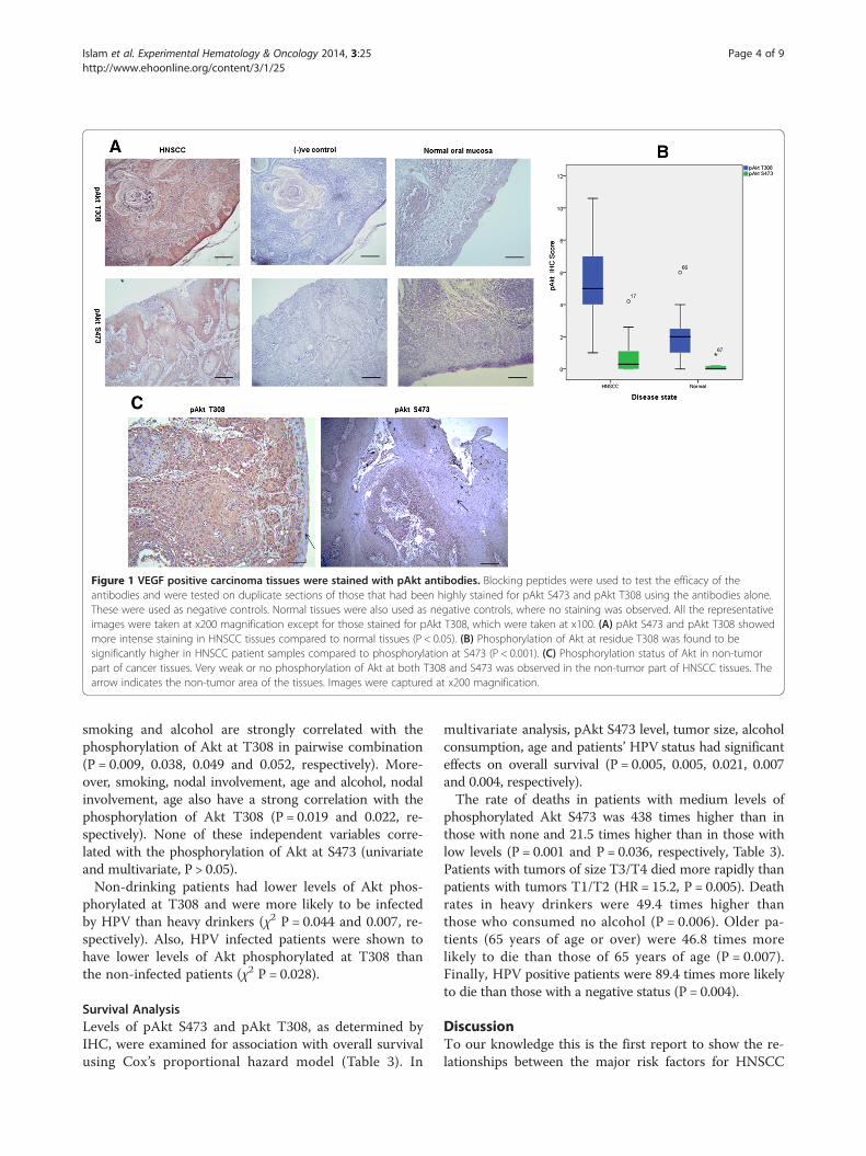
Figure 1 VEGF positive carcinoma tissues were stained with pAkt antibodies. Blocking peptides were used to test the efficacy of theantibodies and were tested on duplicate sections of those that had been highly stained for pAkt S473 and pAkt T308 using the antibodies alone.These were used as negative controls. Normal tissues were also used as negative controls, where no staining was observed. All the representativeimages were taken at x200 magnification except for those stained for pAkt T308, which were taken at x100. (A) pAkt S473 and pAkt T308 showedmore intense staining in HNSCC tissues compared to normal tissues (P < 0.05). (B) Phosphorylation of Akt at residue T308 was found to besignificantly higher in HNSCC patient samples compared to phosphorylation at S473 (P < 0.001). (C) Phosphorylation status of Akt in non-tumorpart of cancer tissues. Very weak or no phosphorylation of Akt at both T308 and S473 was observed in the non-tumor part of HNSCC tissues. Thearrow indicates the non-tumor area of the tissues. Images were captured at x200 magnification.
Islam et al. Experimental Hematology & Oncology 2014, 3:25 Page 4 of 9http://www.ehoonline.org/content/3/1/25
smoking and alcohol are strongly correlated with thephosphorylation of Akt at T308 in pairwise combination(P = 0.009, 0.038, 0.049 and 0.052, respectively). More-over, smoking, nodal involvement, age and alcohol, nodalinvolvement, age also have a strong correlation with thephosphorylation of Akt T308 (P = 0.019 and 0.022, re-spectively). None of these independent variables corre-lated with the phosphorylation of Akt at S473 (univariateand multivariate, P > 0.05).Non-drinking patients had lower levels of Akt phos-
phorylated at T308 and were more likely to be infectedby HPV than heavy drinkers (χ2 P = 0.044 and 0.007, re-spectively). Also, HPV infected patients were shown tohave lower levels of Akt phosphorylated at T308 thanthe non-infected patients (χ2 P = 0.028).
Survival AnalysisLevels of pAkt S473 and pAkt T308, as determined byIHC, were examined for association with overall survivalusing Cox’s proportional hazard model (Table 3). In
multivariate analysis, pAkt S473 level, tumor size, alcoholconsumption, age and patients’ HPV status had significanteffects on overall survival (P = 0.005, 0.005, 0.021, 0.007and 0.004, respectively).The rate of deaths in patients with medium levels of
phosphorylated Akt S473 was 438 times higher than inthose with none and 21.5 times higher than in those withlow levels (P = 0.001 and P = 0.036, respectively, Table 3).Patients with tumors of size T3/T4 died more rapidly thanpatients with tumors T1/T2 (HR = 15.2, P = 0.005). Deathrates in heavy drinkers were 49.4 times higher thanthose who consumed no alcohol (P = 0.006). Older pa-tients (65 years of age or over) were 46.8 times morelikely to die than those of 65 years of age (P = 0.007).Finally, HPV positive patients were 89.4 times more likelyto die than those with a negative status (P = 0.004).
DiscussionTo our knowledge this is the first report to show the re-lationships between the major risk factors for HNSCC

Table 3 COX proportional hazard model- time to death
Unadjust
HR 95% CI
pAkt S473 Overall
Med:No 6.27 0.87, 45
Med:Low 1.83 0.38, 8.8
Tumor size T3/T4:T1/T2 4.59 1.39, 15
Alcohol Overall
Heavy:Non-drinker 15.4 2.74, 86
Heavy:Moderate 3.19 0.62, 16
Age ≥65:<65 5.41 1.17, 25
HPV +ve:−ve
Gender F:M 1.40 0.42, 4.6
Tumor size Overall
Nodal status +ve: −ve 1.49 0.43, 5.1
Smoking No:Yes 1.45 0.42, 4.9
pAkt T308 Overall
Low:High 1.02 0.21, 4.9
Med:High 1.16 0.24, 5.6
Note: Unadjusted HR obtained from univariable analysis and adjusted HR from mulAll the variables are categorical and HR = exp (B). Abbreviations: HR Hazard ratio, 95
Table 2 General Linear Model (Multivariate analysis)
Independent variables F P
Corrected model 4.06 <0.001
Smoking 5.30 0.027
Alcohol 10.56 <0.001
Nodal status 1.31 0.260
Age 4.98 0.031
Smoking * Alcohol 3.20 0.052
Smoking * Nodal status 4.12 0.049
Smoking * Age 0.01 0.919
Alcohol * Nodal status 3.56 0.038
Alcohol * Age 5.31 0.009
Nodal status * Age 2.80 0.102
Smoking * Alcohol * Nodal status 0.17 0.682
Smoking * Alcohol * Age 0.08 0.774
Smoking * Nodal status * Age 6.00 0.019
Alcohol * Nodal status * Age 5.73 0.022
R2 = 0.652
Note: After adjusting Smoking, Alcohol, Nodal Status and Age, this modelaccounts for 65.2% of the total variations in pAkt T308 level. R2 = Coefficient ofdetermination, F = F-statistics.Dependent variable: pAkt T308 score.
Islam et al. Experimental Hematology & Oncology 2014, 3:25 Page 5 of 9http://www.ehoonline.org/content/3/1/25
(alcohol, smoking and HPV) and Akt activation (both atresidues T308 and S473) at the protein level using animmunohistochemical staining method (IHC), in surgi-cally resected specimens. Most studies to date have usedIHC to assess the prognostic value of Akt activation inHNSCC, but have focused only on phosphorylation ofresidue S473. As the differential phosphorylation of Aktat the two sites may modulate downstream substrate se-lectivity and subsequent bioactivity [13], it is not surpris-ing that Akt phosphorylated at any single site couldperform certain cellular bioactivities. Two different mech-anisms are involved in phosphorylating Akt, thereforeoverexpression or amplification of any components inthese mechanisms may result in over-phosphorylation atany one of the two sites. It is therefore worth studying thephosphorylation status of Akt at both sites in HNSCCspecimens to elucidate their different roles.As only VEGFA positive HNSCC biopsy samples were
selected in this study, no statistically significant correl-ation was found between VEGF and pAkt. The presentstudy showed that Akt was significantly phosphorylatedat T308 in VEGF positive HNSCC rather than S473. Al-cohol and smoking were positively correlated with pAktT308 activation but not with pAkt S473. Moreover, Aktactivated at T308 showed a significant relationship withlymph node metastasis, which suggests that pAkt T308may be concerned with invasion and metastasis. Thisdata is similar to our in vitro data concerning the migra-tion of tumor cells in response to VEGF, which suggests
ed HR Adjusted HR
P HR 95% CI P
0.168 0.005
.3 0.069 438 10.9, 1755 0.001
8 0.456 21.5 1.23, 376 0.036
.2 0.013 15.2 2.28, 102 0.005
0.005 0.021
.7 0.002 49.4 3.04, 801 0.006
.5 0.166 2.69 0.35, 20.8 0.343
.05 0.031 46.8 2.81, 781 0.007
89.4 4.05, 1973 0.004
3 0.581
0.310
0 0.529
5 0.556
0.984
2 0.985
3 0.858
tivariable analysis after adjusting tumor size, alcohol, age, HPV and pAkt S473.% CI 95% confidence interval.
Islam et al. Experimental Hematology & Oncology 2014, 3:25 Page 6 of 9http://www.ehoonline.org/content/3/1/25
that migration of oral adeno-squamous cancer cells isdependent on Akt T308 phosphorylation. Our study alsodisclosed that Akt phosphorylated at both residues con-trols oral cancer cell motility [31], but it should be re-membered that studies performed with cultured cells ortissue models may produce different results. In vivo,tumor progression requires both positive and reciprocalfeedback between the components of the tissue micro-environment and cancer cells [32].The activation of Akt in response to alcohol exposure is
an important contributor to the molecular effects of ex-cessive alcohol consumption [33]. In 2003, West showedthat redundant Akt activation by nicotine and nicotine-derived nitrosamine ketone (NNK) could contribute totobacco-related carcinogenesis [29]. A study by the Gon-zalez group in 2005, revealed that Akt activation was cor-related with concomitant PI3K accumulation and PTENdown-regulation in HNSCC, reflecting an early biochem-ical effect in response to nicotine [18]. Combined withthese data, our study supports the basic hypothesis thatAkt activation (especially at T308) is a key step in the pro-gression of HNSCC caused by alcohol and smoking. HPVinfection, another risk factor for HNSCC, was found to benegatively correlated with Akt activation at T308, as HPVpositive HNSCC patients showed lower levels of pAktT308. Non-drinking patients had lower levels of acti-vated Akt at T308 too and there were more HPV posi-tive patients among non-drinkers than amongst theheavy drinkers. Earlier epidemiologic research supportsthis data, that is non-smokers and light or non-drinkersare more likely to have tumors positive for HPV than areheavy smokers and drinkers [34]. Molinolo et al. [35]showed in their study that HPV positive HNSCC patientsover-activate Akt at S473 and mTOR. Although we havenot found any association between HPV infection andpAkt S473 activation, this may suggest that there are twodifferent mechanisms of cancer progression initiated byalcohol, smoking and HPV. The Kelsey group study(2007), strongly supports the emerging view that theetiology of HPV related HNSCC is distinct from thatof HNSCC tumors associated with smoking and drink-ing [36]. Increased Akt activation at T308 by excessivealcohol and smoking may be responsible for cancer devel-opment and progression, including metastasis, whereasHNSCC by HPV infection may over-activate Akt at S473and be responsible for poor survival.In this study we show that increased pAkt S473 levels in
HNSCC are a strong predictor for poor patient outcome.In the multivariate Cox proportional hazard model, ad-justed for well recognized prognostic indicators (e.g. tumorsize and age), pAkt S473 status remained a strong pre-dictor. This is corroborated by three other studies whichhave shown that pAkt activated at S473 is associated withpoor prognosis in oral cancer [37-39]. Although further
molecular analysis is needed to investigate the mechan-ism of smoking and alcohol related HNSCC development,we can propose pAkt T308 as a reliable biomarker forsmoking and alcohol induced HNSCC progression.
ConclusionsThe predictive role of Akt activation in HNSCC suggeststhat targeting PI3K/Akt and mTORC2/Akt pathway alongwith RTK (receptor tyrosine kinase) might be a usefulstrategy for therapy in this disease. A large cohort with alonger follow-up of pre-neoplastic and HNSCC lesions isneeded to more accurately define the role of Akt activa-tion in carcinogenesis and to integrate this data into a riskmodel for carcinoma development and progression. Inconclusion, our findings suggest that targeting Akt activa-tion might be of interest as part of a combination therapyin HNSCC, as described earlier [40].
MethodsPatientsEthical approval (ID: LEC271/03) was granted for theprospective collection of tissues which were stored atthe Tayside Tissue Bank. In total 64 HNSCC and 11 nor-mal oral mucosal tissues (from non-tumor patients)were collected from patients treated at Ninewells Hos-pital Tayside. Baseline data obtained from patient chartsincluded age, sex, histology, site, drinking and smokingstatus, nodal involvement, survival and follow-up data.Patients were followed-up for a total of 66 months(median, 40 months) after diagnosis.Some continuous variables (such as age, drinking and
smoking status) were changed into categorical variablesin this study with clear justification (statistical and/orclinical reasons). Age was grouped as ‘<65 years of age’and ‘≥65 years’ because approximately 50% patients wereabove/below 65 and a growing number of patients withHead and Neck Squamous Cell Carcinoma (HNSCC)are aged 65 and older [41]. Patients who consumed alco-hol were referred to as ‘drinkers’ throughout this article.Drinkers were categorized as non-drinkers, medium ormoderate drinkers (less than 7 units per week for womenand 14 units per week for men or occasional or social,regarded as low risk group by NIAAA), and heavydrinkers (over 7 units per week for women and 14 unitsper week for men) according to the National Institute ofAlcohol Abuse and Alcoholism [42]. Smoking status wasclassified as non- or light smokers (less than 5 cigarettesper day) and smokers (more than 5 cigarettes per day)after reviewing the literature [43-45].HNSCC tissues were stained for VEGFA expression by
IHC [31] and tissues with IHC scores of more than 3were selected and regarded as positive. Tissues were alsoanalyzed for HPV DNA by PCR, automated DNA se-quencing and the SPF10-LiPA25 method [46].
Islam et al. Experimental Hematology & Oncology 2014, 3:25 Page 7 of 9http://www.ehoonline.org/content/3/1/25
ImmunohistochemistryThe paraffin-embedded tissues were cut into 5 μm sections,dewaxed in xylene and then rehydrated in serial ethanolsolutions, before washing in distilled water for 5 minutes.58 VEGFA positive HNSCC and 11 normal mucosal sam-ples were then probed with pAkt T308 (#2965) and pAktS473 (#4060) antibodies according to the manufacturer’sinstructions (Cell Signaling Technology Inc., Danvers, MA,USA). In brief, after the deparaffinization and rehydra-tion process, antigens were unmasked by boiling in10mM Sodium citrate buffer (pH 6.0) using a micro-wave, followed by maintenance at a sub-boiling temperaturefor 10 minutes and then cooling for 30 minutes on thebench top. 3% (v/v) H2O2 was then used as a peroxidaseblocker and TBST (Tris buffered saline with 0.1% v/v Tween20) for washing. Sections were then blocked with 5% (v/v)normal goat serum (NGS) plus TBST for 1 hour at roomtemperature. Sections were then incubated with antibodiesagainst pAkt S473 (1:50) and pAkt T308 (1:50) diluted in5% (v/v) NGS/TBST in a humidified chamber overnight at4°C. After equilibration, sections were then washed threetimes with TBST and then incubated in signal stain boostdetection reagent (HRP, rabbit #8114, Cell Signaling Tech-nology) for 30 minutes at room temperature. Visualizationwas achieved by incubation with 3,3’-diaminobenzidine(DAB) (Sigma-Aldrich, MO, USA) for 5 minutes andcounterstaining with Mayer’s haematoxylin (Sigma) andeosin. Rehydration and mounting processes were thenfollowed as described in the instruction manual (CellSignaling Technology). Normal oral mucosal tissues wereused as negative controls. The pAkt S473 and pAkt T308antibodies were blocked using the respective blockingpeptide (#1140 and 1145B, respectively, Cell SignalingTechnology) by adding twice the volume of peptide asvolume of antibody used, in a total volume of 100 μl.These tissues were also used as negative control.
IHC scoreAccording to the scoring systems that have been re-ported previously in the literature, [47,48] with somemodifications, pAkt staining scoring was performed asfollows: stained sections were visualized using a lightmicroscope at high power field and were evaluated bythree observers without prior knowledge of the patients’characteristics. An intra-class correlation (inter-observercorrelation) analysis using a mixed model and testing forconsistency gave a Chronbach’s alpha of more than 0.8.The cells showing cytoplasmic and/or nuclear stainingwere judged as positive. Five high power fields were se-lected randomly under the microscope. The average per-centage of positive staining was calculated for each field.The average percentage of tissue staining was designatedas 0 when less than 10% was stained, 1 when 10-25%, 2when 25-50%, 3 when 50-75% and 4 when >75% of
tissues stained. The intensity of tissue staining positivelywas categorized as follows: 0, no appreciable staining intissues; 1, barely detectable staining as compared withstromal elements; 2, readily appreciable brown stainingdistinctly marking cell cytoplasm and/or nucleus; and 3,dark brown staining in tissues completely obscuring cyto-plasm and/or nucleus. Scoring was performed accordingto the product of staining intensity and average percentageof tissue staining positively ranging from 0–12. In thefollowing analysis, the level of Akt phosphorylationwas evaluated using the pAkt index either as a con-tinuous variable directly or categorized as no phos-phorylation (IHC score 0), low phosphorylation (IHCscore 0.1-2.0), medium phosphorylation (IHC score 2.1-5.0),high phosphorylation (IHC score 5.1-12.0) after reviewing anumber of studies [37,49-60].
StatisticsData were analyzed using the statistical package IBMSPSS 19.0. Comparisons between the tissues (HNSCCand normal) regarding the Akt phosphorylation werecarried out using a Mann–Whitney U test. Associationsbetween categorical demographic, pathological and be-havioral factors were investigated using cross tabulationand Pearson chi-square test. Associations between thesevariables and continuous immunohistochemical parame-ters were evaluated with general linear models (bothunivariate and multivariate). Bonferroni’s correction formultiple comparisons was applied where appropriate.Patients’ characteristics and pAkt IHC score were ana-
lyzed for possible association with overall survival by uni-variate and multivariate Cox proportional hazard models.Overall survival was defined as the time between diagnosisdate and death or last follow-up date. Initially, explanatoryfactors were screened for univariate associations withdeath, using a method appropriate to the distribution ofthe data. If the two-sided P value was <0.300 for any vari-able it was considered as a candidate in multiple regres-sion models (Hosmer-Lemeshow criterion). Variables withP ≥ 0.300 were discarded at this stage. The assumption ofproportional hazards was checked for independent vari-ables by plotting the logarithm of the cumulative hazardsfunctions. Starting with the set of variables identified forinclusion from the previous steps, a multiple Cox regres-sion model was built using a step-wise approach.All tests were two-sided, using the 5% significance
level.
AbbreviationsHNSCC: Head and neck squamous cell carcinoma; pAkt T308: Aktphosphorylated at Threonine 308; pAkt S473: Akt phosphorylated at Serine473; VEGF: Vascular endothelial growth factor; HPV: Human papilloma virus.
Competing interestsThe authors declare that they have no competing interests.

Islam et al. Experimental Hematology & Oncology 2014, 3:25 Page 8 of 9http://www.ehoonline.org/content/3/1/25
Author contributionMI participated in the design of the study, carried out theimmunohistochemistry and drafted the manuscript. MI performed thestatistical analyses with and under direction of LC. MM was granted ethicalapproval for collection and use of the biopsies and directed the associatedVEGF and HPV studies. IE and SJJ conceived of the study, and participated inits design and coordination and helped to draft the manuscript. IE and SJJwere granted funding for the study and MI’s studentship. All authors haveread and approved the final manuscript.
AcknowledgementsThe authors would like to acknowledge The University of Dundee forsupporting Mohammad Islam with a Scholarship (ORSAS) and The TattersallFund and Anonymous Trust for financial support. The laboratory work wasfacilitated by the technical expertise of Jacqueline Cox and MargaretFlorence.
Author details1Division of Oral and Maxillofacial Clinical Sciences, The Dental School,University of Dundee, Dundee DD1 4HR, UK. 2Division of Population HealthScience, Medical Research Institute, University of Dundee, Dundee DD2 4BF,UK.
Received: 11 August 2014 Accepted: 7 October 2014Published: 17 October 2014
References1. Boyle P, Levin B (Eds): World Cancer Report 2008. IARC: Lyon, France; 2008.2. Al-Sarraf M: Treatment of locally advanced head and neck cancer:
historical and critical review. Cancer Control 2002, 9:387–399.3. Neville BW, Day TA: Oral cancer and precancerous lesions. CA Cancer J Clin
2002, 52:195–215.4. Gillison ML, Castellsague X, Chaturvedi A, Goodman MT, Snijders P,
Tommasino M, Arbyn M, Franceschi S: Comparative epidemiology of HPVinfection and associated cancers of the head and neck and cervix. Int JCancer 2013, 134:497–507.
5. Mao L, Hong WK, Papadimitrakopoulou VA: Focus on head and neckcancer. Cancer Cell 2004, 5:311–316.
6. Tan M, Myers JN, Agrawal N: Oral cavity and oropharyngeal squamouscell carcinoma genomics. Otolaryngol Clin North Am 2013, 46:545–566.
7. Bellacosa A, Kumar CC, Di Cristofano A, Testa JR: Activation of AKT kinasesin cancer: implications for therapeutic targeting. Adv Cancer Res 2005,94:29–86.
8. Altomare DA, Guo K, Cheng JQ, Sonoda G, Walsh K, Testa JR: Cloning,chromosomal localization and expression analysis of the mouse Akt2oncogene. Oncogene 1995, 11:1055–1060.
9. Fresno Vara JA, Casado E, de Castro J, Cejas P, Belda-Iniesta C, Gonzalez-Baron M:PI3K/Akt signalling pathway and cancer. Cancer Treat Rev 2004, 30:193–204.
10. Rodriguez-Viciana P, Warne PH, Dhand R, Vanhaesebroeck B, Gout I, Fry MJ,Waterfield MD, Downward J: Phosphatidylinositol-3-OH kinase as a directtarget of Ras. Nature 1994, 370:527–532.
11. Alessi DR, Cohen P: Mechanism of activation and function of proteinkinase B. Curr Opin Genet Dev 1998, 8:55–62.
12. Sarbassov DD, Guertin DA, Ali SM, Sabatini DM: Phosphorylation andregulation of Akt/PKB by the rictor-mTOR complex. Science 2005,307:1098–1101.
13. Bozulic L, Hemmings BA: PIKKing on PKB: regulation of PKB activity byphosphorylation. Curr Opin Cell Biol 2009, 21:256–261.
14. Song MS, Salmena L, Pandolfi PP: The functions and regulation of thePTEN tumour suppressor. Nat Rev Mol Cell Biol 2012, 13:283–296.
15. Sweeny L, Zimmermann TM, Liu Z, Rosenthal EL: Evaluation of tyrosinereceptor kinases in the interactions of head and neck squamous cellcarcinoma cells and fibroblasts. Oral Oncol 2012, 48:1242–1249.
16. Thariat J, Etienne-Grimaldi MC, Grall D, Bensadoun RJ, Cayre A,Penault-Llorca F, Veracini L, Francoual M, Formento JL, Dassonville O,De Raucourt D, Geoffrois L, Giraud P, Racadot S, Moriniere S, Milano G,Van Obberghen-Schilling E: Epidermal growth factor receptor proteindetection in head and neck cancer patients: a many-faceted picture.Clin Cancer Res 2012, 18:1313–1322.
17. Squarize CH, Castilho RM, Abrahao AC, Molinolo A, Lingen MW, Gutkind JS:PTEN deficiency contributes to the development and progression ofhead and neck cancer. Neoplasia 2013, 15:461–471.
18. Pedrero JM, Carracedo DG, Pinto CM, Zapatero AH, Rodrigo JP, Nieto CS,Gonzalez MV: Frequent genetic and biochemical alterations of the PI 3-K/AKT/PTEN pathway in head and neck squamous cell carcinoma. Int JCancer 2005, 114:242–248.
19. Amornphimoltham P, Patel V, Molinolo A, Gutkind JS: Head and NeckCancer and PI3K/Akt/mTOR Signaling Network: Novel MolecularTargeted Therapy. In Signaling Pathways in Squamous Cancer. Edited byGlick AB, Van Waes C. New York: Springer Science+Business Media, LLC;2011:407–430.
20. Mandal M, Younes M, Swan EA, Jasser SA, Doan D, Yigitbasi O, McMurphey A,Ludwick J, El-Naggar AK, Bucana C, Mills GB, Myers JN: The Akt inhibitor KP372-1inhibits proliferation and induces apoptosis and anoikis in squamous cellcarcinoma of the head and neck. Oral Oncol 2006, 42:430–439.
21. Moral M, Paramio JM: Akt pathway as a target for therapeuticintervention in HNSCC. Histol Histopathol 2008, 23:1269–1278.
22. Lindsley CW: The Akt/PKB family of protein kinases: a review of smallmolecule inhibitors and progress towards target validation: a 2009update. Curr Top Med Chem 2010, 10:458–477.
23. Grandis JR, Drenning SD, Zeng Q, Watkins SC, Melhem MF, Endo S, Johnson DE,Huang L, He Y, Kim JD: Constitutive activation of Stat3 signaling abrogatesapoptosis in squamous cell carcinogenesis in vivo. Proc Natl Acad Sci USA2000, 97:4227–4232.
24. Liotta LA, Steeg PS, Stetler-Stevenson WG: Cancer metastasis andangiogenesis: an imbalance of positive and negative regulation.Cell 1991, 64:327–336.
25. Mao L, Lee JS, Fan YH, Ro JY, Batsakis JG, Lippman S, Hittelman W, HongWK: Frequent microsatellite alterations at chromosomes 9p21 and 3p14in oral premalignant lesions and their value in cancer risk assessment.Nat Med 1996, 2:682–685.
26. Papadimitrakopoulou VA, Izzo J, Mao L, Keck J, Hamilton D, Shin DM, El-Naggar A, den Hollander P, Liu D, Hittelman WN, Hong WK: Cyclin D1 andp16 alterations in advanced premalignant lesions of the upper aerodigestivetract: role in response to chemoprevention and cancer development.Clin Cancer Res 2001, 7:3127–3134.
27. Rosin MP, Cheng X, Poh C, Lam WL, Huang Y, Lovas J, Berean K, Epstein JB,Priddy R, Le ND, Zhang L: Use of allelic loss to predict malignant risk forlow-grade oral epithelial dysplasia. Clin Cancer Res 2000, 6:357–362.
28. Slaughter DP, Southwick HW, Smejkal W: Field cancerization in oralstratified squamous epithelium; clinical implications of multicentricorigin. Cancer 1953, 6:963–968.
29. West KA, Brognard J, Clark AS, Linnoila IR, Yang X, Swain SM, Harris C,Belinsky S, Dennis PA: Rapid Akt activation by nicotine and a tobaccocarcinogen modulates the phenotype of normal human airway epithelialcells. J Clin Invest 2003, 111:81–90.
30. Amornphimoltham P, Sriuranpong V, Patel V, Benavides F, Conti CJ, Sauk J,Sausville EA, Molinolo AA, Gutkind JS: Persistent activation of the Aktpathway in head and neck squamous cell carcinoma: a potential targetfor UCN-01. Clin Cancer Res 2004, 10:4029–4037.
31. Islam MR, Jones SJ, Macluskey M, Ellis IR: Is there a pAkt between VEGFand oral cancer cell migration? Cell Signal 2014, 26:1294–1302.
32. Cirri P, Chiarugi P: Cancer associated fibroblasts: the dark side of the coin.Am J Cancer Res 2011, 1:482–497.
33. Neasta J, Ben Hamida S, Yowell QV, Carnicella S, Ron D: AKT signalingpathway in the nucleus accumbens mediates excessive alcohol drinkingbehaviors. Biol Psychiatry 2011, 70:575–582.
34. Lindel K, Beer KT, Laissue J, Greiner RH, Aebersold DM: Human papillomaviruspositive squamous cell carcinoma of the oropharynx: a radiosensitivesubgroup of head and neck carcinoma. Cancer 2001, 92:805–813.
35. Molinolo AA, Marsh C, El Dinali M, Gangane N, Jennison K, Hewitt S, Patel V,Seiwert TY, Gutkind JS: mTOR as a molecular target in HPV-associated oraland cervical squamous carcinomas. Clin Cancer Res 2012, 18:2558–2568.
36. Applebaum KM, Furniss CS, Zeka A, Posner MR, Smith JF, Bryan J, Eisen EA,Peters ES, McClean MD, Kelsey KT: Lack of association of alcohol andtobacco with HPV16-associated head and neck cancer. J Natl Cancer Inst2007, 99:1801–1810.
37. Massarelli E, Liu DD, Lee JJ, El-Naggar AK, Lo Muzio L, Staibano S, De Placido S,Myers JN, Papadimitrakopoulou VA: Akt activation correlates with adverseoutcome in tongue cancer. Cancer 2005, 105:2430–2436.

Islam et al. Experimental Hematology & Oncology 2014, 3:25 Page 9 of 9http://www.ehoonline.org/content/3/1/25
38. Yu Z, Weinberger PM, Sasaki C, Egleston BL, Speier WF, Haffty B, Kowalski D,Camp R, Rimm D, Vairaktaris E, Burtness B, Psyrri A: Phosphorylation of Akt(Ser473) predicts poor clinical outcome in oropharyngeal squamous cellcancer. Cancer Epidemiol Biomarkers Prev 2007, 16:553–558.
39. Lim J, Kim JH, Paeng JY, Kim MJ, Hong SD, Lee JI, Hong SP: Prognosticvalue of activated Akt expression in oral squamous cell carcinoma. J ClinPathol 2005, 58:1199–1205.
40. LoPiccolo J, Blumenthal GM, Bernstein WB, Dennis PA: Targeting the PI3K/Akt/mTOR pathway: effective combinations and clinical considerations.Drug Resist Updat 2008, 11:32–50.
41. VanderWalde NA, Meyer AM, Liu H, Tyree SD, Zullig LL, Carpenter WR,Shores CD, Weissler MC, Hayes DN, Fleming M, Chera BS: Patterns of carein older patients with squamous cell carcinoma of the head and neck: asurveillance, epidemiology, and end results-medicare analysis. J GeriatrOncol 2013, 4:262–270.
42. Moderate and Binge Drinking. [http://www.niaaa.nih.gov/alcohol-health/overview-alcohol-consumption/moderate-binge-drinking]
43. Fagan P, Rigotti NA: Light and intermittent smoking: the road lesstraveled. Nicotine Tob Res 2009, 11:107–110.
44. Husten CG: How should we define light or intermittent smoking? does itmatter? Nicotine Tob Res 2009, 11:111–121.
45. Shiffman S: Light and intermittent smokers: background and perspective.Nicotine Tob Res 2009, 11:122–125.
46. Sailan AT: HPV and p16 in head and neck cancer. In PhD Thesis. Dundee:University of Dundee, School of Dentistry; 2010.
47. Malik SN, Brattain M, Ghosh PM, Troyer DA, Prihoda T, Bedolla R, Kreisberg JI:Immunohistochemical demonstration of phospho-Akt in high Gleasongrade prostate cancer. Clin Cancer Res 2002, 8:1168–1171.
48. Tang J-M, He Q-Y, Guo R-X, Chang X-J: Phosphorylated Akt overexpressionand loss of PTEN expression in non-small cell lung cancer confers poorprognosis. Lung Cancer 2006, 51:181–191.
49. Bose S, Chandran S, Mirocha JM, Bose N: The Akt pathway in humanbreast cancer: a tissue-array-based analysis. Mod Pathol 2006, 19:238–245.
50. Glynn S, Prueitt R, Ridnour L, Boersma B, Dorsey T, Wink D, Goodman J,Yfantis H, Lee D, Ambs S: COX-2 activation is associated with Aktphosphorylation and poor survival in ER-negative, HER2-positive breastcancer. BMC Cancer 2010, 10:626.
51. Kirkegaard T, Witton CJ, McGlynn LM, Tovey SM, Dunne B, Lyon A, Bartlett JM:AKT activation predicts outcome in breast cancer patients treated withtamoxifen. J Pathol 2005, 207:139–146.
52. Lim WT, Zhang WH, Miller CR, Watters JW, Gao F, Viswanathan A, Govindan R,McLeod HL: PTEN and phosphorylated AKT expression and prognosis inearly- and late-stage non-small cell lung cancer. Oncol Rep 2007, 17:853–857.
53. Messersmith W, Oppenheimer D, Peralba J, Sebastiani V, Amador M, Jimeno A,Embuscado E, Hidalgo M, Iacobuzio-Donahue C: Assessment of EpidermalGrowth Factor Receptor (EGFR) signaling in paired colorectal cancer andnormal colon tissue samples using computer-aided immunohistochemicalanalysis. Cancer Biol Ther 2005, 4:1381–1386.
54. Ogino S, Meyerhardt JA, Cantor M, Brahmandam M, Clark JW, Namgyal C,Kawasaki T, Kinsella K, Michelini AL, Enzinger PC: Molecular alterations intumors and response to combination chemotherapy with gefitinib foradvanced colorectal cancer. Clin Cancer Res 2005, 11:6650–6656.
55. Scartozzi M, Giampieri R, Maccaroni E, Mandolesi A, Biagetti S, Alfonsi S,Giustini L, Loretelli C, Faloppi L, Bittoni A, Bianconi M, Del Prete M, Bearzi I,Cascinu S: Phosphorylated AKT and MAPK expression in primary tumoursand in corresponding metastases and clinical outcome in colorectalcancer patients receiving irinotecan-cetuximab. J Transl Med 2012, 10:71.
56. Schmitz KJ, Grabellus F, Callies R, Otterbach F, Wohlschlaeger J, Levkau B,Kimmig R, Schmid KW, Baba HA: High expression of focal adhesion kinase(p125FAK) in node-negative breast cancer is related to overexpression ofHER-2/neu and activated Akt kinase but does not predict outcome.Breast Cancer Res 2005, 7:R194–R203.
57. Stal O, Perez-Tenorio G, Akerberg L, Olsson B, Nordenskjold B, Skoog L,Rutqvist LE: Akt kinases in breast cancer and the results of adjuvanttherapy. Breast Cancer Res 2003, 5:R37–R44.
58. Tokunaga E, Kimura Y, Mashino K, Oki E, Kataoka A, Ohno S, Morita M, Kakeji Y,Baba H, Maehara Y: Activation of PI3K/Akt signaling and hormone resistancein breast cancer. Breast Cancer 2006, 13:137–144.
59. Wu Y, Mohamed H, Chillar R, Ali I, Clayton S, Slamon D, Vadgama J: Clinicalsignificance of Akt and HER2/neu overexpression in African-Americanand Latina women with breast cancer. Breast Cancer Res 2008, 10:R3.
60. Zhang W, Hart J, McLeod HL, Wang HL: Differential expression of the AP-1transcription factor family members in human colorectal epithelial andneuroendocrine neoplasms. Am J Clin Pathol 2005, 124:11–19.
doi:10.1186/2162-3619-3-25Cite this article as: Islam et al.: Activation of Akt at T308 and S473 inalcohol, tobacco and HPV-induced HNSCC: is there evidence to supporta prognostic or diagnostic role? Experimental Hematology & Oncology2014 3:25.
Submit your next manuscript to BioMed Centraland take full advantage of:
• Convenient online submission
• Thorough peer review
• No space constraints or color figure charges
• Immediate publication on acceptance
• Inclusion in PubMed, CAS, Scopus and Google Scholar
• Research which is freely available for redistribution
Submit your manuscript at www.biomedcentral.com/submit