RESEARCH Open Access Accumulation of prelamin A … · RESEARCH Open Access Accumulation of...
9
RESEARCH Open Access Accumulation of prelamin A compromises NF-κB-regulated B-lymphopoiesis in a progeria mouse model Baohua Liu 1,2 , Shuangcheng Zhou 1 , Xinguang Liu 2 , Keyuan Zhou 2 , Fengju Zhang 3 and Zhongjun Zhou 1* Abstract Background: Alteration in the immune system is one of the most profound aspects of aging. Progressive changes in the number of B lymphocyte progenitors during aging have been reported but the underlying mechanisms are still elusive. A heterozygous G608G mutation in the LMNA gene leads to a deletion of 50 amino acids in lamin A protein, termed progerin, and is the predominant cause of Hutchinson-Gilford progeria syndrome (HGPS). Lack of Zmpste24, a metalloproteinase responsible for prelamin A processing, leads to progeroid features resembling HGPS. Therefore Zmpste24-deficient mice provide an ideal mouse model to study the impact of lamin A and (premature) aging on the aging-related decline of B lymphopoiesis. Results: Analysis of bone marrow (BM) nucleated cells revealed a decline of early B cell progenitors in Zmpste24 −/− mice. BM transplantation in a congenic strain completely rescued the defects in B lymphopoiesis, indicating that the decline in B cell progenitors in Zmpste24 −/− mice is attributable to defective BM microenvironments rather than to cell-intrinsic defects. Further investigation revealed downregulation of a set of important early B lymphopoiesis factors in Zmpste24 −/− bone marrow stromal cells (BMSCs), such as Vcam-1, SDF-1α, Flt3L and TSLP, and most of them are under transcriptional control of NF-κB signaling. Though TNFα stimulates IκBα degradation and NF-κB nuclear translocation in Zmpste24 −/− BMSCs, NF-κB fails to stimulate IκBα re-expression, which mediates a negative feedback loop of NF-κB signaling in wild-type BMSCs. Conclusions: Our data demonstrate a cell-extrinsic defect of B cell development in a progeroid mouse model and a critical role for lamin A in the regulation of NF-κB signaling and cytokines that are essential for lymphopoiesis. Keywords: Aging, Hutchinson-Gilford progeria syndrome (HGPS), Zmpste24, Lamin A, B-lymphopoiesis, NF-κB Background Aging is a progressive deterioration of physiological functions that are necessary for survival and fertility [1]. Alteration in the immune system is one of the most pro- found aspects of aging, including shrinkage of the diverse repertoire of immunoglobins in both B and T lymphocytes [2,3], compromised immune responsiveness to pathogens, which is marked by greater proportion of low-affinity anti- bodies and involves both humoral and cell-mediated immune response [4,5], and increased auto-reactive cells leading to higher risk of autoimmune diseases [6]. B lymphocytes are one of the main components of the adaptive immune system and are responsible for the generation of B cell receptors (BCRs, also known as immunoglobulins), which recognize a large repertoire of antigens [7]. The B cell development is a highly ordered process orchestrated by differentiation from hematopoietic stem cells (HSCs). The initial commitment to the B cell lineage is characterized by the expression of CD45R/B220, leading to the earliest fraction of B cell progenitors, precur- sor of B cell progenitor (pre-pro-B) [8,9]. Pre-pro-B cells give rise to progenitor B (pro-B) cells [10,11]. The following stage is B cell precursors (pre-B), comprising mainly small resting cells. The subsequent expression of surface im- munoglobulin M (sIgM) is the hallmark of the progression from pre-B cells to immature B cells when they start to leave bone marrow (BM) niches and enter the peripheral * Correspondence: [email protected] 1 Department of Biochemistry, Li Ka Shing Faculty of Medicine, The University of Hong Kong, 21 Sassoon Road, Pokfulam, Hong Kong Full list of author information is available at the end of the article © 2013 Liu et al.; licensee BioMed Central Ltd. This is an Open Access article distributed under the terms of the Creative Commons Attribution License (http://creativecommons.org/licenses/by/2.0), which permits unrestricted use, distribution, and reproduction in any medium, provided the original work is properly cited. Liu et al. Longevity & Healthspan 2013, 2:1 http://www.longevityandhealthspan.com/content/2/1/1
Transcript of RESEARCH Open Access Accumulation of prelamin A … · RESEARCH Open Access Accumulation of...
Liu et al. Longevity & Healthspan 2013, 2:1http://www.longevityandhealthspan.com/content/2/1/1
RESEARCH Open Access
Accumulation of prelamin A compromisesNF-κB-regulated B-lymphopoiesis in a progeriamouse modelBaohua Liu1,2, Shuangcheng Zhou1, Xinguang Liu2, Keyuan Zhou2, Fengju Zhang3 and Zhongjun Zhou1*
Abstract
Background: Alteration in the immune system is one of the most profound aspects of aging. Progressive changesin the number of B lymphocyte progenitors during aging have been reported but the underlying mechanisms arestill elusive. A heterozygous G608G mutation in the LMNA gene leads to a deletion of 50 amino acids in lamin Aprotein, termed progerin, and is the predominant cause of Hutchinson-Gilford progeria syndrome (HGPS). Lack ofZmpste24, a metalloproteinase responsible for prelamin A processing, leads to progeroid features resembling HGPS.Therefore Zmpste24-deficient mice provide an ideal mouse model to study the impact of lamin A and (premature)aging on the aging-related decline of B lymphopoiesis.
Results: Analysis of bone marrow (BM) nucleated cells revealed a decline of early B cell progenitors in Zmpste24−/−
mice. BM transplantation in a congenic strain completely rescued the defects in B lymphopoiesis, indicating thatthe decline in B cell progenitors in Zmpste24−/− mice is attributable to defective BM microenvironments rather thanto cell-intrinsic defects. Further investigation revealed downregulation of a set of important early B lymphopoiesisfactors in Zmpste24−/− bone marrow stromal cells (BMSCs), such as Vcam-1, SDF-1α, Flt3L and TSLP, and most ofthem are under transcriptional control of NF-κB signaling. Though TNFα stimulates IκBα degradation and NF-κBnuclear translocation in Zmpste24−/− BMSCs, NF-κB fails to stimulate IκBα re-expression, which mediates a negativefeedback loop of NF-κB signaling in wild-type BMSCs.
Conclusions: Our data demonstrate a cell-extrinsic defect of B cell development in a progeroid mouse model anda critical role for lamin A in the regulation of NF-κB signaling and cytokines that are essential for lymphopoiesis.
Keywords: Aging, Hutchinson-Gilford progeria syndrome (HGPS), Zmpste24, Lamin A, B-lymphopoiesis, NF-κB
BackgroundAging is a progressive deterioration of physiologicalfunctions that are necessary for survival and fertility [1].Alteration in the immune system is one of the most pro-found aspects of aging, including shrinkage of the diverserepertoire of immunoglobins in both B and T lymphocytes[2,3], compromised immune responsiveness to pathogens,which is marked by greater proportion of low-affinity anti-bodies and involves both humoral and cell-mediatedimmune response [4,5], and increased auto-reactive cellsleading to higher risk of autoimmune diseases [6].
* Correspondence: [email protected] of Biochemistry, Li Ka Shing Faculty of Medicine, The Universityof Hong Kong, 21 Sassoon Road, Pokfulam, Hong KongFull list of author information is available at the end of the article
© 2013 Liu et al.; licensee BioMed Central Ltd.Commons Attribution License (http://creativecreproduction in any medium, provided the or
B lymphocytes are one of the main components of theadaptive immune system and are responsible for thegeneration of B cell receptors (BCRs, also known asimmunoglobulins), which recognize a large repertoire ofantigens [7]. The B cell development is a highly orderedprocess orchestrated by differentiation from hematopoieticstem cells (HSCs). The initial commitment to the B celllineage is characterized by the expression of CD45R/B220,leading to the earliest fraction of B cell progenitors, precur-sor of B cell progenitor (pre-pro-B) [8,9]. Pre-pro-B cellsgive rise to progenitor B (pro-B) cells [10,11]. The followingstage is B cell precursors (pre-B), comprising mainly smallresting cells. The subsequent expression of surface im-munoglobulin M (sIgM) is the hallmark of the progressionfrom pre-B cells to immature B cells when they start toleave bone marrow (BM) niches and enter the peripheral
This is an Open Access article distributed under the terms of the Creativeommons.org/licenses/by/2.0), which permits unrestricted use, distribution, andiginal work is properly cited.

Liu et al. Longevity & Healthspan 2013, 2:1 Page 2 of 9http://www.longevityandhealthspan.com/content/2/1/1
blood for further maturation. Contrasting to myeloidcompartments, which are relatively intact during aging, Blymphopoiesis declines significantly with age [12].However, the underlying mechanisms remain elusive.Hutchinson-Gilford progeria syndrome (HGPS) is an
extremely rare genetic disorder of early onset prematureaging. Patients with HGPS can only live for 12 to 16years and are clinically characterized with early growthretardation, small body size, lipodystrophy, loss of hair, stiffjoints, reduced bone density, dilated cardiomyopathy andatherosclerosis [13,14]. HGPS is predominantly caused by ade novo p.G608G lamin A mutation. Lamin A is firstsynthesized as prelamin A with an additional 18 aminoacids on the C-terminus, which dictates a series of proces-sing events involving farnesylation, proteolysis and methyla-tion [15-17]. ZMPSTE24, a metalloprotease, is required forthe proteolytic cleavages during lamin A maturation [18].The G608G mutation activates a cryptic splicing donorsignal in exon 11, leading to a 150-nucleotide deletion inthe LMNA transcript and a 50-residue truncation in theprelamin A protein, referred to as progerin. Progerin lacksthe second proteolytic cleavage site of ZMPSTE24 butretains the CAAX motif [19,20]. Mice lacking Lmna surferfrom growth retardation and muscle dystrophy, resemblingEmery-Dreifuss muscular dystrophy (EDMD) [21]; deplet-ing Zmpste24 in mice recapitulates many progeroid fea-tures found in HGPS patients [16]. Lmna−/−Zmpste24−/−
double knockout mice phenotypically resemble Lmnasingle knockouts, while depleting only one allele of Lmnaameliorates progeroid phenotypes and extends lifespan inZmpste24−/− mice [22,23]. This suggests that prelaminA is most likely the only substrate of Zmpste24, andunprocessed prelamin A is the direct cause of prematureaging imposed by Zmpste24 deficiency.Alternate splicing also happens at the wild type LMNA
locus, leading to expression of low levels of progerin [24]and indicating that HGPS might share, at least partially,mechanism(s) with normal aging process. This idea is sup-ported because the expression of ectopic progerin resultsin defective proliferation and premature senescence inhuman cells [25,26]; the number of progerin positive cellsgradually increase during aging in normal individuals [27];and telomere shortening or dysfunction activates progerinproduction [28]. Therefore Zmpste24-deficient mice pro-vide an ideal mouse model to study the impact of lamin Aand (premature) aging on the decline of B lymphopoiesis.It has been recently reported that loss of Lmna in micecauses non-cell autonomous defects of B cell development,possibly attributable to compromised bone marrowstromal cells (BMSCs) and/or the overall unhealthiness ofthe mutants [29]. In the current study, we asked whetherthe accumulation of prelamin A affects B lymphopoiesis inZmpste24−/− mice. We found an extrinsic decline of earlyB lymphocyte progenitors in Zmpste24−/− mice. Defects in
early B lymphopoiesis are most likely attributable to de-fective BM niches as in vitro cultured Zmpste24−/− BMSCsare compromised in NF-κB signaling and the secretion ofcytokines necessary for early B lymphopoiesis, includingVcam-1, SDF-1α, Flt3L, TSLP, etcetera. Our data reveal acritical role for lamin A in regulating NF-κB signaling thatis essential for lymphopoiesis.
ResultsDefective B lymphopoiesis in Zmpste24−/− miceWe first examined whether there is any difference in the Bcell development in Zmpste24−/− mice. As shown inFigure 1A, the number of nucleated BM cells from femursand tibias decreased in Zmpste24−/− mice when comparingwith Zmpste24+/− mice, which are phenotypically indistin-guishable from their Zmpste24+/+ littermates. Of note,while the number of nucleated BM cells increased alongwith age in Zmpste24+/− mice, no further increase wasobserved after 3 months of age in Zmpste24−/− mice. Wethen performed FACS analysis on Zmpste24−/− mice at 1month, 3 months and 5 months of age using theirsex-matched heterozygous littermates as controls.While 1-month-old Zmpste24−/− mice had a similarprofile of early B cells to that of their heterozygouslittermates, Zmpste24−/− mice of 3 months and 5months of age showed a significant decrease in thenumber of B lymphocytes as well as the percentageof pro-B (identified as B220loCD43+) and pre-B(identified as B220loCD43−) populations (Figure 1B-D), sug-gesting a defect in the generation of B lineage lymphocyteprogenitors, which is consistent with the previous reportson physiologically senescent mice [30-32].
Defective B lymphopoiesis in Zmpste24−/− mice is notcell-intrinsicThe defective B lymphopoiesis observed in Zmpste24−/−
mice could be either cell-intrinsic or cell-extrinsic. Toidentify this, the BM transplantation test was performedwith congenic strain B6 as recipients. A comparablenumber of nucleated BM cells from either 6-month-oldZmpste24 null mice or their wild-type littermates wastransplanted into lethally irradiated B6 mice and keptfor 2 months before fluorescence-activated cell sorting(FACS) analysis. The BM cells of donor origin (CD45.2+)are identified with FITC-CD45.2 antibody. Two monthsafter the transplantation, FACS analysis showed thatthe engraftment efficiency is between 90% and 96%(Figure 2A). However, the donor-derived cells ofZmpste24 null mice showed no significant difference inthe profiling of early B cell development, including theproportions of B lineage progenitors and circulating Bcells when compared with wild-type origin (Figure 2).These results indicate that the defects observed in earlyB cell development in Zmpste24-deficient mice are not
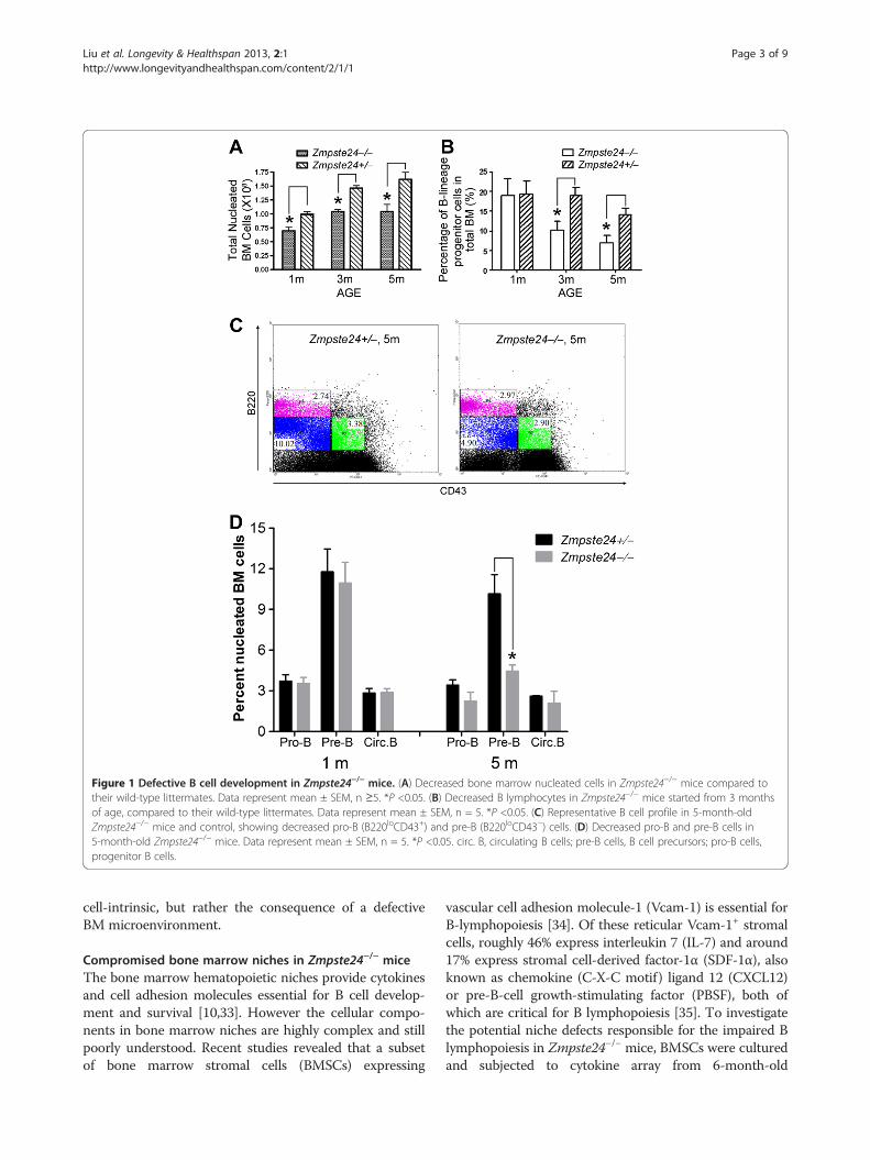
Figure 1 Defective B cell development in Zmpste24−/− mice. (A) Decreased bone marrow nucleated cells in Zmpste24−/− mice compared totheir wild-type littermates. Data represent mean ± SEM, n ≥5. *P <0.05. (B) Decreased B lymphocytes in Zmpste24−/− mice started from 3 monthsof age, compared to their wild-type littermates. Data represent mean ± SEM, n = 5. *P <0.05. (C) Representative B cell profile in 5-month-oldZmpste24−/− mice and control, showing decreased pro-B (B220loCD43+) and pre-B (B220loCD43−) cells. (D) Decreased pro-B and pre-B cells in5-month-old Zmpste24−/− mice. Data represent mean ± SEM, n = 5. *P <0.05. circ. B, circulating B cells; pre-B cells, B cell precursors; pro-B cells,progenitor B cells.
Liu et al. Longevity & Healthspan 2013, 2:1 Page 3 of 9http://www.longevityandhealthspan.com/content/2/1/1
cell-intrinsic, but rather the consequence of a defectiveBM microenvironment.
Compromised bone marrow niches in Zmpste24−/− miceThe bone marrow hematopoietic niches provide cytokinesand cell adhesion molecules essential for B cell develop-ment and survival [10,33]. However the cellular compo-nents in bone marrow niches are highly complex and stillpoorly understood. Recent studies revealed that a subsetof bone marrow stromal cells (BMSCs) expressing
vascular cell adhesion molecule-1 (Vcam-1) is essential forB-lymphopoiesis [34]. Of these reticular Vcam-1+ stromalcells, roughly 46% express interleukin 7 (IL-7) and around17% express stromal cell-derived factor-1α (SDF-1α), alsoknown as chemokine (C-X-C motif) ligand 12 (CXCL12)or pre-B-cell growth-stimulating factor (PBSF), both ofwhich are critical for B lymphopoiesis [35]. To investigatethe potential niche defects responsible for the impaired Blymphopoiesis in Zmpste24−/− mice, BMSCs were culturedand subjected to cytokine array from 6-month-old
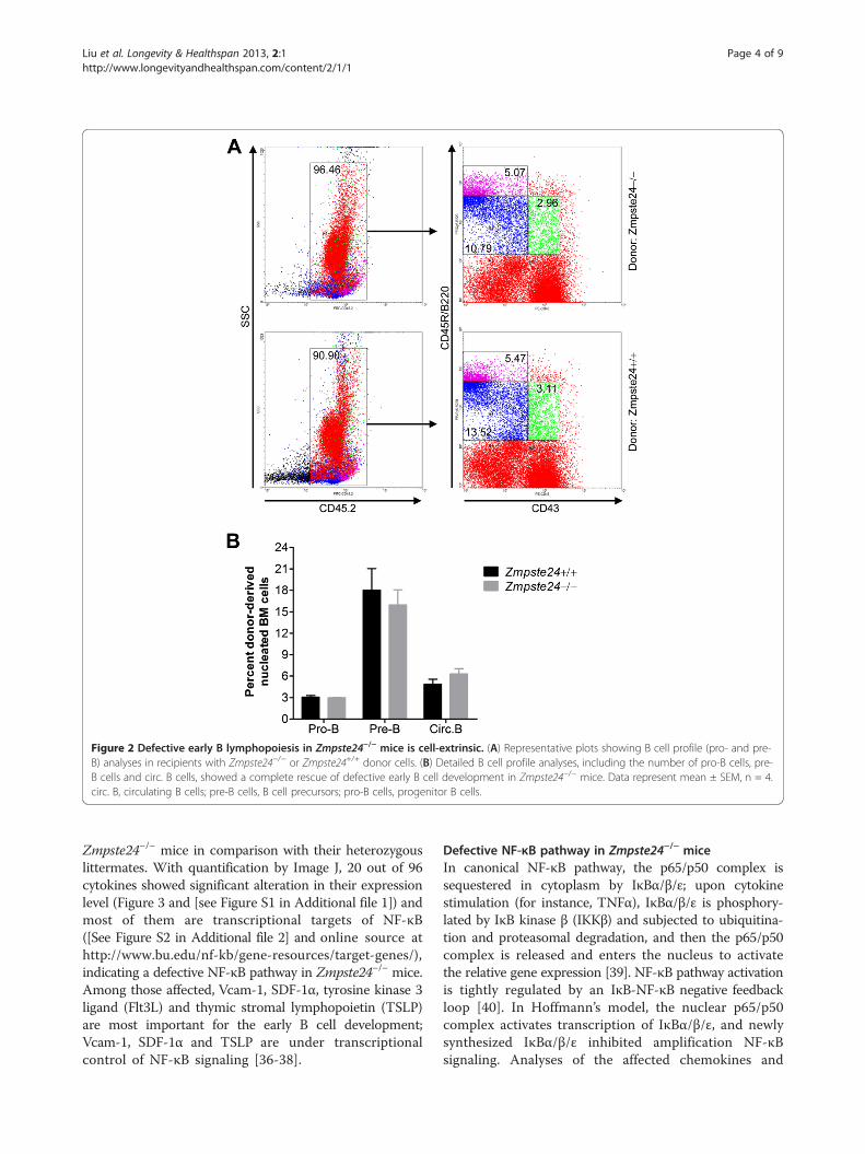
Figure 2 Defective early B lymphopoiesis in Zmpste24−/− mice is cell-extrinsic. (A) Representative plots showing B cell profile (pro- and pre-B) analyses in recipients with Zmpste24−/− or Zmpste24+/+ donor cells. (B) Detailed B cell profile analyses, including the number of pro-B cells, pre-B cells and circ. B cells, showed a complete rescue of defective early B cell development in Zmpste24−/− mice. Data represent mean ± SEM, n = 4.circ. B, circulating B cells; pre-B cells, B cell precursors; pro-B cells, progenitor B cells.
Liu et al. Longevity & Healthspan 2013, 2:1 Page 4 of 9http://www.longevityandhealthspan.com/content/2/1/1
Zmpste24−/− mice in comparison with their heterozygouslittermates. With quantification by Image J, 20 out of 96cytokines showed significant alteration in their expressionlevel (Figure 3 and [see Figure S1 in Additional file 1]) andmost of them are transcriptional targets of NF-κB([See Figure S2 in Additional file 2] and online source athttp://www.bu.edu/nf-kb/gene-resources/target-genes/),indicating a defective NF-κB pathway in Zmpste24−/− mice.Among those affected, Vcam-1, SDF-1α, tyrosine kinase 3ligand (Flt3L) and thymic stromal lymphopoietin (TSLP)are most important for the early B cell development;Vcam-1, SDF-1α and TSLP are under transcriptionalcontrol of NF-κB signaling [36-38].
Defective NF-κB pathway in Zmpste24−/− miceIn canonical NF-κB pathway, the p65/p50 complex issequestered in cytoplasm by IκBα/β/ε; upon cytokinestimulation (for instance, TNFα), IκBα/β/ε is phosphory-lated by IκB kinase β (IKKβ) and subjected to ubiquitina-tion and proteasomal degradation, and then the p65/p50complex is released and enters the nucleus to activatethe relative gene expression [39]. NF-κB pathway activationis tightly regulated by an IκB-NF-κB negative feedbackloop [40]. In Hoffmann’s model, the nuclear p65/p50complex activates transcription of IκBα/β/ε, and newlysynthesized IκBα/β/ε inhibited amplification NF-κBsignaling. Analyses of the affected chemokines and
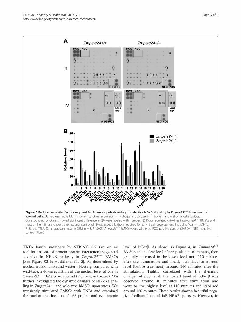
Figure 3 Reduced essential factors required for B lymphopoiesis owing to defective NF-κB signaling in Zmpste24−/− bone marrowstromal cells. (A) Representative blots showing cytokine expression in wild-type and Zmpste24−/− bone marrow stromal cells (BMSCs).Corresponding cytokines showed significant difference in (B) were labeled with number. (B) Downregulated cytokines in Zmpste24−/− BMSCs andmost of them (#) are under transcriptional control of NF-κB, especially those required for early B cell development, including Vcam-1, SDF-1α,Flt3L and TSLP. Data represent mean ± SEM, n = 3. P <0.05, Zmpste24−/− BMSCs versus wild-type. POS, positive control (GAPDH); NEG, negativecontrol (Blank).
Liu et al. Longevity & Healthspan 2013, 2:1 Page 5 of 9http://www.longevityandhealthspan.com/content/2/1/1
TNFα family members by STRING 8.2 (an onlinetool for analysis of protein-protein interaction) suggesteda defect in NF-κB pathway in Zmpste24−/− BMSCs[See Figure S2 in Additional file 2]. As determined bynuclear fractionation and western blotting, compared withwild-type, a downregulation of the nuclear level of p65 inZmpste24−/− BMSCs was found (Figure 4, untreated). Wefurther investigated the dynamic changes of NF-κB signa-ling in Zmpste24−/− and wild-type BMSCs upon stress. Wetransiently stimulated BMSCs with TNFα and examinedthe nuclear translocation of p65 protein and cytoplasmic
level of IκBα/β. As shown in Figure 4, in Zmpste24+/+
BMSCs, the nuclear level of p65 peaked at 10 minutes, thengradually decreased to the lowest level until 110 minutesafter the stimulation and finally stabilized to normallevel (before treatment) around 160 minutes after thestimulation. Tightly correlated with the dynamicchanges of p65 level, the lowest level of IκBα/β wasobserved around 10 minutes after stimulation andwent to the highest level at 110 minutes and stabilizedaround 160 minutes. These results show a beautiful nega-tive feedback loop of IκB-NF-κB pathway. However, in
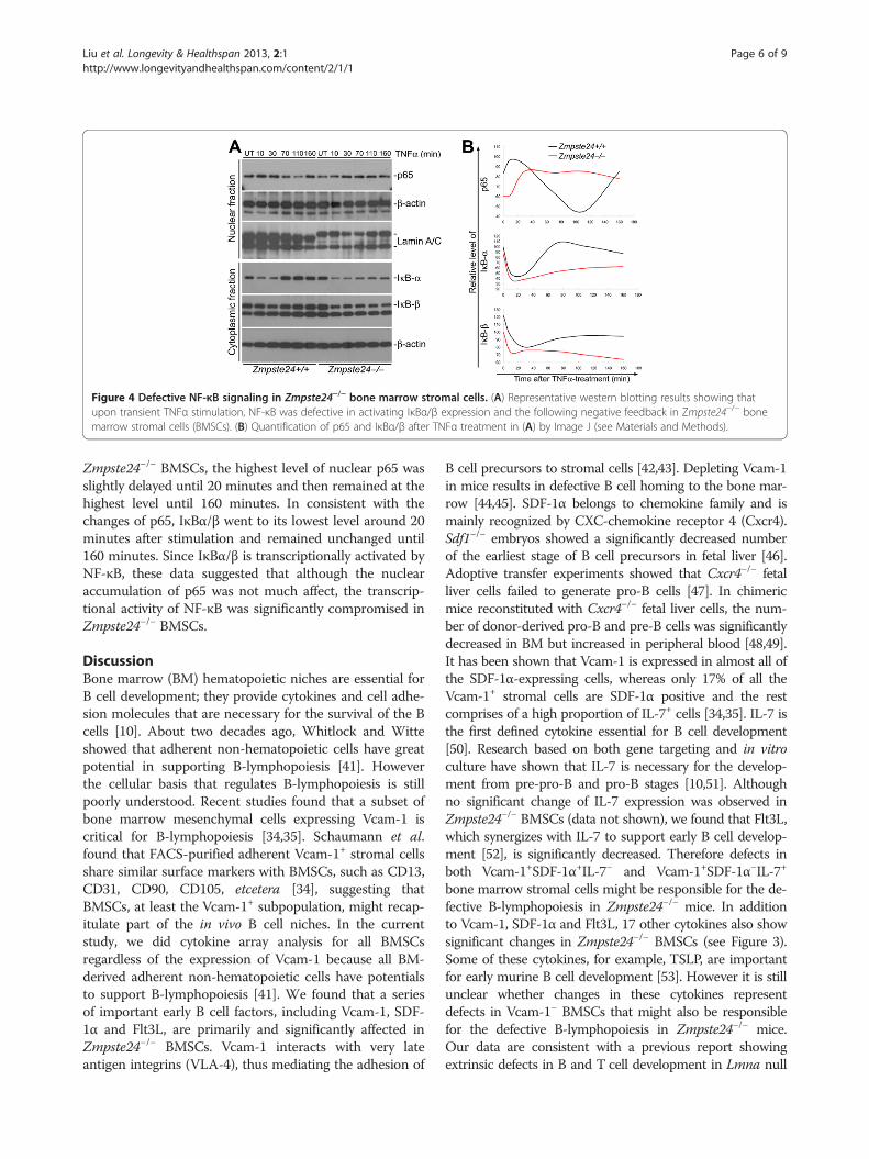
Figure 4 Defective NF-κB signaling in Zmpste24−/− bone marrow stromal cells. (A) Representative western blotting results showing thatupon transient TNFα stimulation, NF-κB was defective in activating IκBα/β expression and the following negative feedback in Zmpste24−/− bonemarrow stromal cells (BMSCs). (B) Quantification of p65 and IκBα/β after TNFα treatment in (A) by Image J (see Materials and Methods).
Liu et al. Longevity & Healthspan 2013, 2:1 Page 6 of 9http://www.longevityandhealthspan.com/content/2/1/1
Zmpste24−/− BMSCs, the highest level of nuclear p65 wasslightly delayed until 20 minutes and then remained at thehighest level until 160 minutes. In consistent with thechanges of p65, IκBα/β went to its lowest level around 20minutes after stimulation and remained unchanged until160 minutes. Since IκBα/β is transcriptionally activated byNF-κB, these data suggested that although the nuclearaccumulation of p65 was not much affect, the transcrip-tional activity of NF-κB was significantly compromised inZmpste24−/− BMSCs.
DiscussionBone marrow (BM) hematopoietic niches are essential forB cell development; they provide cytokines and cell adhe-sion molecules that are necessary for the survival of the Bcells [10]. About two decades ago, Whitlock and Witteshowed that adherent non-hematopoietic cells have greatpotential in supporting B-lymphopoiesis [41]. Howeverthe cellular basis that regulates B-lymphopoiesis is stillpoorly understood. Recent studies found that a subset ofbone marrow mesenchymal cells expressing Vcam-1 iscritical for B-lymphopoiesis [34,35]. Schaumann et al.found that FACS-purified adherent Vcam-1+ stromal cellsshare similar surface markers with BMSCs, such as CD13,CD31, CD90, CD105, etcetera [34], suggesting thatBMSCs, at least the Vcam-1+ subpopulation, might recap-itulate part of the in vivo B cell niches. In the currentstudy, we did cytokine array analysis for all BMSCsregardless of the expression of Vcam-1 because all BM-derived adherent non-hematopoietic cells have potentialsto support B-lymphopoiesis [41]. We found that a seriesof important early B cell factors, including Vcam-1, SDF-1α and Flt3L, are primarily and significantly affected inZmpste24−/− BMSCs. Vcam-1 interacts with very lateantigen integrins (VLA-4), thus mediating the adhesion of
B cell precursors to stromal cells [42,43]. Depleting Vcam-1in mice results in defective B cell homing to the bone mar-row [44,45]. SDF-1α belongs to chemokine family and ismainly recognized by CXC-chemokine receptor 4 (Cxcr4).Sdf1−/− embryos showed a significantly decreased numberof the earliest stage of B cell precursors in fetal liver [46].Adoptive transfer experiments showed that Cxcr4−/− fetalliver cells failed to generate pro-B cells [47]. In chimericmice reconstituted with Cxcr4−/− fetal liver cells, the num-ber of donor-derived pro-B and pre-B cells was significantlydecreased in BM but increased in peripheral blood [48,49].It has been shown that Vcam-1 is expressed in almost all ofthe SDF-1α-expressing cells, whereas only 17% of all theVcam-1+ stromal cells are SDF-1α positive and the restcomprises of a high proportion of IL-7+ cells [34,35]. IL-7 isthe first defined cytokine essential for B cell development[50]. Research based on both gene targeting and in vitroculture have shown that IL-7 is necessary for the develop-ment from pre-pro-B and pro-B stages [10,51]. Althoughno significant change of IL-7 expression was observed inZmpste24−/− BMSCs (data not shown), we found that Flt3L,which synergizes with IL-7 to support early B cell develop-ment [52], is significantly decreased. Therefore defects inboth Vcam-1+SDF-1α+IL-7− and Vcam-1+SDF-1α−IL-7+
bone marrow stromal cells might be responsible for the de-fective B-lymphopoiesis in Zmpste24−/− mice. In additionto Vcam-1, SDF-1α and Flt3L, 17 other cytokines also showsignificant changes in Zmpste24−/− BMSCs (see Figure 3).Some of these cytokines, for example, TSLP, are importantfor early murine B cell development [53]. However it is stillunclear whether changes in these cytokines representdefects in Vcam-1− BMSCs that might also be responsiblefor the defective B-lymphopoiesis in Zmpste24−/− mice.Our data are consistent with a previous report showingextrinsic defects in B and T cell development in Lmna null
Liu et al. Longevity & Healthspan 2013, 2:1 Page 7 of 9http://www.longevityandhealthspan.com/content/2/1/1
mice [29]. However, Hale and colleagues showed thatengrafted Lmna null thymus is comparable in the ability tosupport T lymphopoiesis and concluded that the defectiveT cell development is attributable to the overall unhealthi-ness of the host animal instead of impaired stromal cells. Inthe current study, we employed in vitro expanded BMSCs;therefore, the defects in various cytokines are independentof the overall healthiness of examined animals. In thisregard, the effects of lacking lamin A in Lmna null miceand accumulation of prelamin A in Zmpste24 null mice arelikely different in the regulation of lymphopoietic niches.Our data are also consistent with a report showing de-
fective NF-κB pathway in Lmna−/− mice [54], where lossof Lmna compromised IL-1β-stimulated NF-κB-regu-lated luciferase activity, although the binding of NF-κBto target sequences was increased. It is worthwhile toelucidate the underlying molecular mechanism ofdefective NF-κB signaling in Zmpste24−/− mice in futurestudies. The transcriptional activity of NF-κB can also beregulated by co-activators, including p300/CBP, PCAF,p160 proteins (SRC-1/2/3), etcetera, and co-repressors,such as HDAC1/2/3, SMRT, NcoR, etcetera, whichmodulate local chromatin structure and NF-κB signaling.Given that the chromatin structure is disorganized inHGPS and normal aging cells [24,55,56], one possibilityis that accumulation of unprocessed prelamin A mayaffect local chromatin remodeling and thus impede NF-κB-mediated transcription activation in Zmpste24−/−
BMSCs. This may also explain the finding that only a setof targets are affected by defective NF-κB signaling inZmpste24−/− BMSCs.
ConclusionsIn this study, we found a significant decline in the num-ber of B cell progenitors in Zmpste24−/− mice. Furtherinvestigation revealed that the defective B-lymphopoiesisis most likely attributable to decreased NF-κB signalingin BMSCs, which likely resembles in vivo B cell niches.Of those 20 affected cytokines in Zmpste24−/− BMSCs,Vcam-1, SDF-1α, Flt3L and TSLP are among the mostwell-studied and are essential for early B lymphopoiesis.Collectively, our data demonstrate a cell-extrinsic defectof B cell development in a progeroid mouse model and acritical role for lamin A in the regulation of NF-κBsignaling and essential cytokines in B-lymphopoiesis. Asprogerin accumulates in and contributes to healthyaging, our data also suggest a mechanistic explanationfor aging-related decline in B cell populations inaged individuals.
MethodsAntibodiesPE/Cy5 anti CD45R/B220 (RA3-6B2), R-PE anti CD43and FITC anti-mouse CD45.2 were sourced from
BioLegend (San Diego, CA, USA). Mouse lineagepanel (anti-CD3ε, anti-CD11b, anti-B220, anti-Ly-6G,anti-Ly-6C, and anti-TER-119) and anti Biotin microbeadswere purchased from BD Biosciences (San Jose, CA,USA). Rabbit anti p65, IκB-α, IκB-β and lamin A/Cantibodies were from Santa Cruz (Santa Cruz, CA, USA).Mouse anti β-actin antibody was from Sigma (St. Louis,MO, USA).
AnimalsMouse experiments were performed under the regula-tions and guidelines of the Committee on the Use ofLive Animals in Teaching and Research (CULATR) atthe University of Hong Kong. Zmpste24−/− mice weredescribed previously [16].
B cell profile analysis and bone marrow transplantationBone marrow cells were flushed out into HBSS sup-plemented with 2% FBS, stained with B cell markers(PE/Cy5-anti-CD45R/B220 and R-PE-anti-CD43) andsubjected to FACS analysis. For total bone marrow trans-plantation, 6-month-old B6.SJL/BoyJ mice were irradiatedwith 9 Gy and served as recipients. A total of 1×10⁷ bonemarrow nucleated cells from either Zmpste24−/− mice orwild-type littermates were suspended in 100 μl of HBSSsupplemented with 2% FBS and injected into recipientsvia tail vain. FITC-anti-CD45.2 antibody was used toidentify donor-derived lymphocytes.
Bone marrow stromal cell culture and RayBio™ MouseCytokine Antibody ArrayBMSCs were cultured as described [57]. Briefly, totalbone marrow cells were flushed out with HBSS buffer(containing 2% FBS) and taken into culture with 3 ml α-MEM medium (containing 20% FBS and penicillin-streptomycin) directly in a 60-mm petri dish. Culturemedium was replaced with fresh α-MEM (containing20% FBS and no antibiotics) after 24 h, which clearedmost of the unattached cells (including red blood cells)and cell debris. After 6 days, the P0 culture was trypsi-nized (with 0.025% trypsin and 0.01% EDTA) for 10minutes and the detached cells were transferred into a10-cm petri dish for culture. The cells were then subcul-tured every 3 to 4 days. After passage four, the BMSCcultures were subjected to magnetic sorting with BDmouse lineage panel (anti-CD3ε, anti-CD11b, anti-B220,anti-Ly-6G, anti-Ly-6C, and anti-TER-119) and antiBiotin microbeads according to the manufacture’sinstruction. Cells from three different mice were pooledtogether for the sake of decrease the background noiseand increasing the robustness of the difference betweenmutant and heterozygous mice in the following cytokinearray and protein expression analysis. RayBio™ MouseCytokine Antibody Array III and IV (RayBiotech, USA)
Liu et al. Longevity & Healthspan 2013, 2:1 Page 8 of 9http://www.longevityandhealthspan.com/content/2/1/1
were used to compare the cytokine expression pattern inZmpste24−/− and Zmpste24+/+ BMSCs. The experimentwas performed according to the instruction of themanufacturer.
Protein extraction and western blottingTotal cell lysate was prepared by suspending the cells infive volumes of suspension buffer, and then adding fivevolumes of Laemmli buffer and boiling for 5 minutes.For protein fractionation, cells were suspended in 100 μlice-cold buffer A (10 mM HEPES, pH 7.9, 10 mM KCl,1.5 mM MgCl2, 0.34 M sucrose, 10% glycerol, 1 mMDTT, protease inhibitors). After the addition of 0.1%Triton X-100, the cell suspension was mixed gently,incubated on ice for 5 minutes and centrifuged at 1300×gat 4°C for 4 minutes. The supernatant (S1) was transferredto a new tube and clarified by high-speed centrifugation(12000×g, 10 minutes, 4°C). The remaining nucleipellet (P1) was washed once with 100 μl buffer A andthen resuspended in 100 μl Laemmli buffer andboiled for 5 minutes. Western blotting was performedas described previously [58].
Image quantification and statistics analysisPhotos were processed with Photoshop CSW (AdobeSystems Incorporated, San Jose, CA, USA) when necessary.The pixel intensity of western blotting and dot blotting wasmeasured by Image J gel analysis function [59] andnormalized to housekeeping controls. Student’s t-testwas performed for statistical comparison.
Additional files
Additional file 1: Figure S1. Cytokines that were not affected inZmpste24−/− BMSCs.
Additional file 2: Figure S2. Downregulated cytokines in Zmpste24−/−BMSCs are correlated with NF-κB pathway. (A) Interacting networkamong NF-κB pathway and downregulated factors in Zmpste24−/−BMSCs was predicted by online tool STRING (http://string.embl.de/). (B)Interacting network among those downregulated factors that areessential for B cell development and NF-κB signaling, predicted bySTRING. *Significantly downregulated cytokines in Zmpste24−/− BMSCs.
AbbreviationsBCRs: B cell receptors; BM: Bone marrow; BMSCs: Bone marrow stromal cells;FACS: Fluorescence-activated cell sorting; HGPS: Hutchinson-Gilford progeriasyndrome; HSCs: Hematopoietic stem cells; PBSF: Pre-B-cell growth-stimulating factor; pre-B: B cell precursors; pre-pro-B: Precursor of B cellprogenitor; pro-B: Progenitor B; sIgM: Surface immunoglobulin; TSLP: Thymicstromal lymphopoietin; Vcam-1: Vascular cell adhesion molecule-1.
Competing interestsThe authors declare that they have no competing interests.
Authors’ contributionsBL and ZZ designed experiments. BL and SZ conducted experiments. BL, XL,KZ, FZ and ZZ analyzed the data. BL, SZ and ZZ wrote the manuscript. Allauthors discussed the results and commented on the manuscript. All authorsread and approved the final manuscript.
AcknowledgementsWe thank Ms. Alice Lui for technical assistance. This project is supported byresearch grants (HKU7655/06M, CRF/HKU3/07C) from Research Grant Councilof Hong Kong, the 973 Project (2007CB507400) from the Ministry of Scienceand Technology of China, a NSFC grant (30871440) and a Guangdong NSFgrant (8452402301001450). The funders had no role in study design, datacollection and analysis, decision to publish, or preparation of the manuscript.
Author details1Department of Biochemistry, Li Ka Shing Faculty of Medicine, The Universityof Hong Kong, 21 Sassoon Road, Pokfulam, Hong Kong. 2GuangdongMedical College, 1 Xin Cheng Avenue, Dongguan 523808, China. 3EyeCenter, Beijing Tongren Hospital, Capital Medical University, Beijing 100730,China.
Received: 8 June 2012 Accepted: 6 November 2012Published: 2 January 2013
References1. Gilbert SF: Developmental Biology. 6th edition. Sunderland: SINAUER
ASSOCIATES, INC; 2000.2. Ghia P, Melchers F, Rolink AG: Age-dependent changes in B lymphocyte
development in man and mouse. Exp Gerontol 2000, 35:159–165.3. Goronzy JJ, Weyand CM: T cell development and receptor diversity
during aging. Curr Opin Immunol 2005, 17:468–475.4. Solana R, Pawelec G, Tarazona R: Aging and innate immunity. Immunity
2006, 24:491–494.5. Weng NP: Aging of the immune system: how much can the adaptive
immune system adapt? Immunity 2006, 24:495–499.6. Pistoresi-Palencia MC, Romero-Piffiguer M, Moron G, Ferro ME: Effect of
aging on autoimmune response to rat male accessory glands: young,but not aged, antigen-presenting cells efficiently induce suppression inaged rats. Mech Ageing Dev 1994, 76:33–41.
7. Bassing CH, Alt FW: The cellular response to general and programmedDNA double strand breaks. DNA Repair (Amst) 2004, 3:781–796.
8. Ogawa M, ten Boekel E, Melchers F: Identification of CD19(−)B220(+)c-Kit(+)Flt3/Flk-2(+)cells as early B lymphoid precursors before pre-B-I cells injuvenile mouse bone marrow. Int Immunol 2000, 12:313–324.
9. Li YS, Wasserman R, Hayakawa K, Hardy RR: Identification of the earliest Blineage stage in mouse bone marrow. Immunity 1996, 5:527–535.
10. Hardy RR, Carmack CE, Shinton SA, Kemp JD, Hayakawa K: Resolution andcharacterization of pro-B and pre-pro-B cell stages in normal mousebone marrow. J Exp Med 1991, 173:1213–1225.
11. Allman D, Li J, Hardy RR: Commitment to the B lymphoid lineage occursbefore DH-JH recombination. J Exp Med 1999, 189:735–740.
12. Rossi DJ, Bryder D, Zahn JM, Ahlenius H, Sonu R, Wagers AJ, Weissman IL:Cell intrinsic alterations underlie hematopoietic stem cell aging. Proc NatlAcad Sci USA 2005, 102:9194–9199.
13. Hennekam RC: Hutchinson-Gilford progeria syndrome: review of thephenotype. Am J Med Genet A 2006, 140:2603–2624.
14. Pollex RL, Hegele RA: Hutchinson-Gilford progeria syndrome. Clin Genet2004, 66:375–381.
15. Beck LA, Hosick TJ, Sinensky M: Isoprenylation is required for theprocessing of the lamin A precursor. J Cell Biol 1990, 110:1489–1499.
16. Pendas AM, Zhou Z, Cadinanos J, Freije JM, Wang J, Hultenby K, Astudillo A,Wernerson A, Rodriguez F, Tryggvason K, Lopez-Otin C: Defective prelaminA processing and muscular and adipocyte alterations in Zmpste24metalloproteinase-deficient mice. Nat Genet 2002, 31:94–99.
17. Bergo MO, Gavino B, Ross J, Schmidt WK, Hong C, Kendall LV, Mohr A, MetaM, Genant H, Jiang Y, Wisner ER, Van Bruggen N, Carano RA, Michaelis S,Griffey SM, Young SG: Zmpste24 deficiency in mice causes spontaneousbone fractures, muscle weakness, and a prelamin A processing defect.Proc Natl Acad Sci USA 2002, 99:13049–13054.
18. Rusinol AE, Sinensky MS: Farnesylated lamins, progeroid syndromes andfarnesyl transferase inhibitors. J Cell Sci 2006, 119:3265–3272.
19. Eriksson M, Brown WT, Gordon LB, Glynn MW, Singer J, Scott L, Erdos MR,Robbins CM, Moses TY, Berglund P, Dutra A, Pak E, Durkin S, Csoka AB,Boehnke M, Glover TW, Collins FS: Recurrent de novo point mutations inlamin A cause Hutchinson-Gilford progeria syndrome. Nature 2003,423:293–298.
Liu et al. Longevity & Healthspan 2013, 2:1 Page 9 of 9http://www.longevityandhealthspan.com/content/2/1/1
20. Reddel CJ, Weiss AS: Lamin A expression levels are unperturbed at thenormal and mutant alleles but display partial splice site selection inHutchinson-Gilford progeria syndrome. J Med Genet 2004, 41:715–717.
21. Sullivan T, Escalante-Alcalde D, Bhatt H, Anver M, Bhat N, Nagashima K,Stewart CL, Burke B: Loss of A-type lamin expression compromisesnuclear envelope integrity leading to muscular dystrophy. J Cell Biol1999, 147:913–920.
22. Varela I, Cadinanos J, Pendas AM, Gutierrez-Fernandez A, Folgueras AR,Sanchez LM, Zhou Z, Rodriguez FJ, Stewart CL, Vega JA, Tryggvason K, FreijeJM, López-Otín C: Accelerated ageing in mice deficient in Zmpste24protease is linked to p53 signalling activation. Nature 2005, 437:564–568.
23. Fong LG, Ng JK, Meta M, Cote N, Yang SH, Stewart CL, Sullivan T, BurghardtA, Majumdar S, Reue K, Bergo MO, Young SG: Heterozygosity for Lmnadeficiency eliminates the progeria-like phenotypes in Zmpste24-deficient mice. Proc Natl Acad Sci USA 2004, 101:18111–18116.
24. Scaffidi P, Misteli T: Lamin A-dependent nuclear defects in human aging.Science 2006, 312:1059–1063.
25. Candelario J, Sudhakar S, Navarro S, Reddy S, Comai L: Perturbation ofwild-type lamin A metabolism results in a progeroid phenotype. AgingCell 2008, 7:355–367.
26. Kudlow BA, Stanfel MN, Burtner CR, Johnston ED, Kennedy BK: Suppressionof proliferative defects associated with processing-defective lamin Amutants by hTERT or inactivation of p53. Mol Biol Cell 2008,19:5238–5248.
27. McClintock D, Ratner D, Lokuge M, Owens DM, Gordon LB, Collins FS,Djabali K: The mutant form of lamin A that causes Hutchinson-Gilfordprogeria is a biomarker of cellular aging in human skin. PLoS One 2007,2:e1269.
28. Cao K, Blair CD, Faddah DA, Kieckhaefer JE, Olive M, Erdos MR, Nabel EG,Collins FS: Progerin and telomere dysfunction collaborate to triggercellular senescence in normal human fibroblasts. J Clin Invest 2011,121:2833–2844.
29. Hale JS, Frock RL, Mamman SA, Fink PJ, Kennedy BK: Cell-extrinsic defectivelymphocyte development in Lmna(−/−) mice. PLoS One 2010, 5:e10127.
30. Riley RL, Kruger MG, Elia J: B cell precursors are decreased in senescentBALB/c mice, but retain normal mitotic activity in vivo and in vitro. ClinImmunol Immunopathol 1991, 59:301–313.
31. Stephan RP, Sanders VM, Witte PL: Stage-specific alterations in murine Blymphopoiesis with age. Int Immunol 1996, 8:509–518.
32. Johnson KM, Owen K, Witte PL: Aging and developmental transitions inthe B cell lineage. Int Immunol 2002, 14:1313–1323.
33. Wilson A, Trumpp A: Bone-marrow haematopoietic-stem-cell niches. NatRev Immunol 2006, 6:93–106.
34. Schaumann DH, Tuischer J, Ebell W, Manz RA, Lauster R: VCAM-1-positivestromal cells from human bone marrow producing cytokines for Blineage progenitors and for plasma cells: SDF-1, flt3L, and BAFF. MolImmunol 2007, 44:1606–1612.
35. Tokoyoda K, Egawa T, Sugiyama T, Choi BI, Nagasawa T: Cellular nichescontrolling B lymphocyte behavior within bone marrow duringdevelopment. Immunity 2004, 20:707–718.
36. Iademarco MF, McQuillan JJ, Rosen GD, Dean DC: Characterization of thepromoter for vascular cell adhesion molecule-1 (VCAM-1). J Biol Chem1992, 267:16323–16329.
37. Lee HC, Ziegler SF: Inducible expression of the proallergic cytokinethymic stromal lymphopoietin in airway epithelial cells is controlled byNFkappaB. Proc Natl Acad Sci USA 2007, 104:914–919.
38. Dejardin E, Droin NM, Delhase M, Haas E, Cao Y, Makris C, Li ZW, Karin M,Ware CF, Green DR: The lymphotoxin-beta receptor induces differentpatterns of gene expression via two NF-kappaB pathways. Immunity2002, 17:525–535.
39. Vallabhapurapu S, Karin M: Regulation and function of NF-kappaBtranscription factors in the immune system. Annu Rev Immunol 2009,27:693–733.
40. Hoffmann A, Levchenko A, Scott ML, Baltimore D: The IkappaB-NF-kappaBsignaling module: temporal control and selective gene activation. Science2002, 298:1241–1245.
41. Whitlock CA, Witte ON: Long-term culture of B lymphocytes and theirprecursors from murine bone marrow. Proc Natl Acad Sci USA 1982,79:3608–3612.
42. Ryan DH, Nuccie BL, Abboud CN, Winslow JM: Vascular cell adhesionmolecule-1 and the integrin VLA-4 mediate adhesion of human B cell
precursors to cultured bone marrow adherent cells. J Clin Invest 1991,88:995–1004.
43. Dittel BN, McCarthy JB, Wayner EA, LeBien TW: Regulation of human B-cellprecursor adhesion to bone marrow stromal cells by cytokines that exertopposing effects on the expression of vascular cell adhesion molecule-1(VCAM-1). Blood 1993, 81:2272–2282.
44. Koni PA, Joshi SK, Temann UA, Olson D, Burkly L, Flavell RA: Conditionalvascular cell adhesion molecule 1 deletion in mice: impaired lymphocytemigration to bone marrow. J Exp Med 2001, 193:741–754.
45. Leuker CE, Labow M, Muller W, Wagner N: Neonatally induced inactivationof the vascular cell adhesion molecule 1 gene impairs B cell localizationand T cell-dependent humoral immune response. J Exp Med 2001,193:755–768.
46. Egawa T, Kawabata K, Kawamoto H, Amada K, Okamoto R, Fujii N, KishimotoT, Katsura Y, Nagasawa T: The earliest stages of B cell developmentrequire a chemokine stromal cell-derived factor/pre-B cell growth-stimulating factor. Immunity 2001, 15:323–334.
47. Ma Q, Jones D, Borghesani PR, Segal RA, Nagasawa T, Kishimoto T, BronsonRT, Springer TA: Impaired B-lymphopoiesis, myelopoiesis, and derailedcerebellar neuron migration in CXCR4- and SDF-1-deficient mice. ProcNatl Acad Sci USA 1998, 95:9448–9453.
48. Kawabata K, Ujikawa M, Egawa T, Kawamoto H, Tachibana K, Iizasa H,Katsura Y, Kishimoto T, Nagasawa T: A cell-autonomous requirement forCXCR4 in long-term lymphoid and myeloid reconstitution. Proc Natl AcadSci USA 1999, 96:5663–5667.
49. Ma Q, Jones D, Springer TA: The chemokine receptor CXCR4 is requiredfor the retention of B lineage and granulocytic precursors within thebone marrow microenvironment. Immunity 1999, 10:463–471.
50. Namen AE, Lupton S, Hjerrild K, Wignall J, Mochizuki DY, Schmierer A,Mosley B, March CJ, Urdal D, Gillis S: Stimulation of B-cell progenitors bycloned murine interleukin-7. Nature 1988, 333:571–573.
51. von Freeden-Jeffry U, Vieira P, Lucian LA, McNeil T, Burdach SE, Murray R:Lymphopenia in interleukin (IL)-7 gene-deleted mice identifies IL-7 as anonredundant cytokine. J Exp Med 1995, 181:1519–1526.
52. Namikawa R, Muench MO, de Vries JE, Roncarolo MG: The FLK2/FLT3ligand synergizes with interleukin-7 in promoting stromal-cell-independent expansion and differentiation of human fetal pro-B cellsin vitro. Blood 1996, 87:1881–1890.
53. Ray RJ, Furlonger C, Williams DE, Paige CJ: Characterization of thymicstromal-derived lymphopoietin (TSLP) in murine B cell developmentin vitro. Eur J Immunol 1996, 26:10–16.
54. Lammerding J, Schulze PC, Takahashi T, Kozlov S, Sullivan T, Kamm RD,Stewart CL, Lee RT: Lamin A/C deficiency causes defective nuclearmechanics and mechanotransduction. J Clin Invest 2004, 113:370–378.
55. Columbaro M, Capanni C, Mattioli E, Novelli G, Parnaik VK, Squarzoni S,Maraldi NM, Lattanzi G: Rescue of heterochromatin organization inHutchinson-Gilford progeria by drug treatment. Cell Mol Life Sci 2005,62:2669–2678.
56. Shumaker DK, Dechat T, Kohlmaier A, Adam SA, Bozovsky MR, Erdos MR,Eriksson M, Goldman AE, Khuon S, Collins FS, Jenuwein T, Goldman RD:Mutant nuclear lamin A leads to progressive alterations of epigeneticcontrol in premature aging. Proc Natl Acad Sci USA 2006, 103:8703–8708.
57. Meirelles Lda S, Nardi NB: Murine marrow-derived mesenchymal stemcell: isolation, in vitro expansion, and characterization. Br J Haematol2003, 123:702–711.
58. Liu B, Wang J, Chan KM, Tjia WM, Deng W, Guan X, Huang JD, Li KM, ChauPY, Chen DJ, Cao Y, Cheah KS, Tryggvason K, Zhou Z: Genomic instabilityin laminopathy-based premature aging. Nat Med 2005, 11:780–785.
59. Schneider CA, Rasband WS, Eliceiri KW: NIH Image to ImageJ: 25 years ofimage analysis. Nature methods 2012, 9:671–675.
doi:10.1186/2046-2395-2-1Cite this article as: Liu et al.: Accumulation of prelamin A compromisesNF-κB-regulated B-lymphopoiesis in a progeria mouse model. Longevity& Healthspan 2013 2:1.