Research Article Tolerance of Chemoorganotrophic...
10
Research Article Tolerance of Chemoorganotrophic Bioleaching Microorganisms to Heavy Metal and Alkaline Stresses Annick Monballiu, 1 Nele Cardon, 1 Minh Tri Nguyen, 2 Christel Cornelly, 1 Boudewijn Meesschaert, 1 and Yi Wai Chiang 3 1 Laboratory for Microbial and Biochemical Technology (Lab BCT), Cluster for Bioengineering Technology, Department of Microbial and Molecular Systems, KU Leuven @ Brugge-Oostende (Kulab), 8400 Oostende, Belgium 2 Mat´ eriaux et Contrˆ oles Physico-Chimiques, D´ epartement Mesures Physiques, Universit´ e de Bordeaux, 33175 Gradignan, France 3 School of Engineering, University of Guelph, Guelph, ON, Canada N1G 2W1 Correspondence should be addressed to Yi Wai Chiang; [email protected] Received 27 February 2015; Revised 2 June 2015; Accepted 9 June 2015 Academic Editor: Takao Yagi Copyright © 2015 Annick Monballiu et al. is is an open access article distributed under the Creative Commons Attribution License, which permits unrestricted use, distribution, and reproduction in any medium, provided the original work is properly cited. e bioleaching potential of the bacterium Bacillus mucilaginosus and the fungus Aspergillus niger towards industrial residues was investigated by assessing their response towards various heavy metals (including arsenic, cadmium, cobalt, chromium, nickel, lead, and zinc) and elevated pH. e plate diffusion method was performed for each metal to determine the toxicity effect. Liquid batch cultures were set up for more quantitative evaluation as well as for studying the influence of basicity. Growth curves were prepared using bacterial/fungal growth counting techniques such as plate counting, optical density measurement, and dry biomass determination. Cadmium, nickel, and arsenite had a negative influence on the growth of B. mucilaginosus, whereas A. niger was sensitive to cadmium and arsenate. However, it was shown that growth recovered when microorganisms cultured in the presence of these metals were inoculated onto metal-free medium. Based on the findings of the bacteriostatic/fungistatic effect of the metals and the adaptability of the microorganisms to fairly elevated pH values, it is concluded that both strains have potential applicability for further research concerning bioleaching of alkaline waste materials. 1. Introduction e growing world population has resulted in an increase in materials consumption and industrial expansion, causing unsustainable pressures on the planet’s natural resource reserves and the environment. For this reason, investigation concerning the development of sustainable technologies and recycling strategies is becoming favoured. In the last decades, interest in bioleaching, which is the conversion of solid metal values into their aqueous soluble forms using microor- ganisms, has increased significantly [1, 2]. is technology has been commercialized for some metal ores such as the biooxidation of refractory gold ores and in copper recovery [3, 4]. e solubilization of metals can be accomplished by various species of bacteria and fungi and is based on three main mechanisms that can occur simultaneously [2, 5, 6]. Acidolysis (i) is by far the most important one and is quite similar to the mechanism of conventional acid leaching. Organic and inorganic acids are produced by the microor- ganisms, which in turn leach the metals from the solids by protonation. In complexolysis (ii), biogenic agents are excreted from the microbes, and these solubilize metal ions through ligand formation. Citric acid, for instance, is a powerful natural chelating agent [7]. Lastly, in redoxolysis (iii), oxidation and reduction reactions occur that set metals free from the mineral. Bioleaching is influenced by a wide range of parame- ters including physicochemical and microbiological factors. ese affect both the growth of the microorganisms and their leaching behaviour. For bioleaching to be successful, it is obvious that optimal growth conditions must be maintained and the microorganism must be able to leach the material. In addition, and most importantly, the microorganism must Hindawi Publishing Corporation Bioinorganic Chemistry and Applications Volume 2015, Article ID 861874, 9 pages http://dx.doi.org/10.1155/2015/861874
Transcript of Research Article Tolerance of Chemoorganotrophic...
Research ArticleTolerance of Chemoorganotrophic Bioleaching Microorganismsto Heavy Metal and Alkaline Stresses
Annick Monballiu,1 Nele Cardon,1 Minh Tri Nguyen,2 Christel Cornelly,1
Boudewijn Meesschaert,1 and Yi Wai Chiang3
1Laboratory for Microbial and Biochemical Technology (Lab 𝜇BCT), Cluster for Bioengineering Technology,Department of Microbial and Molecular Systems, KU Leuven @ Brugge-Oostende (Kulab), 8400 Oostende, Belgium2Materiaux et Controles Physico-Chimiques, Departement Mesures Physiques, Universite de Bordeaux, 33175 Gradignan, France3School of Engineering, University of Guelph, Guelph, ON, Canada N1G 2W1
Correspondence should be addressed to Yi Wai Chiang; [email protected]
Received 27 February 2015; Revised 2 June 2015; Accepted 9 June 2015
Academic Editor: Takao Yagi
Copyright © 2015 Annick Monballiu et al. This is an open access article distributed under the Creative Commons AttributionLicense, which permits unrestricted use, distribution, and reproduction in any medium, provided the original work is properlycited.
The bioleaching potential of the bacterium Bacillus mucilaginosus and the fungus Aspergillus niger towards industrial residueswas investigated by assessing their response towards various heavy metals (including arsenic, cadmium, cobalt, chromium, nickel,lead, and zinc) and elevated pH. The plate diffusion method was performed for each metal to determine the toxicity effect. Liquidbatch cultures were set up for more quantitative evaluation as well as for studying the influence of basicity. Growth curves wereprepared using bacterial/fungal growth counting techniques such as plate counting, optical density measurement, and dry biomassdetermination. Cadmium, nickel, and arsenite had a negative influence on the growth of B. mucilaginosus, whereas A. niger wassensitive to cadmium and arsenate. However, it was shown that growth recovered when microorganisms cultured in the presenceof these metals were inoculated onto metal-free medium. Based on the findings of the bacteriostatic/fungistatic effect of the metalsand the adaptability of the microorganisms to fairly elevated pH values, it is concluded that both strains have potential applicabilityfor further research concerning bioleaching of alkaline waste materials.
1. Introduction
The growing world population has resulted in an increasein materials consumption and industrial expansion, causingunsustainable pressures on the planet’s natural resourcereserves and the environment. For this reason, investigationconcerning the development of sustainable technologies andrecycling strategies is becoming favoured. In the last decades,interest in bioleaching, which is the conversion of solidmetal values into their aqueous soluble forms using microor-ganisms, has increased significantly [1, 2]. This technologyhas been commercialized for some metal ores such as thebiooxidation of refractory gold ores and in copper recovery[3, 4].
The solubilization of metals can be accomplished byvarious species of bacteria and fungi and is based on threemain mechanisms that can occur simultaneously [2, 5, 6].
Acidolysis (i) is by far the most important one and is quitesimilar to the mechanism of conventional acid leaching.Organic and inorganic acids are produced by the microor-ganisms, which in turn leach the metals from the solidsby protonation. In complexolysis (ii), biogenic agents areexcreted from the microbes, and these solubilize metal ionsthrough ligand formation. Citric acid, for instance, is apowerful natural chelating agent [7]. Lastly, in redoxolysis(iii), oxidation and reduction reactions occur that set metalsfree from the mineral.
Bioleaching is influenced by a wide range of parame-ters including physicochemical and microbiological factors.These affect both the growth of the microorganisms and theirleaching behaviour. For bioleaching to be successful, it isobvious that optimal growth conditions must be maintainedand the microorganism must be able to leach the material.In addition, and most importantly, the microorganism must
Hindawi Publishing CorporationBioinorganic Chemistry and ApplicationsVolume 2015, Article ID 861874, 9 pageshttp://dx.doi.org/10.1155/2015/861874

2 Bioinorganic Chemistry and Applications
be resistant to the metals that are leached out [1]. A suitabletemperature, pH, oxygen, salinity, and the availability ofnutrients are imperative for the growth and maintenanceof microorganisms. These conditions are species dependentand it is principally important to know to which classifica-tion the microorganism belongs (autotrophic/heterotrophic,etc.). Although the optimal temperature mostly variesaround 30–50∘C (for mesophiles), bioleaching performed atlower/higher temperatures (psychro-/thermophiles) is alsoreported, as is the use of aerobic or anaerobicmicroorganisms[1, 8]. The pH of the medium is of great importance forthe quantity and nature of the biogenic acids excreted andit also has an effect on the availability of heavy metals [9].The production of organic acids is strongly dependent onthe medium composition and the conditions of cultivation;in that respect, the carbon source and its concentration areimportant, as well as the presence of essential ions [10].For the leaching of the metals, the properties of the solidsincluding the mineralogical composition, slurry density, andparticle size are key factors [5].
Besides the case for natural resources, bioleaching can bean interesting route for metal recovery from industrial wastematerials, such as incinerator ashes, metallurgical slag, andmineral tailings. It can also be applied in the detoxificationof soils and groundwater contaminated with heavy metalsat various deposit sites [9, 11, 12]. However, challenges lie inthe caustic and toxic nature of many of these waste mate-rials. The presence of heavy metals in bioleached materialscan adversely affect the growth and metabolic processes ofmicroorganisms. Heavy metals are taken up by the bacterialcell through the same ion transport normally required foressential minerals such as magnesium and phosphate [13].This leads to inhibition of specific enzyme activities, dis-placement of essential metals, and disruption of membranetransport processes [14, 15]. Bioleaching over a two-stepprocess, in which there is no direct contact between themicroorganisms and the released heavy metals, is preferableto reduce the toxicity effects; however, it complicates theimplementation of continuous recovery systems [16].
There is no unequivocal answer on the tolerance ofmicroorganisms to the presence of toxic heavy metal ions. Ithas been reported that some microorganisms acquire someform of adaptation mechanism, similar to what is seen uponexposure to antibiotics, to ensure the microbial population’ssurvival and diversity. Metal tolerance mechanisms consistof either extracellular or intracellular sequestration of thetoxic metal and are often gene-regulated by plasmids [17, 18].Adaptation strategies include active transport of metal ionsfrom the inside to the outside of the cell, interaction withextracellular polymeric substances (EPS), formation of cellsurface complexes, and metal reduction to a less toxic state[9, 14, 19–21].
The best known tolerance strategy towards heavy met-als among Bacillus species is the production of extracel-lular polymeric substances (EPS), a complex mixture ofpolysaccharides, proteins, nucleic acids, and lipids. The EPSproduction is influenced by the medium composition andis the bacterium’s protection against environmental stress[22]. During the bioleaching process, it also contributes to
the formation of biofilm, which plays an important role inthe attachment of the bacterial cell to the solid material [19].In the EPS layer, the optimal conditions for pH and soluteconcentration are maintained. Moreover, the EPS is involvedin the capture of metal ions from the solution, preventingtheir entrance into the bacterial cell [23].
In the present study, it is investigated whether elevatedpH and the presence of multiple heavy metals and metalloidshinder the feasibility of alkaline bioleaching of industrialresidues. For this purpose, the tolerance of two chemoorgan-otrophic microbial strains that have shown potential for thebioleaching of waste materials [16], Bacillus mucilaginosusand Aspergillus niger, was assessed. These microorganismsare associated with silica-richminerals in nature, compoundscommonly found in alkaline industrial residues, but in theirnatural environment they are not exposed to, and thus do notbecome adapted to, high levels of mobile heavy metals andmetalloids [24, 25].
2. Materials and Methods
2.1. Microbial Organisms and Chemicals. Bacillus mucilagi-nosus was obtained from China Center of Industrial CultureCollection (CICC, China). Aspergillus niger (DSM-872) wasdelivered by the Deutsche Sammlung von Mikroorganismenund Zellkulturen GmbH (DSMZ, Germany). As recom-mended by the suppliers, B. mucilaginosus was cultured onnutrient agar supplemented with 0.001% MnSO
4⋅H2O for
sporulation enhancement and incubated for 24 hours at 30∘C,whereas A. niger was cultured on potato dextrose agar andincubated at 30∘C for 5 days. Agar slants of each strain werepreserved at 4∘C.
The heavy metals {and respective salts} investigated wereAs(III) {KHAs
2O4}, As(V) {KH
2AsO4}, Cd(II) {Cd(NO
3)2⋅
4H2O}, Co(II) {Co(NO
3)2⋅6H2O}, Cr(III) {Cr(NO
3)3⋅
9H2O}, Cr(VI) {K
2Cr2O7}, Ni(II) {Ni(NO
3)2⋅6H2O}, Pb(II)
{Pb(NO3)2}, and Zn(II) {Zn(NO
3)2⋅6H2O}. Each metal salt
was dissolved in a citric acid/sodium citrate 100mM bufferof pH 6.80. Stock solutions of 1000 ppm metal content wereprepared, and further dilutions were made with the samebuffer.
All growth media and chemicals were of analytical grade,purchased from Sigma Aldrich (Bornem, Belgium), andwere used without any further purification. Growth media,chemical solutions, and glass materials were sterilized for 15minutes at 120∘C prior to use, and subsequent manipulationswere executed under sterile conditions in a laminar flowcabinet.
2.2. Plate Diffusion Method. A first investigation on theeffect of the aforementioned elements on the growth of themicrobial strains was performed using the plate diffusionmethod; more specifically, the central well test was used[26]. In this method, a small cavity is made at the centreof an agar plate, into which an amount of metal solutionis introduced. This allows the formation of a diffusiongradient over the agar plate during incubation, with higherconcentration near the well and essentially nil concentration

Bioinorganic Chemistry and Applications 3
at the outer perimeter. A nutrient or potato dextrose agarplate (90mm) was first abundantly inoculated with a freshlygrown culture of, respectively, B. mucilaginosus or A. nigerstrain. Then, a central well of 1 cm was made. Into this well,100 𝜇L of metal solution with a concentration of 1000 ppm ofthe metal component was added. Each plate was preparedin duplicate, and blank plates using buffer solution withno metal compounds were also made. After the desiredincubation period at 30∘C, the effect of the metals on thegrowth of the two strains was assessed visually by measuringand comparing the diameter of the growth inhibition zones.The plate diffusion test was subsequently repeated with lowerconcentrations of metal solution for those conditions inwhich sensitivity of the microbial strains occurred with the1000 ppm solution, to assess the tolerance threshold.
2.3. Liquid Batch Cultures. The metal that demonstrateda significant impact on the growth of the two microbialstrains during the aforementioned plate diffusion test wasimplemented in liquid cultures. In this test, a desired quantityof the metal stock solution was added to the inoculatedbroth medium (nutrient broth with 0.001% MnSO
4⋅H2O for
B. mucilaginosus and potato dextrose broth for A. niger).Reference blanks were prepared by adding an equal amountof buffer solution rather than metal solution to the brothmedium.
For the experiments concerning the effect of basicity, theinitial pH of the nutrient-rich medium (initial pH 7.15 for thenutrient broth and pH 6.70 for the potato dextrose broth)was increased with NaOH addition until the desired pH (9.0,11.0, 12.0, and 13.0) was reached. The pH was measured witha SevenMulti pH meter (Mettler Toledo).
For each experiment, 200mL or 100mL of the broths, forthemetal-stress and alkaline-stress experiments, respectively,was placed in Erlenmeyer flasks and inoculated with theappropriate strain. Incubation was performed at 30∘C for thedesired duration. The solutions were stirred by a magneticbar.The experimentswere performed in batchmode andwerenot pH-adjusted. The pH was measured every 24 hours.
2.4. Determination of Microbial Growth. Conventional tech-niques were used to detect the microbial growth overtime. For B. mucilaginosus, the spread plate method andmeasurement of optical density were combined. For spreadplating, a 100 𝜇L aliquot of the bacterial culture (whetheror not serially diluted in physiological salt solution, 0.9%NaCl) was transferred to a nutrient agar plate and streakedacross the entire surface of the plate. Every spread plate wasmade in duplicate. After incubation, colonies were manuallycounted and growth was expressed as colony forming unitsper millilitre (CFU/mL). Optical density was measured witha Jenway spectrophotometer at 610 nm wavelength. For thispurpose, 1mL samples of the culture broths were taken and, ifnecessary, diluted with nutrient broth so that the absorbancewas in the linear range of 0–0.6. A zero-point calibration wasperformed with blank nutrient broth.
Growth of A. niger was followed by either the spreadplate method or by the measurement of dried biomass.
The spread plate method was analogous to the one for theBacillus strain, except that potato dextrose agar plates wereused. Measurement of dried biomass involved the fungalbiomass being separated from the liquid culture with aBiofuge Stratos Centrifuge (Heraeus Instruments), followedby overnight drying at 110∘C. The mass of the dried biomassis a measure for the growth of the fungal cells. These liquidbatch cultures were set up with 400mL of the broth medium.Additionally, the concentration of metal in the supernatantphase was determined and compared to the initial metalconcentration. Metal concentration was determined usingatomic absorption spectrometry (SpectrAA 220, Varian, Bel-gium).
3. Results and Discussion
3.1. Growth of Bacillus mucilaginosus
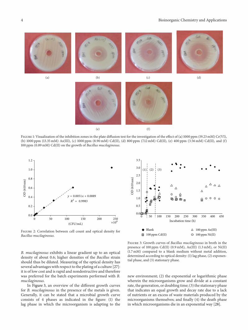
3.1.1. Influence of Heavy Metals. The results of the platediffusion test showed that the buffer solution has no influenceon the growth of both B. mucilaginosus and A. niger. InFigure 1 the most important observations of heavy metalinhibition on B. mucilaginosus are shown, these being thepresence of Cr(VI), As(III), and Cd(II). The other metalsthat were tested did not have a significant influence on thegrowth of B. mucilaginosus and are consequently not shownhere. These tests were performed with 1000 ppm of metalcompound; therefore, for each metal, the molar concentra-tion differed. In the present case, the molar concentrationsin descending order are 19.2mM for Cr(VI), 13.3mM forAs(III), and 8.9mM for Cd(II). As indicated by the size of theinhibition zones, the largest sensitivity occurred with Cd(II)(Figure 1(a)) followed by As(III) (Figure 1(b)); Cr(VI) hadminimal effect (Figure 1(c)). Based on these findings, it isconcluded that Cd(II) is the most toxic metal for the growthof B. mucilaginosus.
The plate diffusion test was repeated for Cd(II) withdecreasing metal concentrations (800, 400, and 100 ppm);these results are shown in Figures 1(d) to 1(e).The decreasingsensitivity with respect to the decreasing concentration of themetal is clearly visible through the behaviour of the inhibitionzones. Another interesting observation made was that oldercultures seemed to bemore sensitive to the heavymetal stress.Yet, over time, some of these colonies adapted and grewunderthe heavy metal stress. The age of the biomass culture cancontribute to an altered and delayed adaptation strategy dueto changes in cell size, wall composition, and extracellularproduct formation [9].
Further investigation on the growth of B. mucilaginosusunder heavy metal stress was performed in liquid batchcultures under the influence of Cd(II) and As(III) at 100 ppmconcentration. In addition, 100 ppm of Ni(II) was also stud-ied. The results were compared with a blank incubated batchwhere no metal was added to the inoculated nutrient-richgrowth medium. Growth was measured over the incubationtime in order to plot a bacterial growth curve. Figure 2demonstrates the correlation between cell count based onspread plating and the measurement of the optical density.
4 Bioinorganic Chemistry and Applications
(a) (b) (c) (d)
(e) (f)
Figure 1: Visualization of the inhibition zones in the plate diffusion test for the investigation of the effect of (a) 1000 ppm (19.23mM) Cr(VI),(b) 1000 ppm (13.35mM) As(III), (c) 1000 ppm (8.90mM) Cd(II), (d) 800 ppm (7.12mM) Cd(II), (e) 400 ppm (3.56mM) Cd(II), and (f)100 ppm (0.89mM) Cd(II) on the growth of Bacillus mucilaginosus.
0.0
0.2
0.4
0.6
0.8
1.0
1.2
0 50 100 150 200 250(CFU/mL)
OD
(610
nm)
y = 0.0051x + 0.0089
R2= 0.9983
×106
Figure 2: Correlation between cell count and optical density forBacillus mucilaginosus.
B. mucilaginosus exhibits a linear gradient up to an opticaldensity of about 0.6; higher densities of the Bacillus strainshould thus be diluted. Measuring of the optical density hasseveral advantageswith respect to the plating of a culture [27]:it is of low cost and is rapid and nondestructive and thereforewas preferred for the batch experiments performed with B.mucilaginosus.
In Figure 3, an overview of the different growth curvesfor B. mucilaginosus in the presence of the metals is given.Generally, it can be stated that a microbial growth curveconsists of 4 phases as indicated in the figure: (1) thelag phase in which the microorganism is adapting to the
0.0
0.5
1.0
1.5
2.0
2.5
3.0
3.5
0 50 100 150 200 250 300 350 400 450Incubation time (h)
Blank
(1) (2) (3)
OD
(610
nm)
100ppm Cd(II)100ppm As(III)100ppm Ni(II)
Figure 3: Growth curves of Bacillus mucilaginosus in broth in thepresence of 100 ppm Cd(II) (0.9mM), As(III) (1.3mM), or Ni(II)(1.7mM) compared to a blank medium without metal addition,determined according to optical density: (1) lag phase, (2) exponen-tial phase, and (3) stationary phase.
new environment; (2) the exponential or logarithmic phasewherein the microorganisms grow and divide at a constantrate, the generation, or doubling time; (3) the stationary phasethat indicates an equal growth and decay rate due to a lackof nutrients or an excess of waste materials produced by themicroorganisms themselves; and finally (4) the death phasein which microorganisms die in an exponential way [28].

Bioinorganic Chemistry and Applications 5
(a) (b)
Figure 4: Growth of Bacillus mucilaginosus on nutrient agar plates.(a) Blank reference strain (after 72 hours of growth in batch culture,resulting in typical colonymorphology on agar plate). (b) Strain thathadprevious contactwithCd(II) in a batch culture for 72 hours (notesmaller colonies and the absence of mucous generation).
6.5
7.0
7.5
8.0
8.5
9.0
9.5
10.0
0 100 200 300 400Incubation time (h)
pH (—
)
Blank100ppm Cd(II)
100ppm As(III)100ppm Ni(II)
Figure 5:Variation of pHduring growth ofBacillusmucilaginosus ina blank nutrient brothmediumand undermetal stress from 100 ppmCd(II) (0.9mM), As(III) (1.3mM), or Ni(II) (1.7mM) additions.
B. mucilaginosus grew under the influence of the metals;however, the lower optical density values obtained in thepresence of metals reveal that the bacterial growth is muchlower compared to the case where no metal is present. Incomparison with the blank, growth in the presence of Cd(II)and Ni(II) was approximately one-third as large, while inthe presence of As(III) approximately half of the maximumbiomass concentration was reached. Taking into account themolar concentrations, once againCd(II) (0.9mM) is themosttoxic metal over As(III) (1.3mM) and Ni(II) (1.7mM) duringthis test.
Each time the OD was measured, duplicate streak plateswith metal-free agar medium were made for counting ofthe colonies. These tests showed that the number of CFUdid not differ drastically for the cultures with and withoutmetals, although the appearance of the colonies changed.Typically, B. mucilaginosus grows on the nutrient agar plates
as large, smooth colonies with significant mucous generation(e.g., in Figure 4(a)). In the cases where the bacterial strainhad contact with a heavy metal in the batch cultures, thecolonies detected were much smaller, with apparently nomucous generation (e.g., in Figure 4(b)). It has been reportedthat, as part of the produced EPS, specific proteins can beformed that are capable of binding metal ions that promotethe tolerance of the bacteria to heavy metals. The presenceof such biomaterial can, however, also inhibit the microbialgrowth, for instance, through creating a delay in oxygentransfer or decreasing the removal of metabolic productsfrom the bacterial cells [29]. The present results indicate thatthe surface or the surface complexes of B. mucilaginosuswere modified when the bacterium was initially grown in amediumwithCd(II) and subsequently was grown on ametal-free medium. It appears that the mucous metabolism ceasedor was heavily disturbed.
As shown in Figure 5, the pH during growth of B.mucilaginosus increased from 7 to 10 for both the blankculture and the cultures withmetals.This can be explained bythe citrate metabolism of B. mucilaginosus where citric acid,available from the buffer solution in which the metal saltswere dissolved, is utilized as a carbon source, resulting in thealkalinization of the medium. It thus appears that this citricacid metabolism is not influenced by the presence of metals.It should be mentioned that, at such high pH, the metals canprecipitate in their hydroxide forms [30]. This may explainwhy the bacteria do not become entirely inactivated, as themetal may no longer be bioavailable in solution.
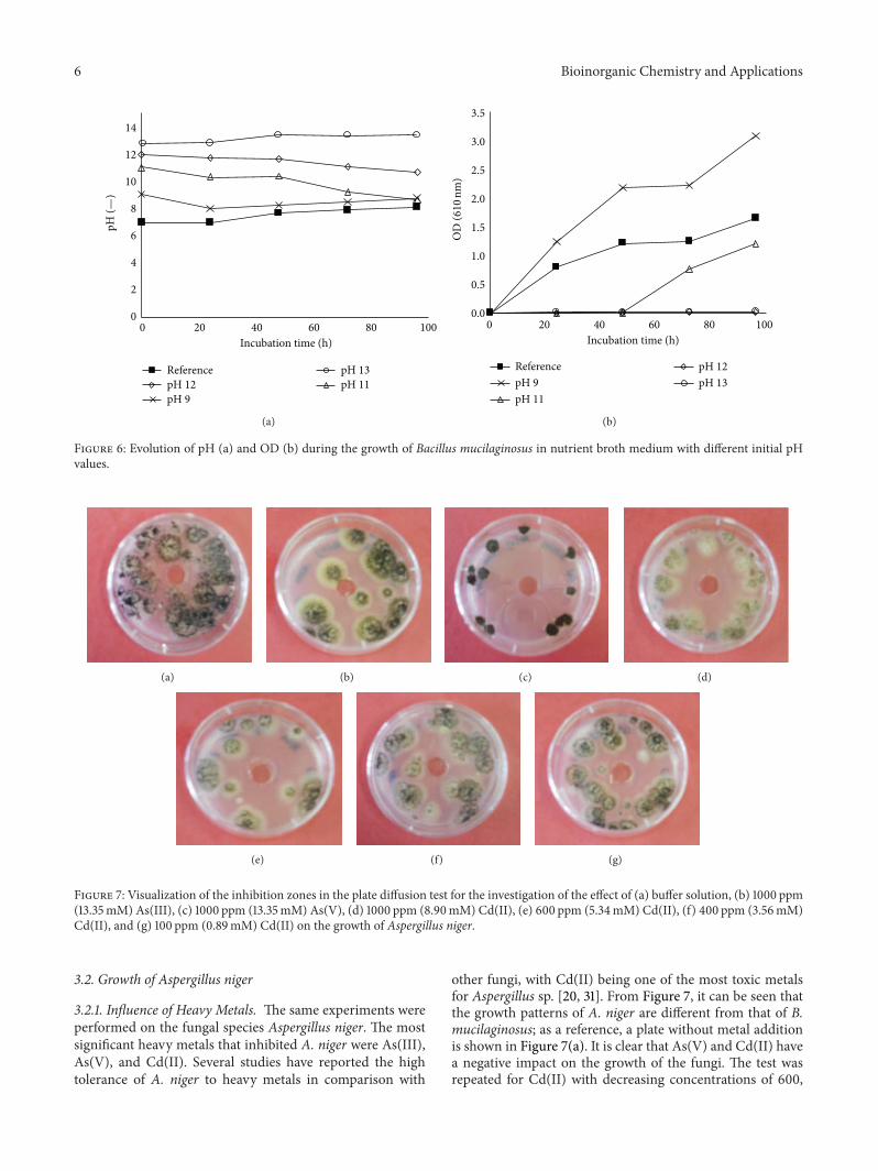
3.1.2. Influence of Initial pH. In addition to the investigationof the impact of heavy metals, the influence of the initialpH value was examined. Metal-rich waste materials suchas metallurgical slag and incineration ashes are typicallycharacterized by their high basicity, and for this reason theBacillus strain was cultivated under the initial pH values of9, 11, 12, and 13. Figure 6 shows the evolution of the pH andthe growth of the Bacillus strain over the incubation time.Based on the optical density measurement, there is moregrowth of B. mucilaginosuswhen the pH of the nutrient brothwas increased to 9 in comparison with the reference nutrientbroth medium with a pH of 7. There was hardly any growthobserved when the initial pH of themediumwas increased to12 and 13. For the culture with the initial pH of 11, exponentialgrowth was observed starting from day 3; notably, at thatmoment the pH had decreased to about 9. These results areconfirmed by themeasurement of theweight of dried biomassat the end of these experiments (Table 1). No biomass wasrecovered for the cultures with the initial pH values of 12 and13, while 0.88 g/L of biomass was found for the culture withinitial pH of 9 and 0.26 g/L biomass for the one with initialpH of 11. Therefore growth of B. mucilaginosus was favouredat an initial pH of 9, whereas growth was delayed at the initialpH of 11; it appears that the bacteria secrete acidic compoundsto adjust the pH to its best growth condition. An initial pH of12 or 13 resulted in complete inactivation of the bacteria; inthis case, the bacteria died off before being able to sufficientlyacidify the medium.
6 Bioinorganic Chemistry and Applications
0
2
4
6
8
10
12
14
0 20 40 60 80 100Incubation time (h)
Reference
pH 9pH 11pH 12pH 13
pH (—
)
(a)
0 20 40 60 80 100Incubation time (h)
ReferencepH 9pH 11
pH 12pH 13
0.0
0.5
1.0
1.5
2.0
2.5
3.0
3.5
OD
(610
nm)
(b)
Figure 6: Evolution of pH (a) and OD (b) during the growth of Bacillus mucilaginosus in nutrient broth medium with different initial pHvalues.
(a) (b) (c) (d)
(e) (f) (g)
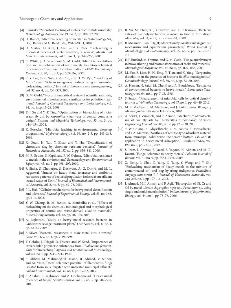
Figure 7: Visualization of the inhibition zones in the plate diffusion test for the investigation of the effect of (a) buffer solution, (b) 1000 ppm(13.35mM) As(III), (c) 1000 ppm (13.35mM) As(V), (d) 1000 ppm (8.90mM) Cd(II), (e) 600 ppm (5.34mM) Cd(II), (f) 400 ppm (3.56mM)Cd(II), and (g) 100 ppm (0.89mM) Cd(II) on the growth of Aspergillus niger.
3.2. Growth of Aspergillus niger
3.2.1. Influence of Heavy Metals. The same experiments wereperformed on the fungal species Aspergillus niger. The mostsignificant heavy metals that inhibited A. niger were As(III),As(V), and Cd(II). Several studies have reported the hightolerance of A. niger to heavy metals in comparison with
other fungi, with Cd(II) being one of the most toxic metalsfor Aspergillus sp. [20, 31]. From Figure 7, it can be seen thatthe growth patterns of A. niger are different from that of B.mucilaginosus; as a reference, a plate without metal additionis shown in Figure 7(a). It is clear that As(V) and Cd(II) havea negative impact on the growth of the fungi. The test wasrepeated for Cd(II) with decreasing concentrations of 600,

Bioinorganic Chemistry and Applications 7
0
2
4
6
8
10
0 100 200 300 400
Log
CFU
Incubation time (h)
Blank100ppm Cd(II)
(a)
Incubation time (h)
0
2
4
6
8
10
0 100 200 300 400 500
Blank100ppm Cd(II)
pH (—
)
(b)
Figure 8: Growth of Aspergillus niger determined by spread plate colony counting (a) and evolution of pH (b) over incubation time in blankpotato dextrose broth medium and in the presence of 100 ppm Cd(II) (0.9mM).
Table 1: Biomass dry weight concentration (g/L) after 96 hours asa function of initial pH during the growth of Bacillus mucilaginosusin nutrient brothmedium and ofAspergillus niger in potato dextrosebroth medium.
pH Bacillus mucilaginosus Aspergillus niger9.0 0.88 0.5511.0 0.26 1.2312.0 No growth 1.3613.0 No growth Negligible
400, and 100 ppm. These results are shown in Figures 7(e)to 7(g), in which significantly greater growth is seen for thelower concentrations.
A liquid batch culture was prepared for A. niger with100 ppm of Cd(II). Just as in the case for B. mucilaginosus,fungal growth and pH were measured over the incubationtime and compared to a metal-free reference culture. Theseresults are provided in Figure 8, in which the fungal growthis calculated on the basis of the number of colony formingunits. The results are similar to that of B. mucilaginosus;there is no significant difference in the CFU values, butthe colony structure clearly changed. While A. niger usuallygrows as large, rough colonies with visible black spores, in thecase of the strain that had contact with Cd(II), the colonieswere appreciably smaller and lacking visible sporulation.Thisvisualization is shown in Figure 9. Such results suggest thatthe metals are bound to substances on the cell surface ascomplexes, which dissociate once growth conditions returnto optimal.
Figure 8 shows that the pH of the reference cultureeventually dropped below 3, while for the culture exposed toCd(II) the pH increased up to nearly 10. A. niger is knownto produce organic acids during its metabolic activity, suchas citric acid and gluconic acid [12]. The pH increase of the
(a) (b)
Figure 9: Comparison of growth of Aspergillus niger inoculatedon potato dextrose agar plates after batch incubation of 10 days in(a) blank medium (normal growth) and (b) medium containing100 ppm Cd(II) (0.9mM).
Cd(II) doped culture does not necessarily mean that therewas no acid production, but it possibly means that basiccompounds were produced in greater quantity. However,previous work performed on Penicillium chrysogenum [32]points to the disruption of the acid production metabolismbeing the primary cause, as the glucose oxidase activity isseverely affected by heavy metals. This enzyme catalyzes theoxidation from glucose to gluconic acid. The reduction orabsence of gluconic acid production could thus explain thepH increase.
An additional experiment using A. niger was performedto determine the dry weight of the produced biomass. Thefungi were grown in 100 ppm Cd(II) broth and comparedagainst a reference culture. After 15 days of growth, the fungalbiomass was harvested. The reference culture contained3.02 g/L dried biomass, while the culture grown in Cd(II)presence only amounted to 0.96 g/L of dried biomass. Theseresults further support the conclusion that Cd(II) negativelyinfluences the growth of A. niger. Notably, at the end of this

8 Bioinorganic Chemistry and Applications
0
2
4
6
8
10
12
14
0 40 80 120 160Incubation time (h)
pH (—
)
ReferencepH 9pH 11
pH 12pH 13
Figure 10: Evolution of pH during the growth ofAspergillus niger inpotato dextrose broth medium with different initial pH values.
experiment, the Cd(II) concentration decreased from the ini-tial 100 ppm to roughly 80 ppm, indicating sorptive behaviourof the biomass to this metal. The biosorption capacity offilamentous fungi, such as Aspergillus sp. and Penicillium sp.,has been demonstrated and is effective with both living anddead biomass material [33]. Fungal cell walls are complexstructures consisting of chitins, proteins, polysaccharides,and other derivatives, providing many functional groups thatare able to bind metal ions. This has even given rise totechnological approacheswhere the biomass is altered to yieldhigher bioadsorption [9].
3.2.2. Influence of Initial pH. For the investigation of higherinitial pH values, the Aspergillus strain was grown in potatodextrose broths with initial pH values of 9, 11, 12, and 13,and growth was compared with the reference culture at pH6.7. As can be seen in Figure 10, which shows the evolutionof the pH during the incubation, the pH decreased for thecultures prepared in initial pH lower than or equal to 12.In the culture with the initial pH of 13, the pH remainedunchanged, which indicates the absence of microbial activity.Still, the pH of the reference culture decreased to the lowestvalue. At the end of the experiments, the dry weight of thebiomasses was determined (Table 1): for the culture withinitial pH of 9, 0.55 g/L of biomass was measured, whilethe biomass concentrations for the cultures with initial pHvalues of 11 and 12 were 1.23 g/L and 1.36 g/L, respectively.Hardly any growth was seen for the culture at pH 13 (massnot measured). It has been reported that A. niger presentsadvantages for bioleaching of incinerator fly ash becauseof its ability to thrive at higher basicity. In such case, theconditions reportedly become more favourable for gluconicacid production, which becomes the main bioleaching agentover citric acid [12]. From the present results it also appearsthat biomass growth is promoted at higher pH, as long as itdoes not exceed a certain limit.
4. Conclusions
The purpose of this study was to evaluate whether heavymetals and high basicity values hinder the growth of twochemoorganotrophic microorganisms, the bacterium Bacil-lus mucilaginosus and the fungus Aspergillus niger. Growthwas determined in agar plates and in liquid batch cultures,both providedwith the essential nutrients.The results showedthat cadmium, nickel, and arsenite (As(III)) had a negativeinfluence on the growth of B. mucilaginosus, whereas A.niger was sensitive to cadmium and arsenate (As(V)). Theseinfluences were characterized by delayed or reduced growth,but in none of the batch cultures a total inactivation or dyingoff of the microorganisms was observed for the 100 ppmconcentration being used. Both microorganisms exhibitedstrong adaptation abilities, and growth was restored oncethe metal was removed. However, the colony appearancechanged; therefore it seems that the metals cause modifica-tions to exterior cell structures.Moreover, with theAspergillusstrain, a modification of the metabolism was detected. Theincubation at higher pH values proved that both microor-ganisms are able to grow at higher basicity, up to pH 9 forB. mucilaginosus and even pH 12 for A. niger, values thatare reasonably within the range of bioleaching of alkalinematerials.
In conclusion, the investigated metals had a bacterio-static and fungistatic effect on Bacillus mucilaginosus andAspergillus niger, respectively, but did not act as bactericidesor fungicides. With respect to the bioleaching of industrialresidues, growth at elevated pH should not be an obstacle.Hence bothmicroorganisms herein studied provide opportu-nities for application in the remediation of heavy metal ladenwaste materials, soils, or sediments.
Conflict of Interests
The authors declare that there is no conflict of interestsregarding the publication of this paper.
Acknowledgment
The authors thank the K.U. Leuven Industrial ResearchFund (IOF) for the funding of the Knowledge Platformon Sustainable Materialization of Residues from ThermalProcesses into Products (SMaRT-Pro2) in which this workwas performed.
References
[1] K. Bosecker, “Bioleaching: metal solubilization by microorgan-isms,” FEMSMicrobiology Reviews, vol. 20, no. 3-4, pp. 591–604,1997.
[2] W. Krebs, C. Brombacher, P. P. Bosshard, R. Bachofen, andH. Brandl, “Microbial recovery of metals from solids,” FEMSMicrobiology Reviews, vol. 20, no. 3-4, pp. 605–617, 1997.
[3] G. J. Olson, J. A. Brierley, and C. L. Brierley, “Bioleachingreview part B: progress in bioleaching: applications ofmicrobialprocesses by the minerals industries,” Applied Microbiology andBiotechnology, vol. 63, no. 3, pp. 249–257, 2003.
Bioinorganic Chemistry and Applications 9
[4] I. Suzuki, “Microbial leaching of metals from sulfide minerals,”Biotechnology Advances, vol. 19, no. 2, pp. 119–132, 2001.
[5] H. Brandl, “Microbial leaching of metals,” in Biotechnology Set,H.-J. Rehm and G. Reed, Eds., Wiley-VCH, 2001.
[6] D. Mishra, D. Kim, J. Ahn, and Y. Rhee, “Bioleaching: amicrobial process of metal recovery; a review,” Metals andMaterials International, vol. 11, no. 3, pp. 249–256, 2005.
[7] C. White, J. A. Sayer, and G. M. Gadd, “Microbial solubiliza-tion and immobilization of toxic metals: key biogeochemicalprocesses for treatment of contamination,” FEMS MicrobiologyReviews, vol. 20, no. 3-4, pp. 503–516, 1997.
[8] E. Y. Lee, S.-R. Noh, K.-S. Cho, and H. W. Ryu, “Leaching ofMn, Co, and Ni from manganese nodules using an anaerobicbioleaching method,” Journal of Bioscience and Bioengineering,vol. 92, no. 4, pp. 354–359, 2001.
[9] G. M. Gadd, “Biosorption: critical review of scientific rationale,environmental importance and significance for pollution treat-ment,” Journal of Chemical Technology and Biotechnology, vol.84, no. 1, pp. 13–28, 2009.
[10] T.-J. Xu and Y.-P. Ting, “Optimisation on bioleaching of incin-erator fly ash by Aspergillus niger—use of central compositedesign,” Enzyme and Microbial Technology, vol. 35, no. 5, pp.444–454, 2004.
[11] K. Bosecker, “Microbial leaching in environmental clean-upprogrammes,” Hydrometallurgy, vol. 59, no. 2-3, pp. 245–248,2001.
[12] X. Quan, H. Tan, Y. Zhao, and Y. Hu, “Detoxification ofchromium slag by chromate resistant bacteria,” Journal ofHazardous Materials, vol. 137, no. 2, pp. 836–841, 2006.
[13] M. R. Bruins, S. Kapil, and F. W. Oehme, “Microbial resistancetometals in the environment,” Ecotoxicology and EnvironmentalSafety, vol. 45, no. 3, pp. 198–207, 2000.
[14] S. Sinha, S. Chatterjee, S. Dushyant, A. G. Dimri, and M. L.Aggarwal, “Studies on heavy metal tolerance and antibioticresistance patterns of bacterial population isolated from effluenttreated water of Delhi,” Journal of Biomedical and Pharmaceuti-cal Research, vol. 2, no. 5, pp. 69–76, 2013.
[15] J. L. Hall, “Cellular mechanisms for heavy metal detoxificationand tolerance,” Journal of Experimental Botany, vol. 53, no. 366,pp. 1–11, 2002.
[16] Y. W. Chiang, R. M. Santos, A. Monballiu et al., “Effects ofbioleaching on the chemical, mineralogical and morphologicalproperties of natural and waste-derived alkaline materials,”Minerals Engineering, vol. 48, pp. 116–125, 2013.
[17] A. Rajbanshi, “Study on heavy metal resistant bacteria inGuheswori sewage treatment plant,” Our Nature, vol. 6, no. 1,pp. 52–57, 2009.
[18] S. Silver, “Bacterial resistances to toxic metal ions: a review,”Gene, vol. 179, no. 1, pp. 9–19, 1996.
[19] T. Gehrke, J. Telegdi, D. Thierry, and W. Sand, “Importance ofextracellular polymeric substances from Thiobacillus ferrooxi-dans for bioleaching,” Applied and Environmental Microbiology,vol. 64, no. 7, pp. 2743–2747, 1998.
[20] S. Akhtar, M. Mahmood-ul-Hassan, R. Ahmad, V. Suthor,and M. Yasin, “Metal tolerance potential of filamentous fungiisolated from soils irrigated with untreated municipal effluent,”Soil and Environment, vol. 32, no. 1, pp. 55–62, 2013.
[21] S. Anahid, S. Yaghmaei, and Z. Ghobadinejad, “Heavy metaltolerance of fungi,” Scientia Iranica, vol. 18, no. 3, pp. 502–508,2011.
[22] B. Vu, M. Chen, R. J. Crawford, and E. P. Ivanova, “Bacterialextracellular polysaccharides involved in biofilm formation,”Molecules, vol. 14, no. 7, pp. 2535–2554, 2009.
[23] B.Mo andB. Lian, “Hg(II) adsorption byBacillusmucilaginosus:mechanism and equilibrium parameters,” World Journal ofMicrobiology and Biotechnology, vol. 27, no. 5, pp. 1063–1070,2011.
[24] E. P. Burford,M. Fomina, andG.M.Gadd, “Fungal involvementin bioweathering and biotransformation of rocks andminerals,”Mineralogical Magazine, vol. 67, no. 6, pp. 1127–1155, 2003.
[25] M. Yao, B. Lian, H. H. Teng, Y. Tian, and X. Yang, “Serpentinedissolution in the presence of bacteria Bacillus mucilaginosus,”Geomicrobiology Journal, vol. 30, no. 1, pp. 72–80, 2013.
[26] A. Hassen, N. Saidi, M. Cherif, and A. Boudabous, “Resistanceof environmental bacteria to heavy metals,” Bioresource Tech-nology, vol. 64, no. 1, pp. 7–15, 1998.
[27] S. Sutton, “Measurement of microbial cells by optical density,”Journal of Validation Technology, vol. 17, no. 1, pp. 46–49, 2011.
[28] M. T. Madigan, J. M. Martinko, and J. Parker, Brock Biology ofMicroorganisms, Pearson Education, 2003.
[29] A. Seidel, Y. Zimmels, and R. Armon, “Mechanism of bioleach-ing of coal fly ash by Thiobacillus thiooxidans,” ChemicalEngineering Journal, vol. 83, no. 2, pp. 123–130, 2001.
[30] Y. W. Chiang, K. Ghyselbrecht, R. M. Santos, B. Meesschaert,and J. A.Martens, “Synthesis of zeolitic-type adsorbentmaterialfrom municipal solid waste incinerator bottom ash and itsapplication in heavy metal adsorption,” Catalysis Today, vol.190, no. 1, pp. 23–30, 2012.
[31] S. Iram, I. Ahmad, B. Javed, S. Yaqoob, K. Akhtar, and M. R.Kazmi, “Fungal tolerance to heavy metals,” Pakistan Journal ofBotany, vol. 41, no. 5, pp. 2583–2594, 2009.
[32] X. Deng, L. Chai, Z. Yang, C. Tang, Y. Wang, and Y. Shi,“Bioleaching mechanism of heavy metals in the mixture ofcontaminated soil and slag by using indigenous Penicilliumchrysogenum strain F1,” Journal of Hazardous Materials, vol.248-249, no. 1, pp. 107–114, 2013.
[33] I. Ahmad, M. I. Ansari, and F. Aqil, “Biosorption of Ni, Cr andCd by metal tolerant Aspergillus niger and Penicillium sp. usingsingle andmulti-metal solution,” Indian Journal of ExperimentalBiology, vol. 44, no. 1, pp. 73–76, 2006.

Submit your manuscripts athttp://www.hindawi.com
Hindawi Publishing Corporationhttp://www.hindawi.com Volume 2014
Inorganic ChemistryInternational Journal of
Hindawi Publishing Corporation http://www.hindawi.com Volume 2014
International Journal ofPhotoenergy
Hindawi Publishing Corporationhttp://www.hindawi.com Volume 2014
Carbohydrate Chemistry
International Journal of
Hindawi Publishing Corporationhttp://www.hindawi.com Volume 2014
Journal of
Chemistry
Hindawi Publishing Corporationhttp://www.hindawi.com Volume 2014
Advances in
Physical Chemistry
Hindawi Publishing Corporationhttp://www.hindawi.com
Analytical Methods in Chemistry
Journal of
Volume 2014
Bioinorganic Chemistry and ApplicationsHindawi Publishing Corporationhttp://www.hindawi.com Volume 2014
SpectroscopyInternational Journal of
Hindawi Publishing Corporationhttp://www.hindawi.com Volume 2014
The Scientific World JournalHindawi Publishing Corporation http://www.hindawi.com Volume 2014
Medicinal ChemistryInternational Journal of
Hindawi Publishing Corporationhttp://www.hindawi.com Volume 2014
Chromatography Research International
Hindawi Publishing Corporationhttp://www.hindawi.com Volume 2014
Applied ChemistryJournal of
Hindawi Publishing Corporationhttp://www.hindawi.com Volume 2014
Hindawi Publishing Corporationhttp://www.hindawi.com Volume 2014
Theoretical ChemistryJournal of
Hindawi Publishing Corporationhttp://www.hindawi.com Volume 2014
Journal of
Spectroscopy
Analytical ChemistryInternational Journal of
Hindawi Publishing Corporationhttp://www.hindawi.com Volume 2014
Journal of
Hindawi Publishing Corporationhttp://www.hindawi.com Volume 2014
Quantum Chemistry
Hindawi Publishing Corporationhttp://www.hindawi.com Volume 2014
Organic Chemistry International
ElectrochemistryInternational Journal of
Hindawi Publishing Corporation http://www.hindawi.com Volume 2014
Hindawi Publishing Corporationhttp://www.hindawi.com Volume 2014
CatalystsJournal of