
Research Article Timing of Pars Plana Vitrectomy in...
9
Research Article Timing of Pars Plana Vitrectomy in Management of Gunshot Perforating Eye Injury: Observational Study Hammouda Hamdy Ghoraba, 1,2 Mohamed Amin Heikal, 3 Hosam Osman Mansour, 4 Haithem Mamon Abdelfattah, 2,5 Emad Mohamed Elgemai, 2,6 and Adel Galal Zaky 7 1 Tanta University, Tanta, Egypt 2 Magrabi Eye Hospital, Tanta, Egypt 3 Benha University, Benha, Egypt 4 Al-Azhar University, Domyat, Egypt 5 Benha Teaching Hospital, Benha, Egypt 6 Damanhour Teaching Hospital, Damanhur, Egypt 7 Menoufia University, Shebin El Kom, Egypt Correspondence should be addressed to Mohamed Amin Heikal; [email protected] Received 10 June 2016; Accepted 29 August 2016 Academic Editor: Anselm G.M. Juenemann Copyright © 2016 Hammouda Hamdy Ghoraba et al. is is an open access article distributed under the Creative Commons Attribution License, which permits unrestricted use, distribution, and reproduction in any medium, provided the original work is properly cited. e aim of this study is to report the difference in either anatomical or functional outcome of vitreoretinal intervention in cases of gunshot perforating eye injury if done 2–4 weeks or aſter the 4th week aſter the original trauma. Patients were treated with pars plana vitrectomy and silicon oil. Surgeries were performed in the period from February 2011 until the end of December 2014. 253 eyes of 237 patients were reviewed. 46 eyes were excluded. 207 eyes of 197 patients were analyzed. e included eyes were classified based on the timing of vitrectomy in relation to the initial trauma into two groups: 149 eyes (the first group) operated on between the 3rd and the 4th week and 58 eyes (the second group) operated on aſter the 4th week aſter the trauma. Following one surgical intervention, in the first group, attached retina was achieved in 93.28% of patients. In the second group, attached retina was achieved in 96.55% of patients. All RD cases could be attached by a second surgery. Visual acuity improved in 81.21% of patients, did not change in 15.43% of patients, and declined in 3.35% of patients. In the second group, visual acuity improved in 81.03% of patients, did not change in 12.06% of patients, and worsened in 6.89% of patients. ere was no statistically significant difference between the two groups in either anatomical or functional results. We recommend interfering before the 5th week aſter the trauma as retinal detachment is encountered more in cases operated on aſter the 4th week. e visual outcome depends on the site of entry and exit (the route of gunshot). 1. Introduction Mechanical injuries of the globe (open and closed) are classified according to Pieramic et al. [1] system that relies on four variables: type of injury, grade of injury, pupillary response, and zone of injury [1]. Open globe injuries are a common and oſten preventable cause of permanent visual impairment and visual loss [2]. Perforating ocular injuries are “through-and-through” globe defects with entry and exit sites. is is in contrast to penetrating injuries, which have a point of entry into the globe but no exit wound [3, 4]. Histopathological studies revealed that posterior vitreous detachment (PVD) usually occurred at 1 to 2 weeks aſter trauma [5]. e peripheral tractional retinal detachment then developed between 7 and 11 weeks, due to contractile fibrovascular ingrowth from the wound along the vitreous scaffold to the vitreous base and from preretinal membranes in the peripheral and equatorial retina. e end result at 4 months was tractional total retinal detachment and fibrous cyclitic, epiretinal, and subretinal membranes [6]. If not operated on it ends by phthisis bulbi [7]. Hindawi Publishing Corporation Journal of Ophthalmology Volume 2016, Article ID 1487407, 8 pages http://dx.doi.org/10.1155/2016/1487407
Transcript of Research Article Timing of Pars Plana Vitrectomy in...

Research ArticleTiming of Pars Plana Vitrectomy in Management ofGunshot Perforating Eye Injury: Observational Study
Hammouda Hamdy Ghoraba,1,2 Mohamed Amin Heikal,3 Hosam Osman Mansour,4
Haithem Mamon Abdelfattah,2,5 Emad Mohamed Elgemai,2,6 and Adel Galal Zaky7
1Tanta University, Tanta, Egypt2Magrabi Eye Hospital, Tanta, Egypt3Benha University, Benha, Egypt4Al-Azhar University, Domyat, Egypt5Benha Teaching Hospital, Benha, Egypt6Damanhour Teaching Hospital, Damanhur, Egypt7Menoufia University, Shebin El Kom, Egypt
Correspondence should be addressed to Mohamed Amin Heikal; [email protected]
Received 10 June 2016; Accepted 29 August 2016
Academic Editor: Anselm G.M. Juenemann
Copyright © 2016 Hammouda Hamdy Ghoraba et al. This is an open access article distributed under the Creative CommonsAttribution License, which permits unrestricted use, distribution, and reproduction in any medium, provided the original work isproperly cited.
The aim of this study is to report the difference in either anatomical or functional outcome of vitreoretinal intervention in cases ofgunshot perforating eye injury if done 2–4 weeks or after the 4th week after the original trauma. Patients were treated with parsplana vitrectomy and silicon oil. Surgeries were performed in the period from February 2011 until the end of December 2014. 253eyes of 237 patients were reviewed. 46 eyes were excluded. 207 eyes of 197 patients were analyzed.The included eyes were classifiedbased on the timing of vitrectomy in relation to the initial trauma into two groups: 149 eyes (the first group) operated on betweenthe 3rd and the 4th week and 58 eyes (the second group) operated on after the 4th week after the trauma. Following one surgicalintervention, in the first group, attached retina was achieved in 93.28% of patients. In the second group, attached retina was achievedin 96.55% of patients. All RD cases could be attached by a second surgery. Visual acuity improved in 81.21% of patients, did notchange in 15.43% of patients, and declined in 3.35% of patients. In the second group, visual acuity improved in 81.03% of patients,did not change in 12.06% of patients, and worsened in 6.89% of patients. There was no statistically significant difference betweenthe two groups in either anatomical or functional results. We recommend interfering before the 5th week after the trauma as retinaldetachment is encountered more in cases operated on after the 4th week. The visual outcome depends on the site of entry and exit(the route of gunshot).
1. Introduction
Mechanical injuries of the globe (open and closed) areclassified according to Pieramic et al. [1] system that relieson four variables: type of injury, grade of injury, pupillaryresponse, and zone of injury [1]. Open globe injuries are acommon and often preventable cause of permanent visualimpairment and visual loss [2]. Perforating ocular injuriesare “through-and-through” globe defects with entry and exitsites. This is in contrast to penetrating injuries, which have apoint of entry into the globe but no exit wound [3, 4].
Histopathological studies revealed that posterior vitreousdetachment (PVD) usually occurred at 1 to 2 weeks aftertrauma [5]. The peripheral tractional retinal detachmentthen developed between 7 and 11 weeks, due to contractilefibrovascular ingrowth from the wound along the vitreousscaffold to the vitreous base and from preretinal membranesin the peripheral and equatorial retina. The end result at 4months was tractional total retinal detachment and fibrouscyclitic, epiretinal, and subretinal membranes [6]. If notoperated on it ends by phthisis bulbi [7].
Hindawi Publishing CorporationJournal of OphthalmologyVolume 2016, Article ID 1487407, 8 pageshttp://dx.doi.org/10.1155/2016/1487407
2 Journal of Ophthalmology
Experimental and clinical studies suggest that, among alltypes of globe wounds, perforating injuries have the worstprognosis [8–10].The factors limiting visual recovery includedirect injury to the optic nerve or macula, intraocularscarring and fibrosis with secondary retinal detachment,and severe ocular disorganization [11, 12]. Several predictivefactors affect the prognosis for final visual acuity [13].
Previous reports showed that vitreoretinal surgery inperforating injury prevents phthisis bulbi and achieves somefunctional result [7]. There is controversy to do vitreoretinalsurgery early days or weeks after repair of the primary injury[14] or to use encircling scleral band or no [15].
The aim of this study was to compare the results of PPVduring the 3rd to 4th or later than the 4th week after traumain gunshot perforating eye injury.
2. Patients and Methods
This is a retrospective observational study of 207 eyes of197 patients with perforating eye injuries caused by gunshotstreated by pars plana vitrectomy and silicon oil with orwithout buckle.
Surgeries were performed in the period from February2011 to the end of December 2014 during the period ofpolitical instability in Egypt. All surgeries were performed bya single surgeon (HG) at a single center.
All patients had preoperative evaluation included: bestcorrected visual acuity, intraocular pressure measurement,anterior segment examination using the slit lamp, and dilatedfundus examination using indirect ophthalmoscope if themedia were clear. The ocular trauma score (OTS) was retro-spectively calculated.
Investigations done were B/scan ultrasonography andComputerized Tomography (CT) to locate the gunshot andfor medicolegal aspect. Visual Evoked Potential (VEP) wasrequested for cases with no light perception to justify nosurgical intervention.
Inclusion Criteria. Perforating gunshot ocular injury withat least light perception vision with minimum follow-up 6months after the last surgical intervention was the inclusioncriterion.
Exclusion Criteria
Patients with visual acuity of no light perceptionPatients with retained intraocular foreign body (gun-shot)Patients with endophthalmitisPatients with follow-up less than 6 months after thelast surgical intervention.
2.1. Surgical Procedures. Primary repair was done elsewherein all cases in the same day of trauma. Our plan forvitreoretinal intervention in such cases was to operate on atleast 2 weeks after the primary repair to allow entry woundhealing, suprachoroidal hemorrhage to liquefy if present andposterior vitreous detachment to occur. In this series, somefactors made the time of intervention variable; for example,
patients with scleral and limbal entry were operated onduring the 3rd week after trauma. Patients with a centralcorneal wound or suprachoroidal hemorrhage were operatedon in the 4th week from injury to allow more time for properwound healing. Patients referred after the 4th week of traumawere operated on once they were presented to us.
The surgical technique was the same in all cases andwas done by the same surgeon (HG). Three-port pars planavitrectomy (PPV) was done using conventional 20 gauges ortransconjunctival cannulated 20 or 23 gauges with or withoutscleral buckling.
Lensectomyusing fragmentation or vitrectomyprobewasdone if there was cataract interfering with proper visualiza-tion or if lens touch occurred; otherwise the lens was spared.
A central vitrectomy was performed until central PVDwas achieved if not already present. Perfluorocarbon (PFC)was injected to flatten the retina in cases with retinal detach-ment, to get a better view, to elevate the residual vitreous andto displace subretinal hemorrhage anteriorly.
Vitrectomy was completed as safe as possible anteriorlyleaving an amalgam of tissue around the exit site to preventPFC, air, and silicon from escaping into the orbit. Nochorioretinectomy was done in those cases.
Vitrectomy under air was used frequently in the presenceof bleeding. Laser was applied to any retinal break, 360degrees and around the exit site if it was outside the macula.Air PFC exchange was done followed by silicon oil injectionof 2,000 or 5,000 cSt. The bright illumination and wide fieldvisualization systems allowed us to do all surgeries withoutthe need of penetrating keratoplasty.
All patients were examined in the 1st postoperative day.Fundus examination and color fundus photography weredone if possible. Postoperative follow-up was scheduled at 1week, 3 weeks, 6 weeks, and then every 8 weeks.
In cases of development of retinal detachment afterthe primary surgery, the surgical procedure consisted ofsilicon oil removal, triamcinolone-assisted removal of anyresidual vitreous cortex, and removal of epiretinal membraneif involving the macula. Relaxing retinotomy was done incases with excessive retinal proliferation preventing retinalattachment or subretinal S.O. PFC was injected. Laser wasadded to any break and to the edge of retinotomy followedby air PFC exchange. Silicon oil 5,000 cSt was injected at theend of surgery.
Statistical tests used are mean, standard deviation, chi-square test, and 𝑃 value. 𝑃 value was considered signif-icant if <0.05. Statistical analysis was performed using acommercially available statistical software package (SPSS forwindows, version 20).
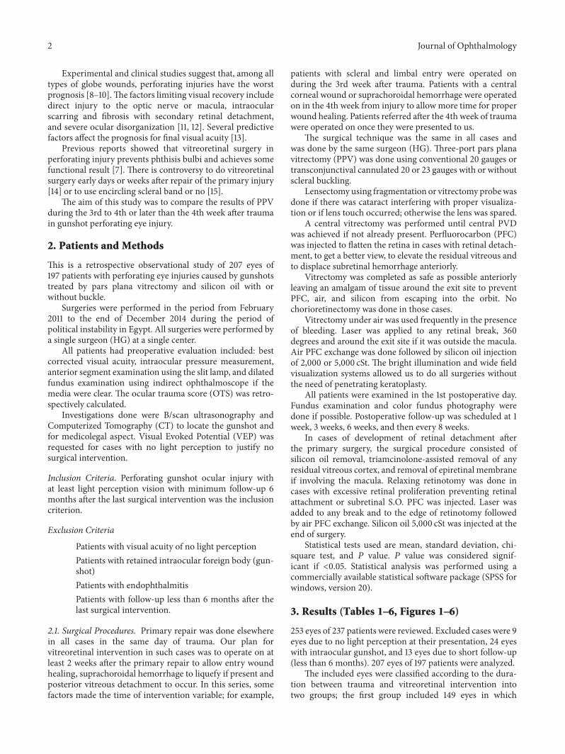
3. Results (Tables 1–6, Figures 1–6)
253 eyes of 237 patients were reviewed. Excluded cases were 9eyes due to no light perception at their presentation, 24 eyeswith intraocular gunshot, and 13 eyes due to short follow-up(less than 6 months). 207 eyes of 197 patients were analyzed.
The included eyes were classified according to the dura-tion between trauma and vitreoretinal intervention intotwo groups; the first group included 149 eyes in which
Journal of Ophthalmology 3
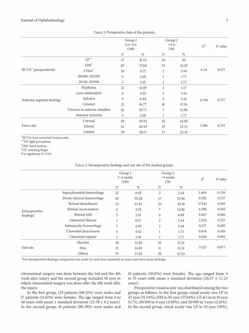
Table 1: Preoperative data of the patients.
Group 12w–4w(149)
Group 2>4w(58) 𝑋
2𝑃 value
𝑁 % 𝑁 %
BCVA∗ preoperatively
LP∗∗ 47 31.54 29 50
6.34 0.175HM† 85 57.04 25 43.10CF1m‡ 10 6.71 2 3.44
20/100–20/200 4 2.68 1 1.7220/40–20/100 3 2.01 1 1.72
Anterior segment findings
Hyphema 21 14.09 3 5.17
4.746 0.577
Lens subluxation 6 4.02 2 3.44Aphakia 9 6.04 2 3.44Cataract 25 16.77 10 17.24
Vitreous in anterior chamber 16 10.73 7 12.06Anterior synechia 4 2.68 1 1.72
Entry siteCorneal 58 38.92 26 44.82
1.186 0.553Scleral 61 40.94 19 32.75Limbal 30 20.13 13 22.41
∗BCVA: best corrected visual acuity.∗∗LP: light perception.†HM: hand motion.‡CF: counting finger.𝑃 is significant if <0.05.
Table 2: Intraoperative findings and exit site of the studied groups.
Group 12–4 weeks
(149)
Group 2>4 weeks
(58) 𝑋2
𝑃 value
𝑁 % 𝑁 %
Intraoperativefindings∗
Suprachoroidal hemorrhage 12 8.05 2 3.44 1.404 0.236Dense vitreous hemorrhage 45 30.20 15 25.86 0.382 0.537
Retinal detachment 23 15.43 25 43.10 17.942 0.001Retinal incarceration 6 4.02 7 12.06 4.588 0.032
Retinal fold 3 2.01 4 6.89 3.047 0.081Subretinal fibrosis 1 0.67 2 3.44 2.254 0.133
Submacular hemorrhage 7 4.69 2 3.44 0.157 0.692Choroidal detachment 6 4.02 1 1.72 0.678 0.410Choroidal rupture 3 2.01 1 1.72 0.018 0.892
Exit siteMacular 49 32.88 10 17.24
5.125 0.077Disc 21 14.09 9 15.51Others 79 53.02 39 67.24
∗For intraoperative findings comparison was made for each item separately as some cases have many findings.
vitreoretinal surgery was done between the 3rd and the 4thweek after injury and the second group included 58 eyes inwhich vitreoretinal surgery was done after the 4th week afterthe injury.
In the first group, 129 patients (88.35%) were males and17 patients (11.65%) were females. The age ranged from 4 to48 years with mean ± standard deviation (25.58 ± 8.2 years).In the second group, 41 patients (80.39%) were males and
10 patients (19.61%) were females. The age ranged from 4to 55 years with mean ± standard deviation (26.57 ± 11.23years).
Preoperative visual acuity was distributed among the twogroups as follows: in the first group, visual acuity was LP in47 eyes (31.54%), HM in 85 eyes (57.04%), CF at 1m in 10 eyes(6.71), 20/200 in 4 eyes (2.68%), and 20/100 in 3 eyes (2.01%).In the second group, visual acuity was LP in 29 eyes (50%),

4 Journal of Ophthalmology
Table 3: Postoperative complications.
Group 1 Group 2𝑋2𝑃 value
𝑛 = 149 % 𝑛 = 58 %Cataract 19 12.75 10 17.24
7.115 0.715
Hypotony 5 3.35 4 6.89Recurrentretinaldetachment
7 4.69 2 3.44
Retinalproliferation 10 6.71 6 10.34
Macularpucker 18 12.08 5 8.62
Corneal scar 21 14.09 6 10.34Bandkeratopathy 5 3.35 3 5.17
Persistenthigh IOP∗despitetreatment
5 3.35 2 3.44
Silicon oil inA/C∗∗ 2 1.34 2 3.44
Subretinalfibrosis 8 5.37 5 8.62
∗IOP: intraocular pressure.∗∗A/C: anterior chamber.𝑃 is significant if <0.05.
Figure 1: Color photo of left eye showing parafoveal exit withmacular dragging.
HM in 25 eyes (43.10%), CF at 1m in 2 eyes (3.44%), 20/200in 1 eye (1.72%), and 20/100 in 1 eye (1.72%).
In the first group, 38.92% of entry sites were corneal,40.94% were scleral, and 20.13% were limbal. In the secondgroup, 44.82%of entry sites were corneal, 32.75%were scleral,and 22.41% were limbal.
Meanpreoperative ocular trauma score (OTS)was 43.96±12.64 in group 1 and 41.72 ± 12.94 in group 2. No statisticallysignificant difference was found between both groups (𝑃 =0.622).
The exit site was found at the macula in 49 eyes (32.88%)in the first group and 10 eyes (17.24%) in the second group.Optic nerve exit was observed in 21 eyes (14.09%) in the firstgroup and 9 eyes (15.51%) in the second group. The exit site
Figure 2: Color photo of the right eye showing temporal exit withmacular dragging.
Figure 3: Color photo of the right eye showing superior exit.
other than macula and optic nerve was present in 79 eyes(53.02%) in the first group and 39 eyes (67.24%) in the secondgroup.
Retinal detachment and retinal incarceration were seenmore frequent in group two with statistically significantdifference (𝑃 values 0.001 and 0.032, resp.).
Most of the cases with retinal detachment in the twogroups were accompanied with vitreous hemorrhage (78.26%in group 1 and 53.57% in group 2).
Operative findings are mentioned in Table 2.By one operation anatomical results in the first group
revealed attached retina in 139 eyes (93.28%) and 10 eyes(6.72%) developed RD. In the second group attached retinawas achieved in 56 eyes (96.55%) and 2 eyes (3.45%) devel-oped RD. All retinal detachment cases could be reattached bya second surgery. No eyes developed phthisis bulbi during thefollow-up period.
There was no statistically significant difference between thetwo groups regarding anatomical results.
We did not notice any escape of either S.O. or PFCLinto the orbit either during or after surgery. The reportedpostoperative complications were presented in Table 3.
In the first group, postoperative VA was LP in 12 eyes(8.05%),HM in 52 eyes (34.89%), CF at 1m in 38 eyes (25.5%),20/200 in 38 eyes (25.5%), and 20/100 in 9 eyes (6.04%).Visual acuity improved in 121 eyes (81.21%), unchanged in 23eyes (15.43%), and declined in 5 eyes (3.35%).
In the second group, VA was LP in 4 eyes (6.89%), HMin 26 eyes (44.82%), CF at 1m in 13 eyes (22.41%), 20/200 in

Journal of Ophthalmology 5
Table 4: Postoperative anatomical and functional results in both groups.
Group 12–4w(149)
Group 2>4w(58) 𝑋
2𝑃 value
𝑁 % 𝑁 %
Anatomical success Attached retina by one operation 139 93.28 56 96.55 0.814 0.367Attached retina by 2nd operation 149 100 58 100
Postoperative BCVA∗
LP∗∗ 12 8.05 4 6.89
1.78 0.77HM† 52 34.89 26 44.82CF1m‡ 38 25.5 13 22.4120/200 38 25.5 12 20.6820/100 9 6.04 3 5.17
∗BCVA: best corrected visual acuity.∗∗LP: light perception.†HM: hand motion.‡CF: counting finger.𝑃 is significant if <0.05.
Table 5: Comparison between pre- and postoperative BCVA in bothgroups.
BCVA∗Preoperative207 eyes
Postoperative207 eyes 𝑋
2𝑃 value
Number % Number %LP∗∗ 76 36.71 16 7.73
109.5 0.001HM† 110 53.14 78 37.68CF 1m‡ 12 5.79 51 24.6320/100–20/200 5 2.41 50 24.1520/40–20/100 4 1.93 12 5.79∗BCVA: best corrected visual acuity.∗∗LP: light perception.†HM: hand motion.‡CF: counting finger.𝑃 is significant if <0.05.
Figure 4: Color photo of right eye showing retinal incarceration atthe exit site and lower RD under silicon oil.
12 eyes (20.68%), and 20/100 in 3 eyes (5.17%). Visual acuityimproved in 47 eyes (81.03%), unchanged in 7 eyes (12.06%),and worsened in 4 eyes (6.89%).
There was no statistically significant difference between thetwo groups regarding functional results.
Figure 5: Color photo of right eye showing macular exit.
Figure 6: Color photo of left eye showing macular exit with retinalincarceration.
The postoperative anatomical and functional results wereshown in Table 4.
The best corrected visual acuity improved in the twogroups as compared to the preoperative VA (Table 5).
The main cause of low visual outcome was the centralroute of the gunshot, central corneal entry, and macular or

6 Journal of Ophthalmology
Table 6: (a) Relation of the gunshot and visual acuity in the 2 groups. (b) Postoperative visual acuity in patients with corneal entry. (c)Postoperative visual acuity in patients with macula exit. (d) Postoperative visual acuity in patients with optic disc exit.
(a)
BCVA∗ postoperatively Corneal entry Macular exit Disc exit𝑋2
𝑃 valueNumber % Number % Number %
LP∗∗ 9 10.71 8 13.55 11 36.6617.295 0.002HM† 47 55.95 34 57.62 18 60
CF 1m‡ 28 33.33 17 28.81 1 3.33∗BCVA: best corrected visual acuity.∗∗LP: light perception.†HM: hand motion.‡CF: counting finger.𝑃 is significant if <0.05.
(b)
BCVA∗ postoperatively Group 1 = 58 Group 2 = 26𝑋2
𝑃 valueNumber % Number %
LP∗∗= 8 13.79 1 3.842.597 0.273HM† 33 56.89 14 53.84
CF 1m‡ 17 29.31 11 42.30∗BCVA: best corrected visual acuity.∗∗LP: light perception.†HM: hand motion.‡CF: counting finger.𝑃 is significant if <0.05.
(c)
BCVA∗ postoperatively Group 1 = 49 Group 2 = 10𝑋2
𝑃 valueNumber % Number %
LP∗∗= 7 14.3 1 100.757 0.685HM† 27 55.1 7 70
CF 1m‡ 15 30.6 2 20∗BCVA: best corrected visual acuity.∗∗LP: light perception.†HM: hand motion.‡CF: counting finger.𝑃 is significant if <0.05.
(d)
BCVA∗ postoperatively Group 1 = 21 Group 2 = 9𝑋2
𝑃 valueNumber % Number %
LP∗∗ 10 47.6 1 11.14.507 0.105HM† 10 47.6 8 88.9
CF 1m‡ 1 4.8 0 0∗BCVA: best corrected visual acuity.∗∗LP: light perception.†HM: hand motion.‡CF: counting finger.𝑃 is significant if <0.05.
optic nerve exit.This was shown in Tables 6(a), 6(b), 6(c), and6(d). There was no statistically significant difference betweenthe two groups.
4. Discussion
Perforating injuries of the globe account for a small portionof open globe injuries [2]. The incidence increased in Egypt
since January 2011 due to political instability. The standardapproach to treating perforating injuries is primary repairto restore the structural integrity of the globe at the earliestopportunity [2]. Previous reports showed the benefit ofvitreoretinal surgery in such cases in preventing phthisisbulbi and achieving some visual result [7].
Controversy remains about the best timing of secondaryintervention [14].There are 3 opinions regarding the timing of
Journal of Ophthalmology 7
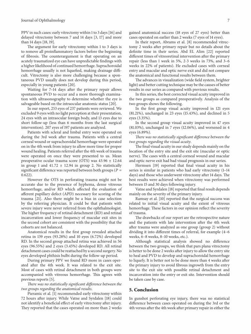
PPV in such cases: early vitrectomywithin 1 to 3 days [16] anddelayed vitrectomy between 7 and 14 days [3, 17] and morethan 14 days [18, 19].
The argument for early vitrectomy within 1 to 3 days isto remove all proinflammatory factors before the beginningof fibrosis. The counterargument is that operating on anacutely traumatized eye can have unpredictable findings witha higher likelihood of continued hemorrhage. Suprachoroidalhemorrhage usually is not liquefied, making drainage diffi-cult. Vitrectomy is also more challenging because a spon-taneous PVD usually does not develop during this period,especially in young patients [20].
Waiting for 7–14 days after the primary repair allowsspontaneous PVD to occur and a more thorough examina-tion with ultrasonography to determine whether the eye issalvageable based on the intraocular anatomic status [20].
In our report, 253 eyes of 237 patients were reviewed. Weexcluded 9 eyes with no light perception at their presentation,24 eyes with an intraocular foreign body, and 13 eyes due toshort follow-up (less than 6 months from the last surgicalintervention). 207 eyes of 197 patients are analyzed.
Patients with scleral and limbal entry were operated onduring the 3rd week after trauma. Patients with a centralcorneal wound or suprachoroidal hemorrhage were operatedon in the 4th week from injury to allow more time for properwound healing. Patients referred after the 4th week of traumawere operated on once they were presented to us. Meanpreoperative ocular trauma score (OTS) was 43.96 ± 12.64in group 1 and 41.72 ± 12.94 in group 2. No statisticallysignificant difference was reported between both groups (𝑃 =0.622).
However the OTS in perforating trauma might not beaccurate due to the presence of hyphema, dense vitreoushemorrhage, and/or RD which affected the evaluation ofafferent pupillary defect (APD) necessary for scoring of thetrauma [21]. Also there might be a bias in case selectionby the referring physician. It could be that patients withsevere injury were never referred from the ophthalmologist.The higher frequency of retinal detachment (RD) and retinalincarceration and lower frequency of macular exit sites inthe second cohort are consistent with the possibility that thecohorts are not balanced.
Anatomical results in the first group revealed attachedretina in 139 eyes (93.28%) and 10 eyes (6.72%) developedRD. In the second group attached retina was achieved in 56eyes (96.55%) and 2 eyes (3.45%) developed RD. All retinaldetachment cases could be reattached by a second surgery.Noeyes developed phthisis bulbi during the follow-up period.
During primary PPV we found RD more in cases oper-ated after the 4th week. It was related to the exit site.Most of cases with retinal detachment in both groups wereaccompanied with vitreous hemorrhage. This agrees withprevious reports [5].
There was no statistically significant difference between thetwo groups regarding the anatomical results.
Pieramic et al. [1] recommended early vitrectomy within72 hours after injury. While Vatne and Syrdalen [18] couldnot identify a beneficial effect of early vitrectomy after injury.They reported that the cases operated on more than 2 weeks
gained anatomical success (18 eyes of 27 eyes) better thancases operated on earlier than 2 weeks (7 eyes of 14 eyes).
In their report, Abrams et al. [8] recommended vitrec-tomy 2 weeks after primary repair but no details about thedefinite time in their series. Abd EL Alim [22] reporteddifferent times of vitreoretinal intervention after the primaryrepair (less than 1 week in 5%, 2-3 weeks in 73%, and 3–6weeks in 22% of patients). He excluded cases with cornealentry and macular and optic nerve exit and did not comparethe anatomical and functional results between them.
The advances in visualization (wide field system, brighterlight) and better cutting techniquemay be the causes of betterresults in our series as compared with previous results.
In this series, the best corrected visual acuity improved inthe two groups as compared preoperatively. Analysis of thetwo groups shows the following.
In the first group visual acuity improved in 121 eyes(81.21%), unchanged in 23 eyes (15.43%), and declined in 5eyes (3.35%).
In the second group visual acuity improved in 47 eyes(81.03%), unchanged in 7 eyes (12.06%), and worsened in 4eyes (6.89%).
There was no statistically significant difference between thetwo groups regarding the visual acuity.
The final visual acuity in our study dependsmainly on thelocation of the entry site and the exit site (macular or opticnerve). The cases with a central corneal wound and macularand optic nerve exit had bad visual prognosis in our series.
Hermsen [19] reported that final visual acuity in hisseries is similar in patients who had early vitrectomy (1–14days) and those who underwent vitrectomy after 14 days.Thebest results were achieved when vitrectomy was performedbetween 15 and 30 days following injury.
Vatne and Syrdalen [18] reported that final result dependsmainly on the severity of the primary injury.
Ramsay et al. [10] reported that the surgical success wasrelated to initial visual acuity and the extent of vitreoushemorrhage. These factors in our opinion reflect the severityof trauma.
The drawbacks of our report are the retrospective natureand the patients with late intervention after the 4th weekafter trauma were analyzed as one group (group 2) withoutdividing it into different times of referral, for example (4–6weeks, 6–8 weeks, 8–10 weeks, etc.).
Although statistical analysis showed no differencebetween the two groups, we think that pars plana vitrectomyis better to be done 2 weeks after injury to allow the entry siteto heal and PVD to develop and suprachoroidal hemorrhageto liquefy. It is better not to be done more than 4 weeks afterthe primary injury to avoid fibrous ingrowth from the entrysite to the exit site with possible retinal detachment andincarceration into the entry or exit site. Intervention shouldbe taken case by case.
5. Conclusion
In gunshot perforating eye injury, there was no statisticaldifference between cases operated on during the 3rd or the4th versus after the 4th week after primary repair in either the

8 Journal of Ophthalmology
anatomical or functional results. However, we recommendinterfering before the 5th week after the trauma as retinaldetachment is encountered more in cases operated on afterthe 4th week.The visual outcome depends on the site of entryand exit (the route of gunshot).
Disclosure
The study was performed in Magrabi Eye Hospital, Tanta,Egypt. This work was self-funded by the authors.
Competing Interests
The authors declare that they have no competing interests.
References
[1] D. J. Pieramic, P. Jr. Sternberg, T. M. Aaberg et al., “A system forclassifyingmechanical injuries of the globe.TheOcular TraumaClassification Group,” American Journal of Ophthalmology, vol.123, no. 6, pp. 820–831, 1997.
[2] I. Rahman, A. Maino, D. Devadason, and B. Leatherbarrow,“Open globe injuries: factors predictive of poor outcome,” Eye,vol. 20, no. 12, pp. 1336–1341, 2006.
[3] D. F. Martin, T. A. Meredith, T. M. Topping, P. Sternberg Jr.,and H. J. Kaplan, “Perforating (through-and-through) injuriesof the globe: surgical results with vitrectomy,” Archives ofOphthalmology, vol. 109, no. 7, pp. 951–956, 1991.
[4] F. Kuhn, R. Morris, C. D. Witherspoon, and V. Mester, “Birm-ingham Eye Trauma Terminology system (BETT),” JournalFrancais d’Ophtalmologie, vol. 27, no. 2, pp. 206–210, 2004.
[5] P. E. Cleary and S. J. Ryan, “Histology of wound, vitreous, andretina in experimental posterior penetrating eye injury in therhesus monkey,” American Journal of Ophthalmology, vol. 88,no. 2, pp. 221–231, 1979.
[6] P. E. Cleary and S. J. Ryan, “Method of production and naturalhistory of experimental posterior penetrating eye injury in therhesus monkey,” American Journal of Ophthalmology, vol. 88,no. 2, pp. 212–220, 1979.
[7] H. H. Ghoraba, A. F. Ellakwa, A. A. Ghali, and H. M. AbdelFattah, “Long-term results of 360A scleral buckling and vitrec-tomy with silicone oil tamponade for management of gunshot-perforating ocular injury,” Eye, vol. 26, no. 10, pp. 1318–1323,2012.
[8] G. W. Abrams, T. M. Topping, and R. Machemer, “Vitrectomyfor injury. The effect on intraocular proliferation followingperforation of the posterior segment of the rabbit eye,” Archivesof Ophthalmology, vol. 97, no. 4, pp. 743–748, 1979.
[9] E. De Juan Jr., P. Sternberg Jr., R. G. Michels, and C. Auer,“Evaluation of vitrectomy in penetrating ocular trauma. A case-control study,” Archives of Ophthalmology, vol. 102, no. 8, pp.1160–1163, 1984.
[10] R. C. Ramsay, H. L. Cantrill, and W. H. Knobloch, “Vitrectomyfor double penetrating ocular injuries,” American Journal ofOphthalmology, vol. 100, no. 4, pp. 586–589, 1985.
[11] R. E. Morris, C. D. Witherspoon, R. M. Feist, J. B. Byrne Jr.,andD. E.Ottemiller, “Bilateral ocular shotgun injury,”AmericanJournal of Ophthalmology, vol. 103, no. 5, pp. 695–700, 1987.
[12] P. Sternberg, E. de Juan Jr., W. R. Green, L. W. Hirst, and A.Sommer, “Ocular BB injuries,” Ophthalmology, vol. 91, no. 10,pp. 1269–1277, 1984.
[13] G. W. Schmidt, A. T. Broman, H. B. Hindman, and M. P. Grant,“Vision survival after open globe injury predicted by classifica-tion and regression tree analysis,”Ophthalmology, vol. 115, no. 1,pp. 202–209, 2008.
[14] G. W. Aylward, “Vitreous management in penetrating trauma:primary repair and secondary intervention,” Eye, vol. 22, no. 10,pp. 1366–1369, 2008.
[15] H. H. Ghoraba, H. O. Mansour, M. A. Heikal, H. M. Abdelfat-tah, and E. M. Elgemai, “Comparison between pars plana vit-rectomy with versus without a 360∘ episcleral band in themanagement of gunshot perforating eye injury,” Retina, vol. 36,pp. 596–602, 2016.
[16] D. J. Coleman, “Early vitrectomy in the management of theseverely traumatized eye,” American Journal of Ophthalmology,vol. 93, no. 5, pp. 543–551, 1982.
[17] S. J. Ryan, “Guidelines in themanagement of penetrating oculartrauma with emphasis on the role and timing of pars planavitrectomy,” International Ophthalmology, vol. 1, no. 2, pp. 105–108, 1979.
[18] H. O. Vatne and P. Syrdalen, “Vitrectomy in double penetratingeye injuries,” Acta Ophthalmologica, vol. 63, pp. 552–556, 1985.
[19] V. Hermsen, “Vitrectomy in severe ocular trauma,”Ophthalmo-logica, vol. 189, no. 1-2, pp. 86–92, 1984.
[20] Y. Yonekawa, J. Chodosh, and D. Eliott, “Surgical techniques inthe management of perforating injuries of the globe,” Interna-tional Ophthalmology Clinics, vol. 53, no. 4, pp. 127–137, 2013.
[21] S. Ozdek,M. Hasanreisoglu, and E. Yuksel, “Chorioretinectomyfor perforating eye injuries,” Eye, vol. 27, no. 6, pp. 722–727, 2013.
[22] A. M. Abd EL Alim, “Vitrectomy in double perforating gunshotinjury,” Journal of Clinical Ophthalmology, vol. 7, pp. 2219–2224,2013.

Submit your manuscripts athttp://www.hindawi.com
Stem CellsInternational
Hindawi Publishing Corporationhttp://www.hindawi.com Volume 2014
Hindawi Publishing Corporationhttp://www.hindawi.com Volume 2014
MEDIATORSINFLAMMATION
of
Hindawi Publishing Corporationhttp://www.hindawi.com Volume 2014
Behavioural Neurology
EndocrinologyInternational Journal of
Hindawi Publishing Corporationhttp://www.hindawi.com Volume 2014
Hindawi Publishing Corporationhttp://www.hindawi.com Volume 2014
Disease Markers
Hindawi Publishing Corporationhttp://www.hindawi.com Volume 2014
BioMed Research International
OncologyJournal of
Hindawi Publishing Corporationhttp://www.hindawi.com Volume 2014
Hindawi Publishing Corporationhttp://www.hindawi.com Volume 2014
Oxidative Medicine and Cellular Longevity
Hindawi Publishing Corporationhttp://www.hindawi.com Volume 2014
PPAR Research
The Scientific World JournalHindawi Publishing Corporation http://www.hindawi.com Volume 2014
Immunology ResearchHindawi Publishing Corporationhttp://www.hindawi.com Volume 2014
Journal of
ObesityJournal of
Hindawi Publishing Corporationhttp://www.hindawi.com Volume 2014
Hindawi Publishing Corporationhttp://www.hindawi.com Volume 2014
Computational and Mathematical Methods in Medicine
OphthalmologyJournal of
Hindawi Publishing Corporationhttp://www.hindawi.com Volume 2014
Diabetes ResearchJournal of
Hindawi Publishing Corporationhttp://www.hindawi.com Volume 2014
Hindawi Publishing Corporationhttp://www.hindawi.com Volume 2014
Research and TreatmentAIDS
Hindawi Publishing Corporationhttp://www.hindawi.com Volume 2014
Gastroenterology Research and Practice
Hindawi Publishing Corporationhttp://www.hindawi.com Volume 2014
Parkinson’s Disease
Evidence-Based Complementary and Alternative Medicine
Volume 2014Hindawi Publishing Corporationhttp://www.hindawi.com