Research Article Silver Core-Shell Nanoclusters Exhibiting...
8
Research Article Silver Core-Shell Nanoclusters Exhibiting Strong Growth Inhibition of Plant-Pathogenic Fungi Viet Anh Ho, 1 Phuong Thu Le, 2 Thi Phuong Nguyen, 1 Cuu Khoa Nguyen, 1 Vinh Truong Nguyen, 3 and Ngoc Quyen Tran 1 1 Department of Materials and Pharmaceutical Chemistry, Institute of Applied Materials Science, Vietnam Academy of Science and Technology, Ho Chi Minh City 700000, Vietnam 2 University of Science and Technology of Hanoi, Hanoi 100000, Vietnam 3 School of Agriculture and Forestry, Hue University, Hue 530000, Vietnam Correspondence should be addressed to Ngoc Quyen Tran; [email protected] Received 17 November 2014; Accepted 19 January 2015 Academic Editor: Antonios Kelarakis Copyright © 2015 Viet Anh Ho et al. is is an open access article distributed under the Creative Commons Attribution License, which permits unrestricted use, distribution, and reproduction in any medium, provided the original work is properly cited. We introduced a novel method to prepare silver core-shell nanoclusters (NCs) in which 3,4-dihydroxyphenyl acetic-conjugated oligochitosan (DHPAC) reduced silver salt and subsequently protected the produced nanosilver via mussel adhesion mechanism. Results indicated that the degree of conjugation was 14 dihydroxyphenyl acetamide moieties over 100 glucosamine units of oligochitosan. We used chitosan-catechol derivative to prepare the well-defined silver core-shell NCs and applied UV-visible spectroscopy, transmission electron microscopy (TEM), and X-ray diffraction (XRD) techniques to characterize the NCs. e core-shell NCs exhibited strong growth inhibition of plant-pathogenic fungi such as Phytophthora capsici, Phytophthora nicotianae, and Phytophthora colocasiae. ese positive results may offer great potential to produce silver core-DHPAC shell NCs for several biomedical applications. 1. Introduction Usage of metallic nanoparticles (NPs) for biomedical and industrial applications has recently had much attention due to several novel properties, such as optical, catalytic, and antimi- crobial properties, compared to their bulk metallic forms [1– 3]. Several kinds of metallic nanoparticles like silver, copper, and gold exhibit activity against some microbes, pathogenic fungi, and microorganisms [4–8]. Among them, silver NPs also had strong activity against various plant-pathogenic fungi, such as Phytophthora and Corticium fungi [9–12]. is is the reason why industrial and agricultural fields have studied silver nanoparticles (AgNPs) for their antibacterial or fungicide applications. In tropical countries, Phytophthora has caused much damage to agriculture, especially economic plants. e Phy- tophthora (P.) fungi caused serious damage to durian tree (P. palmivora), black pepper/chili/tomato plant (P. capsici), tobacco, citrus (P. nicotianae), and taro (P. colocasiae) which resulted in dead plants and significant decrease in productiv- ity [13, 14]. e plant-pathogenic fungi are controlled mainly by treatment with chemical fungicides, such as fosetyl- aluminium, metalaxyl and potassium phosphonate; however, results showed that these fungicides exhibited a low economic efficacy due to high cost, toxicity to humans, and environ- ment pollution, when used in large scale. For these reasons, finding an environment-friendly antimicrobial agent against pathogenic fungi is important in the sustainable agriculture development. is is why AgNPs-based antimicrobial agents for agricultural field have recently received much attention, especially when the AgNPs can be prepared by simple methods and without using toxic reducing agents [10, 12]. In this study, we introduced a simple method to prepare silver (Ag) core-shell nanoclusters (NCs) in which DHPAC reduced silver salt and subsequently protected the produced AgNPs via its surface adhesion with 3,4-dihydroxyphenyl acetamide moieties (demonstrated in Figure 1). is is Hindawi Publishing Corporation Journal of Nanomaterials Volume 2015, Article ID 241614, 7 pages http://dx.doi.org/10.1155/2015/241614
Transcript of Research Article Silver Core-Shell Nanoclusters Exhibiting...

Research ArticleSilver Core-Shell Nanoclusters Exhibiting Strong GrowthInhibition of Plant-Pathogenic Fungi
Viet Anh Ho,1 Phuong Thu Le,2 Thi Phuong Nguyen,1 Cuu Khoa Nguyen,1
Vinh Truong Nguyen,3 and Ngoc Quyen Tran1
1Department of Materials and Pharmaceutical Chemistry, Institute of Applied Materials Science,Vietnam Academy of Science and Technology, Ho Chi Minh City 700000, Vietnam2University of Science and Technology of Hanoi, Hanoi 100000, Vietnam3School of Agriculture and Forestry, Hue University, Hue 530000, Vietnam
Correspondence should be addressed to Ngoc Quyen Tran; [email protected]
Received 17 November 2014; Accepted 19 January 2015
Academic Editor: Antonios Kelarakis
Copyright © 2015 Viet Anh Ho et al. This is an open access article distributed under the Creative Commons Attribution License,which permits unrestricted use, distribution, and reproduction in any medium, provided the original work is properly cited.
We introduced a novel method to prepare silver core-shell nanoclusters (NCs) in which 3,4-dihydroxyphenyl acetic-conjugatedoligochitosan (DHPAC) reduced silver salt and subsequently protected the produced nanosilver via mussel adhesion mechanism.Results indicated that the degree of conjugation was 14 dihydroxyphenyl acetamide moieties over 100 glucosamine units ofoligochitosan. We used chitosan-catechol derivative to prepare the well-defined silver core-shell NCs and applied UV-visiblespectroscopy, transmission electron microscopy (TEM), and X-ray diffraction (XRD) techniques to characterize the NCs. Thecore-shell NCs exhibited strong growth inhibition of plant-pathogenic fungi such as Phytophthora capsici, Phytophthora nicotianae,and Phytophthora colocasiae. These positive results may offer great potential to produce silver core-DHPAC shell NCs for severalbiomedical applications.
1. Introduction
Usage of metallic nanoparticles (NPs) for biomedical andindustrial applications has recently hadmuch attention due toseveral novel properties, such as optical, catalytic, and antimi-crobial properties, compared to their bulk metallic forms [1–3]. Several kinds of metallic nanoparticles like silver, copper,and gold exhibit activity against some microbes, pathogenicfungi, and microorganisms [4–8]. Among them, silver NPsalso had strong activity against various plant-pathogenicfungi, such as Phytophthora and Corticium fungi [9–12]. Thisis the reason why industrial and agricultural fields havestudied silver nanoparticles (AgNPs) for their antibacterialor fungicide applications.
In tropical countries, Phytophthora has caused muchdamage to agriculture, especially economic plants. The Phy-tophthora (P.) fungi caused serious damage to durian tree(P. palmivora), black pepper/chili/tomato plant (P. capsici),tobacco, citrus (P. nicotianae), and taro (P. colocasiae) which
resulted in dead plants and significant decrease in productiv-ity [13, 14]. The plant-pathogenic fungi are controlled mainlyby treatment with chemical fungicides, such as fosetyl-aluminium, metalaxyl and potassium phosphonate; however,results showed that these fungicides exhibited a low economicefficacy due to high cost, toxicity to humans, and environ-ment pollution, when used in large scale. For these reasons,finding an environment-friendly antimicrobial agent againstpathogenic fungi is important in the sustainable agriculturedevelopment. This is why AgNPs-based antimicrobial agentsfor agricultural field have recently received much attention,especially when the AgNPs can be prepared by simplemethods and without using toxic reducing agents [10, 12].
In this study, we introduced a simple method to preparesilver (Ag) core-shell nanoclusters (NCs) in which DHPACreduced silver salt and subsequently protected the producedAgNPs via its surface adhesion with 3,4-dihydroxyphenylacetamide moieties (demonstrated in Figure 1). This is
Hindawi Publishing CorporationJournal of NanomaterialsVolume 2015, Article ID 241614, 7 pageshttp://dx.doi.org/10.1155/2015/241614
2 Journal of Nanomaterials
+ ++ +
++ +
+
+ +
+ ++ +
++ +
+
+ +
O
O
HO
OHAg Dopamine-mediated
adhesive bonding
Ag
HO
OHHO
OH
Ag++
Figure 1: Preparation of Ag core-DHPAC shell via mussel adhesion mechanism.
O O O
O O
OH OHOH
O O
HO HO HO NH
O
O O O
O OO
O
OO
O
OH
OHHO
Carbodiimide coupling agent
HH H
H H H
OHOH
OH
OH
OH
HOHO HONH NH
NH2
CH3
NH2
CH3
NH2
RT, N2
Scheme 1: Synthetic route of DHPAC.
a biomimetic adhesion mechanism like mussel adhesiveproteins [15, 16]. The Ag core-shell NCs were characterizedby UV-Vis spectroscopy, TEM, and XRD techniques. Theiractivities against plant-pathogenic fungi were evaluated on P.capsici, P. nicotianae, and P. colocasiae.
2. Experimental
2.1. Materials. Silver nitrate (AgNO3) and 3,4-dihydroxy-
phenyl acetic acid (DHPA) were purchased from AcrosOrganics (Belgium). Oligochitosan (45,000D, 85% deacety-lation) was prepared in the Department of Materials
and Pharmaceutical Chemistry (Vietnam). We obtained 1-ethyl-3-(3-dimethylaminopropyl)-carbodiimide (EDC) fromSigma Aldrich (United States). P. capsici, P. nicotianae, andP. colocasiae were isolated in the School of Agriculture andForestry, Hue University (Vietnam).
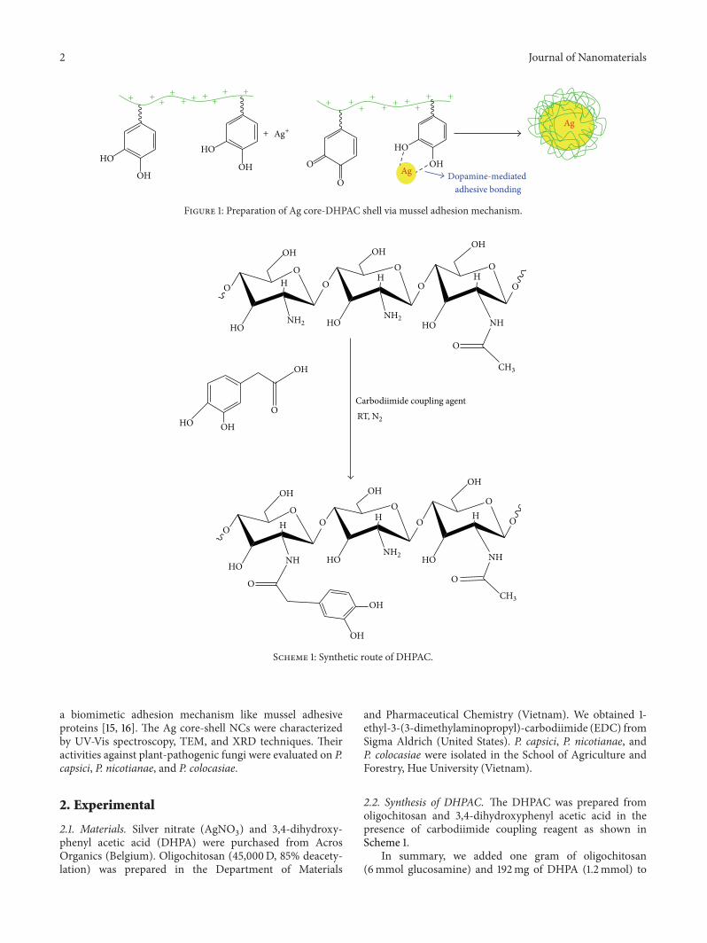
2.2. Synthesis of DHPAC. The DHPAC was prepared fromoligochitosan and 3,4-dihydroxyphenyl acetic acid in thepresence of carbodiimide coupling reagent as shown inScheme 1.
In summary, we added one gram of oligochitosan(6mmol glucosamine) and 192mg of DHPA (1.2mmol) to

Journal of Nanomaterials 3
50mL distilled water under stirring and then added 290mgof EDC (1.4mmol) to the mixture. The reaction mixturewas stirred under nitrogen atmosphere at room tempera-ture overnight and dialyzed with membrane with molecularweight cutoff of 6000–8000 afterward. After two days ofdialysis, the polymer solution was lyophilized using a freeze-drying machine to obtain DHPAC. The degree of DHPAsubstitution was estimated by 1H NMR spectrum.
2.3. Preparation of Ag Core-DHPAC Shell NCs. Silver core-DHPAC shell NCs were prepared by mixing 50mL DHPACsolution (1%) containing around 1600 ppm of DHPA (cal-culated according to 1H NMR) and 50mL AgNO
3solution
containing 800 ppm of silver ion and then stirred for 30minutes at room temperature. The Ag core-DHPAC shellNCs could be formed (as demonstrated in Figure 1) andcharacterized using UV-Vis, TEM, and XRD. For controlsample, DHPA was the reducing agent without DHPAC.The experiment was conducted in the same manner withpreparation of Ag core-shell NCs.
2.4. Characterization of Ag Core-DHPAC Shell NCs. UV-Visabsorption spectrum of the colloidal solutions was measuredusing Jasco V670. TEM images were collected using a JEM-1400 instrument (JEM-1400, JEOL) which was operated atan accelerating voltage of 100 kV. Samples for TEM mea-surement were prepared by dropping AgNPs solution ontoa carbon-coated copper grid. The colloidal solution-coveredgrids were allowed to dry for several hours. The XRD resultwas characterized using D8 advanced Bragg X-ray with CuK𝛼 radiation. For sample handling, glass slide was used asa substrate for measurement. Cleaned substrate was coveredwith the colloidal NCs and dried in air.
2.5. Studies on Growth Inhibition Ability of Plant-PathogenicFungi. The antifungal activity against pathogen was evalu-ated by using the in vitro plate dilution method.The colloidalNPs were mixed with 25mL of melting potato dextroseagar (PDA) medium and then were poured into Petri disheswith final concentrations of 3, 6, and 9 ppm. The controldishes contained distilled water. The fungus was transferredequally onto the center point of the prepared Petri dishesand incubated at room temperature for six days. The growthinhibition of the fungus was evaluated by measuring thediameter of colony growth and calculated with the followingformula: growth inhibition (%) = ((𝑑
1− 𝑑2)/𝑑1) × 100,
where 𝑑1and 𝑑
2are diameters of the colony of control and
NCs-containing samples, respectively. All experiments wereperformed in triplicate.
3. Results and Discussion
3.1. Characterization of DHPAC. Structure of DHPA-conjugated oligochitosan was well determined by the 1HNMR spectrum. Figure 2 showed three aromatic protonsof the conjugated DHPA with chemical shift (𝛿H) at 6.78,6.34, and 6.73 ppm. Some typical glucosamine protons of
HeHd
Habc
Hd
Ha Hb
HcHeHd
Habc
Hd
Ha Hb
Hc
O
OO
O
HO
OH
NH
OH OH OH
HO HO NHO
OOHHH O
CH3
NH2
OH
8 7 6 5 4 3 2
𝛿 (ppm)
O
H2
Figure 2: 1H NMR spectra of chitosan-catechol derivative.
oligochitosan appear at 2 ppm (acetyl protons), 3.02 ppm(H2proton), and 3.5–4 ppm (H
1–6 protons). These resultsconfirm that theDHPAwas grafted to the chitosan effectively.According to integral values of DHPA aromatic protons andmethyl protons of chitosan, the degree of DHPA substitution(DS) was calculated as 14 DHPA/100 glucosamine units.
3.2. Preparation of Ag Core-DHPAC Shell NCs. Preparationof AgNPs using natural extracts containing polyphenolcompounds or catechol-functionalized polymers has recentlybeen reported that is regarded as a green method [17–19]. Inour study, DHPAmoieties were utilized to reduce Ag+ into ametallic form and then protected the produced AgNPs. Aftermixing DHPAC solution and AgNO
3solution, the color of
the mixed solution immediately changed. After shaking for15minutes, UV-Vis absorbance of the solution performed twoUV-Vis absorption peaks at wavelength ranging from 270 to320 nm and 375 to 430 nm that was ascribed to absorbanceof the DHPA moiety/oxidized DHPA product and surfaceplasmon resonance of AgNPs, respectively (as shown inFigure 3). The result indicated formation of the AgNPs.
Figure 4 showed XRD diffractogram of the Ag core-DHPAC shell NCs and the crystalline phase of metallic silverat 38.0, 44.2, 64.4, 77.6, and 81.6∘. These peaks correspondedto the typical face centered cubic structure of Ag with millerindices at (111), (200), (220), (311), and (222), respectively[4, 20].
Figure 5 showed clearly that the core-shell NCs wereformed in which Ag cores had the size distribution of 26 ±9 nm and polymer shell layer of 18±8 nm of thickness.Thesevalues were estimated by mean value of 50 selected nanopar-ticles. The experiment preparing only silver nanoparticlewithout shell had also been conducted to compare differencein morphology of AgNPs but the reaction mixture showedprecipitation after AgNO
3and DHPAwere added.This could
be an aggregate of the formed AgNPs which was not wellprotected by polymer shell layer.
Formation of the polymer layer coating on the surfaceof the AgNPs could be explained as shown in Figure 6,in which some DHPA moieties on DHPAC reduced ionicsilver to AgNPs and quinone moieties. Some DHPAmoietiessimultaneously adhered to the formed AgNPs via dopamine-like adhesion mechanism which was well proven in someprevious report [15, 16, 21, 22]. The quinone moieties fromoxidized DHPAC easily reacted with amine groups of other

4 Journal of Nanomaterials
0
1
2
3
4
200 300 400 500 600 700Wavelength (nm)
Abs
AgNPs 25ppmAgNPs 50ppmAgNPs 100ppm
AgNPs 200ppmAgNPs 400ppm
Figure 3: UV-Vis absorbance of Ag core-DHPAC shell NCs solution.
0
10
20
30
40
50
60
40 50 60 70 80
Lin
(CPs
)
2𝜃-scale
d=2.35837
d=2.05520
d=1.44604
d=1.23137
d=1.17806
Figure 4: XRD pattern of the Ag core-DHPAC shell NCs.
Figure 5: TEM images of the Ag core-DHPAC shell NCs: red and green arrows indicate Ag core and polymer shell layer, respectively.

Journal of Nanomaterials 5
+ +
+ +
++ +
+
+ + + +
+ +
++ +
+
+ +
++
+ +
+
+
+
+
+ +
+
Schiff-basereaction
Michael reaction
Biomimetic adhesion like mussel adhesive proteins
HOOH
OHOH
OO
OHOH
OHOH
OH
OH
HN
HO OH
Ag
Ag
NH2
NH2NH2
H2N
Ag+
H2N
H2N
H2N
H2N
H2N
Gel layer
External quinone or amine moieties
bond to other core-shell NPs forming
Ag nanoclusters
O
O
Figure 6: Core-shell AgNPs formation via biomimetic adhesion and chemical reactions.
DHPAC chains via Michael reaction between amine groupof chitosan and double bond next to carbonyl of oxidizedcatechol or Schiff-base reaction between amine group ofchitosan and carbonyl of oxidized catechol which resulted information of a polymeric hydrogel network. In the process,some functional groups on the core-shell AgNPs could bondtogether to form a nanocluster containing these AgNPs.
3.3. Antibacterial Activity of Ag Core-DHPAC Shell NCs. TheAg core-DHPAC shell NCs exhibited strong growth inhibi-tion of plant-pathogenic fungi such as P. capsici, P. nicotianae,and P. colocasiae. Figure 7 showed results obtained fromantifungal experiments of the AgNCs colloidal solutions.There was strong growth of these fungi in control samplewithout AgNCs. A high colony diameter of the fungi wasrecorded. In the presence of small amount of AgNCs (3 ppm),growth of the fungi was significantly decreased.
Figure 7 showed growth inhibition of these fungi, inwhichP. capsici funguswas inhibited at 80% in the presence of9 ppmAgNCs. Growth inhibition of P. nicotianae and P. colo-casiae was approximated. Fifty percent growth inhibition ofthese fungi was recorded at six ppm of AgNCs approximately,which was a low concentration compared to effective dose(ED50) of inhibition for plant-pathogenicCorticium salmoni-color at 27.2 ppm of AgNPs [23]. In additional experiment,
antifungal activity of oligochitosan has also been conducted.In the same concentration with DHPAC used in preparationof AgNCs, the chitosan was diluted in the same manner tothe highest concentration at 45 ppm. In the concentration,chitosan did not show antifungal activity. It was in agreementwith a previous report in which oligochitosan polymersolution showed antibacterial activity at high concentration(100 ppm) [22].
Different behaviors might be due to differences in orga-nizations, structures, and functions of these plant-pathogenicfungi or due to different interaction of the core-shell AgNPsand oligochitosan with the fungi (Figure 8).
For the Ag core-DHPAC shell NCs, chitosan-basedshell layer might improve antifungal activity of the AgNCsbecause of cationic polymer possessing antimicrobial orantipathogenic activity. Xu et al. reported that oligochi-tosan exhibited the highest inhibitory growth with P. capsicicompared to other phytopathogens. Fifty percent inhibitorygrowth of P. capsici was reported at 100 ppm of oligochitosan[24]. Although the activity was much lower than that ofAgNPs, the polymer layer could act as an active targeting siteand result in increasing interaction of the cationic chitosanshell layer on Ag core in the NCs and phospholipid layeron bacterial membrane via electrostatic interaction. Thisbrought the Ag core-DHPAC shell NCs to the surface of themicrobes and sustainedly released Ag+ ions could kill the

6 Journal of Nanomaterials
(a) (b)
(c)
Figure 7: Growth of the fungi in diffrent PDA media with and without AgNCs: P. capsici (a), P. nicotianae (b), and P. colocasiae (c).
0
20
40
60
80
100
0
20
40
60
80
100
0 3 6 9
Col
ony
diam
eter
(mm
)
Gro
wth
inhi
bitio
n (%
)
Concentration of Ag (ppm)
(a)
0
20
40
60
80
0
20
40
60
80
100
0 3 6 9
Col
ony
diam
eter
(mm
)
Gro
wth
inhi
bitio
n (%
)
Concentration of Ag (ppm)
(b)
Figure 8: Growth inhibition of Ag core-DHPAC shell NCs on the plant-pathogenic fungi: P. capsici (a) and P. nicotianae (b).
fungi. Slade and Pegg reported that several kinds of Phy-tophthora fungi were killed by Ag+ in the range 46–460 nM(5–50 ppb) [25]. Sustained release of Ag+ ions from the Agcore-DHPAC shell NCs could be significant to inhibit growthof the plant-pathogenic fungi and protect plants from Phy-tophthora fungi.
4. Conclusion
In this work, silver core-chitosan shell nanoclusters wereprepared via chemical reduction using chitosan derivativeas a reducing and protecting agent. Low concentration ofthe nanoparticles exhibited a powerful activity against plantpathogens, such as P. capsici, P. nicotianae, and P. colocasiae.Positive results show the potential application as an inexpen-sive ecofungicide for agriculture.
Conflict of Interests
The authors declare that there is no conflict of interestsregarding the publication of this paper.
Acknowledgment
This research was supported by Vietnam National Founda-tion for Science and TechnologyDevelopment (NAFOSTED)under Grant no. 104.04-2011.49.
References
[1] M. de, P. S. Ghosh, and V. M. Rotello, “Applications of nanopar-ticles in biology,” Advanced Materials, vol. 20, no. 22, pp. 4225–4241, 2008.

Journal of Nanomaterials 7
[2] M. Abhilash, “Potential applications of nanoparticles,” Interna-tional Journal of Pharma and Bio Sciences, vol. 1, no. 1, pp. 1–12,2010.
[3] M. Sharon, A. K. Choudhary, and R. Kumar, “Nanotechnologyin agricultural disease and food safety,” Journal of Phyto-medicine, vol. 2, no. 4, pp. 83–92, 2010.
[4] J. P. Ruparelia, A. K. Chatterjee, S. P. Duttagupta, and S.Mukherji, “Strain specificity in antimicrobial activity of silverand copper nanoparticles,” Acta Biomaterialia, vol. 4, no. 3, pp.707–716, 2008.
[5] L. Rastogi and J. Arunachalam, “Synthesis and characterizationof bovine serum albumin-copper nanocomposites for antibac-terial applications,” Colloids and Surfaces B: Biointerfaces, vol.108, pp. 134–141, 2013.
[6] K. Adavallan and N. Krishnakumar, “Mulberry leaf extractmediated synthesis of gold nanoparticles and its anti-bacterialactivity against human pathogens,” Advances in Natural Sci-ences: Nanoscience and Nanotechnology, vol. 5, no. 2, Article ID025018, 2014.
[7] V. D. Cao, P. P. Nguyen, V. Q. Khuong et al., “Ultrafine coppernanoparticles exhibiting a powerful antifungal/killing activityagainstCorticium salmonicolor,”Bulletin of the Korean ChemicalSociety, vol. 35, no. 9, pp. 2645–2648, 2014.
[8] X. Wang, F. Cheng, J. Gao, and L. Wang, “Antibacterial wounddressing from chitosan/polyethylene oxide nanofibers matsembedded with silver nanoparticles,” Journal of BiomaterialsApplications, 2014.
[9] Q. H. Tran, V. Q. Nguyen, and A.-T. Le, “ Silver nanoparticles:synthesis, properties, toxicology, applications and perspectives,”Advances in Natural Sciences: Nanoscience and Nanotechnology,vol. 4, Article ID 033001, 2013.
[10] R.M.Tripathi, R.K.Gupta,A. Shrivastav,M. P. Singh, B. R. Shri-vastav, and P. Singh, “Trichoderma koningii assisted biogenicsynthesis of silver nanoparticles and evaluation of their antibac-terial activity,” Advances in Natural Sciences: Nanoscience andNanotechnology, vol. 4, no. 3, Article ID 035005, 2013.
[11] H.-J. Park, S. H. Kim, H. J. Kim, and S.-H. Choi, “A new com-position of nanosized silica-silver for control of various plantdiseases,” Plant Pathology Journal, vol. 22, no. 3, pp. 295–302,2006.
[12] T. S. Cu, V. D. Cao, C. K. Nguyen, and N. Q. Tran, “Prepara-tion of silver core-chitosan shell nanoparticles using catechol-functionalized chitosan and antibacterial studies,”Macromolec-ular Research, vol. 22, no. 4, pp. 418–423, 2014.
[13] N. V. Truong, L. W. Burgess, and E. C. Y. Liew, “Greenhouseand field evaluations of potassium phosphonate: the control ofPhytophthora foot rot of black pepper in Vietnam,” Archives ofPhytopathology and Plant Protection, vol. 45, no. 6, pp. 724–739,2012.
[14] N. V. Truong, L.W. Burgess, and E. C. Y. Liew, “Cross-infectivityand genetic variation of Phytophthora capsici isolates from chilliand black pepper inVietnam,”Australasian Plant Pathology, vol.41, no. 4, pp. 439–447, 2012.
[15] H. Lee, S. M. Dellatore, W. M. Miller, and P. B. Messersmith,“Mussel-inspired surface chemistry for multifunctional coat-ings,” Science, vol. 318, no. 5849, pp. 426–430, 2007.
[16] J. H. Waite, “Surface chemistry: mussel power,” Nature Materi-als, vol. 7, pp. 7–9, 2008.
[17] E. Rodrıguez-Leon, R. Iniguez-Palomares, R. E. Navarroet al., “Synthesis of silver nanoparticles using reducingagents obtained from natural sources (Rumex hymenosepalus
extracts),” Nanoscale Research Letters, vol. 8, no. 1, article 318,2013.
[18] F. Cataldo, “Green synthesis of silver nanoparticles by theaction of black or green tea infusions on silver ions,” EuropeanChemical Bulletin, vol. 3, no. 3, pp. 280–289, 2014.
[19] K. C. L. Black, Z. Liu, and P. B. Messersmith, “Catechol redoxinduced formation of metal core-polymer shell nanoparticles,”Chemistry of Materials, vol. 23, no. 5, pp. 1130–1135, 2011.
[20] S. Zaki, M. Etarahony, M. Elkady, and D. Abd-El-Haleem,“The use of bioflocculant and bioflocculant-producing Bacillusmojavensis strain 32A to synthesize silver nanoparticles,” Jour-nal of Nanomaterials, vol. 2014, Article ID 431089, 7 pages, 2014.
[21] Y. Lee, H. Lee, P. B. Messersmith, and T. G. Park, “A bioinspiredpolymeric template for 1D assembly of metallic nanoparticles,semiconductor quantum dots, and magnetic nanoparticles,”Macromolecular Rapid Communications, vol. 31, no. 24, pp.2109–2114, 2010.
[22] H. Lee, N. F. Scherer, and P. B. Messersmith, “Single-moleculemechanics of mussel adhesion,” Proceedings of the NationalAcademy of Sciences of the United States of America, vol. 103, no.35, pp. 12999–13003, 2006.
[23] D. van Phu, V. T. K. Lang, N. T. K. Lan et al., “Synthesis andantimicrobial effects of colloidal silver nanoparticles in chitosanby 𝛾-irradiation,” Journal of Experimental Nanoscience, vol. 5,no. 2, pp. 169–179, 2010.
[24] J. Xu, X. Zhao, X. Han, and Y. Du, “Antifungal activity ofoligochitosan against Phytophthora capsici and other plantpathogenic fungi in vitro,” Pesticide Biochemistry and Physiol-ogy, vol. 87, no. 3, pp. 220–228, 2007.
[25] S. J. Slade and G. F. Pegg, “The effect of silver and other metalions on the in vitro growth of root-rotting Phytophthora andother fungal species,” Annals of Applied Biology, vol. 122, no. 2,pp. 233–251, 1993.

Submit your manuscripts athttp://www.hindawi.com
ScientificaHindawi Publishing Corporationhttp://www.hindawi.com Volume 2014
CorrosionInternational Journal of
Hindawi Publishing Corporationhttp://www.hindawi.com Volume 2014
Polymer ScienceInternational Journal of
Hindawi Publishing Corporationhttp://www.hindawi.com Volume 2014
Hindawi Publishing Corporationhttp://www.hindawi.com Volume 2014
CeramicsJournal of
Hindawi Publishing Corporationhttp://www.hindawi.com Volume 2014
CompositesJournal of
NanoparticlesJournal of
Hindawi Publishing Corporationhttp://www.hindawi.com Volume 2014
Hindawi Publishing Corporationhttp://www.hindawi.com Volume 2014
International Journal of
Biomaterials
Hindawi Publishing Corporationhttp://www.hindawi.com Volume 2014
NanoscienceJournal of
TextilesHindawi Publishing Corporation http://www.hindawi.com Volume 2014
Journal of
NanotechnologyHindawi Publishing Corporationhttp://www.hindawi.com Volume 2014
Journal of
CrystallographyJournal of
Hindawi Publishing Corporationhttp://www.hindawi.com Volume 2014
The Scientific World JournalHindawi Publishing Corporation http://www.hindawi.com Volume 2014
Hindawi Publishing Corporationhttp://www.hindawi.com Volume 2014
CoatingsJournal of
Advances in
Materials Science and EngineeringHindawi Publishing Corporationhttp://www.hindawi.com Volume 2014
Smart Materials Research
Hindawi Publishing Corporationhttp://www.hindawi.com Volume 2014
Hindawi Publishing Corporationhttp://www.hindawi.com Volume 2014
MetallurgyJournal of
Hindawi Publishing Corporationhttp://www.hindawi.com Volume 2014
BioMed Research International
MaterialsJournal of
Hindawi Publishing Corporationhttp://www.hindawi.com Volume 2014
Nano
materials
Hindawi Publishing Corporationhttp://www.hindawi.com Volume 2014
Journal ofNanomaterials


