Research Article Sagittal and Vertical Craniofacial Growth...
8
Research Article Sagittal and Vertical Craniofacial Growth Pattern and Timing of Circumpubertal Skeletal Maturation: A Multiple Regression Study Giuseppe Perinetti, Luigi Rosso, Riccardo Riatti, and Luca Contardo Department of Medical, Surgical and Health Sciences, School of Dentistry, University of Trieste, Trieste, Italy Correspondence should be addressed to Giuseppe Perinetti; [email protected] Received 28 August 2016; Accepted 26 October 2016 Academic Editor: Alberto Baldini Copyright © 2016 Giuseppe Perinetti et al. is is an open access article distributed under the Creative Commons Attribution License, which permits unrestricted use, distribution, and reproduction in any medium, provided the original work is properly cited. e knowledge of the associations between the timing of skeletal maturation and craniofacial growth is of primary importance when planning a functional treatment for most of the skeletal malocclusions. is cross-sectional study was thus aimed at evaluating whether sagittal and vertical craniofacial growth has an association with the timing of circumpubertal skeletal maturation. A total of 320 subjects (160 females and 160 males) were included in the study (mean age, 12.3±1.7 years; range, 7.6–16.7 years). ese subjects were equally distributed in the circumpubertal cervical vertebral maturation (CVM) stages 2 to 5. Each CVM stage group also had equal number of females and males. Multiple regression models were run for each CVM stage group to assess the significance of the association of cephalometric parameters (ANB, SN/MP, and NSBa angles) with age of attainment of the corresponding CVM stage (in months). Significant associations were seen only for stage 3, where the SN/MP angle was negatively associated with age ( coefficient, −0.7). ese results show that hyperdivergent and hypodivergent subjects may have an anticipated and delayed attainment of the pubertal CVM stage 3, respectively. However, such association remains of little entity and it would become clinically relevant only in extreme cases. 1. Introduction e knowledge of the associations between the timing of skeletal maturation and craniofacial growth is of primary importance when planning a functional treatment for most of the skeletal malocclusions, including those on the sagittal [1] and vertical dimensions [2, 3]. Although being a controversial issue [4–6], functional treatment for Class II malocclusion would induce clinically relevant mandibular elongation when performed during the pubertal growth phase [7, 8], while, Class III malocclusion requires early treatment [1]. Finally, both excessive vertical facial growth [2] and deepbite [3] have also been reported to be best treated during the pubertal growth phase. ese aspects are of particular importance also in consideration that skeletal Class III malocclusion [9] and vertical facial growth pattern [10] tend to aggravate when not treated. erefore, the knowledge of whether attainment of a specific growth phase is also dependent on the different sagittal and vertical craniofacial growth pattern has a clinical relevance in terms of timing of intervention. In this regard, the most common procedures to monitor the different growth phases are the radiographic methods of maturational stages of the cervical vertebral maturation (CVM) [1, 11] and hand-and-wrist maturation (HWM) (for review, see [12]). To date very little research has focused on the possible association between the timing of the circumpubertal skeletal maturation phases and sagittal craniofacial growth, that is, skeletal class [13–15]. Moreover, none of these previous studies investigated possible associations of vertical cran- iofacial facial growth and timing of attainment of skeletal maturation phases. ese studies were further limited by the use of univariate analyses [14, 15] with only one exception, where a multivariate model was used [13]. Finally, a further study [16] used an overall craniofacial composite measured, derived from multiple measurements; thus, it was not able to discriminate between sagittal and vertical growth patterns. Hindawi Publishing Corporation BioMed Research International Volume 2016, Article ID 1728712, 7 pages http://dx.doi.org/10.1155/2016/1728712
Transcript of Research Article Sagittal and Vertical Craniofacial Growth...

Research ArticleSagittal and Vertical Craniofacial GrowthPattern and Timing of Circumpubertal Skeletal Maturation:A Multiple Regression Study
Giuseppe Perinetti, Luigi Rosso, Riccardo Riatti, and Luca Contardo
Department of Medical, Surgical and Health Sciences, School of Dentistry, University of Trieste, Trieste, Italy
Correspondence should be addressed to Giuseppe Perinetti; [email protected]
Received 28 August 2016; Accepted 26 October 2016
Academic Editor: Alberto Baldini
Copyright © 2016 Giuseppe Perinetti et al. This is an open access article distributed under the Creative Commons AttributionLicense, which permits unrestricted use, distribution, and reproduction in any medium, provided the original work is properlycited.
The knowledge of the associations between the timing of skeletal maturation and craniofacial growth is of primary importancewhen planning a functional treatment formost of the skeletalmalocclusions.This cross-sectional studywas thus aimed at evaluatingwhether sagittal and vertical craniofacial growth has an associationwith the timing of circumpubertal skeletal maturation. A total of320 subjects (160 females and 160males) were included in the study (mean age, 12.3±1.7 years; range, 7.6–16.7 years).These subjectswere equally distributed in the circumpubertal cervical vertebral maturation (CVM) stages 2 to 5. Each CVM stage group also hadequal number of females and males. Multiple regression models were run for each CVM stage group to assess the significanceof the association of cephalometric parameters (ANB, SN/MP, and NSBa angles) with age of attainment of the correspondingCVM stage (in months). Significant associations were seen only for stage 3, where the SN/MP angle was negatively associatedwith age (𝛽 coefficient, −0.7). These results show that hyperdivergent and hypodivergent subjects may have an anticipated anddelayed attainment of the pubertal CVM stage 3, respectively. However, such association remains of little entity and it would becomeclinically relevant only in extreme cases.
1. Introduction
The knowledge of the associations between the timing ofskeletal maturation and craniofacial growth is of primaryimportancewhen planning a functional treatment formost ofthe skeletal malocclusions, including those on the sagittal [1]and vertical dimensions [2, 3]. Although being a controversialissue [4–6], functional treatment for Class II malocclusionwould induce clinically relevantmandibular elongationwhenperformed during the pubertal growth phase [7, 8], while,Class III malocclusion requires early treatment [1]. Finally,both excessive vertical facial growth [2] and deepbite [3] havealso been reported to be best treated during the pubertalgrowth phase. These aspects are of particular importancealso in consideration that skeletal Class III malocclusion[9] and vertical facial growth pattern [10] tend to aggravatewhen not treated. Therefore, the knowledge of whetherattainment of a specific growth phase is also dependent on the
different sagittal and vertical craniofacial growth pattern hasa clinical relevance in terms of timing of intervention. In thisregard, themost commonprocedures tomonitor the differentgrowth phases are the radiographic methods of maturationalstages of the cervical vertebral maturation (CVM) [1, 11] andhand-and-wrist maturation (HWM) (for review, see [12]).
To date very little research has focused on the possibleassociation between the timing of the circumpubertal skeletalmaturation phases and sagittal craniofacial growth, thatis, skeletal class [13–15]. Moreover, none of these previousstudies investigated possible associations of vertical cran-iofacial facial growth and timing of attainment of skeletalmaturation phases. These studies were further limited by theuse of univariate analyses [14, 15] with only one exception,where a multivariate model was used [13]. Finally, a furtherstudy [16] used an overall craniofacial composite measured,derived from multiple measurements; thus, it was not ableto discriminate between sagittal and vertical growth patterns.
Hindawi Publishing CorporationBioMed Research InternationalVolume 2016, Article ID 1728712, 7 pageshttp://dx.doi.org/10.1155/2016/1728712

2 BioMed Research International
Investigation on the craniofacial vertical growth pattern andtiming of skeletal maturation becomes of interest also inconsideration of the previous evidence reporting an earlierdental maturation in hyperdivergent subjects [17].
Therefore, through multivariate models, this cross-sectional study was aimed at evaluating whether sagittaland vertical craniofacial growth pattern, as described bycommon cephalometric parameters, has an association withthe timing of circumpubertal skeletal maturation, that is, ageof attainment of the maturation phases as defined by theCVMmethod.
2. Materials and Methods
2.1. Study Population and Design. The database between Jan-uary 2009 and December 2015 of the Sections of Stomatologyof the Department of Medical, Surgical and Health Sciences,University of Trieste, was screened. This study includedsubjects who were seeking orthodontic treatment and whohad never been treated before. As a routine procedure, asigned informed consent for releasing diagnostic material forscientific purposes was obtained from the patients’ parentsprior to entry into treatment, procedures followed adheredto the World Medical Organization Declaration of Helsinki[18], and the protocol was reviewed and approved by thelocal Ethical Committee. In particular, in the first clini-cal session a lateral cephalograms was taken as a part ofthe pretreatment clinical recording. The following inclusioncriteria were applied: (i) age between 7 and 17 years; (ii)circumpubertal skeletal maturation between CVM stages 2and 5; (iii) absence of any craniofacial anomaly or extensivedental caries or restorations; (iv) good general health withno signs of symptoms of temporomandibular disorders; (v)no history of trauma at the craniofacial region; and (vi)Caucasian ethnicity. A dedicated X-ray machine (KODAK8000C; Eastman Kodak Company) was employed for therecording of lateral head cephalograms. Settings were of73–77 kV, 12mA with an exposure time of 0.80 seconds.Images were saved at 300 dpi resolution and radiographsof low quality were excluded. An experienced orthodontist(LC) assisted by a second operator (LR) screened the casesfor inclusion. A further experienced orthodontist (GP) wasinvolved to ensure correct enrollment and, in case of dis-agreement, discussion was made until satisfaction of bothoperators. From an initial sample of over 450 subjects, totalof 320 subjects (160 females and 160 males) were included inthe study (mean age, 12.3 ± 1.7 years; range, 7.6–16.7 years).
2.2. Cephalometric Analysis for the Face and Cervical Verte-brae. A customized digitization regimen and analysis withcephalometric software (Viewbox, version 3.0, dHAL Soft-ware, Kifissia, Greece) were used for all cephalograms exam-ined in this study. The cephalometric analysis of the facerequired the digitization of 9 landmarks (Figure 1) [19].The customized cephalometric analysis included 4 angularmeasurements as follows (Figure 1): maxillary prognathism(SNA angle), mandibular prognathism (SNB angle), maxillo-mandibular relationship (ANB angle), maxillary inclination
ANSPNS
Me
Go
MP
SN
A
B
BaPP
Figure 1: Diagram of the cephalometric measurements of thecraniofacial complex. Landmarks: A, subspinale; B, supramentale;N, nasion; S, centre of the sella turcica; Ba, Basion; ANS, anteriornasal spine; PNS, posterior nasal spine; Me, menton; Go, Gonion;Planes: PP, palatal plane; MP, mandibular plane. See text for details.
Upper width
Lower width
Ant
. hei
ght
Post.
hei
ght
Concavity
Figure 2: Diagram of the cephalometric measurements of thecervical vertebrae. Only a vertebral body is shown for clarity. Incervical vertebra 2, only concavity wasmeasured. See text for details.
relative to the cranial base (SN/PP angle), mandibular incli-nation relative to the cranial base (SN/MP angle), and cranialbase angle (NSBa angle).
Regarding the body of the cervical vertebrae, a quanti-tative assessment of the shape (maturation) was also per-formed. The customized cephalometric analysis includedmeasurements generated from 17 landmarks from which 11linear and 8 angular variables were derived. Among the linearvariables, 3 were for concavities of lower borders of C2–C4and 8 related to the anterior and posterior heights and upperand lower widths for the C3 and C4 (Figure 2) [20]. Amongangular variables, 4 were for the inner angles of the C3 andother 4 were for the inner angles of the C4. The data wereused to calculate presence/absence of concavity and shape ofthe vertebral body through a dedicated Excel data sheetwherelinear and angular measurements were used in combination.The used method was independent of absolute recordings;instead we used relative dimensions to assess the shapes ofthe C3 and C4, while the concavity was assessed when itwas at least 10% of the corresponding posterior height of the
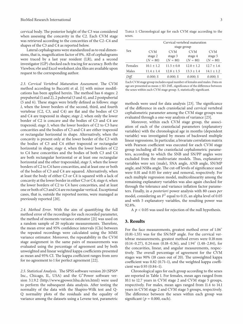
BioMed Research International 3
cervical body. The posterior height of the C3 was consideredwhen assessing the concavity in the C2. Each CVM stagewas retrieved according to the concavities of the C2–C4 andshapes of the C3 and C4 as reported below.
Lateral cephalogramswere standardized as to real dimen-sions, that is, magnification factor of 0%. All of cephalogramswere traced by a last year resident (LR), and a secondinvestigator (GP) checked each tracing for accuracy. Both theViewbox.vbr andExcel worksheet.xlsx files are available uponrequest to the corresponding author.
2.3. Cervical Vertebral Maturation Assessment. The CVMmethod according to Baccetti et al. [1] with minor modifi-cations has been applied herein. The method has 6 stages: 2prepubertal (1 and 2), 2 pubertal (3 and 4), and 2 postpubertal(5 and 6). These stages were briefly defined as follows: stage1, when the lower borders of the second, third, and fourthvertebrae (C2, C3, and C4) are flat and the bodies of C3and C4 are trapezoid in shape; stage 2, when only the lowerborder of C2 is concave and the bodies of C3 and C4 aretrapezoid; stage 3, when the lower borders of C2 to C3 haveconcavities and the bodies of C3 and C4 are either trapezoidor rectangular horizontal in shape. Alternatively, when theconcavity is present only at the lower border of the C3 withthe bodies of C3 and C4 either trapezoid or rectangularhorizontal in shape; stage 4, when the lower borders of C2to C4 have concavities and the bodies of both C3 and C4are both rectangular horizontal or at least one rectangularhorizontal and the other trapezoidal; stage 5, when the lowerborders of C2 to C4 have concavities, and at least one or bothof the bodies of C3 and C4 are squared. Alternatively, whenat least the body of either C3 or C4 is squared with a lack ofconcavity at the lower border in either C3 or C4; stage 6, whenthe lower borders of C2 to C4 have concavities, and at leastone or both of C3 andC4 are rectangular vertical. Exceptionalcases, that is, outside the reported norms, were managed aspreviously reported [20].
2.4. Method Error. With the aim of quantifying the fullmethod error of the recordings for each recorded parameter,the method of moments variance estimator [21] was used ona random sample of 20 replicate measurements. Therefore,the mean error and 95% confidence intervals (CIs) betweenthe repeated recordings were calculated using the MMEvariance estimator. Moreover, the repeatability in the CVMstage assignment in the same pairs of measurements wasevaluated using the percentage of agreement and by bothunweighted and linear weighted kappa coefficients presentedas mean and 95% CI. The kappa coefficient ranges from zerofor no agreement to 1 for perfect agreement [22].
2.5. Statistical Analysis. The SPSS software version 20 (SPSS�Inc., Chicago, IL, USA) and the G∗Power software ver-sion 3.1.9.2 (http://www.gpower.hhu.de/en.html) were usedto perform the subsequent data analysis. After testing thenormality of the data with the Shapiro-Wilk test and Q-Q normality plots of the residuals and the equality ofvariance among the datasets using a Levene test, parametric
Table 1: Chronological age for each CVM stage according to thesexes.
Sex
Cervical vertebral maturationstage group
CVMstage 2(𝑁 = 80)
CVMstage 3(𝑁 = 80)
CVMstage 4(𝑁 = 80)
CVMstage 5(𝑁 = 80)
Females 10.1 ± 1.2 11.5 ± 0.8 12.0 ± 1.2 12.7 ± 1.6
Males 11.4 ± 1.4 12.8 ± 1.5 13.3 ± 1.4 14.1 ± 1.2
Diff. 0.000; S 0.000; S 0.000; S 0.000; SEachCVMstage group includes equal number of females andmales.Data onage are presented as mean ± SD. Diff., significance of the difference betweenthe sexes within each CVM stage group. S, statistically significant.
methods were used for data analysis [23]. The significanceof the difference in each craniofacial and cervical vertebralcephalometric parameter among the CVM stage groups wasevaluated through a one-way analysis of variance [23].
Moreover, within each CVM stage group, the associ-ation of each of the craniofacial parameters (explanatoryvariables) with the chronological age in months (dependentvariable) was investigated by means of backward multiplelinear regressions. In particular, a bivariate correlationmatrixwith Pearson coefficient was executed for each CVM stagegroup including all the craniofacial cephalometric parame-ters, according to which the SNB and SN/PP angles wereexcluded from the multivariate models. Thus, explanatoryvariables were sex (male), SNA angle, ANB angle, SN/MPangle, and NSBa angle. The cut-off levels of significance usedwere 0.01 and 0.05 for entry and removal, respectively. Foreach multiple regression model, multicolinearity among theremaining explanatory variables was also again checked forthrough the tolerance and variance inflation factor parame-ters. Finally, in a posteriori power analysis with 80 cases permodel, considering an 𝐹2 equal to 0.15, an alpha level of 0.05and with 5 explanatory variables, the resulting power was92.8%.
A 𝑝 < 0.05 was used for rejection of the null hypothesis.
3. Results
For the face measurements, greatest method error of 1.06∘(0.81–1.55) was for the SN/MP angle. For the cervical ver-tebrae measurements, greatest method errors were 0.18mm(0.14–0.27), 0.24mm (0.18–0.36), and 1.94∘ (1.48–2.84), forthe concavities, linear, and angular measurements, respec-tively. The overall percentage of agreement for the CVMstages was 90% (18 cases out of 20). The unweighted kappacoefficient was 0.82 (0.71–1), and the weighted kappa coeffi-cient was 0.93 (0.84–1).
Chronological ages for each group according to the sexesare reported in Table 1. For females, mean ages ranged from10.1 to 12.7 years in CVM stage 2 and CVM stage 5 groups,respectively. For males, mean ages ranged from 11.4 to 14.1years in CVM stage 2 and CVM stage 5 groups, respectively.The difference between the sexes within each group wassignificant (𝑝 = 0.000, each).

4 BioMed Research International
Table 2: Descriptive statistics for the craniofacial parameters (in degrees) for each group.
Parameter (degree)Cervical vertebral maturation stage group
Diff.CVM stage 2(𝑁 = 80)
CVM stage 3(𝑁 = 80)
CVM stage 4(𝑁 = 80)
CVM stage 5(𝑁 = 80)
SNA angle 80.7 ± 3.6 81.2 ± 3.5 80.3 ± 3.2 81.2 ± 3.4 0.301; NSSNB angle 77.0 ± 3.9 77.2 ± 3.6 76.7 ± 3.7 77.7 ± 3.7 0.365; NSANB angle 3.7 ± 2.0 3.9 ± 2.1 3.6 ± 2.2 3.5 ± 2.3 0.558; NSSN/PP angle 7.0 ± 3.4 7.1 ± 3.9 7.9 ± 3.3 8.1 ± 2.8 0.200; NSSN/MP angle 31.2 ± 5.6 30.4 ± 5.9 30.8 ± 5.9 30.6 ± 5.3 0.848; NSNSBa angle 129.6 ± 5.2 129.4 ± 4.6 130.0 ± 4.9 130.7 ± 4.7 0.375; NSEach CVM stage group includes equal number of females and males. Data on age are presented as mean ± SD. Diff., significance of the levels of differencesamong the CVM stage groups for each cephalometric parameter. NS, not statistically significant.
Table 3: Results of the backward multiple linear regressions for the association of craniofacial cephalometric parameters with thechronological age (in months) for each CVM stage.
Explanatoryvariable 𝛽 (SE) t Sig.
Model 1: age of attainment of CVM stage2 (N = 80), R2 = 0.213Sex (male) 17.0 (3.6) 4.754 0.000; SANB angle 1.6 (0.9) 1.793 0.077; NSModel 2: age of attainment of CVM stage3 (𝑁 = 80), R2 = 0.269Sex (male) 13.6 (3.2) 4.311 0.000; SMP/SN angle −0.7 (0.3) 2.477 0.015; SModel 3: age of attainment of CVM stage4 (N = 80), R2 = 0.194Sex (male) 15.4 (3.6) 4.339 0.000; SNSBa angle 0.6 (0.4) 1.700 0.093; NSModel 4: age of attainment of CVM stage5 (N = 80), R2 = 0.165Sex (male) 16.3 (4.0) 4.080 0.000; SIndependent variables entered in each model: sex, SNA angle, ANB angle, SN/MP angle, and NSBa angle, with variables having a p value above 0.1 removedfrom the model. Results of the multiple linear regressions are presented as 𝛽 (SE); 𝑅2, coefficient of determination. Sig., level of significance; S, statisticallysignificant; NS, not statistically significant.
Descriptive statistics for each analysed parameter isreported in Table 2. The SNA angle ranged from 80.3∘ ± 3.2(CVM stage 4) to 81.2∘ ± 3.4 (CVM stage 5); the SNB angleranged from 76.7∘ ± 3.7 (CVM stage 4) to 77.7∘ ± 3.7 (CVMstage 5); the ANB angle ranged from 3.5∘ ±2.3 (CVM stage 5)to 3.9∘ ± 2.1∘ (CVM stage 3); the SN/PP angle ranged from7.1∘ ± 3.9 (CVM stage 3) to 8.1∘ ± 2.8 (CVM stage 5); theSN/MP angle ranged from 30.4∘±5.9 (CVM stage 3) to 31.2∘±5.6 (CVM stage 2); the NSBa angle ranged from 129.4∘ ± 4.6(CVM stage 3) to 130.7∘ ± 4.7 (CVM stage 5). For all of thesecraniofacial cephalometric parameters the differences amongthe groups were not statistically significant.
Results of the backwardmultiple linear regressionmodelsaccording to each CVM stage group are reported in Table 3.In the CVM stage 2 group (Model 1) 𝑅2 was of 0.213 withthe sex (male) and ANB angle positively associated with theage of attainment of the CVM stage 2 with 𝛽 coefficients of17.0 and 1.3, respectively. However, only the sex reached the
statistical significance (𝑝 = 0.000), while the ANB angle didnot (𝑝 = 0.077). In the CVM stage 3 group (Model 2) 𝑅2was of 0.269 with the sex (male) and SN/MP angle positivelyand negatively associated with 𝛽 coefficients of 13.6 and −0.7,respectively (𝑝 = 0.015, at least). In the CVM stage 4 group(Model 3) 𝑅2 was of 0.194 with the sex (male) and NSBaangle positively associated with the age of attainment of theCVM stage 4 with 𝛽 coefficients of 15.4 and 0.6, respectively.However, only the sex reached the statistical significance (𝑝 =0.000), while the NSBa angle did not (𝑝 = 0.093). Finally, Inthe CVM stage 5 group (Model 4) 𝑅2 was of 0.165 with onlythe sex (male) positively associatedwith the age of attainmentof the CVM stage 5 with a 𝛽 coefficient of 16.3 (𝑝 = 0.000).
4. Discussion
Throughmultivariatemodels, the present study demonstratesa little association of the sagittal and vertical craniofacial

BioMed Research International 5
growth pattern with the timing of skeletal maturation. Whilefemales had anticipated attainment of each CVM stage ascompared to males (Table 1), the different cephalometricparameters showed no significant differences among theCVM stage groups (Table 2), allowing a more reliable com-parison of the regression models.
The previous investigations [13–15] on sagittal craniofa-cial growth pattern and timing of skeletal maturation werefocused on the CVM stages 3 and 4. Therefore, present dataon the timing of the CVM stages 2 and 5 are not comparablewith previous evidence. Of interest, 𝑅2 retrieved for themodels ranged from 0.165 to 0.269 (Table 3). Although suchvalues were not particularly high, the greatest value was seenfor the pubertal CVM stage 3 while, generally, the valuesdecreased as maturation progresses into the postpubertalphases. Thus, in spite of the significant associations, thedifferent CVM stages, sex, and craniofacial parameters alltogether accounted for no more than ≈27% of the totalvariability of corresponding ages.This evidence demonstrateshow other relevant factors are responsible for the timing ofskeletal maturation such as genetics, ethnicity, nutrition, andsocioeconomic status [24].
As expected, sexwas themost significant factor associatedwith the age of attainment of each CVM stage from 2 to 5(Tables 1 and 3). According to the 𝛽 coefficients, the malesubjects had on average a delayed attainment of the differentstages about 15 months later as compared to females. Thisevidence is in line with previous studies using the CVM [1]or other radiographic maturational methods [25, 26].
Herein, the ANB, SN/MP, and NSBa angles yielded themost relevant associations with the mean age for the attain-ment of the CVM stages 2, 3, and 4, respectively (Table 3). Inparticular, the greater the ANB angle, the greater the meanage for the attainment of the CVM stage 2, while, the greaterthe MP/SN angle, the lower the age for the attainment of theCVM stage 3; finally, the greater the NSBa angle, the greaterthe age for the attainment of the CVM stage 4. However,only the SN/MP angle yields an association that reached astatistically significant level (𝑝 = 0.015), while the ANB andNSBa angles yielded association very close to the significancelevel (𝑝 < 0.1), according to which they were kept inthe final regression models. According to the 𝛽 coefficients,unitary increments in ANB angle would account for about1.6 months’ retardation in the attainment of the CVM stage2; unitary increments in SN/MP would account for about 0.7months’ anticipation of the attainment of the CVM stage 3,and unitary increments of the NSBa angle would account forabout 0.6 months’ anticipation of the attainment of the CVMstage 4. However, the relevance on the ANB angle in the ageof attainment of the CVM stage 2 would also be limited bythe concept that, from a clinical standpoint, the attainmentof the pubertal CVM stages 3 and 4 is of primary importancein most of the functional treatments [1].
It has been suggested that the deficiency [27] andincreased [9] mandibular length in Class II and Class IIIsubjects at the pubertal growth spurt could be linked to thedifferent duration of the pubertal peak in these subjects, ascompared to those of Class I subjects [13–15]. Indeed, shorterand longer pubertal growth spurt, as recorded through the
ages of attainment of CVM stages 3 and 4, have beenreported for untreated Class II [15] and Class III [14] subjects,respectively.
The present results on the ANB angle and age of attain-ment of the CVM stage 2 group, although not statisticallysignificant, are consistent with previous evidence showingthat 8- to 14-year-old subjects with Class II malocclusionexhibited twice as much chance of being in CVM stage 1or 2 than individuals with Class I malocclusion with similarage [13]. Regarding the pubertal stages, the duration of thematuration fromCVMstage 3 to stage 4 has been reported forClass II subjects to be about 4 months shorter as comparedto that of Class I subjects [15]. The present results do notsupport such evidence, with the CVM stages 3 and 4 notshowing association with the craniofacial sagittal growthpattern. Differences in the study designs may explain suchinconsistency (see also below).
In a previous investigation [14], the average age at onsetof the pubertal peak was very similar for both skeletal Class IandClass III subjects.Therefore, the present data on theCVMstage 3 would be consistent with the concept that the sagittalgrowth has no influence on the age of attainment of the CVMstage 3 [14]. On the contrary, herein the sagittal growth hadalso no influence on the age of attainment of the CVM stage4, while it has been reported that this stage is reached byClass III subjects about 5 months later compared to ClassI subjects [14]. Possible explanations for such contrastingevidencewould reside in themultivariate analysis used hereinor in the concept that in the present study only 23 subjectsshowed an ANB angle ≤0∘; thus, a full comparison for ClassIII subjects has to be done with caution. Moreover, the entityof Class III malocclusion also has to be taken into accountalong the concept that previous investigations were limited tosubjects with normal vertical growth, that is, normodivergent[14].
Interestingly, the only previous investigation [13] usingmultiple regression models on the age of attainment ofdifferent CVM stages and sagittal growth of the face reportedno significant difference between the Class I and Class IIIsubjects. However, this study [13] missed the reporting ofdata regarding vertical growth, and this parameter was usedfor adjustments in the multiple regression model. Therefore,the question whether in Class III malocclusion subjects theinterval between the ages of attainment of the CVM stages 3and 4 is longer than that in Class I subjects is still an openissue.
Even considering the duration of each CVM stage from 2to 4 lasting 1 year, as initially proposed [1], inherent error inthe use of such discrete staging systems would make reliableand clinically relevant a variation in age of the attainment ofeach circumpubertal CVM stage when of at least 4–6 months[16]. Considering the mean values of ANB, SN/MP, andNSBa angles seen herein and the corresponding𝛽 coefficients(even those close to the statistical significance), estimations ofranges for these craniofacial parameters, fromwhich relevantage variation in the attainment of the CVM stages is expected,may be carried out. In particular, subjects with expectedage variation of at least 6 months in the attainment of thedifferent stages would be as follows: (i) for the CVM stage

6 BioMed Research International
2, those with an ANB angle at least ±3.8∘ of the samplemean of 3.7∘ (10.0% of the whole group); (ii) for the CVMstage 3, those with an SN/MP angle at least ±8.6∘ of thesample mean of 30.4∘ (11.3% of the whole group); and (iii)for the CVM stage 3, those with an NSBa angle at least±10.0∘ of the sample mean of 130.0∘ (3.8% of the wholegroup). However, the actual duration of each CVM stage issubjected to variability in individual subjects [28] that maynot be uncovered in cross-sectional investigations. Whilethis variability would not compromise the results obtainedby correlation analyses in a group of subjects, it has to betaken into account when dealing with individual patients,especially when little associations are seen. Moreover, unlessraters undergo dedicated training [20], the repeatability ofthe CVM stage assignment may be not satisfactory [29].A further limitation of the present study is related to thecontrasting evidence regarding the reliability of the CVMmethod in detecting the mandibular growth peak [16, 28,30–33]. However, most of the current studies used differentvariants of the CVM method [16, 33, 34], making resultspoorly comparable, or were focused on Class II malocclusionsubjects [35], limiting the external validity. However, suchconclusions may only be applied to the mandibular sagittalgrowth, with correlations of the CVM stage with verticalgrowth still poorly investigated. The present study warrantsfurther investigations using different growth indicators, suchas hand-and-wrist maturation [36] or third finger middlephalanx maturation [37] methods or longitudinal designs.Of note, while potential biases due to temporomandibulardisorders were excluded herein, the present study was basedon a population of subjects seeking orthodontic treatment;thus, the present results have to be extended with caution togeneral population without evident malocclusion.
5. Conclusions
Age variations in the attainment of the different circum-pubertal CVM stages 2 to 5 have been seen mainly forvertical craniofacial growth pattern, as recorded throughthe SN/MP angle, with hyperdivergent and hypodivergentsubjects, having an anticipated and delayed attainment ofthe pubertal CVM stage 3. However, such association wouldbecome clinically relevant only in extreme cases that wouldhave a low prevalence in a population of subjects seekingorthodontic treatment of about 1 case out of 10. Timing forfunctional treatment of vertical discrepancy that requires tobe performed during the pubertal growth spurt may takeadvantage of this evidence.
Competing Interests
The authors declare that there is no conflict of interestsregarding the publication of this paper.
References
[1] T. Baccetti, L. Franchi, and J. A. McNamara Jr., “The cervicalvertebral maturation (CVM) method for the assessment of
optimal treatment timing in dentofacial orthopedics,” Seminarsin Orthodontics, vol. 11, no. 3, pp. 119–129, 2005.
[2] T. Baccetti, L. Franchi, S. O. Schulz, and J. A. McNamara Jr.,“Treatment timing for an orthopedic approach to patients withincreased vertical dimension,” American Journal of Orthodon-tics and Dentofacial Orthopedics, vol. 133, no. 1, pp. 58–64, 2008.
[3] T. Baccetti, L. Franchi, V. Giuntini, C. Masucci, A. Vangelisti,and E. Defraia, “Early vs late orthodontic treatment of deepbite:a prospective clinical trial in growing subjects,” AmericanJournal of Orthodontics and Dentofacial Orthopedics, vol. 142,no. 1, pp. 75–82, 2012.
[4] S. Ehsani, B. Nebbe, D. Normando, M. O. Lagravere, andC. Flores-Mir, “Short-term treatment effects produced by theTwin-block appliance: a systematic review and meta-analysis,”European Journal of Orthodontics, vol. 37, no. 2, pp. 170–176,2014.
[5] E. Marsico, E. Gatto, M. Burrascano, G. Matarese, and G. Cor-dasco, “Effectiveness of orthodontic treatment with functionalappliances on mandibular growth in the short term,” AmericanJournal of Orthodontics and Dentofacial Orthopedics, vol. 139,no. 1, pp. 24–36, 2011.
[6] B.Thiruvenkatachari, J. E. Harrison, H. V.Worthington, and K.D. O’Brien, “Orthodontic treatment for prominent upper frontteeth (Class II malocclusion) in children,”Cochrane Database ofSystematic Reviews, vol. 11, Article ID CD003452, 2013.
[7] G. Perinetti, J. Primozic, L. Franchi, and L. Contardo, “Treat-ment effects of removable functional appliances in pre-pubertaland pubertal Class II patients: a systematic review and meta-analysis of controlled studies,” PLoS ONE, vol. 10, no. 10, ArticleID e0141198, pp. 1–35, 2015.
[8] G. Perinetti, J. Primozic, G. Furlani, L. Franchi, and L. Contardo,“Treatment effects of fixed functional appliances alone or incombination with multibracket appliances: a systematic reviewand meta-analysis,” The Angle Orthodontist, vol. 85, no. 3, pp.480–492, 2015.
[9] B. C. Reyes, T. Baccetti, and J. A. McNamara Jr., “An estimateof craniofacial growth in Class III malocclusion,” The AngleOrthodontist, vol. 76, no. 4, pp. 577–584, 2006.
[10] S.-C. Moon, H.-K. Kim, T.-K. Kwon, S. H. Han, C.-H. An,and Y.-S. Park, “Patterns of vertical facial growth in Koreanadolescents analyzed with mixed-effects regression analysis,”American Journal of Orthodontics and Dentofacial Orthopedics,vol. 143, no. 6, pp. 810–818, 2013.
[11] B. Hassel and A. G. Farman, “Skeletal maturation evaluationusing cervical vertebrae,” American Journal of Orthodontics andDentofacial Orthopedics, vol. 107, no. 1, pp. 58–66, 1995.
[12] C. Flores-Mir, B. Nebbe, and P. W. Major, “Use of skeletalmaturation based on hand-wrist radiographic analysis as apredictor of facial growth: a systematic review,” The AngleOrthodontist, vol. 74, no. 1, pp. 118–124, 2004.
[13] M. C. Armond, R. Generoso, S. G. M. Falci, M. L. Ramos-Jorge, and L. S. Marques, “Skeletal maturation of the cervicalvertebrae: association with various types of malocclusion,”Brazilian Oral Research, vol. 26, no. 2, pp. 145–150, 2012.
[14] M. Kuc-Michalska and T. Baccetti, “Duration of the pubertalpeak in skeletal Class I and Class III subjects,” The AngleOrthodontist, vol. 80, no. 1, pp. 54–57, 2010.
[15] R. Salazar-Lazo, L. E. Arriola-Guillen, and C. Flores-Mir,“Duration of the peak of adolescent growth spurt in class I andII malocclusion subjects using a cervical vertebrae maturationanalisis,” Acta Odontologica Latinoamericana, vol. 27, no. 2, pp.96–101, 2014.

BioMed Research International 7
[16] Z. J. Mellion, R. G. Behrents, and L. E. Johnston Jr., “The patternof facial skeletal growth and its relationship to various commonindexes of maturation,” American Journal of Orthodontics andDentofacial Orthopedics, vol. 143, no. 6, pp. 845–854, 2013.
[17] G. R. P. Janson, D. R. Martins, O. Tavano, and E. A. Dainesi,“Dental maturation in subjects with extreme vertical facialtypes,” European Journal of Orthodontics, vol. 20, no. 1, pp. 73–78, 1998.
[18] World Medical Association, WMA Declaration of Helsinki—Ethical Principles for Medical Research Involving HumanSubjects, http://www.wma.net/en/30publications/10policies/b3/index.html.
[19] L. Perillo, A. Femiano, S. Palumbo, L. Contardo, and G.Perinetti, “Skeletal and dental effects produced by functionalregulator-2 in pre-pubertal class II patients: a controlled study,”Progress in Orthodontics, vol. 14, article 18, 2013.
[20] G. Perinetti, A. Caprioglio, and L. Contardo, “Visual assessmentof the cervical vertebral maturation stages: a study of diagnosticaccuracy and repeatability,”The Angle Orthodontist, vol. 84, no.6, pp. 951–956, 2014.
[21] G. Perinetti, “StaTips Part II: assessment of the repeatability ofmeasurements for continuous data,” South European Journal ofOrthodontics and Dentofacial Research, vol. 3, no. 2, pp. 33–34,2016.
[22] J. R. Landis and G. G. Koch, “The measurement of observeragreement for categorical data,”Biometric, vol. 33, no. 1, pp. 159–174, 1977.
[23] G. Perinetti, “StaTips Part I: choosing statistical test when deal-ing with differences,” South European Journal of Orthodonticsand Dentofacial Research, vol. 3, no. 1, pp. 4–5, 2016.
[24] A.-S. Parent, G. Teilmann, A. Juul, N. E. Skakkebaek, J. Toppari,and J.-P. Bourguignon, “The timing of normal puberty andthe age limits of sexual precocity: variations around the world,secular trends, and changes aftermigration,”Endocrine Reviews,vol. 24, no. 5, pp. 668–693, 2003.
[25] U. Hagg and J. Taranger, “Maturation indicators and thepubertal growth spurt,” American Journal of Orthodontics, vol.82, no. 4, pp. 299–309, 1982.
[26] G. Perinetti, L. Perillo, L. Franchi, R. Di Lenarda, and L.Contardo, “Maturation of themiddle phalanx of the third fingerand cervical vertebrae: a comparative and diagnostic agreementstudy,”Orthodontics and Craniofacial Research, vol. 17, no. 4, pp.270–279, 2014.
[27] F. Stahl, T. Baccetti, L. Franchi, and J. A. McNamara Jr.,“Longitudinal growth changes in untreated subjects with ClassII Division 1 malocclusion,” American Journal of Orthodonticsand Dentofacial Orthopedics, vol. 134, no. 1, pp. 125–137, 2008.
[28] G. Ball, D. Woodside, B. Tompson, W. S. Hunter, and J.Posluns, “Relationship between cervical vertebral maturationandmandibular growth,”American Journal of Orthodontics andDentofacial Orthopedics, vol. 139, no. 5, pp. e455–e461, 2011.
[29] T. S. Nestman, S. D. Marshall, F. Qian, N. Holton, R. G.Franciscus, and T. E. Southard, “Cervical vertebrae maturationmethod morphologic criteria: poor reproducibility,” AmericanJournal of Orthodontics and Dentofacial Orthopedics, vol. 140,no. 2, pp. 182–188, 2011.
[30] G. Perinetti, L. Franchi, A. Castaldo, and L. Contardo, “Gingivalcrevicular fluid protein content and alkaline phosphatase activ-ity in relation to pubertal growth phase,” The Angle Orthodon-tist, vol. 82, no. 6, pp. 1047–1052, 2012.
[31] Y. Gu and J. A. McNamara Jr., “Mandibular growth changes andcervical vertebral maturation. A cephalometric implant study,”The Angle Orthodontist, vol. 77, no. 6, pp. 947–953, 2007.
[32] S. Gray, H. Bennani, J. A. Kieser, andM. Farella, “Morphometricanalysis of cervical vertebrae in relation to mandibular growth,”American Journal of Orthodontics and Dentofacial Orthopedics,vol. 149, no. 1, pp. 92–98, 2016.
[33] L. Franchi, T. Baccetti, and J. A. McNamara Jr., “Mandibulargrowth as related to cervical vertebral maturation and bodyheight,” American Journal of Orthodontics and DentofacialOrthopedics, vol. 118, no. 3, pp. 335–340, 2000.
[34] P. Beit, T. Peltomaki, M. Schatzle, L. Signorelli, and R. Patcas,“Evaluating the agreement of skeletal age assessment basedon hand-wrist and cervical vertebrae radiography,” AmericanJournal of Orthodontics and Dentofacial Orthopedics, vol. 144,no. 6, pp. 838–847, 2013.
[35] T. P. Engel, A. M. Renkema, C. Katsaros, P. Pazera, N. Pandis,and P. S. Fudalej, “The cervical vertebrae maturation (CVM)method cannot predict craniofacial growth in girls with ClassII malocclusion,”The European Journal of Orthodontics, vol. 38,no. 1, pp. 1–7, 2015.
[36] L. S. Fishman, “Radiographic evaluation of skeletal maturation.A clinically oriented method based on hand-wirst films,” AngleOrthodontist, vol. 52, no. 2, pp. 88–112, 1982.
[37] G. Perinetti, V. Sbardella, and L. Contardo, “Diagnostic relia-bility of the third finger middle phalanx maturation (MPM)method in the identification of the mandibular growth peak,”The European Journal of Orthodontics, 2016.

Submit your manuscripts athttp://www.hindawi.com
Hindawi Publishing Corporationhttp://www.hindawi.com Volume 2014
Anatomy Research International
PeptidesInternational Journal of
Hindawi Publishing Corporationhttp://www.hindawi.com Volume 2014
Hindawi Publishing Corporation http://www.hindawi.com
International Journal of
Volume 2014
Zoology
Hindawi Publishing Corporationhttp://www.hindawi.com Volume 2014
Molecular Biology International
GenomicsInternational Journal of
Hindawi Publishing Corporationhttp://www.hindawi.com Volume 2014
The Scientific World JournalHindawi Publishing Corporation http://www.hindawi.com Volume 2014
Hindawi Publishing Corporationhttp://www.hindawi.com Volume 2014
BioinformaticsAdvances in
Marine BiologyJournal of
Hindawi Publishing Corporationhttp://www.hindawi.com Volume 2014
Hindawi Publishing Corporationhttp://www.hindawi.com Volume 2014
Signal TransductionJournal of
Hindawi Publishing Corporationhttp://www.hindawi.com Volume 2014
BioMed Research International
Evolutionary BiologyInternational Journal of
Hindawi Publishing Corporationhttp://www.hindawi.com Volume 2014
Hindawi Publishing Corporationhttp://www.hindawi.com Volume 2014
Biochemistry Research International
ArchaeaHindawi Publishing Corporationhttp://www.hindawi.com Volume 2014
Hindawi Publishing Corporationhttp://www.hindawi.com Volume 2014
Genetics Research International
Hindawi Publishing Corporationhttp://www.hindawi.com Volume 2014
Advances in
Virolog y
Hindawi Publishing Corporationhttp://www.hindawi.com
Nucleic AcidsJournal of
Volume 2014
Stem CellsInternational
Hindawi Publishing Corporationhttp://www.hindawi.com Volume 2014
Hindawi Publishing Corporationhttp://www.hindawi.com Volume 2014
Enzyme Research
Hindawi Publishing Corporationhttp://www.hindawi.com Volume 2014
International Journal of
Microbiology