8.5: Reproductive Hormones Male Reproductive System Female Reproductive System.

Research ArticleReproductive Pathological Changes Associated withExperimental Subchronic Corynebacterium pseudotuberculosisInfection in Nonpregnant Boer Does
A M Othman1 Y Abba23 F F A Jesse14 Y M Ilyasu2 A A Saharee14
A W Haron14 M Zamri-Saad2 and M A M Lila2
1Department of Veterinary Clinical Studies Faculty of Veterinary Medicine Universiti Putra Malaysia43400 Serdang Selangor Malaysia2Department of Pathology and Microbiology Faculty of Veterinary Medicine Universiti Putra Malaysia43400 Serdang Selangor Malaysia3Department of Veterinary Pathology Faculty of Veterinary Medicine University of Maiduguri PMB 1069Maiduguri 600233 Borno State Nigeria4Research Centre for Ruminant Disease Faculty of Veterinary Medicine Universiti Putra Malaysia43400 Serdang Selangor Malaysia
Correspondence should be addressed to F F A Jesse jesseariasamygmailcom
Received 17 November 2015 Accepted 11 January 2016
Academic Editor Hin-Chung Wong
Copyright copy 2016 A M Othman et al This is an open access article distributed under the Creative Commons Attribution Licensewhich permits unrestricted use distribution and reproduction in any medium provided the original work is properly cited
Corynebacterium pseudotuberculosis causes caseous lymphadenitis (CLA) which is a contagious and chronic disease in sheep andgoats In order to assess the histopathological changes observed in the reproductive organs of nonpregnant does infected with thebacteria 20 apparently healthy adult Boer does were divided into four inoculation groups intradermal intranasal oral and controlconsisting of five goats each Excluding the control group which was unexposed other does were inoculated with 107 CFU1mL oflive C pseudotuberculosis through the various routes stated above Thirty days after infection the ovaries uterus and iliac lymphnodes were collected for bacterial recovery and molecular detection as well as histopathological examination The mean changesin necrosis congestion inflammatory cell infiltration and oedema varied in severity among the ovaries uterus and iliac lymphnodes following different inoculation routes Overall the intranasal route of inoculation showed more severe (119901 lt 005) lesions inall the organs examined The findings of this study have shown that C pseudotuberculosis could predispose to infertility resultingfrom pathological lesions in the uterus and ovaries of does
1 Introduction
Corynebacterium pseudotuberculosis is gram-positive facul-tative anaerobic small curved bacillus [1 2] It is generallyregarded as an important animal pathogen causing caseouslymphadenitis (CLA) in sheep and goats Several publicationshave reported that CLA can be transmitted through the oralintradermal intranasal and intraperitoneal routes [3ndash7]Theorganism has also been reported to cause significant eco-nomic losses to farmers due to hide and meat condemnation
[8ndash11] Recently our study showed an increase in proges-terone and estrogen levels in nonpregnant does inoculatedwith the bacterium this may predispose to infertility sincealtered hormonal levels impair ovulation and implantation[7] Based on the above study there is a possibility that thebacteria might affect the reproductive organs of the doe andlead to pathological changes
The pathogenesis of C pseudotuberculosis in the repro-ductive organs is complex and not well understood eventhough it is believed that the bacteria disseminates through
Hindawi Publishing CorporationJournal of PathogensVolume 2016 Article ID 4624509 7 pageshttpdxdoiorg10115520164624509
2 Journal of Pathogens
the afferent lymphatic system to the lymph node and internalorgans before multiplying within the macrophages as itsurvives the action of phagolysosomal enzymes [12 13]However to better understand the mechanism of its patho-genesis with associated infertility it is necessary to inves-tigate the pathological changes in the reproductive organsand associated lymph nodes of hosts inoculated with thebacterium Therefore the present study aimed to investigatethe histopathological changes in the reproductive organsand associated lymph nodes of nonpregnant does whichwere experimentally infected with C pseudotuberculosis viaintradermal intranasal and oral routes
2 Materials and Method
21 Ethical Consideration The experiment was conductedaccording to the guidelines of the international animalcare and use committee Universiti Putra MalaysiaThe experimental procedure was conducted under theapproval of the Animal Care and Use Ethics Committee(UPMIACUCAUP-R292014) Universiti Putra Malaysiaas required in Malaysia by the Animal Welfare Act (2014)
22 Animals and Management Twenty (20) apparentlyhealthy nonpregnant Boer does with no history of CLAwere used in this study The goats were kept in separatepens The temperature and ventilation of the pens werenot regulated and goats were fed with commercial goatpellets (300 ggoatday) Cut Napier grass mineral blockand drinking water were given ad libitum The does wereacclimatized for two weeks Blood samples and swab samples(oral vaginal and nasal) were collected for screening of CLAthrough bacterial isolation and PCR detection as describedby Cetinkaya et al [14] Rectal temperature was monitoreddaily (morning and evening) and pregnancy examinationwas conducted one week before the experiment began usingan ultrasound machine (brand-SIUI CTS 900V and probe50MHZ)
23 Estrus Synchronization In order to avoid variationsin cyclic changes all the does were attempted to estrussynchronization Intravaginal sponge containing 30mgfluro-gesterone acetate (FGA) was inserted for 9 days Forty-eight hours before sponge removal cloprostenol (50 120583g) andpregnant mare serum gonadotropin (PMSG 750 IU) wereinjected intramuscularly [15]
24 Bacterial Source and Reconfirmation Corynebacteriumpseudotuberculosis used in this study was obtained from aprevious outbreak of caseous lymphadenitis in Taman Perta-nian Universiti Universiti Putra Malaysia [16] The isolateswere revived in a brain-heart infusion broth The bacteriawere recultured in 10 sheep blood agar and MacConkeyrsquosagar plates and incubated at 37∘C for 48 hours Furthermorebiochemical tests (catalase test nitrate reduction test ureaseand fermentation of the sugars (glucose sucrose maltoseand xylose)) were performed for further confirmation of thebacterium
25 Inoculum Preparation and Colony Forming Unit CountsFew colonies from the blood agar plate were inoculatedinto a brain-heart infusion broth (BHI) and incubated in anincubator shaker set at 37∘C 150 rpm for 48 hours The BHIbrothwas 10-fold serially dilutedOnemilliliter of 107 dilutionwas used as inoculum and simultaneously plated onto bloodagar plate for colony forming unit (CFU) count described byAlcamo [17]
26 Experimental Design The 20 goats were divided ran-domly into four (Groups 1 2 3 and 4) equal groupsGroups 2 3 and 4 were inoculated with 107 cfu1mL ofC pseudotuberculosis intradermally intranasally and orallyrespectively Group 1 was kept unexposed as a control groupand was given 1mL of sterile phosphate buffered saline (PBS)orally All animals were observed for clinical signs dailyand rectal temperature was monitored throughout the studyperiod Thirty days after infection all animals were culledby exsanguination and postmortem examination was carriedout to harvest the reproductive organs and their associatedlymph nodes for bacterial isolation and identification of Cpseudotuberculosis and subsequent PCR detection
27 Isolation and Detection of C pseudotuberculosis fromTissues Tissue samples from the reproductive organs andiliac lymph nodes were homogenized aseptically and culturedon blood agar and incubated for 48 hours at 37∘C Colonieswith strikingmorphological characteristics (ie small whitedry and crumbly colonies) were subcultured onto new bloodagar to get pure colonies Biochemical tests (catalase testnitrate reduction test urease and fermentation of the sugars(glucose sucrose maltose and xylose)) were performed toconfirm the bacterium
28 DNA Extraction and Polymerase Chain Reaction (PCR)Confirmation DNA was extracted from the bacterialcultures using DNAzolreg according to the manufacturerrsquosinstructions (httpstoolsthermofishercomcontentsfsmanuals10503pdf)The PCR primers andmaster mix were purchasedfrom IDNA Biotechnology Pte Ltd The PCR was performedusing a SensiQuestThermocycler machine and was set for 30cycles of amplification following an initial denaturation stepat 94∘C for 5 minutes Each cycle involved denaturation at94∘C for 1 minute annealing at 56∘C for 1 minute synthesisat 72∘C for 2 minutes and final synthesis at 72∘C for 2minutes For each PCR reaction tube a total of 50 120583Lreaction volume containing 2 120583L DNA as template 2120583Lof 25Mm MgCl
2 2 120583L of 10x Taq buffer with (NH
4)2SO4
05 120583L of 10mM dNTP mix 05120583L forward primer 05120583Lreverse primer 05 120583L Taq DNA polymerase (5 120583120583L) and42 120583L sterile distilled water were added respectively Theamplified products were analyzed by electrophoresis on 1(wv) agarose gel with the addition of 15 120583L FloroSafe DNAstain and run at 60 volts (V) 350 milliampere (mA) for70 minutes The oligonucleotide primer used in this studywas 16S rRNA gene [14] The forward primer used was (51015840-CCGCACTTTAGTGTGTGTG-31015840) and the reversed primerwas (51015840-TCTCTACGCCGATCTTGTAT-31015840) respectivelyThe PCR products were detected as C pseudotuberculosis
Journal of Pathogens 3
Table 1 Mean scores of histopathological changes in the ovaries of nonpregnant Boer does experimentally inoculated with Cpseudotuberculosis
Parameters Inoculation groupsControl Intradermal Intranasal Oral
Necrosis 00 plusmn 000a 240 plusmn 069b 260 plusmn 052b 240 plusmn 052b
Inflammatory cells 00 plusmn 000a 10 plusmn 000b 290 plusmn 032c 250 plusmn 053c
Congestion 00 plusmn 000a 180 plusmn 042b 240 plusmn 052b 150 plusmn 053cabcMeans with different superscripts within the same row are significantly different from each other at 119901 lt 005
according to molecular size of 816 bp as documented byCetinkaya et al [14]
29 Histopathology The isolated reproductive organs (ovaryand uterus) and the associated lymph node (iliac lymphnode) were collected and fixed at 10 buffered formalinfor 3 days before processing The tissues were processed inan automatic tissue processor (Leica TP 1020 Germany)and then embedded in paraffin blocks sectioned at 5 120583 andstained with standard hematoxylin and eosin (HampE) stain forlight microscopic evaluation [18]
210 Histological Lesion Scoring Based on the methodsdescribed by Henry et al [19] and Hair-Bejo et al [20] thehistopathological scoring was performed following observa-tion of ten slides from each organ About six microscopicareas for each slide were observed at different magnification(200x and 400x) The lesion of the three inoculated groupsand control was scored on a scale of 0ndash3 based on the presenceof oedema necrosis congestion infiltration of neutrophilsdegeneration and necrosis Score 0 indicates normal (nolesion observed) 1 mild (less than 30 lesion observed) 2moderate (less than 60 of the lesion observed) and 3 severe(more than 60 of the lesion observed)
211 Statistical Analysis Data were analyzed using statisticalsoftware Graph Pad Prism (version 60) One way ANOVAwith Kruskal-Wallis nonparametric test was used to test thedifferences between the lesion scores in the different inocula-tion groups The differences were considered as significant at119901 lt 005
3 Results
31 Bacterial Reconfirmation The biochemical test carriedout confirmed the bacteria as C pseudotuberculosis Theresults were catalase positive nitrate reduction and ureasepositive
32 PCR Confirmation and Analysis The 816 bp segment ofthe 16S RNA is unique to C pseudotuberculosis and wasdetected by PCR Bacterial isolations from tissues of goats(ovary uterus and iliac lymph nodes) inoculated through theintranasal and oral inoculation routes tested positive for the16S RNA while goatsrsquo inoculated intradermally tested PCRpositive only from bacterial cultures of the iliac lymph node
33Histopathological Changes Themean changes in necrosisand congestion in the ovaries were moderate to severe inall groups and were not significant (119901 gt 005) between thethree inoculation groups (Table 1) However inflammatorycell infiltration in the ovarywasmild in the intradermal groupand moderate to severe in both oral and intranasal groups(Figures 1(a) 1(b) and 1(c))
The mean changes in necrosis inflammatory cell andcongestion in the uterus were mild to moderate followingintradermal and oral inoculation and moderate to severefollowing intranasal inoculation (Table 2) These changeswere significantly higher (119901 lt 005) in the intranasal groupand comparable in the intradermal and oral groups On theother hand edema was comparable in the uterus followingintranasal and oral inoculation (119901 gt 005) and significantlylower (119901 lt 005) in the intradermal group (Figures 2(a) 2(b)and 2(c))
In the iliac lymph nodes distribution of abscess wasmod-erate to severe and comparable (119901 gt 005) in the intradermaland oral groups while it was severe in the intranasal group(Table 3) However distribution of inflammatory cells andcongestion is mild to moderate in the intradermal groupwhile it was severe and comparable in both intranasal and oralinoculation groups (Figures 3(a) 3(b) and 3(c))
4 Discussion
The present study reports for the first time the changesobserved in the reproductive organs and associated lymphnodes in nonpregnant does experimentally infected withC pseudotuberculosis via intradermal intranasal and oralroutes All animals inoculatedwith the organism through dif-ferent routes developed significant histopathological changesin the reproductive organs compared to the control groupshowever goats inoculated through the intranasal routeshowed severe lesion in the reproductive organs compared toother routes
The histopathological lesions we observed in the ovarywere comparable with those observed in the uterine hornThese lesions may have been induced by either the bacteriaor its phospholipase D and might explain the hormonalalterations observed in our earlier study [7] All the lesionsobserved in the ovary in the present study were similarto those described by Khuder et al [21] in female miceexperimentally inoculated with C pseudotuberculosis and itsphospholipase D (PLD) The authors observed infiltration ofleukocytes in the lumen of ovulated follicles and generalizedcongestion thrombosis degeneration and necrosis of the
4 Journal of Pathogens
100120583m
(a)
100120583m
(b)
100120583m
(c)
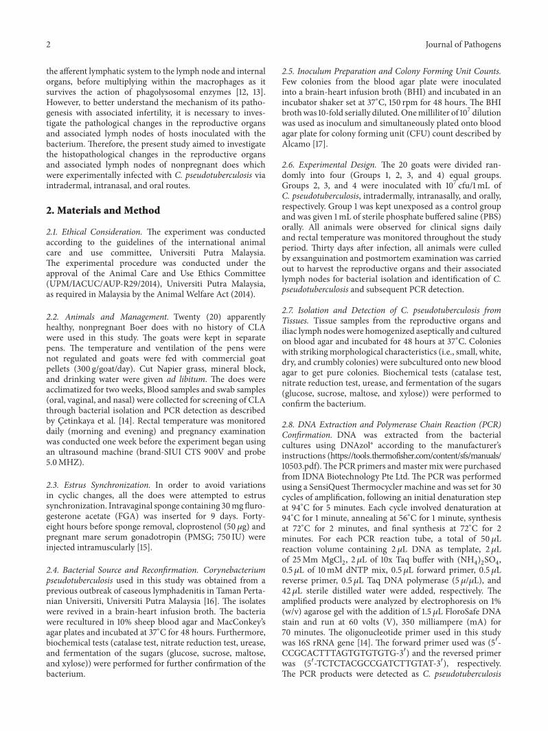
Figure 1 Photomicrograph section of the ovary of nonpregnant does inoculated with Corynebacterium pseudotuberculosis (a) inflammatoryand necrotic cells (circle) following intranasal inoculation (b) inflammatory and necrotic cells (circle) congestion (brown arrows) followingoral inoculation (c) congestion (black arrows) inflammatory and necrotic cells (black circle) following intradermal inoculation (HampEtimes200)
Table 2 Mean scores of histopathological changes in uterus of nonpregnant Boer does experimentally inoculated withC pseudotuberculosis
Parameters GroupControl Intradermal Intranasal Oral
Necrosis 00 plusmn 000a 030 plusmn 048a 300 plusmn 000b 100 plusmn 000c
Inflammatory cell 00 plusmn 000a 10 plusmn 000b 250 plusmn 053c 150 plusmn 053b
Congestion 00 plusmn 000a 120 plusmn 092b 300 plusmn 000c 200 plusmn 000b
Edema 00 plusmn 000a 080 plusmn 042b 280 plusmn 042c 260 plusmn 069cabcMeans with different superscripts within the same row are significantly different from each other at 119901 lt 005
stromal cells of the ovary Changes observed in the ovaries ofall does inoculated through the different routes can be asso-ciated with severe biological activity such as dermonecrosiscomplete capillary destruction caused by the bacterial exo-toxin [22ndash24] However this explains the presence of necrosisand congestion of the blood vessel in the present studyEdema found in the uterus of all goats after inoculation mayalso be due to the presence of the exotoxin phospholipaseD (PLD) Carne and Onon [25] stated that hydrolysis ofsphingomyelin and leakage of plasma protein into surround-ing tissues may occur as a result of increase in vascularendothelial membrane permeability In our previous study[7] we investigated the changes in reproductive hormonallevels of nonpregnant does inoculated with live C pseudo-tuberculosis and reported increased levels of progesteroneand estrogen thirty days after infection in all inoculation
groups These hormonal changes could be attributed to thepathological conditions we observed in the ovary in thisstudy The abnormal increase of progesterone may also betranslated as a sign of pseudo pregnancy thus impairingnormal follicular development and ovulation Foster [26]stated that stated that pseudo pregnancy in nonpregnantanimals may result from mucometra or hydrometra in theuterus which may lead to increase in progesterone secretionMicroabscesses were observed in the iliac lymph nodes andare considered among the typical signs of the disease Thesefindings are supported by Adza Rina et al [6] who foundthat goats inoculated with 107 concentrations of live C pseu-dotuberculosis through intranasal route developed abscessesin the mesenteric and supramammary lymph nodes com-pared to oral and intradermal routes Similarly Mahmoodet al [27] observed severe abscess formation congestion
Journal of Pathogens 5
100120583m
(a)
60120583m
(b)
10120583m
(c)
Figure 2 Photomicrograph section of the uterus of nonpregnant does inoculated with Corynebacterium pseudotuberculosis Note (a) theinflammatory and necrotic cells (brown arrows) degeneration of endometrial glandular epithelium (blue arrows) following intranasalinoculation (b) degeneration of endometrial glandular cells (black arrows) inflammatory cells (orange arrow) following oral inoculation (c)inflammatory cells in the submucosa (blue circle) degeneration and necrosis of endometrial glandular epithelium (black arrow) followingintradermal inoculation (HampE times200)
Table 3Mean scores of pathological changes in lymph node of nonpregnant Boer does experimentally inoculated withC pseudotuberculosis
Parameters GroupControl Intradermal Intranasal Oral
Abscess 000 plusmn 00a 220 plusmn 042b 300 plusmn 00c 230 plusmn 048b
Inflammatory cells 000 plusmn 00a 160 plusmn 052b 300 plusmn 00c 300 plusmn 000c
Congestion 000 plusmn 00a 160 plusmn 052b 300 plusmn 00c 300 plusmn 00cabcMeans with different superscripts within the same row are significantly different from each other at 119901 lt 005
hemorrhage degeneration and necrosis and inflammatorycellular infiltration in Boer goats subcutaneously inoculatedwith C pseudotuberculosis as compared to those inoculatedintravenously with its phospholipase D However all of thesestudies failed to investigate the association of the iliac lymphnodes and pathological changes in the female reproductiveorgans such as the uterus and ovaries
Bacterial isolation showed presence of the organism inall the tissues following oral and intranasal inoculation butonly in the lymph node following intradermal inoculationSince macrophages and dendritic cells are the first to comein contact with an organism following inoculation throughthe intradermal route these phagocytes likely engulfed thebacteria and were carried to the lymph nodes through thelymphatic drainage and the bacteria eventually multiplied inthe nodes in the other tissues Valli and Parry [28] reported
that CLA lesions are normally present in the internal organsudder and less common in the uterus testis and scrotum Ina related study Junior et al [29] reported the developmentof mastitis and increased systemic neutrophilia followingintradermal inoculation ofC pseudotuberculosis in themam-mary gland of goats The researchers also observed enlargedsupramammary lymph nodes without abscess formation
5 Conclusion
This study showed that the intranasal route was the mosteffective route for CLA infection of the reproductive systemas evidenced by pathological lesions observed in these tissuesThis study further corroborates with our previous findingsthat CLA infection in nonpregnant does may lead to infer-tility resulting from pathological changes in the reproductive
6 Journal of Pathogens
50120583m
(a)
50120583m
(b)
50120583m
(c)
Figure 3 Photomicrograph section of the lymph node of nonpregnant does inoculated with Corynebacterium pseudotuberculosis Note (a)the neutrophil infiltration and microabscesses with a central necrotic areas (black and orange arrows) following intranasal inoculation(b) inflammatory cell infiltration characterized by neutrophils and macrophages (black arrows) congestion (blue arrow) following oralinoculation (c) neutrophil infiltration andmicroabscesses with central necrotic areas (black arrows) following intradermal inoculation (HampEtimes400)
organs as well as an imbalance in reproductive hormonallevels
Conflict of Interests
The authors have no conflict of interests to declare
Acknowledgments
Theauthorswish to thankMrMohammed Jefri BinNorsidinMr Yap Keng Chee Puan Latifah Mohd Hanan and PuanJamilah Jahari for their technical assistanceThe research wasfunded by Ministry of Education (MOE) Malaysia
References
[1] M C Fontaine and G J Baird ldquoCaseous lymphadenitisrdquo SmallRuminant Research vol 76 no 1-2 pp 42ndash48 2008
[2] F F A Jesse S L Sang A A Saharee and S ShahirudinldquoPathological changes in the organs of mice model inoculatedwith Corynebacterium pseudotuberculosis organismrdquo PertanikaJournal of Tropical Agricultural Science vol 34 no 1 pp 145ndash149 2011
[3] A C Goldberger B A Lipsky and J J Plorde ldquoSuppurativegranulomatous lymphadenitis caused by Corynebacterium ovis(pseudotuberculosis)rdquo American Journal of Clinical Pathologyvol 76 no 4 pp 486ndash490 1981
[4] S G Stoops H W Renshaw and J P Thilsted ldquoOvine caseouslymphadenitis disease prevalence lesion distribution and tho-racic manifestations in a population of mature culled sheepfrom western United Statesrdquo American Journal of VeterinaryResearch vol 45 no 3 pp 557ndash561 1984
[5] M G Collett G F Bath and C M Cameron ldquoCorynebac-terium pseudotuberculosis infectionsrdquo in Infectious Diseases ofLivestock with Special Reference to Southern Africa J CoetzerG R Thomson and R C Tustin Eds pp 1387ndash1395 OxfordUniversity Press Cape Town South Africa 2nd edition 1994
[6] M N Adza Rina M Zamri-Saad F F A Jesse A A SahareeA W Haron and S Shahirudin ldquoClinical and pathologicalchanges in goats inoculated Corynebacterium pseudotubercu-losis by intradermal intranasal and oral routesrdquo Journal ofVeterinary Research vol 17 no 2 pp 73ndash81 2013
[7] AMOthman F F A Jesse L Adamu et al ldquoChanges in serumprogesterone and estrogen concentrations in non-pregnantboer does following experimental infection with Corynebac-terium pseudotuberculosisrdquo Journal of Veterinary Advances vol5 pp 524ndash528 2014
[8] L HWilliamson ldquoClinical small ruminantsrdquoVeterinary Clinicsof North America vol 17 pp 359ndash371 2001
[9] MW Paton S BWalker I R Rose andG FWatt ldquoPrevalenceof caseous lymphadenitis and usage of caseous lymphadenitisvaccines in sheep flocksrdquo Australian Veterinary Journal vol 81no 1-2 pp 91ndash95 2003
Journal of Pathogens 7
[10] J Arsenault C Girard P Dubreuil et al ldquoPrevalence of andcarcass condemnation from maedi-visna paratuberculosis andcaseous lymphadenitis in culled sheep from Quebec CanadardquoPreventive Veterinary Medicine vol 59 no 1-2 pp 67ndash81 2003
[11] E Peterhans T Greenland J Badiola et al ldquoRoutes of transmis-sion and consequences of small ruminant lentiviruses (SRLVs)infection and eradication schemesrdquoVeterinary Research vol 35no 3 pp 257ndash274 2004
[12] F A Dorella L G C Pacheco S C Oliveira AMiyoshi and VAzevedo ldquoCorynebacterium pseudotuberculosis microbiologybiochemical properties pathogenesis and molecular studies ofvirulencerdquo Veterinary Research vol 37 no 2 pp 201ndash218 2006
[13] G J Baird and M C Fontaine ldquoCorynebacterium pseudotuber-culosis and its role in ovine caseous lymphadenitisrdquo Journal ofComparative Pathology vol 137 no 4 pp 179ndash210 2007
[14] B Cetinkaya M Karahan E Atil R Kalin T De Baere andM Vaneechoutte ldquoIdentification of Corynebacterium pseudo-tuberculosis isolates from sheep and goats by PCRrdquo VeterinaryMicrobiology vol 88 no 1 pp 75ndash83 2002
[15] V J F Freitas G Baril and J Saumande ldquoEstrus synchroniza-tion in dairy goats use of fluorogestone acetate vaginal spongesor norgestomet ear implantsrdquoAnimal Reproduction Science vol46 no 3-4 pp 237ndash244 1997
[16] F F A Jesse C M Azlan A A Saharee et al ldquoControlof Caseous Lymphadenitis (CLA) in goat at UPM Farmrdquo inProceedings of the 20th Veterinary Association Malaysia (VAMrsquo08) 2008
[17] I E Alcamo Fundamentals of Microbiology Addison WesleyLongman 5th edition 1998
[18] J D Bancroft and A Stevens ldquoThe haematoxylin and eosinrdquo inTheory and Practice of Histology Techniques vol 4th chapter 6pp 99ndash112 1996
[19] C W Henry R B N Brewer and S A Edger ldquoStudies oninfectious bursal diseases in chickensrdquo Poultry Science vol 59pp 1006ndash1017 1980
[20] M Hair-Bejo S Salina H Hafiza and S Julaida ldquoIn ovo vac-cination against infectious bursal disease in broiler chickensrdquoJournal of Veterinary Malaysia vol 12 pp 63ndash69 2000
[21] Z Khuder Y O Abdulnasir F F A Jesse et al ldquoSex hormoneprofiles and cellular changes of reproductive organs of miceexperimentally infected with C pseudotuberculosis and itsexotoxin phospholipase D (PLD)rdquo IOSR Journal of Agricultureand Veterinary Science vol 1 no 3 pp 24ndash29 2012
[22] H R Carne ldquoThe toxin of Corynebacterium ovisrdquo The Journalof Pathology and Bacteriology vol 51 no 2 pp 199ndash212 1940
[23] C AMuckle and C L Gyles ldquoExotoxic activities ofCorynebac-terium pseudotuberculosisrdquo Current Microbiology vol 13 no 2pp 57ndash60 1986
[24] K A Brogden and R L Engen ldquoAlterations in the phospholipidcomposition and morphology of ovine erythrocytes after intra-venous inoculation of Corynebacterium pseudotuberculosisrdquoAmerican Journal of Veterinary Research vol 51 no 6 pp 874ndash877 1990
[25] H R Carne and E O Onon ldquoAction of Corynebacterium ovisexotoxin on endothelial cells of blood vesselsrdquo Nature vol 271no 5642 pp 246ndash248 1978
[26] R A Foster ldquoFemale reproductive system and mammaryglandrdquo in Pathologic Basis of Veterinary Disease J F Zacharyand M D McGavin Eds p 1103 Elsevier Health Sciences 5thedition 2013
[27] Z K H Mahmood F F Jesse A A Saharee S Jasni RYusoff and H Wahid ldquoClinio-pathological changes in goatschallenged with Corynebacterium Peudotuberculosis and itsexotoxin (PLD)rdquo American Journal of Animal and VeterinarySciences vol 10 no 3 pp 112ndash132 2015
[28] V E O Valli and B W Parry ldquoCaseous lymphadenitisrdquo inPathology of Domestic Animals K V F Jubb P C Kennedy andN Palmer Eds vol 3 pp 238ndash240 Academic Press SanDiegoCalif USA 4th edition 1993
[29] J P Junior A A F Oliveira F S F Alves L B G Silva S S ARabelo and R A Mota ldquoCorynebacterium pseudotuberculosisexperimental infection of goats mammary glandrdquo Arquivos doInstituto Biologico vol 73 no 4 pp 395ndash400 2006
Submit your manuscripts athttpwwwhindawicom
Stem CellsInternational
Hindawi Publishing Corporationhttpwwwhindawicom Volume 2014
Hindawi Publishing Corporationhttpwwwhindawicom Volume 2014
MEDIATORSINFLAMMATION
of
Hindawi Publishing Corporationhttpwwwhindawicom Volume 2014
Behavioural Neurology
EndocrinologyInternational Journal of
Hindawi Publishing Corporationhttpwwwhindawicom Volume 2014
Hindawi Publishing Corporationhttpwwwhindawicom Volume 2014
Disease Markers
Hindawi Publishing Corporationhttpwwwhindawicom Volume 2014
BioMed Research International
OncologyJournal of
Hindawi Publishing Corporationhttpwwwhindawicom Volume 2014
Hindawi Publishing Corporationhttpwwwhindawicom Volume 2014
Oxidative Medicine and Cellular Longevity
Hindawi Publishing Corporationhttpwwwhindawicom Volume 2014
PPAR Research
The Scientific World JournalHindawi Publishing Corporation httpwwwhindawicom Volume 2014
Immunology ResearchHindawi Publishing Corporationhttpwwwhindawicom Volume 2014
Journal of
ObesityJournal of
Hindawi Publishing Corporationhttpwwwhindawicom Volume 2014
Hindawi Publishing Corporationhttpwwwhindawicom Volume 2014
Computational and Mathematical Methods in Medicine
OphthalmologyJournal of
Hindawi Publishing Corporationhttpwwwhindawicom Volume 2014
Diabetes ResearchJournal of
Hindawi Publishing Corporationhttpwwwhindawicom Volume 2014
Hindawi Publishing Corporationhttpwwwhindawicom Volume 2014
Research and TreatmentAIDS
Hindawi Publishing Corporationhttpwwwhindawicom Volume 2014
Gastroenterology Research and Practice
Hindawi Publishing Corporationhttpwwwhindawicom Volume 2014
Parkinsonrsquos Disease
Evidence-Based Complementary and Alternative Medicine
Volume 2014Hindawi Publishing Corporationhttpwwwhindawicom
2 Journal of Pathogens
the afferent lymphatic system to the lymph node and internalorgans before multiplying within the macrophages as itsurvives the action of phagolysosomal enzymes [12 13]However to better understand the mechanism of its patho-genesis with associated infertility it is necessary to inves-tigate the pathological changes in the reproductive organsand associated lymph nodes of hosts inoculated with thebacterium Therefore the present study aimed to investigatethe histopathological changes in the reproductive organsand associated lymph nodes of nonpregnant does whichwere experimentally infected with C pseudotuberculosis viaintradermal intranasal and oral routes
2 Materials and Method
21 Ethical Consideration The experiment was conductedaccording to the guidelines of the international animalcare and use committee Universiti Putra MalaysiaThe experimental procedure was conducted under theapproval of the Animal Care and Use Ethics Committee(UPMIACUCAUP-R292014) Universiti Putra Malaysiaas required in Malaysia by the Animal Welfare Act (2014)
22 Animals and Management Twenty (20) apparentlyhealthy nonpregnant Boer does with no history of CLAwere used in this study The goats were kept in separatepens The temperature and ventilation of the pens werenot regulated and goats were fed with commercial goatpellets (300 ggoatday) Cut Napier grass mineral blockand drinking water were given ad libitum The does wereacclimatized for two weeks Blood samples and swab samples(oral vaginal and nasal) were collected for screening of CLAthrough bacterial isolation and PCR detection as describedby Cetinkaya et al [14] Rectal temperature was monitoreddaily (morning and evening) and pregnancy examinationwas conducted one week before the experiment began usingan ultrasound machine (brand-SIUI CTS 900V and probe50MHZ)
23 Estrus Synchronization In order to avoid variationsin cyclic changes all the does were attempted to estrussynchronization Intravaginal sponge containing 30mgfluro-gesterone acetate (FGA) was inserted for 9 days Forty-eight hours before sponge removal cloprostenol (50 120583g) andpregnant mare serum gonadotropin (PMSG 750 IU) wereinjected intramuscularly [15]
24 Bacterial Source and Reconfirmation Corynebacteriumpseudotuberculosis used in this study was obtained from aprevious outbreak of caseous lymphadenitis in Taman Perta-nian Universiti Universiti Putra Malaysia [16] The isolateswere revived in a brain-heart infusion broth The bacteriawere recultured in 10 sheep blood agar and MacConkeyrsquosagar plates and incubated at 37∘C for 48 hours Furthermorebiochemical tests (catalase test nitrate reduction test ureaseand fermentation of the sugars (glucose sucrose maltoseand xylose)) were performed for further confirmation of thebacterium
25 Inoculum Preparation and Colony Forming Unit CountsFew colonies from the blood agar plate were inoculatedinto a brain-heart infusion broth (BHI) and incubated in anincubator shaker set at 37∘C 150 rpm for 48 hours The BHIbrothwas 10-fold serially dilutedOnemilliliter of 107 dilutionwas used as inoculum and simultaneously plated onto bloodagar plate for colony forming unit (CFU) count described byAlcamo [17]
26 Experimental Design The 20 goats were divided ran-domly into four (Groups 1 2 3 and 4) equal groupsGroups 2 3 and 4 were inoculated with 107 cfu1mL ofC pseudotuberculosis intradermally intranasally and orallyrespectively Group 1 was kept unexposed as a control groupand was given 1mL of sterile phosphate buffered saline (PBS)orally All animals were observed for clinical signs dailyand rectal temperature was monitored throughout the studyperiod Thirty days after infection all animals were culledby exsanguination and postmortem examination was carriedout to harvest the reproductive organs and their associatedlymph nodes for bacterial isolation and identification of Cpseudotuberculosis and subsequent PCR detection
27 Isolation and Detection of C pseudotuberculosis fromTissues Tissue samples from the reproductive organs andiliac lymph nodes were homogenized aseptically and culturedon blood agar and incubated for 48 hours at 37∘C Colonieswith strikingmorphological characteristics (ie small whitedry and crumbly colonies) were subcultured onto new bloodagar to get pure colonies Biochemical tests (catalase testnitrate reduction test urease and fermentation of the sugars(glucose sucrose maltose and xylose)) were performed toconfirm the bacterium
28 DNA Extraction and Polymerase Chain Reaction (PCR)Confirmation DNA was extracted from the bacterialcultures using DNAzolreg according to the manufacturerrsquosinstructions (httpstoolsthermofishercomcontentsfsmanuals10503pdf)The PCR primers andmaster mix were purchasedfrom IDNA Biotechnology Pte Ltd The PCR was performedusing a SensiQuestThermocycler machine and was set for 30cycles of amplification following an initial denaturation stepat 94∘C for 5 minutes Each cycle involved denaturation at94∘C for 1 minute annealing at 56∘C for 1 minute synthesisat 72∘C for 2 minutes and final synthesis at 72∘C for 2minutes For each PCR reaction tube a total of 50 120583Lreaction volume containing 2 120583L DNA as template 2120583Lof 25Mm MgCl
2 2 120583L of 10x Taq buffer with (NH
4)2SO4
05 120583L of 10mM dNTP mix 05120583L forward primer 05120583Lreverse primer 05 120583L Taq DNA polymerase (5 120583120583L) and42 120583L sterile distilled water were added respectively Theamplified products were analyzed by electrophoresis on 1(wv) agarose gel with the addition of 15 120583L FloroSafe DNAstain and run at 60 volts (V) 350 milliampere (mA) for70 minutes The oligonucleotide primer used in this studywas 16S rRNA gene [14] The forward primer used was (51015840-CCGCACTTTAGTGTGTGTG-31015840) and the reversed primerwas (51015840-TCTCTACGCCGATCTTGTAT-31015840) respectivelyThe PCR products were detected as C pseudotuberculosis
Journal of Pathogens 3
Table 1 Mean scores of histopathological changes in the ovaries of nonpregnant Boer does experimentally inoculated with Cpseudotuberculosis
Parameters Inoculation groupsControl Intradermal Intranasal Oral
Necrosis 00 plusmn 000a 240 plusmn 069b 260 plusmn 052b 240 plusmn 052b
Inflammatory cells 00 plusmn 000a 10 plusmn 000b 290 plusmn 032c 250 plusmn 053c
Congestion 00 plusmn 000a 180 plusmn 042b 240 plusmn 052b 150 plusmn 053cabcMeans with different superscripts within the same row are significantly different from each other at 119901 lt 005
according to molecular size of 816 bp as documented byCetinkaya et al [14]
29 Histopathology The isolated reproductive organs (ovaryand uterus) and the associated lymph node (iliac lymphnode) were collected and fixed at 10 buffered formalinfor 3 days before processing The tissues were processed inan automatic tissue processor (Leica TP 1020 Germany)and then embedded in paraffin blocks sectioned at 5 120583 andstained with standard hematoxylin and eosin (HampE) stain forlight microscopic evaluation [18]
210 Histological Lesion Scoring Based on the methodsdescribed by Henry et al [19] and Hair-Bejo et al [20] thehistopathological scoring was performed following observa-tion of ten slides from each organ About six microscopicareas for each slide were observed at different magnification(200x and 400x) The lesion of the three inoculated groupsand control was scored on a scale of 0ndash3 based on the presenceof oedema necrosis congestion infiltration of neutrophilsdegeneration and necrosis Score 0 indicates normal (nolesion observed) 1 mild (less than 30 lesion observed) 2moderate (less than 60 of the lesion observed) and 3 severe(more than 60 of the lesion observed)
211 Statistical Analysis Data were analyzed using statisticalsoftware Graph Pad Prism (version 60) One way ANOVAwith Kruskal-Wallis nonparametric test was used to test thedifferences between the lesion scores in the different inocula-tion groups The differences were considered as significant at119901 lt 005
3 Results
31 Bacterial Reconfirmation The biochemical test carriedout confirmed the bacteria as C pseudotuberculosis Theresults were catalase positive nitrate reduction and ureasepositive
32 PCR Confirmation and Analysis The 816 bp segment ofthe 16S RNA is unique to C pseudotuberculosis and wasdetected by PCR Bacterial isolations from tissues of goats(ovary uterus and iliac lymph nodes) inoculated through theintranasal and oral inoculation routes tested positive for the16S RNA while goatsrsquo inoculated intradermally tested PCRpositive only from bacterial cultures of the iliac lymph node
33Histopathological Changes Themean changes in necrosisand congestion in the ovaries were moderate to severe inall groups and were not significant (119901 gt 005) between thethree inoculation groups (Table 1) However inflammatorycell infiltration in the ovarywasmild in the intradermal groupand moderate to severe in both oral and intranasal groups(Figures 1(a) 1(b) and 1(c))
The mean changes in necrosis inflammatory cell andcongestion in the uterus were mild to moderate followingintradermal and oral inoculation and moderate to severefollowing intranasal inoculation (Table 2) These changeswere significantly higher (119901 lt 005) in the intranasal groupand comparable in the intradermal and oral groups On theother hand edema was comparable in the uterus followingintranasal and oral inoculation (119901 gt 005) and significantlylower (119901 lt 005) in the intradermal group (Figures 2(a) 2(b)and 2(c))
In the iliac lymph nodes distribution of abscess wasmod-erate to severe and comparable (119901 gt 005) in the intradermaland oral groups while it was severe in the intranasal group(Table 3) However distribution of inflammatory cells andcongestion is mild to moderate in the intradermal groupwhile it was severe and comparable in both intranasal and oralinoculation groups (Figures 3(a) 3(b) and 3(c))
4 Discussion
The present study reports for the first time the changesobserved in the reproductive organs and associated lymphnodes in nonpregnant does experimentally infected withC pseudotuberculosis via intradermal intranasal and oralroutes All animals inoculatedwith the organism through dif-ferent routes developed significant histopathological changesin the reproductive organs compared to the control groupshowever goats inoculated through the intranasal routeshowed severe lesion in the reproductive organs compared toother routes
The histopathological lesions we observed in the ovarywere comparable with those observed in the uterine hornThese lesions may have been induced by either the bacteriaor its phospholipase D and might explain the hormonalalterations observed in our earlier study [7] All the lesionsobserved in the ovary in the present study were similarto those described by Khuder et al [21] in female miceexperimentally inoculated with C pseudotuberculosis and itsphospholipase D (PLD) The authors observed infiltration ofleukocytes in the lumen of ovulated follicles and generalizedcongestion thrombosis degeneration and necrosis of the
4 Journal of Pathogens
100120583m
(a)
100120583m
(b)
100120583m
(c)
Figure 1 Photomicrograph section of the ovary of nonpregnant does inoculated with Corynebacterium pseudotuberculosis (a) inflammatoryand necrotic cells (circle) following intranasal inoculation (b) inflammatory and necrotic cells (circle) congestion (brown arrows) followingoral inoculation (c) congestion (black arrows) inflammatory and necrotic cells (black circle) following intradermal inoculation (HampEtimes200)
Table 2 Mean scores of histopathological changes in uterus of nonpregnant Boer does experimentally inoculated withC pseudotuberculosis
Parameters GroupControl Intradermal Intranasal Oral
Necrosis 00 plusmn 000a 030 plusmn 048a 300 plusmn 000b 100 plusmn 000c
Inflammatory cell 00 plusmn 000a 10 plusmn 000b 250 plusmn 053c 150 plusmn 053b
Congestion 00 plusmn 000a 120 plusmn 092b 300 plusmn 000c 200 plusmn 000b
Edema 00 plusmn 000a 080 plusmn 042b 280 plusmn 042c 260 plusmn 069cabcMeans with different superscripts within the same row are significantly different from each other at 119901 lt 005
stromal cells of the ovary Changes observed in the ovaries ofall does inoculated through the different routes can be asso-ciated with severe biological activity such as dermonecrosiscomplete capillary destruction caused by the bacterial exo-toxin [22ndash24] However this explains the presence of necrosisand congestion of the blood vessel in the present studyEdema found in the uterus of all goats after inoculation mayalso be due to the presence of the exotoxin phospholipaseD (PLD) Carne and Onon [25] stated that hydrolysis ofsphingomyelin and leakage of plasma protein into surround-ing tissues may occur as a result of increase in vascularendothelial membrane permeability In our previous study[7] we investigated the changes in reproductive hormonallevels of nonpregnant does inoculated with live C pseudo-tuberculosis and reported increased levels of progesteroneand estrogen thirty days after infection in all inoculation
groups These hormonal changes could be attributed to thepathological conditions we observed in the ovary in thisstudy The abnormal increase of progesterone may also betranslated as a sign of pseudo pregnancy thus impairingnormal follicular development and ovulation Foster [26]stated that stated that pseudo pregnancy in nonpregnantanimals may result from mucometra or hydrometra in theuterus which may lead to increase in progesterone secretionMicroabscesses were observed in the iliac lymph nodes andare considered among the typical signs of the disease Thesefindings are supported by Adza Rina et al [6] who foundthat goats inoculated with 107 concentrations of live C pseu-dotuberculosis through intranasal route developed abscessesin the mesenteric and supramammary lymph nodes com-pared to oral and intradermal routes Similarly Mahmoodet al [27] observed severe abscess formation congestion
Journal of Pathogens 5
100120583m
(a)
60120583m
(b)
10120583m
(c)
Figure 2 Photomicrograph section of the uterus of nonpregnant does inoculated with Corynebacterium pseudotuberculosis Note (a) theinflammatory and necrotic cells (brown arrows) degeneration of endometrial glandular epithelium (blue arrows) following intranasalinoculation (b) degeneration of endometrial glandular cells (black arrows) inflammatory cells (orange arrow) following oral inoculation (c)inflammatory cells in the submucosa (blue circle) degeneration and necrosis of endometrial glandular epithelium (black arrow) followingintradermal inoculation (HampE times200)
Table 3Mean scores of pathological changes in lymph node of nonpregnant Boer does experimentally inoculated withC pseudotuberculosis
Parameters GroupControl Intradermal Intranasal Oral
Abscess 000 plusmn 00a 220 plusmn 042b 300 plusmn 00c 230 plusmn 048b
Inflammatory cells 000 plusmn 00a 160 plusmn 052b 300 plusmn 00c 300 plusmn 000c
Congestion 000 plusmn 00a 160 plusmn 052b 300 plusmn 00c 300 plusmn 00cabcMeans with different superscripts within the same row are significantly different from each other at 119901 lt 005
hemorrhage degeneration and necrosis and inflammatorycellular infiltration in Boer goats subcutaneously inoculatedwith C pseudotuberculosis as compared to those inoculatedintravenously with its phospholipase D However all of thesestudies failed to investigate the association of the iliac lymphnodes and pathological changes in the female reproductiveorgans such as the uterus and ovaries
Bacterial isolation showed presence of the organism inall the tissues following oral and intranasal inoculation butonly in the lymph node following intradermal inoculationSince macrophages and dendritic cells are the first to comein contact with an organism following inoculation throughthe intradermal route these phagocytes likely engulfed thebacteria and were carried to the lymph nodes through thelymphatic drainage and the bacteria eventually multiplied inthe nodes in the other tissues Valli and Parry [28] reported
that CLA lesions are normally present in the internal organsudder and less common in the uterus testis and scrotum Ina related study Junior et al [29] reported the developmentof mastitis and increased systemic neutrophilia followingintradermal inoculation ofC pseudotuberculosis in themam-mary gland of goats The researchers also observed enlargedsupramammary lymph nodes without abscess formation
5 Conclusion
This study showed that the intranasal route was the mosteffective route for CLA infection of the reproductive systemas evidenced by pathological lesions observed in these tissuesThis study further corroborates with our previous findingsthat CLA infection in nonpregnant does may lead to infer-tility resulting from pathological changes in the reproductive
6 Journal of Pathogens
50120583m
(a)
50120583m
(b)
50120583m
(c)
Figure 3 Photomicrograph section of the lymph node of nonpregnant does inoculated with Corynebacterium pseudotuberculosis Note (a)the neutrophil infiltration and microabscesses with a central necrotic areas (black and orange arrows) following intranasal inoculation(b) inflammatory cell infiltration characterized by neutrophils and macrophages (black arrows) congestion (blue arrow) following oralinoculation (c) neutrophil infiltration andmicroabscesses with central necrotic areas (black arrows) following intradermal inoculation (HampEtimes400)
organs as well as an imbalance in reproductive hormonallevels
Conflict of Interests
The authors have no conflict of interests to declare
Acknowledgments
Theauthorswish to thankMrMohammed Jefri BinNorsidinMr Yap Keng Chee Puan Latifah Mohd Hanan and PuanJamilah Jahari for their technical assistanceThe research wasfunded by Ministry of Education (MOE) Malaysia
References
[1] M C Fontaine and G J Baird ldquoCaseous lymphadenitisrdquo SmallRuminant Research vol 76 no 1-2 pp 42ndash48 2008
[2] F F A Jesse S L Sang A A Saharee and S ShahirudinldquoPathological changes in the organs of mice model inoculatedwith Corynebacterium pseudotuberculosis organismrdquo PertanikaJournal of Tropical Agricultural Science vol 34 no 1 pp 145ndash149 2011
[3] A C Goldberger B A Lipsky and J J Plorde ldquoSuppurativegranulomatous lymphadenitis caused by Corynebacterium ovis(pseudotuberculosis)rdquo American Journal of Clinical Pathologyvol 76 no 4 pp 486ndash490 1981
[4] S G Stoops H W Renshaw and J P Thilsted ldquoOvine caseouslymphadenitis disease prevalence lesion distribution and tho-racic manifestations in a population of mature culled sheepfrom western United Statesrdquo American Journal of VeterinaryResearch vol 45 no 3 pp 557ndash561 1984
[5] M G Collett G F Bath and C M Cameron ldquoCorynebac-terium pseudotuberculosis infectionsrdquo in Infectious Diseases ofLivestock with Special Reference to Southern Africa J CoetzerG R Thomson and R C Tustin Eds pp 1387ndash1395 OxfordUniversity Press Cape Town South Africa 2nd edition 1994
[6] M N Adza Rina M Zamri-Saad F F A Jesse A A SahareeA W Haron and S Shahirudin ldquoClinical and pathologicalchanges in goats inoculated Corynebacterium pseudotubercu-losis by intradermal intranasal and oral routesrdquo Journal ofVeterinary Research vol 17 no 2 pp 73ndash81 2013
[7] AMOthman F F A Jesse L Adamu et al ldquoChanges in serumprogesterone and estrogen concentrations in non-pregnantboer does following experimental infection with Corynebac-terium pseudotuberculosisrdquo Journal of Veterinary Advances vol5 pp 524ndash528 2014
[8] L HWilliamson ldquoClinical small ruminantsrdquoVeterinary Clinicsof North America vol 17 pp 359ndash371 2001
[9] MW Paton S BWalker I R Rose andG FWatt ldquoPrevalenceof caseous lymphadenitis and usage of caseous lymphadenitisvaccines in sheep flocksrdquo Australian Veterinary Journal vol 81no 1-2 pp 91ndash95 2003
Journal of Pathogens 7
[10] J Arsenault C Girard P Dubreuil et al ldquoPrevalence of andcarcass condemnation from maedi-visna paratuberculosis andcaseous lymphadenitis in culled sheep from Quebec CanadardquoPreventive Veterinary Medicine vol 59 no 1-2 pp 67ndash81 2003
[11] E Peterhans T Greenland J Badiola et al ldquoRoutes of transmis-sion and consequences of small ruminant lentiviruses (SRLVs)infection and eradication schemesrdquoVeterinary Research vol 35no 3 pp 257ndash274 2004
[12] F A Dorella L G C Pacheco S C Oliveira AMiyoshi and VAzevedo ldquoCorynebacterium pseudotuberculosis microbiologybiochemical properties pathogenesis and molecular studies ofvirulencerdquo Veterinary Research vol 37 no 2 pp 201ndash218 2006
[13] G J Baird and M C Fontaine ldquoCorynebacterium pseudotuber-culosis and its role in ovine caseous lymphadenitisrdquo Journal ofComparative Pathology vol 137 no 4 pp 179ndash210 2007
[14] B Cetinkaya M Karahan E Atil R Kalin T De Baere andM Vaneechoutte ldquoIdentification of Corynebacterium pseudo-tuberculosis isolates from sheep and goats by PCRrdquo VeterinaryMicrobiology vol 88 no 1 pp 75ndash83 2002
[15] V J F Freitas G Baril and J Saumande ldquoEstrus synchroniza-tion in dairy goats use of fluorogestone acetate vaginal spongesor norgestomet ear implantsrdquoAnimal Reproduction Science vol46 no 3-4 pp 237ndash244 1997
[16] F F A Jesse C M Azlan A A Saharee et al ldquoControlof Caseous Lymphadenitis (CLA) in goat at UPM Farmrdquo inProceedings of the 20th Veterinary Association Malaysia (VAMrsquo08) 2008
[17] I E Alcamo Fundamentals of Microbiology Addison WesleyLongman 5th edition 1998
[18] J D Bancroft and A Stevens ldquoThe haematoxylin and eosinrdquo inTheory and Practice of Histology Techniques vol 4th chapter 6pp 99ndash112 1996
[19] C W Henry R B N Brewer and S A Edger ldquoStudies oninfectious bursal diseases in chickensrdquo Poultry Science vol 59pp 1006ndash1017 1980
[20] M Hair-Bejo S Salina H Hafiza and S Julaida ldquoIn ovo vac-cination against infectious bursal disease in broiler chickensrdquoJournal of Veterinary Malaysia vol 12 pp 63ndash69 2000
[21] Z Khuder Y O Abdulnasir F F A Jesse et al ldquoSex hormoneprofiles and cellular changes of reproductive organs of miceexperimentally infected with C pseudotuberculosis and itsexotoxin phospholipase D (PLD)rdquo IOSR Journal of Agricultureand Veterinary Science vol 1 no 3 pp 24ndash29 2012
[22] H R Carne ldquoThe toxin of Corynebacterium ovisrdquo The Journalof Pathology and Bacteriology vol 51 no 2 pp 199ndash212 1940
[23] C AMuckle and C L Gyles ldquoExotoxic activities ofCorynebac-terium pseudotuberculosisrdquo Current Microbiology vol 13 no 2pp 57ndash60 1986
[24] K A Brogden and R L Engen ldquoAlterations in the phospholipidcomposition and morphology of ovine erythrocytes after intra-venous inoculation of Corynebacterium pseudotuberculosisrdquoAmerican Journal of Veterinary Research vol 51 no 6 pp 874ndash877 1990
[25] H R Carne and E O Onon ldquoAction of Corynebacterium ovisexotoxin on endothelial cells of blood vesselsrdquo Nature vol 271no 5642 pp 246ndash248 1978
[26] R A Foster ldquoFemale reproductive system and mammaryglandrdquo in Pathologic Basis of Veterinary Disease J F Zacharyand M D McGavin Eds p 1103 Elsevier Health Sciences 5thedition 2013
[27] Z K H Mahmood F F Jesse A A Saharee S Jasni RYusoff and H Wahid ldquoClinio-pathological changes in goatschallenged with Corynebacterium Peudotuberculosis and itsexotoxin (PLD)rdquo American Journal of Animal and VeterinarySciences vol 10 no 3 pp 112ndash132 2015
[28] V E O Valli and B W Parry ldquoCaseous lymphadenitisrdquo inPathology of Domestic Animals K V F Jubb P C Kennedy andN Palmer Eds vol 3 pp 238ndash240 Academic Press SanDiegoCalif USA 4th edition 1993
[29] J P Junior A A F Oliveira F S F Alves L B G Silva S S ARabelo and R A Mota ldquoCorynebacterium pseudotuberculosisexperimental infection of goats mammary glandrdquo Arquivos doInstituto Biologico vol 73 no 4 pp 395ndash400 2006
Submit your manuscripts athttpwwwhindawicom
Stem CellsInternational
Hindawi Publishing Corporationhttpwwwhindawicom Volume 2014
Hindawi Publishing Corporationhttpwwwhindawicom Volume 2014
MEDIATORSINFLAMMATION
of
Hindawi Publishing Corporationhttpwwwhindawicom Volume 2014
Behavioural Neurology
EndocrinologyInternational Journal of
Hindawi Publishing Corporationhttpwwwhindawicom Volume 2014
Hindawi Publishing Corporationhttpwwwhindawicom Volume 2014
Disease Markers
Hindawi Publishing Corporationhttpwwwhindawicom Volume 2014
BioMed Research International
OncologyJournal of
Hindawi Publishing Corporationhttpwwwhindawicom Volume 2014
Hindawi Publishing Corporationhttpwwwhindawicom Volume 2014
Oxidative Medicine and Cellular Longevity
Hindawi Publishing Corporationhttpwwwhindawicom Volume 2014
PPAR Research
The Scientific World JournalHindawi Publishing Corporation httpwwwhindawicom Volume 2014
Immunology ResearchHindawi Publishing Corporationhttpwwwhindawicom Volume 2014
Journal of
ObesityJournal of
Hindawi Publishing Corporationhttpwwwhindawicom Volume 2014
Hindawi Publishing Corporationhttpwwwhindawicom Volume 2014
Computational and Mathematical Methods in Medicine
OphthalmologyJournal of
Hindawi Publishing Corporationhttpwwwhindawicom Volume 2014
Diabetes ResearchJournal of
Hindawi Publishing Corporationhttpwwwhindawicom Volume 2014
Hindawi Publishing Corporationhttpwwwhindawicom Volume 2014
Research and TreatmentAIDS
Hindawi Publishing Corporationhttpwwwhindawicom Volume 2014
Gastroenterology Research and Practice
Hindawi Publishing Corporationhttpwwwhindawicom Volume 2014
Parkinsonrsquos Disease
Evidence-Based Complementary and Alternative Medicine
Volume 2014Hindawi Publishing Corporationhttpwwwhindawicom