RESEARCH ARTICLE Open Access Exploration of microRNAs in … · 2017-04-05 · RESEARCH ARTICLE...
19
RESEARCH ARTICLE Open Access Exploration of microRNAs in porcine milk exosomes Ting Chen † , Qian-Yun Xi † , Rui-Song Ye, Xiao Cheng, Qi-En Qi, Song-Bo Wang, Gang Shu, Li-Na Wang, Xiao-Tong Zhu, Qing-Yan Jiang and Yong-Liang Zhang * Abstract Background: Breast milk contains complex nutrients and facilitates the maturation of various biological systems in infants. Exosomes, membranous vesicles of endocytic origin found in different body fluids such as milk, can mediate intercellular communication. We hypothesized that microRNAs (miRNAs), a class of non-coding small RNAs of 18–25 nt which are known to be packaged in exosomes of human, bovine and porcine milk, may play important roles in the development of piglets. Results: In this study, exosomes of approximately 100 nm in diameter were isolated from porcine milk through serial centrifugation and ultracentrifugation procedures. Total RNA was extracted from exosomes, and 5S ribosomal RNA was found to be the major RNA component. Solexa sequencing showed a total of 491 miRNAs, including 176 known miRNAs and 315 novel mature miRNAs (representing 366 pre-miRNAs), which were distributed among 30 clusters and 35 families, and two predicted novel miRNAs were verified targeting 3’UTR of IGF-1R by luciferase assay. Interestingly, we observed that three miRNAs (ssc-let-7e, ssc-miR-27a, and ssc-miR-30a) could be generated from miRNA-offset RNAs (moRNAs). The top 10 miRNAs accounted for 74.5% (67,154 counts) of total counts, which were predicted to target 2,333 genes by RNAhybrid software. Gene Ontology and Kyoto Encyclopedia of Genes and Genomes (KEGG) pathway analyses using DAVID bioinformatics resources indicated that the identified miRNAs targeted genes enriched in transcription, immunity and metabolism processes, and 14 of the top 20 miRNAs possibly participate in regulation of the IgA immune network. Conclusions: Our findings suggest that porcine milk exosomes contain a large number of miRNAs, which potentially play an important role in information transfer from sow milk to piglets. The predicted miRNAs of porcine milk exosomes in this study provide a basis for future biochemical and biophysical function studies. Keywords: Porcine milk exosomes, Solexa sequencing, miRNA Background Milk, as the sole source of nutrition for infants, contains a potent mixture of diverse components such as milk fat globules (MFG) [1], immune competent cells and soluble proteins, for instance IgA, cytokines and antimicrobial pep- tides [2], which can provide protection against infections in newborns [3]. In addition, breast milk may have a role in tolerance induction [1] and may protect infants from devel- oping allergies [4]. Exosomes are small (30–100 nm) membrane vesicles of endocytic origin that are released into the extracellu- lar environment upon fusion of multivesicular bodies (MVB) with the plasma membrane [5]. Many cells have the capacity to release exosomes, including reticulocytes [6], dendritic cells [7], B cells [8], T cells [9], mast cells [10], epithelial cells [11] and tumor cells [12]. In addition, exosomes have been found in physiological fluids, such as saliva [13,14], plasma [15], urine [16], am- niotic fluid [17], malignant ascites [18], bronchoalveolar lavage fluid [19] and synovial fluids [20]. Several studies have suggested that exosomes, which contain proteins, mRNA and microRNA (miRNA), stimulate and transfer surface receptors to target cells [21-23], as well as serve as novel vehicles for genetic exchange between cells * Correspondence: [email protected] † Equal contributors Guandong Provincial Key Lab of Agro-Animal Genomics And Molecular Breeding, College of Animal Science, ALLTECH-SCAU Animal Nutrition Control Research Alliance, National Engineering Research Center For Breeding Swine Industry, South China Agricultural University, Guangzhou 510642, China © 2014 Chen et al.; licensee BioMed Central Ltd. This is an open access article distributed under the terms of the Creative Commons Attribution License (http://creativecommons.org/licenses/by/2.0), which permits unrestricted use, distribution, and reproduction in any medium, provided the original work is properly cited. Chen et al. BMC Genomics 2014, 15:100 http://www.biomedcentral.com/1471-2164/15/100
Transcript of RESEARCH ARTICLE Open Access Exploration of microRNAs in … · 2017-04-05 · RESEARCH ARTICLE...
Chen et al. BMC Genomics 2014, 15:100http://www.biomedcentral.com/1471-2164/15/100
RESEARCH ARTICLE Open Access
Exploration of microRNAs in porcine milkexosomesTing Chen†, Qian-Yun Xi†, Rui-Song Ye, Xiao Cheng, Qi-En Qi, Song-Bo Wang, Gang Shu, Li-Na Wang,Xiao-Tong Zhu, Qing-Yan Jiang and Yong-Liang Zhang*
Abstract
Background: Breast milk contains complex nutrients and facilitates the maturation of various biological systems ininfants. Exosomes, membranous vesicles of endocytic origin found in different body fluids such as milk, canmediate intercellular communication. We hypothesized that microRNAs (miRNAs), a class of non-coding small RNAsof 18–25 nt which are known to be packaged in exosomes of human, bovine and porcine milk, may play importantroles in the development of piglets.
Results: In this study, exosomes of approximately 100 nm in diameter were isolated from porcine milk throughserial centrifugation and ultracentrifugation procedures. Total RNA was extracted from exosomes, and 5S ribosomalRNA was found to be the major RNA component. Solexa sequencing showed a total of 491 miRNAs, including 176known miRNAs and 315 novel mature miRNAs (representing 366 pre-miRNAs), which were distributed among 30clusters and 35 families, and two predicted novel miRNAs were verified targeting 3’UTR of IGF-1R by luciferase assay.Interestingly, we observed that three miRNAs (ssc-let-7e, ssc-miR-27a, and ssc-miR-30a) could be generated frommiRNA-offset RNAs (moRNAs). The top 10 miRNAs accounted for 74.5% (67,154 counts) of total counts, which werepredicted to target 2,333 genes by RNAhybrid software. Gene Ontology and Kyoto Encyclopedia of Genes andGenomes (KEGG) pathway analyses using DAVID bioinformatics resources indicated that the identified miRNAstargeted genes enriched in transcription, immunity and metabolism processes, and 14 of the top 20 miRNAspossibly participate in regulation of the IgA immune network.
Conclusions: Our findings suggest that porcine milk exosomes contain a large number of miRNAs, which potentiallyplay an important role in information transfer from sow milk to piglets. The predicted miRNAs of porcine milkexosomes in this study provide a basis for future biochemical and biophysical function studies.
Keywords: Porcine milk exosomes, Solexa sequencing, miRNA
BackgroundMilk, as the sole source of nutrition for infants, contains apotent mixture of diverse components such as milk fatglobules (MFG) [1], immune competent cells and solubleproteins, for instance IgA, cytokines and antimicrobial pep-tides [2], which can provide protection against infections innewborns [3]. In addition, breast milk may have a role intolerance induction [1] and may protect infants from devel-oping allergies [4].
* Correspondence: [email protected]†Equal contributorsGuandong Provincial Key Lab of Agro-Animal Genomics And MolecularBreeding, College of Animal Science, ALLTECH-SCAU Animal Nutrition ControlResearch Alliance, National Engineering Research Center For Breeding SwineIndustry, South China Agricultural University, Guangzhou 510642, China
© 2014 Chen et al.; licensee BioMed Central LCommons Attribution License (http://creativecreproduction in any medium, provided the or
Exosomes are small (30–100 nm) membrane vesiclesof endocytic origin that are released into the extracellu-lar environment upon fusion of multivesicular bodies(MVB) with the plasma membrane [5]. Many cells havethe capacity to release exosomes, including reticulocytes[6], dendritic cells [7], B cells [8], T cells [9], mastcells [10], epithelial cells [11] and tumor cells [12]. Inaddition, exosomes have been found in physiologicalfluids, such as saliva [13,14], plasma [15], urine [16], am-niotic fluid [17], malignant ascites [18], bronchoalveolarlavage fluid [19] and synovial fluids [20]. Several studieshave suggested that exosomes, which contain proteins,mRNA and microRNA (miRNA), stimulate and transfersurface receptors to target cells [21-23], as well as serveas novel vehicles for genetic exchange between cells
td. This is an open access article distributed under the terms of the Creativeommons.org/licenses/by/2.0), which permits unrestricted use, distribution, andiginal work is properly cited.
Chen et al. BMC Genomics 2014, 15:100 Page 2 of 19http://www.biomedcentral.com/1471-2164/15/100
[24]. As with other biological fluids, microvesicle-likeparticles are also present in mouse milk [25] and humanmilk [26]. Recent published studies have isolated mRNAsand miRNAs from bovine milk-derived microvesicles [27].One study via deep sequencing technology identified 602unique miRNAs originating from 452 miRNA precursors(pre-miRNAs) in human breast milk exosomes and foundthat, out of 87 well-characterized immune-related pre-miRNAs, 59 (67.82%) were enriched in breast milk exo-somes [28]. Recently, porcine milk was reported to contain180 pre-miRNAs, including 140 known and 40 novel por-cine pre-miRNAs, altogether encoding 237 mature miR-NAs [29].MiRNAs are widespread among eukaryotes and represent
key components of a conserved system of RNA-based generegulation [30-33]. Many studies have demonstrated thatmiRNAs are key post-transcriptional regulators of geneexpression and play important roles in a wide range ofphysiological and pathological processes [34], includingdevelopment, differentiation, proliferation and immune re-sponses. It is believed that about 60% of mammalian genesare regulated by miRNAs [35-39].Aside from being important farm livestock, pigs are
also model animals for medical research. In the presentstudy, we investigated miRNAs in milk exosomes ofLandrace pigs in order to provide new information forinvestigations into the physiological functions of porcinemilk.
MethodsMilk samplesPorcine milk samples were collected between day 1 to 5after parturition from healthy lactating Landrace femalepigs bred in the breeding farm of the Livestock ResearchInstitute (Guangzhou, China). Milk samples were frozenimmediately after milking and were kept at-80°C untiluse.
Preparation of exosomes from milkPorcine milk samples were centrifuged first at 2,000 × g for30 min at 4°C to remove MFGs as well as mammary gland-derived cells. Defatted samples were then subjected tocentrifugations at 4°C for 30 min at 12,000 × g to removeresidual MFGs, casein and other debris. Subsequently, fromthe final supernatant (so-called whey or milk serum), themembrane fraction was prepared by ultracentrifugation at110,000 × g for 2 h in an SW41T rotor (Beckman CoulterInstruments, Fullerton, CA) [40].
Transmission electron microscopy (TEM)The final fraction obtained as described above was di-luted with 0.01 M PBS and ultracentrifuged again torecover microvesicles as pellets. Following fixation in2% glutaraldehyde, microvesicles were negatively stained
with uranyl acetate and observed by TEM (JEOLJEM2000EX, Tokyo, Japan).
RNA isolation and Solexa sequencingTotal RNA was isolated from samples collected afterultracentrifugation using Trizol reagent (Invitrogen,Carlsbad, CA) according to the manufacturer’s protocol.The quality of RNA was examined by 2% agarose gelelectrophoresis and with a Biophotometer 6131 (Eppendorf,Germany), as well as further confirmed by using aBioanalyzer (Agilent Technologies, Santa Clara, CA).Small RNAs (18–30 nt) were obtained from the totalRNA, 5’ and 3’ adaptors were ligated to the smallRNAs, then the adaptor-ligated RNAs were subse-quently transcribed into cDNA by RT-PCR, and thesamples were amplified by PCR using primers comple-mentary to the two adaptors. The PCR products werepurified and subjected to Solexa sequencing (Illumina,CA) at the Beijing Genomics Institute (BGI, Shenzhen,China).
Sequence data analysisThe raw reads obtained from Solexa sequencing wereprocessed to obtain clean reads by summarizing dataproduction, evaluating sequencing quality, calculatingthe length distribution of small RNA reads, removinglow quality reads and adaptor sequences as described inprevious paper [41]. All the clean reads were alignedagainst non-coding RNAs from the GenBank and Rfam(11.0) (ftp.sanger.ac.uk/pub/databases/Rfam) database toannotate and classify rRNA, tRNA, snRNA and otherncRNA sequences using tag2 annotation software (devel-oped by BGI). Then the selected sequences were mappedto the pig genome (sscrofa9, www.ensembl.org/Sus_scrofa/)using SOAPv1.11 software [42] to analyze their expressionand distribution. Subsequently, the miRNA candidates werefurther analyzed by miRDeep 2 against all known miRNAsand porcine miRNA precursors (miRBase 20.0). All re-maining candidates who did not map to any miRNAs inmiRBase 20.0 were considered as potential novel miRNAs.To further identify these potential novel miRNA candi-dates, software MIREAPv0.2 (http://sourceforge.net/pro-jects/mireap) [43] developed by BGI was used to predictnovel miRNA by exploring the secondary structure, theDicer cleavage site and the minimum free energy of theannotated small RNAs which could be mapped to genome.In briefly, the sequence length should be between 18–26nt, maximal free energy allowed for a miRNA precursorwas −18 kcal/mol, maximal space between miRNA andmiRNA* was 35 nt, and flank sequence length ofmiRNA precursor should be 10 nt. Finally, all remainingnovel miRNA candidates were further subjected toMiPred (http://www.bioinf.seu.edu.cn/miRNA/) to filterout pseudo-pre-miRNAs. The minimum free energy

Table 1 PCR primers for miRNAs
miRNAs name Primer sequence Renaturationtemperature
ssc-let-7e TGAGGTAGGAGGTTGTATAGTT 59.5°C
ssc-miR-21 GCTAGCTTATCAGACTGATGTTG 59.5°C
ssc-miR-206 TGGAATGTAAGGAAGTGTGTG 59.5°C
ssc- let-7i GCCGCTGAGGTAGTAGTTTGTGCT 59.5°C
ssc-miR-140 GACAGTGGTTTTACCCTATGGTA 59.5°C
ssc-miR-92b-5p TTATAGGGACGGGACGCGGTG 59.5°C
ssc-miR-22b-3p AAGCTGCCAGTTGAAGAACTG 59.5°C
ssc-miR-28-5p GAAGGAGCTCACACTCTATTGA 59.5°C
ssc-miR-205 TCCTTCATTCCACCGGAGTCT 59.5°C
ssc-miR-451 AAACCGTTACCATTACTGAGTT 59.5°C
ssc-miR-125b TCCCTGAGACCCTAACTTGTG 59.5°C
ssc-miR-9 GCGGTCTTTGGTTATCTAGCTGT 59.5°C
ssc-let-7c TGAGGTAGTAGGTTGTATGGT 59.5°C
P-m0281-5p(PS-16) TCTCCCAACCCTTGTACCA 58°C
P-m0124-3p(PC-280) TGTTCCGAGATTGGGCTG 58°C
P-m0227-5p(PC-291) TTCCTGAGTCGGACTGGG 58°C
P-m0355-5p(PC-82) CCCAGGATCAGAGGATGG 58°C
P-m0338-3p(PC-241) TCTGTGAACTAGAAACCTCTGG 58°C
P-m0105-3p(PC-72) CATTTGATTCAGTTGGACACT 58°C
P-m0113-3p(PC-130) CTATGGATCTAGGAGGACGC 58°C
P-m0129-5p(PC-129) CTATGGATCTAAGAGGACACCC 58°C
P-m0058-5p(PC-276) TGTGTGTGATCGTTAATGTGC 58°C
P-m0279-5p(PC-192) GTCCTTGGTGAGTCGGATG 58°C
P-m0103-3p(PC-70) CATTGCTTTGATCGTCTGG 58°C
P-m0265-3p(PC-139) CTGGAAGGATTTGGGTAGG 58°C
P-m0210-5p(PC-277) TGTGTGTTCTGTCGGATGAG 58°C
P-m0186-5p(PC-73) CATTTTAAGGATCGTGTGGG 58°C
P-m0070-3p(PC-63) CAGTAGGGATGAGAGGACACT 58°C
Chen et al. BMC Genomics 2014, 15:100 Page 3 of 19http://www.biomedcentral.com/1471-2164/15/100
must be > −20 kcal/mol or P-value was >0.05 [44], andtheir secondary structures were also checked using theMfold3.2 software [45]. All data for analysis in this studyhave been deposited in https://mynotebook.labarchives.com/share/allinchen/MTkuNXwxMzMxMS8xNS0yL1RyZWVOb2RlLzE1NzEyODU2fDQ5LjU= with a DOI:10.6070/H4DN432G.
PCR and qRT-PCR identification of known and novel miRNAsTotal RNA (identical sample to that of the Solexasequencing sample) was first digested with DNase I(Invitrogen), and 2 μg of total RNA was reverse transcribedto poly (A) tail-added cDNA using the One Step Prime-Script miRNA cDNA Synthesis Kit (TaKaRa, Dalian) ac-cording to the manufacturer’s instructions. Briefly, a 60-ntadaptor containing a poly (A) structure was added to the 3’sequence of miRNAs, which were then reverse transcribedto an 80-bp cDNA sequence [46]. The cDNA was diluted5-fold with ddH2O, and PCR was performed on a Bio-Radsystem (BIO-RAD,USA )in a final 20 μL volume reactioncontaining 2 μl PCR cDNA, 10 μL of 2× PCR Mix (Toyobo,Osaka, Japan) and 1 mM of each primer. The real-timePCR thermal profile was as follows: 5 min at 95°C, 40 cyclesof 30 s at 94°C, 30 s at the corresponding annealingtemperature (Tm) and 72°C for 30 s, followed by 72°Cat 10 min. PCR products were examined on an agar-ose gel to confirm that a single PCR product was gen-erated, and 5S ribosomal RNA was used as an internalcontrol for the PCR. The miRNA forward primer wasdesigned with Primer 5.0 (Table 1), and the reverseprimer for miRNAs was the Uni-miR qPCR Primer of-fered by the kit One Step PrimeScript miRNA cDNASynthesis Kit (TaKaRa, Dalian).
miRNAs target prediction and plasmid constructionTwo predicted novel miRNAs, named miR-PC-86 andmiR-PC-263, were selected to predict their target genesin pig genome using the RNAhybrid software (http://bibiserv.techfak.uni-bielefeld.de/rnahybrid/) with its ownalgorithm. The 3’-UTR sequences of porcine transcripts inwhole genome were obtained from ensemble gene 66(sscorfa 9, www.ensembl.org/Sus_scrofa/). The 3’-UTR ofIGF-1R contains the highly conserved binding sites for thetwo miRNAs, and the sequence (104 bp) is as follows:TCCTGGATCCCGATCCCGTGCAAACAGTACCGTGCGCACGCGGGCGGGCGGGGGGAGAGTTTTAACAATCTATTCACAAGCCTCCTGTACCTCAGTGGATCTTC. Fur-ther, the 3’-UTR sequence was inserted into pmirGLOVector (Promega) with XhoI and XbaI double digestion toconstruct recombinant Dual-Luciferase reporter vector,named as pGLO-IGF-1R-3’UTR (Figure 1A). Meanwhile, aplasmid containing mutant IGF-1R 3′-UTR, named aspGLO-IGF-1R-3’UTR-delete (Figure 1A), was generated bydeleting the core sequence of the two miRNA binding sites
through DNA synthesis (Sangon Biotech (Shanghai) Co.,Ltd.), the sequence is as follows: TCCTGGATCCCGATCCCGTGCAAACAGTACCGTGCGCACGCGGGCGGGCGGGGGGAGAGTTTTAACAATCTATTCACAAGCCTCCTGTACCC.
Leuciferase reporter assayIPEC-J2 cells were maintained in DMEM/F12 (1:1)(GIBCO) and supplemented with 10% fetal bovine serum(FBS, GIBCO), 5 ng/ml EGF (peprotech, USA) and 5 ug/ml insulin (Sigma, USA). Lipofectamine 2000 (Invitro-gen) was used for transfection. Cells (10,000) were platedin a 96-well plate. After 24 h cultivation, cells weretransfected with a mixture including 500 ng pGLO-IGF-1R-3’UTR or pGLO-IGF-1R-3’UTR-delete construct and30pM of miR-PC-86 or miR-PC-263 mimics (GenePharma,Shanghai, China). For control, 500 ng of pmirGLO-

Figure 1 MiR-PC-86 and MiR-PC-263 directly regulate IGF-1R expression via 3’ UTR sites. (A) Schematic of IGF-1R mRNA and the luciferasereporter plasmids containing the miR-PC-86 and miR-PC-263 binding sites of IGF-1R mRNA. The 3’ UTR sites were inserted downstream of theluciferase reporter, as indicated. TCAGTGG was the predicted target site of miR-PC-86, GGATCTT was the predicted target site of miR-PC-263. (B)miR-PC-86 and miR-PC-263 sequences and predicted binding site between miR-PC-86 and miR-PC-263 and IGF-1R mRNA. IGF-1R mRNA has oneputative binding site for miR-PC-86/ miR-PC-263 on the 3’ UTR. Twelve nucleotides TCAGTGGATCTT of IGF-1R 3’ UTR (underlined) were delete inorder to disrupt the binding with miR-PC-86 and miR-PC-263 seed regions. (C) IPEC-J2 cells were transfected with each of the constructedplasmids, together with miR-PC-86/ miR-PC-263and Renilla luciferase reporter plasmid (*P < 0.05, n = 6).
Chen et al. BMC Genomics 2014, 15:100 Page 4 of 19http://www.biomedcentral.com/1471-2164/15/100
scramble including a scrambled sequence of the miRNAtarget sequence was used. Cells were collected 48 hafter transfection, and luciferase activity was measuredusing a Dual-GLO luciferase reporter assay system(Promega). Statistical differences between treatmentand control groups were determined using Student’st-test, at P < 0.05.
Bioinformatics analysisChromosomal localization and cluster analysis of miRNAsPre-miRNAs of all miRNAs (known miRNAs andnovel miRNAs) were mapped to the porcine genome(sscrofa9, www.ensembl.org/Sus_scrofa/) according to their
positions on the chromosomes. Pre-miRNA positions lessthan 10 kb apart were considered to belong to the samemiRNA cluster.
Target prediction and Gene Ontology (GO) and KyotoEncyclopedia of Genes and Genomes (KEGG) pathwayanalysesPorcine miRNA targets were obtained at the genomelevel. In brief, miRNA targets were predicted using theRNAhybrid software algorithm (http://bibiserv.techfak.uni-bielefeld.de/rnahybrid/) in 3’-UTR sequences oftranscripts from the whole pig genome obtained fromEnsembl Gene 66 database (sscrofa9, www.ensembl.org/Sus_scrofa/). Strict criteria (perfect match of 2–8 nt in

Figure 2 Exosomes detected by TEM. The exosomes appeared as round or oval microvesicles (A, B), with a diameter of 50–150 nm andheavier density at the center than on the margin.
Chen et al. BMC Genomics 2014, 15:100 Page 5 of 19http://www.biomedcentral.com/1471-2164/15/100
the seed region; no more than −25 kcal/mol low freeenergy of miRNAs-mRNA binding) were applied to thetarget prediction procedure. GO and KEGG pathwayanalyses were performed using DAVID bioinformaticsresources (http://david.abcc.ncifcrf.gov/).
ResultsIdentification of exosomesExosomes were obtained from porcine milk by ultra-centrifugation. After negative staining, approximatelyround-shaped porcine milk exosomes with diametersof ~50-100 nm were observed by TEM, showing agreater density at the center than at the boundary(Figure 2A, B).
Porcine milk exosomes contain RNATo further ascertain whether the porcine milk exosomescollected by ultracentrifugation contained RNA, we ex-tracted the samples using Trizol reagent and then exam-ined the recovered product by electrophoresis on a 2%agarose gel. To exclude the possibility of DNA contam-ination, total RNA was incubated at 37°C for 30 min
Figure 3 Milk-derived exosomes containing RNA. (A) Total RNA was ex(DL 2000), RNA without any treatment, RNA treated with DNase I and RNAAgilent Bioanalyzer 2100.
with DNase I or RNase. The extracted RNA could notbe digested by DNase I, while it could be degraded byRNase (Figure 3A). These results demonstrated thatthe porcine milk exosomes contained RNA. More inter-estingly, total RNA of porcine milk exosomes wereenriched with 5S rRNA (Figure 3B), consistent withprevious studies [27,28,47].
Solexa sequencing and analysisSolexa sequencingThe sRNAs were enriched from porcine exosomes to con-struct a library for Solexa sequencing. We obtained9,033,167 raw reads and 6,013,724 high qualities reads afterremoval of low quality and contaminant reads. Amongthe high quality reads, there were 4,964,542 clean reads(82.55%), representing 1,691,655 unique sRNAs. The major-ity of the sRNAs in porcine milk exosomes were 18–25 ntin length (74.89%, Figure 4), with 2,458,894 reads (49.53%)representing 872,096 unique sRNAs (51.55%), includingmiRNAs and other sRNAs, such as rRNA, tRNA, snRNA,snoRNA, scRNA, small recognition particle RNA (srpRNA),repetitive sequence elements and unannotated sequences,
tracted from porcine exosomes. M, 1, 2 and 3 represent the markertreated with RNase, respectively. (B) RNA sample analyzed by the

Figure 4 Length distribution of miRNAs reads from Solexa sequencing. A total of 4,964,542 clean reads were obtained, ranging from 10 to32 nt, most of which were 18–25 nt in length (accounting for 74.89%).
Chen et al. BMC Genomics 2014, 15:100 Page 6 of 19http://www.biomedcentral.com/1471-2164/15/100
which could be mapped to the pig genome. BLAST search-ing with the miRbase 20.0, identified a total of 230,216 reads,representing 1,555 unique known miRNAs. Due to RNAbias editing, 5’ modifications and 3’ modifications, manypre-miRNAs produce multiple mature miRNA isoforms,namely isomiRNAs, as described in many studies [48-50]. Inthe subsequent analysis, all isomiRNAs generated from thesame precursor were considered one type of miRNA. Con-sequently, these 1,555 unique miRNAs corresponded to 176known mature miRNAs (205 pre-miRNAs, all the detailwere listed in Additional file 1: Table S1). In addition, we
Table 2 Porcine novel miRNAs conserved in other species (mi
Unique ID miRNAs name Count Sequence
PS-1 miR-290-5p 14 ACTCAAACTGTGGGG
PS-2 miR-378c 33 ACTGGACTTGGAGTC
PS-3 miR-20b-3p 4 ACTGTAGTGTGGGCA
PS-4 miR-219-3p 11 AGAATTGTGGCTGGA
PS-6 miR-138-5p 42 AGCTGGTGTTGTGAA
PS-7 miR-31-5p 5 AGGCAAGATGCTGG
PS-9 let-7f-1-3p 2 CTATACAATCTATTG
PS-11 miR-874-3p 25 CTGCCCTGGCCCGAG
PS-12 miR-551a 225 GCGACCCACTCTTGG
PS-13 miR-138-3p 1 GCTACTTCACAACAC
PS-14 miR-182-3p 1 GGTGGTTCTAGACTT
PS-15 miR-5003-3p 58 TATTTAATAGGTTGTT
PS-16 miR-150-5p 164 TCTCCCAACCCTTGT
PS-17 miR-2411-3p 7 TGAACTGTCATACTC
PS-18 let-7f-5p 20 TGAGGTAGTAGATTG
PS-19 miR-31-3p 3 TGCTATGCCAACATA
PS-20 miR-182 46 TTTGGCAATGGTAGA
PS-21 miR-96-5p 65 TTTGGCACTAGCACA
#: mmu, Mus musculus; hsa, Homo sapiens; bta, Bos taurus; dre, Danio rerio; rno, Ra“sub”, “delete”, “add” represents nucleotide substitution, deletion, addition (at 5-end of m
identified 315 novel mature miRNAs (generated from 366pre-miRNAs, detail in Additional file 2: Table S2). Amongthe 315 novel miRNAs, 18 have not been deposited as por-cine miRNAs in miRbase 20.0, but had very high similaritywith miRNA sequences of other species. These 18 miRNAsare labeled “PS” (Table 2), while 298 miRNAs that have notbeen deposited in miRbase 20.0 for any organism are labeledas “PC” and presented in Additional file 2: Table S2.There were 73 miRNAs with more than 100 counts
and 264 miRNAs with less than 10 counts. The top 10,top 20, top 50 and top 100 miRNAs accounted for
RBase release 20.0)
Size Conservation Match
GCACTTT 22 mmu(#) 1nt sub(#)
AGAAGT 21 hsa 4nt delete
CTTCCAGT 23 hsa 1nt add, 1nt sub
CATCT 20 bta 1nt delete
TCAGGCCG 23 mmu perfect
CATAGCT 21 has perfect
CCTTCC 21 rno perfect
GGACCGA 22 mmu perfect
TTTCC 20 hsa 1nt delete
CAGGGT 21 hsa 1nt sub, 1delete
GCCAACT 22 mmu 1nt insert
GGGA 20 hsa 2nt sub, 2nt delete
ACCAGT 21 mmu 1nt delete
CCACATC 22 bta 3nt delete, 1nt sub
TATAGTTG 23 hsa 1nt insert
TTGCCA 21 has 1nt delete
ACTCACA 22 dre perfect
TTTTTGCT 23 hsa perfect
ttus norvegicus;iRNAs), respectively perfect stands for perfect match with reference miRNAs.

Figure 5 Distribution of miRNA reads and top 10 miRNAs. (A) The distribution of miRNA reads showed that the top 10, top 20, top 50 andtop 100 miRNAs accounted for 74.5%, 85.2% and 95.3% and 98.3% of total reads. (B) Cumulative proportions of top 10 miRNAs. MiR-193-3pranked first, accounting for 29.6% of total reads.
Chen et al. BMC Genomics 2014, 15:100 Page 7 of 19http://www.biomedcentral.com/1471-2164/15/100
74.5%, 85.2%, 95.3% and 98.3% of the reads, respectively(Figure 5A). The top 10 miRNAs were ssc-miR-193a-3p,ssc-miR-423-5p, ssc-miR-320, ssc-miR-181a, ssc-miR-30a-3p, ssc-miR-378, ssc-miR-191, ssc-let-7a, ssc-let-7f and ssc-let-7c. With 67,154 counts (29.6%, average count: 460.6)(Figure 5B), ssc-miR-193a-3p ranked first among allmiRNAs reads.
Identification of miRNAs by PCR and sequencingTo verify the deep sequencing results, we selectedrandomly 13 known miRNAs and 15 predicted novelmiRNAs for PCR amplification (Figure 6A, B). Subse-quently, the 15 newly predicted miRNAs were clonedand sequenced. The results showed that 8 sequences
Figure 6 MiRNAs detected randomly in porcine milk. (A) KnownmiRNAs from miRBase (18.0), from M to 14, respectively: marker(DL 2000), ssc-let-7e, ssc-miR-21, ssc-miR-206, ssc-let-7i, ssc-miR-140,ssc-miR-92b-5p, ssc-miR-22-3p, ssc-miR-28-5p, ssc-miR-205,ssc-miR-451, ssc-miR-125b, ssc-miR-9, ssc-let-7c and 5 s (control).(B) Top 15 predicted novel miRNAs, from M to 16, respectively:marker (DL 2000), P-m0227-5p, P-m0338-3p, P-m0105-3p,P-m0058-5p, P-m0281-5p, P-m0265-3p, P-m0279-5p, P-m0103-3p,P-m0113-3p, P-m0129-5p, P-m0355-5p, P-m0210-5p, P-m0070-3p,P-m0124-3p, P-m0186-5p and 5 s (control).
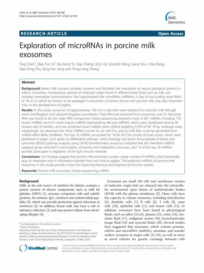
were fully matched, while 7 sequences had one or twomismatched nucleotides (Table 3). However their seedsequences remained unchanged. Simultaneously, theabundance of some novel miRNAs predicted by Solexasequencing was confirmed by quantitative real-timePCR. The abundance of most miRNAs observed byqPCR of the sample pool and by sequencing were gen-erally consistent (Figure 7).
Target verification of miR-PC-86 and miR-PC-263 against3’UTR of IGF-1R using luciferase report assayTo investigate whether the predicted miR-PC-86 andmiR-PC-263 (Figure 1) were functional novel miRNAs,target genes were predicted, and miR-PC-86/ miR-PC-263 were found to directly target IGF-1R 3’UTR se-quence. The full-length 3’UTR of IGF-1R mRNA wasinserted downstream of the luciferase gene in the pmir-GLO Dual-Luciferase miRNA Target Expression Vectorreporter plasmid, and the seed sequence was also deleteto disrupt miR-PC-86/ miR-PC-263 binding (Figure 1B).The wild-type (pGLO-IGF-1R-3’UTR) or delete (pGLO-IGF-1R-3’UTR-delete) plasmid was co-transfected withthe miR-PC-86 and miR-PC-263 mimics into IPEC-J2cells. Forty-eight hours after transfection, the luciferaseactivity of the miR-PC-86 and miR-PC-263 group weresignificantly lower than that of the NC group (P < 0.05)respectively, and the reduction was rescued in the deletegroup (Figure 1C). Thus, IGF-1R was initially confirmedas the target of miR-PC-86 and miR-PC-263.
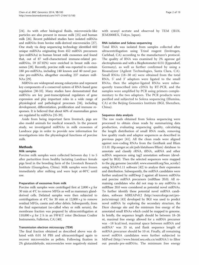
Genomic localization of pre-miRNAsTo further establish the presence of miRNA precursorsin the genome, all mature miRNAs (176 known and 315novels) were mapped to the S. scrofa genome (Figure 8).As a result, 176 known mature miRNAs were mapped to205 pre-miRNAs, and 315 novel miRNAs were mappedto 366 pre-miRNAs on the chromosomes. Our analysis

Table 3 miRNAs matched to sequecing
Predict new miRNA Matched sequence
P-m0281-5p
P-m0124-3p
P-m0227-5p
P-m0355-5p
P-m0338-3p
P-m0105-3p
P-m0113-3p
P-m0129-5p
P-m0058-5p
Table 3 miRNAs matched to sequecing (Continued)
P-m0279-5p
P-m0103-3p
P-m0265-3p
P-m0210-5p
P-m0186-5p
P-m0070-3p
Chen et al. BMC Genomics 2014, 15:100 Page 8 of 19http://www.biomedcentral.com/1471-2164/15/100
revealed that the genomic density distribution of porcinemilk pre-miRNAs (number of pre-miRNAs per Mb ofeach chromosome) was heterogeneous (Figure 8), ran-ging from 0.45 to 0.11 pre-miRNAs for 1 M of genomicsequence. Chromosomes with the highest and lowestdensities of pre-miRNAs were chromosome 12 (29 pre-miRNAs per 64 Mbp) and chromosome 13 (25 pre-miRNAs per 219 Mbp), respectively. Interestingly, themedium-length X chromosome (144 Mbp, ranking 10thin length among the 19 chromosomes in pigs) was anexception by encoding an intermediate number (25 outof 366, 6.8%) of pre-miRNAs, corresponding to 0.17 pre-miRNAs for 1 M of genome sequence, but yet containedthe most clusters.In addition, we observed many mature miRNAs having
multiple miRNA precursors located in the same or dif-ferent chromosomes. Of the novel predicted miRNAs,
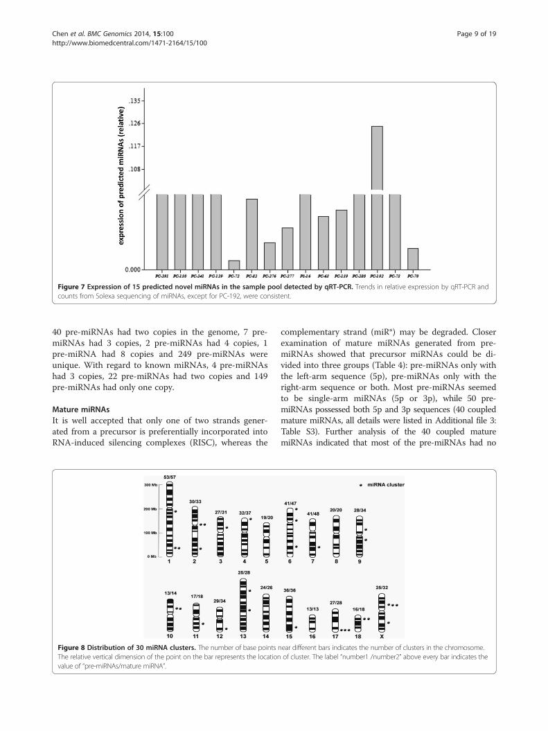
Figure 7 Expression of 15 predicted novel miRNAs in the sample pool detected by qRT-PCR. Trends in relative expression by qRT-PCR andcounts from Solexa sequencing of miRNAs, except for PC-192, were consistent.
Chen et al. BMC Genomics 2014, 15:100 Page 9 of 19http://www.biomedcentral.com/1471-2164/15/100
40 pre-miRNAs had two copies in the genome, 7 pre-miRNAs had 3 copies, 2 pre-miRNAs had 4 copies, 1pre-miRNA had 8 copies and 249 pre-miRNAs wereunique. With regard to known miRNAs, 4 pre-miRNAshad 3 copies, 22 pre-miRNAs had two copies and 149pre-miRNAs had only one copy.
Mature miRNAsIt is well accepted that only one of two strands gener-ated from a precursor is preferentially incorporated intoRNA-induced silencing complexes (RISC), whereas the
Figure 8 Distribution of 30 miRNA clusters. The number of base points nThe relative vertical dimension of the point on the bar represents the locationvalue of “pre-miRNAs/mature miRNA”.
complementary strand (miR*) may be degraded. Closerexamination of mature miRNAs generated from pre-miRNAs showed that precursor miRNAs could be di-vided into three groups (Table 4): pre-miRNAs only withthe left-arm sequence (5p), pre-miRNAs only with theright-arm sequence or both. Most pre-miRNAs seemedto be single-arm miRNAs (5p or 3p), while 50 pre-miRNAs possessed both 5p and 3p sequences (40 coupledmature miRNAs, all details were listed in Additional file 3:Table S3). Further analysis of the 40 coupled maturemiRNAs indicated that most of the pre-miRNAs had no
ear different bars indicates the number of clusters in the chromosome.of cluster. The label “number1 /number2” above every bar indicates the
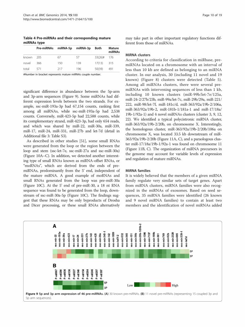
Table 4 Pre-miRNAs and their corresponding maturemiRNAs type
Pre-miRNAs miRNA-5p miRNA-3p Both MaturemiRNAs
known 205 67 57 33(26)# 176
novel 366 150 139 17(13) 315
total 571 217 196 50(39) 491
#Number in bracket represents mature miRNAs couple number.
Chen et al. BMC Genomics 2014, 15:100 Page 10 of 19http://www.biomedcentral.com/1471-2164/15/100
significant difference in abundance between the 5p-armand 3p-arm sequences (Figure 9). Some miRNAs had dif-ferent expression levels between the two strands. For ex-ample, ssc-miR-193a-3p had 67,154 counts, ranking firstamong all miRNAs, while ssc-miR-193a-5p had 2,538counts. Conversely, miR-423-5p had 22,588 counts, whileits complementary strand, miR-423-3p, had only 654 reads,and which was shared by miR-22, miR-30a, miR-339,miR-17, miR-24, miR-331, miR-27b and let-7d (detail inAdditional file 3: Table S3).As described in other studies [51], some small RNAs
were generated from the loop or the region between theloop and stem (ssc-let-7e, ssc-miR-27a and ssc-miR-30a)(Figure 10A–C). In addition, we detected another interest-ing type of small RNAs known as miRNA-offset RNAs, or“moRNAs”, which are derived from the ends of pre-miRNAs, predominantly from the 5’ end, independent ofthe mature miRNA. A good example of moRNAs andsmall RNAs generated from the loop was pre-miR-30a(Figure 10C). At the 5’ end of pre-miR-30, a 18 nt RNAsequence was found to be generated from the loop, down-stream of ssc-miR-30a-5p (Figure 10C). The findings sug-gest that these RNAs may be only byproducts of Droshaand Dicer processing, or these small RNAs alternatively
Figure 9 5p and 3p arm expression of 46 pre-miRNAs. (A) 30 known p5p arm sequences).
may take part in other important regulatory functions dif-ferent from those of miRNAs.
MiRNA clustersAccording to criteria for classification in miRbase, pre-miRNAs located on a chromosome with an interval ofless than 10 kb are defined as belonging to an miRNAcluster. In our analysis, 30 (including 11 novel and 19known) (Figure 8) clusters were detected (Table 5).Among all miRNAs clusters, there were several pre-miRNAs with intervening sequences of less than 1 kb,including 10 known clusters (miR-99b/let-7e/125a,miR-24-2/27b/23b, miR-99a/let-7c, miR-29b/29a, miR-221/222, miR-98/let-7f, miR-181c/d, miR-363/92a/19b-2/106a,miR-363/92a/19b-2, miR-181b-1/181a-1 and miR-17/18a/19b-1/92a-1) and 4 novel miRNAs clusters (cluster 3, 9, 12,22). We identified a typical polycistronic miRNA cluster,miR-363/92a/19b-2/20b, on chromosome X. Interestingly,the homologous cluster, miR-363/92a/19b-2/20b/106a onchromosome X, was located 33.5 kb downstream of miR-363/92a/19b-2/20b (Figure 11A, C), and a paraologous clus-ter miR-17/18a/19b-1/92a-1 was found on chromosome 11(Figure 11B, C). The organization of miRNA precursors inthe genome may account for variable levels of expressionand regulation of mature miRNAs.
MiRNA familiesIt is widely believed that the members of a given miRNAfamily regulate very similar sets of target genes. Apartfrom miRNA clusters, miRNA families were also recog-nized in the miRNAs of exosomes. Based on seed se-quences, 35 miRNA families were identified (26 knownand 9 novel miRNA families) to contain at least twomembers and the identification of novel miRNAs added
re-miRNAs. (B) 11 novel pre-miRNAs (representing 15 coupled 3p and

Figure 10 Three distinctive pre-miRNAs identified in porcine milk exosomes. (A) The ssc-miR-27a precursor produced a 3p-arm miRNAsequence and a loop-derived small RNA. (B) The ssc-let-7e precursor produced a 5p-arm sequence, a 3p-arm miRNA sequence and a loop-derivedsmall RNA. (C) The ssc-miR-30a precursor produced a 5p-arm sequence, a 3p-arm miRNA sequence and a loop-derived small RNA. In addition,ssc-moRNA-3, belonging to new type of miRNA termed moRNA, was found at the 5’ end of pre-miR-30.
Chen et al. BMC Genomics 2014, 15:100 Page 11 of 19http://www.biomedcentral.com/1471-2164/15/100
new members to 5 known families (Table 6). In ourstudy, 8 miRNA families (let-7, mir-1, mir-17, mir-181,mir-148, mir-30, mir-92 and mir-99) were found with atleast 3 members among all exosome miRNAs. The let-7family had 9 members, miR-181 family had 4 members(miR-181a/b/c/d) and miR-30 family had 5 members(miR-30a/b/c/d/e). Most importantly, these miRNAswere highly expressed, and let-7 family members (let-7a/f/c), miR-181a and miR-30a-3p were enriched amongthe top 10 miRNAs. However, members in the samefamily were highly differentially expressed. In themiR-181 family, miR-181a and miR-181b were dominanttypes with 13,345 reads and 3,333 reads, respectively.Similarly, miR-30a was the most abundant in the miR-30family. The differential expression of members in thesame family may be partly due to regulation of their pre-cursors [52]. On the other hand, combined with thecluster analysis, we also observed that some miRNAsshared not only the same cluster but also the same fam-ilies. These miRNAs included 181a/b, let-7f/miR-98,181c/d, let-7a/f-5p/d-5p, 30b/d, 30c/e and miR-221/222.More interestingly, family members of the same clusterseemed to share expression patterns (Figure 12A–E).As mentioned above, miR-17-5p, miR-363, miR-106a,
miR-18a, miR-19b, miR-92a, miR-20b and miR-92b formeda complex cluster and family network, and they alsoshowed different expression patterns. MiR-92a, miR-19band miR-363 were found to be highly expressed, whilemiR-17-5p, miR-18a, miR-20b and miR-106a werelowly expressed. The difference in abundance of thehomologous or paralogous clusters may be attributedto the copy number of miRNA precursor itself or tothe post-transcriptional regulation of the processof generating a mature miRNA from the precursormiRNA.In addition, many miRNA families showed low expres-
sion (count number <100) in milk exosomes, such as themiR-1, miR-130, miR-17, miR-10, miR-29, miR-374,mir-9, miR-15 and miR-491 families (Figure 12F), whichare routinely expressed in specific tissues [53-56]. Inter-estingly, all 9 novel miRNAs families showed extremelylow expression levels (Figure 12G, count number <50),indicating that these miRNAs may only be expressed incertain physiology processes and may be the reason forwhy these miRNAs have not been detected until now.MiR-148a was reported to be an important biomarkerfor milk exosome miRNAs [28,57]. In this study, threemembers of this family (miR-148a, miR-148b and
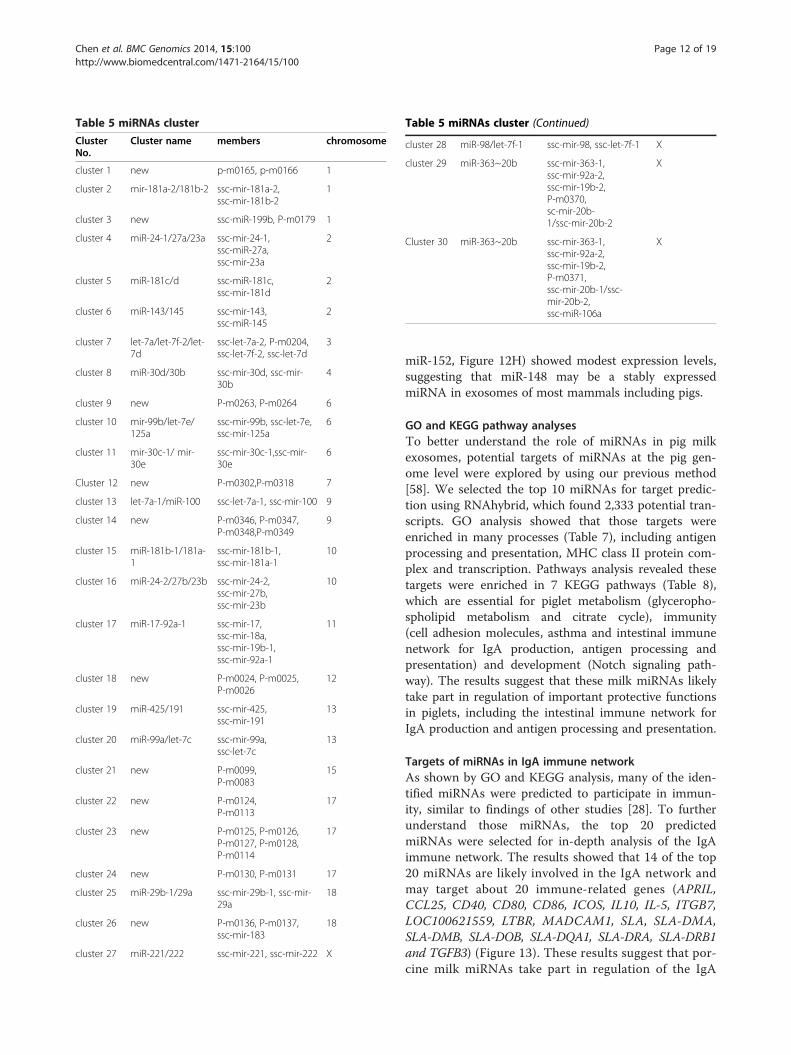
Table 5 miRNAs cluster
ClusterNo.
Cluster name members chromosome
cluster 1 new p-m0165, p-m0166 1
cluster 2 mir-181a-2/181b-2 ssc-mir-181a-2,ssc-mir-181b-2
1
cluster 3 new ssc-miR-199b, P-m0179 1
cluster 4 miR-24-1/27a/23a ssc-mir-24-1,ssc-miR-27a,ssc-mir-23a
2
cluster 5 miR-181c/d ssc-miR-181c,ssc-mir-181d
2
cluster 6 miR-143/145 ssc-mir-143,ssc-miR-145
2
cluster 7 let-7a/let-7f-2/let-7d
ssc-let-7a-2, P-m0204,ssc-let-7f-2, ssc-let-7d
3
cluster 8 miR-30d/30b ssc-mir-30d, ssc-mir-30b
4
cluster 9 new P-m0263, P-m0264 6
cluster 10 mir-99b/let-7e/125a
ssc-mir-99b, ssc-let-7e,ssc-mir-125a
6
cluster 11 mir-30c-1/ mir-30e
ssc-mir-30c-1,ssc-mir-30e
6
Cluster 12 new P-m0302,P-m0318 7
cluster 13 let-7a-1/miR-100 ssc-let-7a-1, ssc-mir-100 9
cluster 14 new P-m0346, P-m0347,P-m0348,P-m0349
9
cluster 15 miR-181b-1/181a-1
ssc-mir-181b-1,ssc-mir-181a-1
10
cluster 16 miR-24-2/27b/23b ssc-mir-24-2,ssc-mir-27b,ssc-mir-23b
10
cluster 17 miR-17-92a-1 ssc-mir-17,ssc-mir-18a,ssc-mir-19b-1,ssc-mir-92a-1
11
cluster 18 new P-m0024, P-m0025,P-m0026
12
cluster 19 miR-425/191 ssc-mir-425,ssc-mir-191
13
cluster 20 miR-99a/let-7c ssc-mir-99a,ssc-let-7c
13
cluster 21 new P-m0099,P-m0083
15
cluster 22 new P-m0124,P-m0113
17
cluster 23 new P-m0125, P-m0126,P-m0127, P-m0128,P-m0114
17
cluster 24 new P-m0130, P-m0131 17
cluster 25 miR-29b-1/29a ssc-mir-29b-1, ssc-mir-29a
18
cluster 26 new P-m0136, P-m0137,ssc-mir-183
18
cluster 27 miR-221/222 ssc-mir-221, ssc-mir-222 X
Table 5 miRNAs cluster (Continued)
cluster 28 miR-98/let-7f-1 ssc-mir-98, ssc-let-7f-1 X
cluster 29 miR-363~20b ssc-mir-363-1,ssc-mir-92a-2,ssc-mir-19b-2,P-m0370,sc-mir-20b-1/ssc-mir-20b-2
X
Cluster 30 miR-363~20b ssc-mir-363-1,ssc-mir-92a-2,ssc-mir-19b-2,P-m0371,ssc-mir-20b-1/ssc-mir-20b-2,ssc-miR-106a
X
Chen et al. BMC Genomics 2014, 15:100 Page 12 of 19http://www.biomedcentral.com/1471-2164/15/100
miR-152, Figure 12H) showed modest expression levels,suggesting that miR-148 may be a stably expressedmiRNA in exosomes of most mammals including pigs.
GO and KEGG pathway analysesTo better understand the role of miRNAs in pig milkexosomes, potential targets of miRNAs at the pig gen-ome level were explored by using our previous method[58]. We selected the top 10 miRNAs for target predic-tion using RNAhybrid, which found 2,333 potential tran-scripts. GO analysis showed that those targets wereenriched in many processes (Table 7), including antigenprocessing and presentation, MHC class II protein com-plex and transcription. Pathways analysis revealed thesetargets were enriched in 7 KEGG pathways (Table 8),which are essential for piglet metabolism (glyceropho-spholipid metabolism and citrate cycle), immunity(cell adhesion molecules, asthma and intestinal immunenetwork for IgA production, antigen processing andpresentation) and development (Notch signaling path-way). The results suggest that these milk miRNAs likelytake part in regulation of important protective functionsin piglets, including the intestinal immune network forIgA production and antigen processing and presentation.
Targets of miRNAs in IgA immune networkAs shown by GO and KEGG analysis, many of the iden-tified miRNAs were predicted to participate in immun-ity, similar to findings of other studies [28]. To furtherunderstand those miRNAs, the top 20 predictedmiRNAs were selected for in-depth analysis of the IgAimmune network. The results showed that 14 of the top20 miRNAs are likely involved in the IgA network andmay target about 20 immune-related genes (APRIL,CCL25, CD40, CD80, CD86, ICOS, IL10, IL-5, ITGB7,LOC100621559, LTBR, MADCAM1, SLA, SLA-DMA,SLA-DMB, SLA-DOB, SLA-DQA1, SLA-DRA, SLA-DRB1and TGFB3) (Figure 13). These results suggest that por-cine milk miRNAs take part in regulation of the IgA

Figure 11 MiR-363~20b~106a homologous or paralogous cluster and expression level. (A) The miR-363/92a/19b/20b cluster and itshomologous cluster miR-363/92a/19b/20b/106a on chromosome X were separated by a 33.5 kb DNA fragment. (B) The miR-17/18a/19/92a clusterwas located on chromosome 11. In the genome, miR-92a/19b showed three copies; miR-363 and miR-20b had two copies; while miR-17, miR-18aand miR-106a had one copy. (C) Expression of mature miRNAs produced from the miR-363~20b~106a cluster and miR-17~92 cluster.
Chen et al. BMC Genomics 2014, 15:100 Page 13 of 19http://www.biomedcentral.com/1471-2164/15/100
network and immunity of piglets. In addition, severalmiRNAs shared the same target gene. Interestingly, allof the let-7 family members (let-7a, let-7c and let-7f )could target CCL25. Those miRNAs are proposed toplay a key role in IgA production in the piglet digestivetract and deserve further exploration, as mucosal im-munity is critically important for the protection ofnewborn piglets.
DiscussionIn the present study, a comprehensive miRNA expres-sion profile of porcine breast milk exosomes was ex-plored via a deep sequencing approach. We found intotal 176 known miRNAs (miRBase 20.0) and 366 pre-miRNAs producing 315 mature miRNAs. Luciferasereporter assay was used to explore the targets of twopredicted novel miRNAs in this study. Results indicatedboth of them down-regulated the luciferase expressionby targeting 3’UTR of IGF-1R. All these pre-miRNAswere distributed in 30 clusters (11 novel and 19 knownclusters), and the mature miRNAs could be assigned to35 families (26 known and 9 unknown families). GO andKEGG pathway analyses show that those miRNAs mayparticipate in many different immune-related processes.An analysis of the top 20 miRNAs showed that 14 ofthem may be involved in many regulatory aspects of theIgA immune network.
A recent study of exosome miRNAs in Yorkshire sowmilk discovered 180 pre-miRNAs, including 140 knownporcine pre-miRNAs and 40 novel pre-miRNAs, whichencode 237 mature miRNAs (234 unique miRNAs) [29]In the current study, we discovered 205 known porcinepre-miRNAs (176 mature miRNAs) and 366 novel pre-miRNAs (315 mature miRNAs), approximately 254 moremature miRNAs than were revealed in the former report[29]. Therefore, our results substantially supplement theknown pig miRNAs, particularly milk exosome miRNAs.Interestingly, most of the novel miRNAs were low inabundance (312 miRNAs with less than 100 reads andonly 3 miRNAs with >100 reads), which is possibly thereason for why these miRNAs were not detected in aprevious study by Gu et al. [29]. Further comparison re-vealed that miR-191 and let-7a, which potentially play avital role in immunity, were found in both that studyand in the top 10 miRNAs of our study. Other miRNAsidentified previously (miR-30a-5p, miR-25-3p, miR-182-5p, miR-200c-3p and miR-375-3p) were not detected inour study. Furthermore, miR-148a, a potential biomarkerfor quality control in bovine milk [57] and human milk[28], which was found to be highly expressed throughoutthe lactation period of Yorkshire sows [29], was only de-tected at a moderate level in Landrace pigs in our study.MiR-148a has been reported to be a tumor metastasissuppressor in gastric cancer [59], and ectopic expressionof miR-148a was shown to induce apoptosis and silence

Table 6 miRNAs family
Family Seed Number Members
Family 1 let-7 GAGGTAG 9 ssc-let-7i, ssc-miR-98,ssc-let-7a,ssc-let-7f,ssc-let-7c, ssc-let-7 g,ssc-let-7e, ssc-let7d-5p
Family 2 mir-1 GGAATGT 3 ssc-miR-1, ssc-miR-206,PC-117
Family 3 mir-10 ACCCTGT 2 ssc-miR-10b,ssc-miR-10a
Family 4 mir-103 GCAGCAT 2 ssc-miR-107,ssc-miR-103
Family 5 mir-125 CCCTGAG 2 ssc-miR-125a,ssc-miR-125b
Family 6 mir-130 AGTGCAA 2 ssc-miR-130b,ssc-miR-130a
Family 7 mir-148 CAGTGCA 3 ssc-miR-148b,ssc-miR-152,ssc-miR-148a
Family 8 mir-17 AAAGTGC 3 ssc-miR-106a,ssc-miR-17-5p,ssc-mir-20b-1/ssc-mir-20b-2
Family 9 mir-181 ACATTCA 4 ssc-miR-181a,ssc-miR-181b,ssc-miR-181c,ssc-miR-181d-5p
Family 10 mir-186 AAAGAAT 2 ssc-miR-186, PC-36
Family 11 mir-193 ACTGGCC 2 ssc-miR-193a-3p, PC-3
Family 12 mir-221 GCTACAT 2 ssc-miR-222, ssc-miR-221
Family 13 mir-23 TCACATT 2 ssc-miR-23b, ssc-miR-23a
Family 14 mir-27 TCACAGT 2 ssc-miR-27a,ssc-miR-27b
Family 15 mir-29 AGCACCA 2 ssc-miR-29b, ssc-miR-29c
Family 16 mir-30 GTAAACA 5 ssc-miR-30e-5p,ssc-miR-30c,ssc-miR-30d,ssc-miR-30b-5p,ssc-miR-30a-5p
Family 17 mir-30(#) TTTCAGT 2 ssc-miR-30a-3p,ssc-miR-30e-3p
Family 18 mir-339 CCCTGTC 2 ssc-miR-339-5p,ssc-miR-4334-3p
Family 19 mir-34 GGCAGTG 2 ssc-miR-34c,ssc-miR-34a,
Family 20 mir-363 ATTGCAC 3 ssc-miR-363,ssc-miR-92b-3p,ssc-miR-92a
Family 21 mir-374 TATAATA 2 ssc-miR-374b-5p,ssc-miR-374a,
Family 22 mir-378 CTGGACT 2 ssc-miR-378, PS-2
Family 23 mir-491 GTGGGGA 2 ssc-miR-491, PC-122
Family 24 mir-497 AGCAGCA 4 ssc-miR-497, ssc-miR-15b,ssc-miR-16, ssc-miR-15a
Family 25 mir-9 CTTTGGT 2 ssc-miR-9-2, ssc-miR-9-1,
Family 26 mir-99 ACCCGTA 3 ssc-miR-99a,ssc-miR-99b,ssc-miR-100
Table 6 miRNAs family (Continued)
Family 27 new-1 CATGATT 2 P-m0040-3p(PC-9),P-m0240-5p(PC-232),
Family 28 new-2 AGAGGGA 2 P-m0048-3p(PC-152),P-m0064-3p(PC-217)
Family 29 new-3 ATTTGAT 2 P-m0084-5p(PC-228),P-m0105-3p(PC-72),
Family 30 new-4 GCTAGGA 2 P-m0110-5p(PC-112),P-m0139-5p(PC-111)
Family 31 new-5 TATGGAT 2 P-m0113-3p(PC-130),P-m0129-5p(PC-129)
Family 32 new-6 CCTGGAT 2 P-m0142-5p(PC-164),P-m0179-3p(PC-165)
Family 33 new-7 CATATTT 2 P-m0161-3p(PC-231),P-m0244-3p(PC-162)
Family 34 new-8 GTTTGGA 2 P-m0183-3p(PC-286),P-m0325-5p(PC-128)
Family 35 new-9 CTTTGGG 2 P-m0229-5p(PC-15),P-m0342-3p(PC-105)
The underline indicated this family contains novel miRNAs. #: due to miRNAsclassification by seed sequence, 3p and 5p of miR-30 represent differentmiRNAs families. PC is unique ID for porcine miRNAs candidate.
Chen et al. BMC Genomics 2014, 15:100 Page 14 of 19http://www.biomedcentral.com/1471-2164/15/100
Bcl-2 in colorectal cancer cells [60]. By bioinformaticsanalysis, miR-148a was determined to be possibly relatedto immunity and gastrointestinal health, but the under-lying regulatory mechanism remains unclear.MiR-92a belongs to the miR-17 ~92 cluster with seven
miRNAs (miR-17-5p, miR-17-3p, miR-18a, miR-19a,miR-19b, miR-20a and miR-92a) and was first described asan oncogenic miRNA cluster involved in B-cell lymphoma[61]. Recent studies indicated that the miRNA-17-92(miR-17-92) cluster directly targets the TGFB pathway incancer cell lines in the mouse embryo stage [62]. Inaddition, the miR-17-92 cluster also participates in normaldevelopment of the heart, lungs and immune system [63].MiR-19 can promote leukemogenesis in Notch1-inducedT-cell acute lymphoblastic leukemia (T-ALL) in vivo [64].Overexpression of the mir-17–mir-18a–mir-19b-1 clusterwas shown to accelerate Myc-induced tumor developmentin a mouse B-cell lymphoma model [61]. The combined re-sults above imply that members of the cluster miR-363/92a/19b-2/20b/106a may be related to cell proliferation anddevelopment. In porcine milk, miR-363/92a/19b-2/20b(miR-363/92a/19b-2/20b/106a) and miR-17/18a/19b-1/92-1 were also detected. The miR-181 (181a/b/c/d) family isrelated to the development of different cells. It was reportedthat miR-181c/d can inhibit cell cycle and proliferation andthat miR-181c regulates TNF-α [65]. The miR-30(b/c/d/e)family regulates kidney development by targeting the tran-scription factor Xlim1/Lhx1 in Xenopus [66]. The well-known let-7(a/b/d/f) family is involved in oncogene expres-sion [67], and let-7/miR-98 family members are expressedlate in mammalian embryonic development [68]. Thus,

Figure 12 Expression of miRNA families. (A) Expression of the let-7f family; let-7f-1, let-7a and let-7d formed a cluster, while miR-98 and let-7eformed a cluster. (B) Expression of miR-30 family; miR-30d clustered with miR-30b, and miR-30c clustered with miR-30e. (C) Expression of miR-181family; miR-181a clustered with miR-181b, and miR-181c clustered with miR-181d. (D) MiR-222 and miR-221; miR-222 and miR-221 belonged tothe same family and the same cluster. (E) MiR-27 family and miR-23 family; miR-27b and miR-23b formed a cluster in the genome. (F) Expressionand clustering of remaining miRNA families; members 1, 2, 3 and 4 represent members of the indicated family. (G) Novel miRNA family with lowexpression. (H) Expression of the miR-148 family containing milk “marker” miRNAs.
Chen et al. BMC Genomics 2014, 15:100 Page 15 of 19http://www.biomedcentral.com/1471-2164/15/100

Table 8 KEGG pathway analysis of potential targets oftop10 miRNAs
Term Count gene p-value
ssc04514: Cell adhesionmolecules (CAMs)
14 CADM3, CD4, CD40,F11R, LOC100521555,SELE, SELL, SELP, SLA,SLA-DMA, SLA-DOA,SLA-DOB, SLA-DRA,SLA-DRB1
1.65E-03
ssc05310: Asthma 7 CD40, SLA, SLA-DMA,SLA-DOA, SLA-DOB,SLA-DRA, SLA-DRB1
5.55E-03
ssc04672: Intestinal immunenetwork for IgA production
8 CD40, SLA, SLA-DMA,SLA-DOA, SLA-DOB,SLA-DRA, SLA-DRB1,TGFB3
2.15 E-02
ssc04330: Notch signalingpathway
5 DLL4, PTCRA,LOC733643,APH1A, NOTCH4
2.33E-02
ssc04612: Antigen processingand presentation
10 CD4, LOC100152370,NFYB, PSME1, SLA,SLA-DMA, SLA-DOA,SLA-DOB, SLA-DRA,SLA-DRB1
3.00E-02
ssc00564:Glycerophospholipidmetabolism
6 AGPAT1, AGPAT4,|AGPAT6, GNPAT,LOC100152491,PCYT1B
4.25E-02
ssc00020:Citrate cycle(TCA cycle)
5 ACO2,DLST, IDH2,LOC100157889,PCK1
4.57E-02
Table 7 Gene ontology analysis of potential targets oftop10 miRNAs
Category Term Count P-Value
Biological process transcription 15 6.70E-03
Biological process regulation of transcription 21 1.00E-02
Biological process antigen processing andpresentation
7 1.50E-02
Biological process regulation of RNAmetabolic process
17 1.70E-02
Biological process regulation of transcription,DNA-dependent
17 1.70E-02
Biological process antigen processing andpresentation of peptideor polysaccharide antigenvia MHC class II
4 2.20E-02
CellularComponent
MHC class II proteincomplex
6 3.70E-04
CellularComponent
MHC protein complex 7 5.00E-03
CellularComponent
large ribosomal subunit 3 2.90E-02
MolecularFunction
transcription regulatoractivity
20 1.80E-03
MolecularFunction
sequence-specificDNA binding
13 2.60E-03
MolecularFunction
ligand-dependent nuclearreceptor activity
7 5.80E-03
MolecularFunction
transcription factor activity 15 1.20E-02
MolecularFunction
steroid hormonereceptor activity
6 2.10E-02
MolecularFunction
DNA binding 20 2.40E-02
MolecularFunction
phosphatase regulatoractivity
4 2.90E-02
MolecularFunction
protein phosphataseregulator activity
4 2.90E-02
Chen et al. BMC Genomics 2014, 15:100 Page 16 of 19http://www.biomedcentral.com/1471-2164/15/100
these miRNAs mentioned above may participate in devel-opment of the piglet digestive tract.Notably, some miRNAs among the top 10 identified
here have been reported to be related to immunity(miR-320, miR-181a, miR-30a-3p, let-7a, let-7f andlet-7c) and development (miR-193a-3p, miR-378 andmiR-191). MiR-193a-3p was demonstrated to regulate cellproliferation, cell cycle progression in vitro and in nudemice [69]. MiR-378 promotes osteoblast differentiation bytargeting polypeptide N-acetylgalactosaminyltransferase 7(GalNAc-T7 or GalNT7) [70], and miR-191 regulates eryth-roid differentiation in mammals by up-regulating erythroid-enriched genes Riok3 and Mxi1 [71]. Meanwhile, miR-320is able to inhibit HL-60 cell proliferation by suppressingreceptor 1 (TfR-1; CD71) [72], and miR-181a was believedto act as an intrinsic antigen sensitivity “rheostat”
during T cell development [73]. MiR-320, miR-181a,miR-30a-3p and let-7 were shown to be downregu-lated in colorectal cancer [74]. Of course, further ex-perimental evidence is needed to verify that thesemiRNAs are indeed related to immunity of the pigletdigestive tract.IgA is a major immunoglobulin in milk [75]. Expres-
sion of the polymeric IgA receptor (pIgR) in mammaryepithelial cells contribute much to the development ofthe immune system at the early stage of lactation [76].In the present study, some miRNAs were predicted totarget genes (CD40, SLA, SLA-DMA, SLA-DOA, SLA-DOB, SLA-DRA, SLA-DRB1 and TGFB3) involved inprocesses of the intestinal immune network for IgAproduction in porcine milk. CD40 is a B-cell antigenactivated during immune responses [77]. CD40 andCD40 ligand (CD40L) expressed on activated T cellsare essential to B cell proliferation [78] and secretionof IgG, IgA and IgE [79]. SLA Class I were found tobe expressed in the epithelial and lamina propriacells of the intestine in adult pigs and to be in-volved in mother-newborn interactions [80]. A studyin humans showed that TGF-β acts as a specific switchfor IgA present at early stages of development of Bcells [81].

Figure 13 MiRNAs targeting the IgA immune network. The top 20 miRNAs were analyzed, and 14 of them were found to participate in theIgA immune network, involving 20 target genes. Different colors and shapes represent various relationships between miRNAs and genes.
Chen et al. BMC Genomics 2014, 15:100 Page 17 of 19http://www.biomedcentral.com/1471-2164/15/100
In the present study, the top 20 miRNAs were usedfor IgA network analysis. APRIL was the predictedtarget of miR-193a-5p, which is essential to triggeringIgA2 class switch in human B cells. Intestinal epithe-lial cells (IECs) release APRIL after sensing bacteriathrough Toll-like receptors, and mucosal vaccines acti-vate IECs to induce more effective IgA2 responses[82]. The let7 family and miR-423-5p were predictedto target CCL25, a potent and selective chemoattract-ant for IgA antibody-secreting cells [83]. CCL25 isknown to selectively modulate immune responses,specifically the localization of T lymphocytes tothe small-intestinal mucosa [84]. CD80 and CD86,which are costimulators of T lymphocytes [85], wereidentified as possible targets of five miRNAs in ourstudy. Let-7a, let-7c, miR-181b, miR-185, miR-378 andmiR-423-5p were predicted to target the inducible co-stimulatory molecule (ICOS), which plays a key rolein regulating T-cell differentiation, T-cell proliferation,and secretion of lymphokines, providing effective helpfor antibody secretion by B cells [86]. We hypothesizethat some miRNAs identified here in porcine milkregulate IgA production in the intestine of piglets,which may play an important role in mucosa immun-ity. However, their regulatory mechanisms warrant fur-ther study.
ConclusionsIn conclusion, the present study revealed 176 knownmiRNAs and 366 (315 mature miRNAs) novel pre-miRNAsin porcine milk, most of which were predicted to be involvedin regulation of digestive tract development and immunity ofnewborn piglets. These findings contribute to an increasedunderstanding of the roles of miRNAs in porcine (S. scrofa)milk exosomes and to building the foundation for under-standing their physiological functions and regulatorymechanisms.
Availability of supporting dataAll the supporting data has been deposited in https://mynotebook.labarchives.com/share/allinchen/MTkuNXwxMzMxMS8xNS0yL1RyZWVOb2RlLzE1NzEyODU2fDQ5LjU= with a DOI:10.6070/H4DN432G.
Additional files
Additional file 1: Table S1. 176 known mature miRNAs.xls.
Additional file 2: Table S2. 315 novel miRNAs.xls.
Additional file 3: Table S3. Pre-miRNAs with 3p and 5p sequence.xls.
Competing interestsWe declared this manuscript have no competing interests.

Chen et al. BMC Genomics 2014, 15:100 Page 18 of 19http://www.biomedcentral.com/1471-2164/15/100
Authors’ contributionsTC, QX carried out the miRNAs Solexa Sequencing and data analysis, andparticipated in drafted the manuscript. XC, QQ carried out the PCR, qPCR. GS,SW participated in the sample collected. XZ, NW performed the Transmissionelectron microscopy. RY, QJ YZ conceived of the study, and participated inits design and coordination and helped to draft the manuscript. All authorsread and approved the final manuscript.
AcknowledgementsThis work was supported by grants from the Key Project of GuangdongProvincial Nature Science Foundation (S2013020012766), National BasicResearch Program of China (973 Program, 2011CB944200, 2009CB941600 and2013CB127304), Natural Science Foundation of China program (31272529)and the Natural Science Foundation of Guangdong Province(S2013010013215). We thank the breeding farm of the Livestock ResearchInstitute (Guangzhou, China) for providing milk samples.
Received: 8 March 2013 Accepted: 31 January 2014Published: 5 February 2014
References1. Strobel S: Immunity induced after a feed of antigen during early life: oral
tolerance v. sensitisation. Proc Nutr Soc 2001, 60(4):437–442.2. Armogida SA, Yannaras NM, Melton AL, Srivastava MD: Identification and
quantification of innate immune system mediators in human breastmilk. Allergy Asthma Proc 2004, 25(5):297–304.
3. Kramer MS, Chalmers B, Hodnett ED, Sevkovskaya Z, Dzikovich I, Shapiro S,Collet JP, Vanilovich I, Mezen I, Ducruet T: Promotion of breastfeedingintervention trial (PROBIT). JAMA 2001, 285(4):413–420.
4. Høst A, Koletzko B, Dreborg S, Muraro A, Wahn U, Aggett P, Bresson J,Hernell O, Lafeber H, Michaelsen K: Dietary products used in infantsfor treatment and prevention of food allergy. Arch Dis Child 1999,81(1):80–84.
5. Van Niel G, Porto-Carreiro I, Simoes S, Raposo G: Exosomes: a commonpathway for a specialized function. J Biochem 2006, 140(1):13–21.
6. Pan BT, Johnstone RM: Fate of the transferrin receptor during maturationof sheep reticulocytes in vitro: selective externalization of the receptor.Cell 1983, 33(3):967–978.
7. Théry C, Regnault A, Garin J, Wolfers J, Zitvogel L, Ricciardi-Castagnoli P,Raposo G, Amigorena S: Molecular characterization of dendritic cell-derivedexosomes. J Cell Biol 1999, 147(3):599–610.
8. Raposo G, Nijman HW, Stoorvogel W, Liejendekker R, Harding CV, Melief C,Geuze HJ: B lymphocytes secrete antigen-presenting vesicles. J Exp Med1996, 183(3):1161–1172.
9. Blanchard N, Lankar D, Faure F, Regnault A, Dumont C, Raposo G, Hivroz C:TCR activation of human T cells induces the production of exosomesbearing the TCR/CD3/ζ complex. J Immunol 2002, 168(7):3235–3241.
10. Raposo G, Tenza D, Mecheri S, Peronet R, Bonnerot C, Desaymard C:Accumulation of major histocompatibility complex class II molecules inmast cell secretory granules and their release upon degranulation.Mol Biol Cell 1997, 8(12):2631–2645.
11. Van Niel G, Raposo G, Candalh C, Boussac M, Hershberg R, Cerf-BensussanN, Heyman M: Intestinal epithelial cells secrete exosome-like vesicles.Gastroenterology 2001, 121(2):337–349.
12. Mears R, Craven RA, Hanrahan S, Totty N, Upton C, Young SL, Patel P,Selby PJ, Banks RE: Proteomic analysis of melanoma‐derived exosomesby two‐dimensional polyacrylamide gel electrophoresis and massspectrometry. Proteomics 2004, 4(12):4019–4031.
13. Gonzalez-Begne M, Lu B, Han X, Hagen FK, Hand AR, Melvin JE, Yates JR III:Proteomic analysis of human parotid gland exosomes bymultidimensional protein identification technology (MudPIT). J ProteomeRes 2009, 8(3):1304–1314.
14. Ogawa Y, Kanai-Azuma M, Akimoto Y, Kawakami H, Yanoshita R: Exosome-likevesicles with dipeptidyl peptidase IV in human saliva. Biol Pharm Bull 2008,31(6):1059–1062.
15. García JM, García V, Peña C, Domínguez G, Silva J, Diaz R, Espinosa P, CitoresMJ, Collado M, Bonilla F: Extracellular plasma RNA from colon cancerpatients is confined in a vesicle-like structure and is mRNA-enriched.RNA 2008, 14(7):1424–1432.
16. Pisitkun T, Shen RF, Knepper MA: Identification and proteomic profiling ofexosomes in human urine. Proc Natl Acad Sci USA 2004, 101(36):13368.
17. Keller S, Rupp C, Stoeck A, Runz S, Fogel M, Lugert S, Hager H, Abdel-BakkyM, Gutwein P, Altevogt P: CD24 is a marker of exosomes secreted intourine and amniotic fluid. Kidney Int 2007, 72(9):1095–1102.
18. Runz S, Keller S, Rupp C, Stoeck A, Issa Y, Koensgen D, Mustea A, Sehouli J, KristiansenG, Altevogt P:Malignant ascites-derived exosomes of ovarian carcinomapatients contain CD24 and EpCAM. Gynecol Oncol 2007, 107(3):563–571.
19. Prado N, Marazuela EG, Segura E, Fernández-García H, Villalba M, Théry C,Rodríguez R, Batanero E: Exosomes from bronchoalveolar fluid of tolerizedmice prevent allergic reaction. J Immunol 2008, 181(2):1519–1525.
20. Simpson RJ, Jensen SS, Lim JWE: Proteomic profiling of exosomes: currentperspectives. Proteomics 2008, 8(19):4083–4099.
21. Deregibus MC, Cantaluppi V, Calogero R, Iacono ML, Tetta C, Biancone L,Bruno S, Bussolati B, Camussi G: Endothelial progenitor cell–derivedmicrovesicles activate an angiogenic program in endothelial cells by ahorizontal transfer of mRNA. Blood 2007, 110(7):2440–2448.
22. Lakkaraju A, Rodriguez-Boulan E: Itinerant exosomes: emerging roles incell and tissue polarity. Trends Cell Biol 2008, 18(5):199–209.
23. Schorey JS, Bhatnagar S: Exosome function: from tumor immunology topathogen biology. Traffic 2008, 9(6):871–881.
24. Valadi H, Ekström K, Bossios A, Sjöstrand M, Lee JJ, Lötvall JO: Exosome-mediated transfer of mRNAs and microRNAs is a novel mechanism ofgenetic exchange between cells. Nat Cell Biol 2007, 9(6):654–659.
25. Nakatani H, Aoki N, Nakagawa Y, Jin-No S, Aoyama K, Oshima K, Ohira S,Sato C, Nadano D, Matsuda T: Weaning-induced expression of a milk-fatglobule protein, MFG-E8, in mouse mammary glands, as demonstratedby the analyses of its mRNA, protein and phosphatidylserine-bindingactivity. Biochem J 2006, 395(Pt 1):21.
26. Admyre C, Johansson SM, Qazi KR, Filén JJ, Lahesmaa R, Norman M, Neve EPA,Scheynius A, Gabrielsson S: Exosomes with immune modulatory features arepresent in human breast milk. J Immunol 2007, 179(3):1969–1978.
27. Hata T, Murakami K, Nakatani H, Yamamoto Y, Matsuda T, Aoki N: Isolationof bovine milk-derived microvesicles carrying mRNAs and microRNAs.Biochem Biophys Res Commun 2010, 396(2):528–533.
28. Zhou Q, Li M, Wang X, Li Q, Wang T, Zhu Q, Zhou X, Gao X, Li X: Immune-related microRNAs are abundant in breast milk exosomes. Int J Biol Sci2012, 8(1):118.
29. Gu Y, Li M, Wang T, Liang Y, Zhong Z, Wang X, Zhou Q, Chen L, Lang Q, HeZ, et al: Lactation-related microRNA expression profiles of porcine breastmilk exosomes. PLoS One 2012, 7(8):e43691.
30. Cullen BR: RNA interference: antiviral defense and genetic tool. NatImmunol 2002, 3(7):597–599.
31. Hutvágner G, Zamore PD: A microRNA in a multiple-turnover RNAienzyme complex. Science 2002, 297(5589):2056–2060.
32. Carrington JC, Ambros V: Role of microRNAs in plant and animaldevelopment. Science 2003, 301(5631):336–338.
33. Cerutti H: RNA interference: traveling in the cell and gaining functions?Trends Genet 2003, 19(1):39–46.
34. Bartel DP: MicroRNAs: target recognition and regulatory functions.Cell 2009, 136(2):215–233.
35. Friedman RC, Farh KKH, Burge CB, Bartel DP: Most mammalian mRNAs areconserved targets of microRNAs. Genome Res 2009, 19(1):92–105.
36. Rodriguez A, Vigorito E, Clare S, Warren MV, Couttet P, Soond DR, vanDongen S, Grocock RJ, Das PP, Miska EA: Requirement of bic/microRNA-155 for normal immune function. Science 2007, 316(5824):608–611.
37. Hyun S, Lee JH, Jin H, Nam J, Namkoong B, Lee G, Chung J, Kim VN:Conserved MicroRNA miR-8/miR-200 and its target USH/FOG2 controlgrowth by regulating PI3K. Cell 2009, 139(6):1096–1108.
38. Lal A, Navarro F, Maher CA, Maliszewski LE, Yan N, O’Day E, Chowdhury D,Dykxhoorn DM, Tsai P, Hofmann O: miR-24 Inhibits cell proliferation bytargeting E2F2, MYC, and other cell-cycle genes via binding to “seedless”3’ UTR microRNA recognition elements. Mol Cell 2009, 35(5):610–625.
39. Chen CZ, Li L, Lodish HF, Bartel DP: MicroRNAs modulate hematopoieticlineage differentiation. Science 2004, 303(5654):83–86.
40. Lässer C, Alikhani VS, Ekström K, Eldh M, Paredes PT, Bossios A, Sjöstrand M,Gabrielsson S, Lötvall J, Valadi H: Human saliva, plasma and breast milkexosomes contain RNA: uptake by macrophages. J Transl Med 2011, 9(1):9.
41. Ji Z, Wang G, Xie Z, Wang J, Zhang C, Dong F, Chen C: Identification of noveland differentially expressed microRNAs of dairy goat mammary gland tissuesusing Solexa sequencing and bioinformatics. PLoS ONE 2012, 7(11):e49463.
42. Li R, Li Y, Kristiansen K, Wang J: SOAP: short oligonucleotide alignmentprogram. Bioinformatics 2008, 24(5):713–714.

Chen et al. BMC Genomics 2014, 15:100 Page 19 of 19http://www.biomedcentral.com/1471-2164/15/100
43. Li Y, Zhang Z, Liu F, Vongsangnak W, Jing Q, Shen B: Performancecomparison and evaluation of software tools for microRNA deep-sequencing data analysis. Nucleic Acids Res 2012, 40(10):4298–4305.
44. Jiang P, Wu H, Wang W, Ma W, Sun X, Lu Z: MiPred: classification of realand pseudo microRNA precursors using random forest prediction modelwith combined features. Nucleic Acids Res 2007, 35(suppl 2):W339–W344.
45. Zuker M: Mfold web server for nucleic acid folding and hybridizationprediction. Nucleic Acids Res 2003, 31(13):3406–3415.
46. Fu H, Tie Y, Xu C, Zhang Z, Zhu J, Shi Y, Jiang H, Sun Z, Zheng X:Identification of human fetal liver miRNAs by a novel method. FEBS Lett2005, 579(17):3849–3854.
47. Kosaka N, Izumi H, Sekine K, Ochiya T: microRNA as a new immune-regulatory agent in breast milk. Silence 2010, 1(1):7.
48. Ebhardt HA, Fedynak A, Fahlman RP: Naturally occurring variations insequence length creates microRNA isoforms that differ in argonauteeffector complex specificity. Silence 2010, 1(1):12.
49. Guo L, Lu Z: Global expression analysis of miRNA gene cluster and familybased on isomiRs from deep sequencing data. Comput Biol Chem 2010,34(3):165–171.
50. Naya L, Khan GA, Sorin C, Hartmann C, Crespi M, Lelandais-Brière C:Cleavage of a non-conserved target by a specific miR156 isoform in rootapexes of Medicago truncatula. Plant Signal Behav 2010, 5(3):328–331.
51. Li M, Xia Y, Gu Y, Zhang K, Lang Q, Chen L, Guan J, Luo Z, Chen H, Li Y:MicroRNAome of porcine pre-and postnatal development. PLoS One2010, 5(7):e11541.
52. He L, Hannon GJ: MicroRNAs: small RNAs with a big role in generegulation. Nat Rev Genet 2004, 5(7):522–531.
53. Lagos-Quintana M, Rauhut R, Yalcin A, Meyer J, Lendeckel W, Tuschl T:Identification of tissue-specific microRNAs from mouse. Curr Biol 2002,12(9):735–739.
54. Tran N, O’Brien CJ, Clark J, Rose B: Potential role of micro‐RNAs in headand neck tumorigenesis. Head Neck 2010, 32(8):1099–1111.
55. Morales Prieto DM, Markert UR: MicroRNAs in pregnancy. J ReprodImmunol 2011, 88(2):106–111.
56. Xie S, Huang T, Shen Y, Li X, Zhang X, Zhu M, Qin H, Zhao S: Identificationand characterization of microRNAs from porcine skeletal muscle.Anim Genet 2010, 41(2):179–190.
57. Chen X, Gao C, Li H, Huang L, Sun Q, Dong Y, Tian C, Gao S, Dong H, GuanD: Identification and characterization of microRNAs in raw milk duringdifferent periods of lactation, commercial fluid, and powdered milkproducts. Cell Res 2010, 20(10):1128–1137.
58. Ye R-S, Xi Q-Y, Qi Q, Cheng X, Chen T, Li H, Kallon S, Shu G, Wang S-B,Jiang Q-Y: Differentially Expressed miRNAs after GnRH Treatment andTheir Potential Roles in FSH Regulation in Porcine Anterior Pituitary Cell.PLoS ONE 2013, 8(2):e57156.
59. Zheng B, Liang L, Wang C, Huang S, Cao X, Zha R, Liu L, Jia D, Tian Q, Wu J:MicroRNA-148a suppresses tumor cell invasion and metastasis bydownregulating ROCK1 in gastric cancer. Clin Cancer Res 2011,17(24):7574–7583.
60. Zhang H, Li Y, Huang Q, Ren X, Hu H, Sheng H, Lai M: MiR-148a promotesapoptosis by targeting Bcl-2 in colorectal cancer. Cell Death Differ 2011,18(11):1702–1710.
61. He L, Thomson JM, Hemann MT, Hernando-Monge E, Mu D, Goodson S,Powers S, Cordon-Cardo C, Lowe SW, Hannon GJ: A microRNA polycistronas a potential human oncogene. Nature 2005, 435(7043):828–833.
62. Li L, Shi JY, Zhu GQ, Shi B: MiR‐17‐92 cluster regulates cell proliferationand collagen synthesis by targeting TGFB pathway in mouse palatalmesenchymal cells. J Cell Biochem 2012, 113(4):1235–1244.
63. Mendell JT: miRiad roles for the miR-17-92 cluster in development anddisease. Cell 2008, 133(2):217–222.
64. Mavrakis KJ, Wolfe AL, Oricchio E, Palomero T, De Keersmaecker K, McJunkinK, Zuber J, James T, Khan AA, Leslie CS: Genome-wide RNA-mediatedinterference screen identifies miR-19 targets in Notch-induced T-cellacute lymphoblastic leukaemia. Nat Cell Biol 2010, 12(4):372–379.
65. Río P, Agirre X, Garate L, Baños R, Álvarez L, San José-Enériz E, Badell I,Casado JA, Garín M, Prósper F: Down-regulated expression of hsa-miR-181c inFanconi anemia patients: implications in TNFα regulation and proliferation ofhematopoietic progenitor cells. Blood 2012, 119(13):3042–3049.
66. Agrawal R, Tran U, Wessely O: The miR-30 miRNA family regulatesXenopus pronephros development and targets the transcription factorXlim1/Lhx1. Development 2009, 136(23):3927–3936.
67. Akao Y, Nakagawa Y, Naoe T: let-7 microRNA functions as a potentialgrowth suppressor in human colon cancer cells. Biol Pharm Bull 2006,29(5):903–906.
68. Shell S, Park SM, Radjabi AR, Schickel R, Kistner EO, Jewell DA, Feig C,Lengyel E, Peter ME: Let-7 expression defines two differentiation stagesof cancer. Proc Natl Acad Sci 2007, 104(27):11400–11405.
69. Ma K, He Y, Zhang H, Fei Q, Niu D, Wang D, Ding X, Xu H, Chen X, Zhu J: DNAmethylation-regulated miR-193a-3p dictates resistance of hepatocellularcarcinoma to 5-fluorouracil via repression of SRSF2 expression.J Biol Chem 2012, 287(8):5639–5649.
70. Kahai S, Lee SC, Lee DY, Yang J, Li M, Wang CH, Jiang Z, Zhang Y, Peng C,Yang BB: MicroRNA miR-378 regulates nephronectin expression modulatingosteoblast differentiation by targeting GalNT-7. PLoS One 2009, 4(10):e7535.
71. Zhang L, Flygare J, Wong P, Lim B, Lodish HF: miR-191 regulates mouseerythroblast enucleation by down-regulating Riok3 and Mxi1. Genes Dev2011, 25(2):119–124.
72. Schaar DG, Medina DJ, Moore DF, Strair RK, Ting Y: miR-320 targetstransferrin receptor 1 (CD71) and inhibits cell proliferation. Exp Hematol2009, 37(2):245–255.
73. Li QJ, Chau J, Ebert PJR, Sylvester G, Min H, Liu G, Braich R, Manoharan M,Soutschek J, Skare P: miR-181a is an intrinsic modulator of T cellsensitivity and selection. Cell 2007, 129(1):147–161.
74. Aslam MI, Patel M, Singh B, Jameson JS, Pringle JH: MicroRNAs are NovelBiomarkers for Detection of Colorectal Cancer. In Biomarker. Edited byKhan TK. Croatia, Rijeka: In Tech; 2012:1–18.
75. Curtis J, Bourne F: Immunoglobulin quantitation in sow serum, colostrumand milk and the serum of young pigs. Biochimica et Biophysica Acta(BBA)-Protein Structure 1971, 236(1):319–332.
76. Kumura H, Sone T, Shimazaki K, Kobayashi E: Sequence analysis of porcinepolymeric immunoglobulin receptor from mammary epithelial cellspresent in colostrum. J Dairy Res 2000, 67(04):631–636.
77. Castigli E, Alt FW, Davidson L, Bottaro A, Mizoguchi E, Bhan AK, Geha RS:CD40-deficient mice generated by recombination-activating gene-2-deficient blastocyst complementation. Proc Natl Acad Sci 1994,91(25):12135–12139.
78. Kawabe T, Naka T, Yoshida K, Tanaka T, Fujiwara H, Suematsu S, Yoshida N,Kishimoto T, Kikutani H: The immune responses in CD40-deficient mice:impaired immunoglobulin class switching and germinal center formation.Immunity 1994, 1(3):167.
79. Durandy A, Schiff C, Bonnefoy JY, Forveille M, Rousset F, Mazzei G, Milili M,Fischer A: Induction by anti‐CD40 antibody or soluble CD40 ligand andcytokines of IgG, IgA and IgE production by B cells from patients withX‐linked hyper IgM syndrome. Eur J Immunol 2005, 23(9):2294–2299.
80. Le Jan C, Chevaleyre C: Reduced expression of SLA Class 1 antigens byintestinal epithelium of newborn piglets. Vet Immunol Immunopathol1996, 50(1):167–172.
81. Van Vlasselaer P, Punnonen J, De Vries J: Transforming growth factor-betadirects IgA switching in human B cells. J Immunol 1992, 148(7):2062–2067.
82. He B, Xu W, Santini PA, Polydorides AD, Chiu A, Estrella J, Shan M,Chadburn A, Villanacci V, Plebani A: Intestinal Bacteria Trigger TCell-Independent Immunoglobulin A < sub > 2</sub > Class Switchingby Inducing Epithelial-Cell Secretion of the Cytokine APRIL. Immunity2007, 26(6):812–826.
83. Bowman EP, Kuklin NA, Youngman KR, Lazarus NH, Kunkel EJ, Pan J, GreenbergHB, Butcher EC: The intestinal chemokine thymus-expressed chemokine(CCL25) attracts IgA antibody-secreting cells. J Exp Med 2002, 195(2):269–275.
84. Svensson M, Marsal J, Ericsson A, Carramolino L, Brodén T, Márquez G,Agace WW: CCL25 mediates the localization of recently activatedCD8alphabeta^+ lymphocytes to the small-intestinal mucosa. J ClinInvestig 2002, 110(8):1113–1122.
85. Lanier LL, O’Fallon S, Somoza C, Phillips JH, Linsley PS, Okumura K, Ito D,Azuma M: CD80 (B7) and CD86 (B70) provide similar costimulatorysignals for T cell proliferation, cytokine production, and generation ofCTL. J Immunol 1995, 154(1):97–105.
86. Hutloff A, Dittrich AM, Beier KC, Eljaschewitsch B, Kraft R, Anagnostopoulos I,Kroczek RA: ICOS is an inducible T-cell co-stimulator structurally andfunctionally related to CD28. Nature 1999, 402:21–24.
doi:10.1186/1471-2164-15-100Cite this article as: Chen et al.: Exploration of microRNAs in porcine milkexosomes. BMC Genomics 2014 15:100.