RESEARCH ARTICLE Open Access Effect of local ... applied a constant linear propulsion (v = 2 mm/min)...
9
RESEARCH ARTICLE Open Access Effect of local zoledronate on implant osseointegration in a rat model David A Back 1,3 , Stephan Pauly 2 , Lisa Rommel 3 , Norbert P Haas 2 , Gerhard Schmidmaier 4 , Britt Wildemann 3 and Stefan H Greiner 2* Abstract Background: An implant coating with poly(D, L-lactide) (PDLLA) releasing incorporated Zoledronic acid (ZOL) has already proven to positively effect osteoblasts, to inhibit osteoclasts and to accelerate fracture healing. Aim of this study was to investigate the release kinetics of the chosen coating and the effect of different concentrations of ZOL locally released from this coating on the osseointegration of implants. Methods: For release kinetics the release of C14-labled ZOL out of the coating was monitored over a period of six weeks in vitro. For testing the osseointegration, titanium Kirschner wires were implanted into the medullary canal of right femurs of 100 Sprague Dawley rats. The animals were divided into five groups receiving implants either uncoated or coated with PDLLA, PDLLA/ZOL low (1.2% w/w) or PDLLA/ZOL high (2% w/w). Additionally, a group with uncoated implants received ZOL intravenously (i.v.). After 56 days animals were sacrificed, femurs dissected and either strength of fixation or histological bone/implant contacts and newly formed bone around the implants were determined. Results: Release kinetics revealed an initial peak in the release of C14-ZOL with a slight further progression over the following weeks. There was no significant enhancement of osseointegration for both groups who received ZOL-coated implants or ZOL i.v. compared to the controls in biomechanical or histological analyses, except for a significant raise in strength of fixation of ZOL i.v. versus PDLLA. Conclusions: Even though the investigated local ZOL application did not enhance the osseointegration of the implant, the findings might support its application in fracture treatment, since fracture stabilization devices are often explanted after consolidation. Background Implants commonly used in orthopedic surgery serve on the one hand as fixation devices after fractures, granting stabilization until fracture healing. On the other hand implants are used for joint replacement and a persistent osseointegration is desired. However, failure of osseoin- tegration in joint replacement is a frequent complica- tion. To address this issue, research has recently been focused on the supplementary use of drugs that are known to influence bone turnover. In particular, the class of Bisphosphonates (BPs) has been evaluated for improvement of both, fracture healing and osseointegra- tion in animal models [1-8] and clinical studies [9,10]. BPs such as Zoledronic acid (ZOL) are in clinical use for prevention and treatment of skeletal diseases asso- ciated with increased bone resorption like malignant tumors [11,12] or osteoporosis [8,9]. As BPs have a strong affinity to Calcium, they almost exclusively con- centrate in bone mineral, mainly in regions of bone resorption or formation. Incorporated by osteoclasts during bone resorption, Nitrogen-containing BPs such as ZOL block the farnesyl pyrophosphate synthase, a crucial enzyme in the mevalonate pathway, which inter- feres with the prenylation of proteins causing functional impairment of osteoclasts and thus a decrease in bone resorption [13]. The influence of BPs on bone healing and the osseoin- tegration of implants have been widely investigated. During bone repair, BPs have been shown to have an anti-catabolic, almost net anabolic effect [14,15]. In * Correspondence: [email protected] 2 Center for Musculoskeletal Surgery, Charité-Universitaetsmedizin, Berlin, Germany Full list of author information is available at the end of the article Back et al. BMC Musculoskeletal Disorders 2012, 13:42 http://www.biomedcentral.com/1471-2474/13/42 © 2012 Back et al; licensee BioMed Central Ltd. This is an Open Access article distributed under the terms of the Creative Commons Attribution License (http://creativecommons.org/licenses/by/2.0), which permits unrestricted use, distribution, and reproduction in any medium, provided the original work is properly cited.
Transcript of RESEARCH ARTICLE Open Access Effect of local ... applied a constant linear propulsion (v = 2 mm/min)...

RESEARCH ARTICLE Open Access
Effect of local zoledronate on implantosseointegration in a rat modelDavid A Back1,3, Stephan Pauly2, Lisa Rommel3, Norbert P Haas2, Gerhard Schmidmaier4, Britt Wildemann3 andStefan H Greiner2*
Abstract
Background: An implant coating with poly(D, L-lactide) (PDLLA) releasing incorporated Zoledronic acid (ZOL) hasalready proven to positively effect osteoblasts, to inhibit osteoclasts and to accelerate fracture healing. Aim of thisstudy was to investigate the release kinetics of the chosen coating and the effect of different concentrations ofZOL locally released from this coating on the osseointegration of implants.
Methods: For release kinetics the release of C14-labled ZOL out of the coating was monitored over a period of sixweeks in vitro. For testing the osseointegration, titanium Kirschner wires were implanted into the medullary canalof right femurs of 100 Sprague Dawley rats. The animals were divided into five groups receiving implants eitheruncoated or coated with PDLLA, PDLLA/ZOL low (1.2% w/w) or PDLLA/ZOL high (2% w/w). Additionally, a groupwith uncoated implants received ZOL intravenously (i.v.). After 56 days animals were sacrificed, femurs dissectedand either strength of fixation or histological bone/implant contacts and newly formed bone around the implantswere determined.
Results: Release kinetics revealed an initial peak in the release of C14-ZOL with a slight further progression overthe following weeks. There was no significant enhancement of osseointegration for both groups who receivedZOL-coated implants or ZOL i.v. compared to the controls in biomechanical or histological analyses, except for asignificant raise in strength of fixation of ZOL i.v. versus PDLLA.
Conclusions: Even though the investigated local ZOL application did not enhance the osseointegration of theimplant, the findings might support its application in fracture treatment, since fracture stabilization devices areoften explanted after consolidation.
BackgroundImplants commonly used in orthopedic surgery serve onthe one hand as fixation devices after fractures, grantingstabilization until fracture healing. On the other handimplants are used for joint replacement and a persistentosseointegration is desired. However, failure of osseoin-tegration in joint replacement is a frequent complica-tion. To address this issue, research has recently beenfocused on the supplementary use of drugs that areknown to influence bone turnover. In particular, theclass of Bisphosphonates (BPs) has been evaluated forimprovement of both, fracture healing and osseointegra-tion in animal models [1-8] and clinical studies [9,10].
BPs such as Zoledronic acid (ZOL) are in clinical usefor prevention and treatment of skeletal diseases asso-ciated with increased bone resorption like malignanttumors [11,12] or osteoporosis [8,9]. As BPs have astrong affinity to Calcium, they almost exclusively con-centrate in bone mineral, mainly in regions of boneresorption or formation. Incorporated by osteoclastsduring bone resorption, Nitrogen-containing BPs suchas ZOL block the farnesyl pyrophosphate synthase, acrucial enzyme in the mevalonate pathway, which inter-feres with the prenylation of proteins causing functionalimpairment of osteoclasts and thus a decrease in boneresorption [13].The influence of BPs on bone healing and the osseoin-
tegration of implants have been widely investigated.During bone repair, BPs have been shown to have ananti-catabolic, almost net anabolic effect [14,15]. In
* Correspondence: [email protected] for Musculoskeletal Surgery, Charité-Universitaetsmedizin, Berlin,GermanyFull list of author information is available at the end of the article
Back et al. BMC Musculoskeletal Disorders 2012, 13:42http://www.biomedcentral.com/1471-2474/13/42
© 2012 Back et al; licensee BioMed Central Ltd. This is an Open Access article distributed under the terms of the Creative CommonsAttribution License (http://creativecommons.org/licenses/by/2.0), which permits unrestricted use, distribution, and reproduction inany medium, provided the original work is properly cited.
most studies, this resulted in the enhancement of frac-ture consolidation [16,17]. Concerning osseointegration,studies focused on the effect of systemically or locallyapplied BPs on implant fixation [10,18]. Locally, BPswere either applied directly to the implant site or linkedto the implant with a coating substance as drug carrier[1,19]. Different coatings were tested using especiallycalcium phosphates like hydroxyapatite [20-22] or cross-linked fibrinogen layer [23-25]. In most cases, applica-tion of BPs showed beneficial biomechanical and histo-logical effects on implant osseointegration [1,23,25-27].However, in some studies, BPs did not improve implantfixation or even worsened it [28-30].Previous in vitro studies investigated the effect of locally
released ZOL from a poly(D, L-lactide) (PDLLA) implantcoating on human osteoblasts and osteoclasts. A dosedependent promoting effect on osteoblasts [31] and adecrease in osteoclast formation and osteoclastic resorptionactivity [32] could be shown. In a coculture of human osteo-blasts and osteoclasts, beneficial effects on osteoblast differ-entiation and protein synthesis and a decrease of osteoclastformation with no significant decrease in osteoclasticresorption activity has also been shown [33]. Moreover, in arat fracture model, local application of ZOL delivered froman intramedullary implant showed a significant increase inbiomechanical strength of the fracture side [17].Aims of the study were first to investigate the release
kinetics of the chosen PDLLA/ZOL coating. Second, toanalyze the effect of the coating on osseointegration ofimplants using an established non-weight-bearing ratimplant model [34]. Third, to detect possible differencesof the local application of ZOL in comparison to sys-temic application.
MethodsRelease kineticsTo detect the release profile of ZOL from PDLLA, an invitro elution experiment was performed. Titanium K-wires were dip coated in a solution with Carbon 14(C14)-labeled ZOL (Novartis Pharma AG, Basel, Switzerland)dissolved in PDLLA/ethyl acetate resulting in an esti-mated concentration of 50 μg per sample. Ten TitaniumK-wires with PDLLA/C14-ZOL coating were sterile incu-bated at 37°C (each in 5.2 mL 0.9% NaCl). At time points0 min, 10 min, 1 h, 6 h, 12 h, 24 h, 48 h, 4 d, 7 d, 14 d, 28d, and 42 d 100 μL were drawn from each solution. Theconcentrations of C14-ZOL were detected indirectly viaradioactivity count as automatically quench-correctedCounts Per Minute (CPM) by a Liquid ScintillationCounter (Wallac Oy, Turku, Finland).
Coating of the implants and groupsCoating of the implants was performed as described pre-viously [35]. After dissolving 100 mg PDLLA in 1.5 ml
ethyl acetate at room temperature, the solution was ster-ile filtered. ZOL was dissolved in ethyl acetate/PDLLAsolution to obtain a concentration of 1.2% w/w or 2%w/w. Sterile Kirschner wires (1.4-mm diameter,Synthes®, Paoli, USA) were used as implants for theexperiments. They were dipped two times into the coat-ing solution and dried under laminar air flow condi-tions. For systemic application ZOL was dissolved inNaCl to obtain a concentration of 0.1 mg/ml. The studycontained five groups:- - Group I (Control)-uncoated K-wire- - Group II (PDLLA)-PDLLA- - Group III (ZOL low)-PDLLA + ZOL low dose (20
μg, 1.2% w/w)- - Group IV (ZOL high)-PDLLA + ZOL high dose
(50 μg, 2% w/w)- - Group V (ZOL i.v.)-uncoated + ZOL i.v. 0.1 mg/kgZoledronic acid as a pure substance was kindly pro-
vided by Novartis Pharma AG (Basel, Switzerland).
Animals and experimental designAnimals used for experiment were 100 five-month-oldfemale Sprague Dawley rats (Harlan-Winkelmann, Ger-many) with a mean body weight of 267 g +/- 25 g (SD).They were kept in groups of five rats each. For anesthesiaisoflurane gas (Forene®) and an intraperitoneal injectionwith a mixture of Ketaminhydrochlorid (100 mg/ml per80 mg/kg body weight) and Xylazin 2% (12 mg/kg bodyweight) were used. The right hind leg was shaved foroperation. A 3 mm longitudinal incision was made tosplit the skin and the patellar ligament. Using a 1.2 mmhand drill, the intracondylar notch of the right femur wasreamed. Thereafter, insertion of the implant into themedullary cavity to the proximal end was performed in aretrograde manner. Radiographs were taken to verify thecorrect position of the implant using the trochantermajor as control point. At the end of operation theextending part of the implant was cut off and skinwounds were sutured. For systemic application of ZOLeither the cephalic vein or the left greater saphenous veinwere used. Immediately after implant insertion, ZOL dis-solved in 1 ml NaCl was injected into the vein.After the operation and on the first postoperative day
all animals received subcutaneous injections of bupre-norphine (Temgesic®, 0.05 mg/kg) for analgesia. Using aprecision scale, body weight was determined and bodytemperature was measured rectally at days 0 (OP) and56. Wound healing and signs of a local or systemicinfection were evaluated regularly. 56 days after opera-tion, animals were sacrificed for biomechanical and his-tological analysis. The national guidelines for the careand use of laboratory animals were observed and thestudy was approved by the Animal Experimental EthicsCommittee of Berlin (approval number G0174/07).
Back et al. BMC Musculoskeletal Disorders 2012, 13:42http://www.biomedcentral.com/1471-2474/13/42
Page 2 of 9
Radiographic evaluationRadiographs were taken in lateral and posterior-anteriorview at day 0 (operation) and at day 56 (sacrifice) withthe use of digital X-ray cassettes (Fuji Photo Film Co.,Fuji, Japan) and a Mobilett Plus X-ray unit (SiemensAG, Munich, Germany). The X-rays were evaluated bytwo observers.
Biomechanical testingAll rats were sacrificed after 56 days and 10 animals ofeach group were taken for biomechanical analysis. Theright femurs were dissected and soft tissue was removed.Afterwards, they were carefully prepared at the proximaland distal end to expose 3 mm of the implant on eachside of the bone. The distal end was embedded withbone cement (Heraeus-Kulzer GmbH, Wehrheim, Ger-many) into a previously described push-out device [34],which was placed into a material testing machine(Zwick 1455, Ulm, Germany). During the test, themachine applied a constant linear propulsion (v = 2mm/min) to the implant and the applied force wasrecorded. For measuring the bone-implant attachmentstrength, the peak force needed to loosen the implantwas used. To minimize differences in the push-out forcedue to a different length of the bone or implant, maxi-mum force was set in ratio to the total bone area incontact with the implanted K-wire [26,34]:
Strength of fixation σu = Fmax/πDH[σu : strength of fixation (Mpa), Fmax : initial push - out force
(N),D : implant diameter(mm),H : Bone length (mm)].
HistomorphometryFor histological assessment the right femur of 10 ani-mals of each group was harvested and cleared of soft tis-sue. The bones were fixed in 10% normal bufferedformaldehyde for 5 days, dehydrated by ascending con-centrations of ethanol and finally embedded in methyl-methacrylate (Technovit 7200, Heraeus-Kulzer GmbH,Wehrheim, Germany). A grinding machine (Exakt, Nor-derstedt, Germany) was used to grind the embeddedspecimens until the implant was visible in full lengthwith maximum diameter. After gluing the ground sidesto microscope slides, a diamond band saw (Exakt, Nor-derstedt, Germany) was used to slice 300 μm sectionswhich were ground down to 80 μm. The staining forhistological analyses of the sections was performed withSafranin-O and von-Kossa. The specimens were scannedwith a microscope (Axioskop 40, Carl Zeiss, GöttingenGermany). Direct bone/implant contact area was deter-mined with analyzing software (AxioVision Rel. 4.7, CarlZeiss AG, Jena, Germany). The region of interest (ROI)was defined by drawing a line of exact the same length(13.7 mm) in each digitalized picture, starting from thenutrient foramen (Figure 1a). Afterwards, bone/implant
contact areas of both cortices were measured along thedistance of the drawn line and set into per cent ratio.Furthermore, trabecular mass was measured within theROI in an 0.3 mm range on both sides of the implantand set in per cent to the entire area of this spatium todetermine bone area/total area ratio (Figure 1b).
StatisticsStatistical differences between all groups were analyzedwith the Kruskal-Wallis test. If significant differenceswere shown, Mann-Whitney U test was used for com-parison between two single groups. Data were controlledwith Bonferroni-Holm correction for multiple compari-sons. Statistical differences were defined at a 95% confi-dence level. SPSS software (14.0; SPSS Inc., Chicago, IL)supported statistical evaluation.
ResultsRelease kineticsThe release of the incorporated C14-ZOL from thePDLLA coating showed a strong increase in detectableCounts Per Minute (CPM) within the first hours. In thefollowing days and weeks, only a slight further progres-sion of CPM took place, indicating that approximately90% of C14-ZOL had been released in an initial peakwithin the first 24 hours (Figure 2).
Body parameter evaluation and x-rayNo significant differences were detected in body tem-perature and raise of average body weight between thegroups over the experimental period. None of the ani-mals showed any clinical signs of infection throughoutthe experimental period.Immediate postoperative X-rays revealed a correct
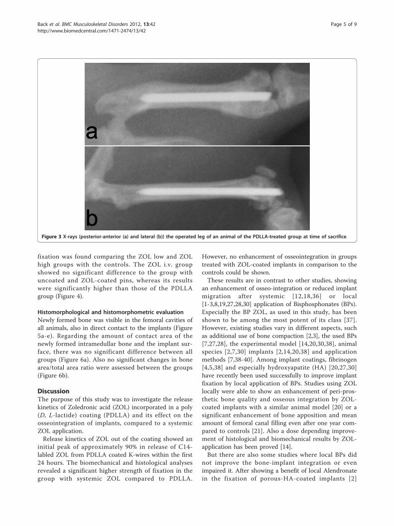
insertion of each K-wire in all femurs. Evaluation cri-teria were here the placement of each implant inside themedullar canal with its tip positioned at the level of thelesser trochanter. The X-ray control after 56 days (Fig-ure 3) showed no signs of dislocation of the implants orzones of osteolysis in all animals. Here, X-rays were ana-lyzed in comparison to the postoperative records toevaluate if the medullar canal had been widened and ifthe position of the implants had varied, determined bythe location of the K-wire tip and two points of contactwith the cortical bone. Radiologically, there was no dif-ference in implant/bone fitting between all groups.
Biomechanical evaluationAfter determination of the strength of fixation by corre-lating the initial push-out force of each implant with thelength of the surrounding bone, all groups were com-pared with each other. The results of the PDLLA groupwere slightly lower than those of the group withuncoated pins. No significant difference in strength of
Back et al. BMC Musculoskeletal Disorders 2012, 13:42http://www.biomedcentral.com/1471-2474/13/42
Page 3 of 9
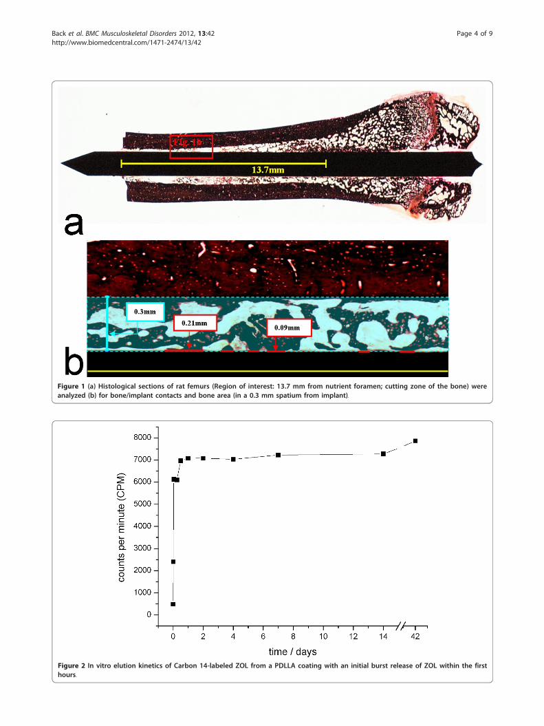
Figure 1 (a) Histological sections of rat femurs (Region of interest: 13.7 mm from nutrient foramen; cutting zone of the bone) wereanalyzed (b) for bone/implant contacts and bone area (in a 0.3 mm spatium from implant).
Figure 2 In vitro elution kinetics of Carbon 14-labeled ZOL from a PDLLA coating with an initial burst release of ZOL within the firsthours.
Back et al. BMC Musculoskeletal Disorders 2012, 13:42http://www.biomedcentral.com/1471-2474/13/42
Page 4 of 9
fixation was found comparing the ZOL low and ZOLhigh groups with the controls. The ZOL i.v. groupshowed no significant difference to the group withuncoated and ZOL-coated pins, whereas its resultswere significantly higher than those of the PDLLAgroup (Figure 4).
Histomorphological and histomorphometric evaluationNewly formed bone was visible in the femoral cavities ofall animals, also in direct contact to the implants (Figure5a-e). Regarding the amount of contact area of thenewly formed intramedullar bone and the implant sur-face, there was no significant difference between allgroups (Figure 6a). Also no significant changes in bonearea/total area ratio were assessed between the groups(Figure 6b).
DiscussionThe purpose of this study was to investigate the releasekinetics of Zoledronic acid (ZOL) incorporated in a poly(D, L-lactide) coating (PDLLA) and its effect on theosseointegration of implants, compared to a systemicZOL application.Release kinetics of ZOL out of the coating showed an
initial peak of approximately 90% in release of C14-labled ZOL from PDLLA coated K-wires within the first24 hours. The biomechanical and histological analysesrevealed a significant higher strength of fixation in thegroup with systemic ZOL compared to PDLLA.
However, no enhancement of osseointegration in groupstreated with ZOL-coated implants in comparison to thecontrols could be shown.These results are in contrast to other studies, showing
an enhancement of osseo-integration or reduced implantmigration after systemic [12,18,36] or local[1-3,8,19,27,28,30] application of Bisphosphonates (BPs).Especially the BP ZOL, as used in this study, has beenshown to be among the most potent of its class [37].However, existing studies vary in different aspects, suchas additional use of bone compaction [2,3], the used BPs[7,27,28], the experimental model [14,20,30,38], animalspecies [2,7,30] implants [2,14,20,38] and applicationmethods [7,38-40]. Among implant coatings, fibrinogen[4,5,38] and especially hydroxyapatite (HA) [20,27,30]have recently been used successfully to improve implantfixation by local application of BPs. Studies using ZOLlocally were able to show an enhancement of peri-pros-thetic bone quality and osseous integration by ZOL-coated implants with a similar animal model [20] or asignificant enhancement of bone apposition and meanamount of femoral canal filling even after one year com-pared to controls [21]. Also a dose depending improve-ment of histological and biomechanical results by ZOL-application has been proved [14].But there are also some studies where local BPs did
not improve the bone-implant integration or evenimpaired it. After showing a benefit of local Alendronatein the fixation of porous-HA-coated implants [2]
Figure 3 X-rays (posterior-anterior (a) and lateral (b)) the operated leg of an animal of the PDLLA-treated group at time of sacrifice.
Back et al. BMC Musculoskeletal Disorders 2012, 13:42http://www.biomedcentral.com/1471-2474/13/42
Page 5 of 9

Jakobsen et al. detected a decrease in implant fixationwhen using soaked morselized allograft with the samedose and BP [29]. This effect, also shown in other stu-dies with Pamidronate [28], could be due to the combi-nation of densely compacted bone and BP [7]. But evenin other study designs locally applied BPs did not alwayslead to significantly increased bone/implant contacts ofBP-coated implants [22] nor did the additional applica-tion of BP enhance the biomechanical properties ofcoated implants [23].Regarding systemic delivery, BPs have been shown to
increase peri-implant bone density and implant-bonecontact ratio in animal [39,40] and clinical [9] studies.Even though the same i.v. ZOL concentration (0.1 mg/kg) was used as by Yu et al. [39], no improvement inosseointegration was detected compared to the groupswith PDLLA/ZOL or uncoated implants, except for asignificant biomechanical enhancement in comparisonto the PDLLA group. A possible reason could be thechosen time point of the application, being one weekafter surgery for Yu et al. vs. immediately during surgeryin the presented study. This might have led to a stron-ger impairment of bone catabolism.There are different explanations for the positive effects
of BP in osseointegration. A reduction of implant migra-tion by BPs, seen by Hilding and Aspenberg [19] wasprobably due to the inhibition of the resorption of peri-prosthetic necrotic bone [18].
Also bone formation around screws coated with fibri-nogen, Pamidronate and Ibandronate was supposed tobe based on reduced bone loss due to BP with theretained bone serving as scaffold for new bone cells[38]. Thus the enhancement of periprosthetic stabiliza-tion by BP appears to depend on the contact to sur-rounding bone and maybe even a press fit position[28,29]. Especially in a press fit situation the bone nextto the implant may be necrotic and prone to resorptionby osteoclasts. This resorption could be inhibited by BP,leading to an enhanced implant fixation.In the present study, however, the implant fixation
was not press fit in the medullary canal but in the cor-tex of the insertion point. Therefore neither local norsystemic inhibition of osteoclasts by ZOL might havesupported additional implant ingrowth.Since the time point of BP application seems to be
decisive for its local effect on bone cells [39], data aboutrelease kinetics is important for a better predictionconcerning the effect of the locally released BPs on theosseointegration of implants. Few other studies dealingwith local release of BPs showed data for the individualspecific elution kinetics [4,37]. Regarding the usedcoating, the present work is the first describing therelease kinetics of ZOL out of the PDLLA coating[15,18-20].However, even though the detected release kinetics
with an initial peak confirms previous findings for the
Figure 4 Push-out strength of fixation (MPa). There were no significant differences between ZOL low/high and the other groups. However,the results of the ZOL i.v. group were significantly higher than those of the PDLLA group (* p = 0.002).
Back et al. BMC Musculoskeletal Disorders 2012, 13:42http://www.biomedcentral.com/1471-2474/13/42
Page 6 of 9

Figure 5 a-Histological section of the femoral bone of an animal of the group without coating. b - Histological sections of the femoralbone of an animal of the PDLLA-treated group. c - Histological sections of the femoral bone of an animal of the group with PDLLA/ZOL lowcoated implants. d - Histological sections of the femoral bone of an animal of the group with ZOL high coated implants. e - Histologicalsections of the femoral bone of an animal of the group which received ZOL intravenously.
Back et al. BMC Musculoskeletal Disorders 2012, 13:42http://www.biomedcentral.com/1471-2474/13/42
Page 7 of 9

PDLLA coating [24] and though comparable kineticsbetween a phosphate buffered saline solution and cellculture medium have been shown [15], the hereobtained releasing curve of ZOL cannot reflect the truerelease dynamics of intramedullary implants. Future stu-dies will also have to investigate if the effect of the sub-stance on bone cells could be improved by modificationof the coating and variation of the release resulting in aslow sustained or delayed release [25]. In this context itcould be tried to use C14-labled ZOL with autoradio-graphic analysis for local in vivo detection of releasedZOL [16] as systemic detection would not be promisingdue to the high affinity of BPs to bones [13].There are some limitations of this study. Among those
should be seen the single time point (56 days), as nopossible effect over time could be detected, even thoughother experiments have shown that osseous integrationof implants was completed after six weeks [36]. Furtherlimiting was the fact that the chosen implant model wasnot weight bearing and thereby effects of direct loadtransfer were not addressed.Retrospectively, the use of a micro-CT with its possibility
of a 3-D detection of newly formed bone or bone/implantcontacts should be seen as best method for this purpose andwould have avoided the danger of harming fragile structureslike trabeculae by histological preparation. Another short-coming was the lack of direct determination of the bioactiv-ity of C14-ZOL after coating and release from PDLLA.However, previously published data has shown bioactivity ofPDLLA-released ZOL on human bone cells [31-33].
ConclusionThe time point of application as well as the way of implantfixation (press fit) seem to be decisive for the effect of BPin osseointegration. The presented model shows that aPDLLA/ZOL coating does not lead to an enhancement ofosseointegration of non-press fit inserted implants. Furtherstudies will be necessary to clarify if the mechanism ofaction of ZOL will lead to an improved osseointegration
in a press fit implant fixation model and if a coating with adelayed release of the substance would lead to differentfindings in osseointegration.However, in fracture fixation where strong bone/
implant integration of intramedullary implants is anundesirable effect, local application of ZOL to stimulatefracture healing, as it has been described before [17],may still be an option. Since fracture stabilizationdevices are often explanted after consolidation, anenhanced osseointegration would pose an undesirableco-effect. Thus, the current findings are reassuring forfurther investigations of this coating in the context offracture treatment with intramedullary implants.
AcknowledgementsWe would like to thank Dr. Christine Kratzel and Kathrin Käppler for theirhighly valuable participation in animal care and operations. This study waspartially supported by the BMBF, Berlin Brandenburg Center for RegenerativeTherapies. Carbon 14-labled ZOL was kindly provided by Novartis PharmaAG, Basel, Switzerland.
Author details1Department of Orthopedics and Traumatology, German Armed ForcesHospital Berlin, Berlin, Germany. 2Center for Musculoskeletal Surgery, Charité-Universitaetsmedizin, Berlin, Germany. 3Julius Wolff Institute and Berlin-Brandenburg Center for Regenerative Therapies, Charité-Universitaetsmedizin,Berlin, Germany. 4Department for Orthopedics, Traumatology andParaplegiology, University of Heidelberg, Heidelberg, Germany.
Authors’ contributionsDB participated study design, coordination, in operations and drafted themanuscript. LR assisted in operations and performed the biomechanical andhistological analyses. SG, SP, NH, GS and BW conceived the study, andparticipated in its design and coordination and helped to draft themanuscript. All authors read and approved the final manuscript.
Competing interestsThe authors declare that they have no competing interests.
Received: 12 September 2011 Accepted: 22 March 2012Published: 22 March 2012
References1. Jakobsen T, Baas J, Kold S, Bechtold JE, Elmengaard B, Soballe K: Local
bisphosphonate treatment increases fixation of hydroxyapatite-coated
Figure 6 a Bone/implant contact area (%). There were no significant differences between the investigated groups. b - Bone area/total area(%). There were no significant differences in bone area (%) in between the investigated groups.
Back et al. BMC Musculoskeletal Disorders 2012, 13:42http://www.biomedcentral.com/1471-2474/13/42
Page 8 of 9
implants inserted with bone compaction. J Orthop Res 2009,27(2):189-194.
2. Jakobsen T, Kold S, Bechtold JE, Elmengaard B, Soballe K: Localalendronate increases fixation of implants inserted with bonecompaction: 12-week canine study. J Orthop Res 2007, 25(4):432-441.
3. Jakobsen T, Kold S, Bechtold JE, Elmengaard B, Soballe K: Effect of topicalalendronate treatment on fixation of implants inserted with bonecompaction. Clin Orthop Relat Res 2006, 444:229-234.
4. Wermelin K, Aspenberg P, Linderback P, Tengvall P: Bisphosphonatecoating on titanium screws increases mechanical fixation in rat tibiaafter two weeks. J Biomed Mater Res A 2008, 86(1):220-227.
5. Wermelin K, Suska F, Tengvall P, Thomsen P, Aspenberg P: Stainless steelscrews coated with bisphosphonates gave stronger fixation and moresurrounding bone. Histomorphometry in rats Bone 2008, 42(2):365-371.
6. Wermelin K, Tengvall P, Aspenberg P: Surface-bound bisphosphonatesenhance screw fixation in rats-increasing effect up to 8 weeks afterinsertion. Acta Orthop 2007, 78(3):385-392.
7. Agholme F, Aspenberg P: Experimental results of combiningbisphosphonates with allograft in a rat model. J Bone Joint Surg Br 2009,91(5):670-675.
8. Garbuz DS, Hu Y, Kim WY, Duan K, Masri BA, Oxland TR, Burt H, Wang R,Duncan CP: Enhanced gap filling and osteoconduction associated withalendronate-calcium phosphate-coated porous tantalum. J Bone JointSurg Am 2008, 90(5):1090-1100.
9. Friedl G, Radl R, Stihsen C, Rehak P, Aigner R, Windhager R: The effect of asingle infusion of zoledronic acid on early implant migration in total hiparthroplasty. A randomized, double-blind, controlled trial. J Bone JointSurg Am 2009, 91(2):274-281.
10. Hansson U, Toksvig-Larsen S, Ryd L, Aspenberg P: Once-weekly oralmedication with alendronate does not prevent migration of kneeprostheses: A double-blind randomized RSA study. Acta Orthop 2009,80(1):41-45.
11. Doggrell SA: Clinical efficacy and safety of zoledronic acid in prostateand breast cancer. Expert Rev Anticancer Ther 2009, 9(9):1211-1218.
12. Lipton A: The safety of zoledronic acid. Expert Opin Drug Saf 2007,6(3):305-313.
13. Russell RG, Watts NB, Ebetino FH, Rogers MJ: Mechanisms of action ofbisphosphonates: similarities and differences and their potentialinfluence on clinical efficacy. Osteoporos Int 2008, 19(6):733-759.
14. Jakobsen T, Baas J, Bechtold JE, Elmengaard B, Soballe K: The Effect ofSoaking Allograft in Bisphosphonate: A Pilot Dose-response Study. ClinOrthop Relat Res 2009.
15. Strobel C, Bormann N, Kadow-Romacker A, Schmidmaier G, Wildemann B:Sequential release kinetics of two (gentamicin and BMP-2) or three(gentamicin, IGF-I and BMP-2) substances from a one-componentpolymeric coating on implants. J Control Release 2011, 156(1):37-45.
16. Amanat N, McDonald M, Godfrey C, Bilston L, Little D: Optimal timing of asingle dose of zoledronic acid to increase strength in rat fracture repair.J Bone Miner Res 2007, 22(6):867-876.
17. Greiner SH, Wildemann B, Back DA, Alidoust M, Schwabe P, Haas NP,Schmidmaier G: Local application of zoledronic acid incorporated in apoly(D, L-lactide)-coated implant accelerates fracture healing in rats.Acta Orthop 2008, 79(5):717-725.
18. Aspenberg P: Bisphosphonates and implants: an overview. Acta Orthop2009, 80(1):119-123.
19. Hilding M, Aspenberg P: Local peroperative treatment with abisphosphonate improves the fixation of total knee prostheses: arandomized, double-blind radiostereometric study of 50 patients. ActaOrthop 2007, 78(6):795-799.
20. Suratwala SJ, Cho SK, van Raalte JJ, Park SH, Seo SW, Chang SS, Gardner TR,Lee FY: Enhancement of periprosthetic bone quality with topicalhydroxyapatite-bisphosphonate composite. J Bone Joint Surg Am 2008,90(10):2189-2196.
21. Bobyn JD, McKenzie K, Karabasz D, Krygier JJ, Tanzer M: Locally deliveredbisphosphonate for enhancement of bone formation and implantfixation. J Bone Joint Surg Am 2009, 91(Suppl 6):23-31.
22. Langhoff JD, Voelter K, Scharnweber D, Schnabelrauch M, Schlottig F,Hefti T, Kalchofner K, Nuss K, von Rechenberg B: Comparison of chemicallyand pharmaceutically modified titanium and zirconia implant surfaces indentistry: a study in sheep. Int J Oral Maxillofac Surg 2008,37(12):1125-1132.
23. Ferguson SJ, Langhoff JD, Voelter K, von Rechenberg B, Scharnweber D,Bierbaum S, Schnabelrauch M, Kautz AR, Frauchiger VM, Mueller TL, et al:Biomechanical comparison of different surface modifications for dentalimplants. Int J Oral Maxillofac Implants 2008, 23(6):1037-1046.
24. Schmidmaier G, Wildemann B, Bail H, Lucke M, Fuchs T, Stemberger A,Flyvbjerg A, Haas NP, Raschke M: Local application of growth factors(insulin-like growth factor-1 and transforming growth factor-beta1) froma biodegradable poly(D, L-lactide) coating of osteosynthetic implantsaccelerates fracture healing in rats. Bone 2001, 28:341-350.
25. Strobel C, Schmidmaier G, Wildemann B: Changing the release kinetics ofgentamicin from poly(D, L-lactide) implant coatings using only onepolymer. Int J Artif Organs 2011, 34:304-316.
26. Berzins A, Summer DR: Implant pushout and pullout tests. In Mechanicaltesting of bone and bone-implant interface. Edited by: An YH, Draughn RA.Washington: CRC Press; 2000:463-475.
27. Peter B, Pioletti DP, Laib S, Bujoli B, Pilet P, Janvier P, Guicheux J,Zambelli PY, Bouler JM, Gauthier O: Calcium phosphate drug deliverysystem: influence of local zoledronate release on bone implantosteointegration. Bone 2005, 36(1):52-60.
28. Jakobsen T, Baas J, Bechtold JE, Elmengaard B, Soballe K: The effect ofsoaking allograft in bisphosphonate: a pilot dose-response study. ClinOrthop Relat Res 2010, 468(3):867-874.
29. Jakobsen T, Baas J, Bechtold JE, Elmengaard B, Soballe K: Soakingmorselized allograft in bisphosphonate can impair implant fixation. ClinOrthop Relat Res 2007, 463:195-201.
30. Stadelmann VA, Gauthier O, Terrier A, Bouler JM, Pioletti DP: Implantsdelivering bisphosphonate locally increase periprosthetic bone densityin an osteoporotic sheep model. A pilot study. Eur Cell Mater 2008,16:10-16.
31. Greiner S, Kadow-Romacker A, Lubberstedt M, Schmidmaier G,Wildemann B: The effect of zoledronic acid incorporated in a poly(D, L-lactide) implant coating on osteoblasts in vitro. J Biomed Mater Res A2007, 80(4):769-775.
32. Greiner S, Kadow-Romacker A, Wildemann B, Schwabe P, Schmidmaier G:Bisphosphonates incorporated in a poly(D, L-lactide) implant coatinginhibit osteoclast like cells in vitro. J Biomed Mater Res A 2007,83(4):1184-1191.
33. Greiner S, Kadow-Romacker A, Schmidmaier G, Wildemann B: Cocultures ofosteoblasts and osteoclasts are influenced by local application ofzoledronic acid incorporated in a poly(D, L-lactide) implant coating. JBiomed Mater Res A 2009, 91(1):288-295.
34. Schmidmaier G, Wildemann B, Schwabe P, Stange R, Hoffmann J,Sudkamp NP, Haas NP, Raschke M: A new electrochemically gradedhydroxyapatite coating for osteosynthetic implants promotes implantosteointegration in a rat model. J Biomed Mater Res 2002, 63(2):168-172.
35. Schmidmaier G, Wildemann B, Stemberger A, Haas NP, Raschke M:Biodegradable poly(D, L-lactide) coating of implants for continuousrelease of growth factors. J Biomed Mater Res 2001, 58:449-455.
36. Branemark R, Ohrnell LO, Nilsson P, Thomsen P: Biomechanicalcharacterization of osseointegration during healing: an experimental invivo study in the rat. Biomaterials 1997, 18(14):969-978.
37. Gao Y, Zou S, Liu X, Bao C, Hu J: The effect of surface immobilizedbisphosphonates on the fixation of hydroxyapatite-coated titaniumimplants in ovariectomized rats. Biomaterials 2009, 30(9):1790-1796.
38. Tengvall P, Skoglund B, Askendal A, Aspenberg P: Surface immobilizedbisphosphonate improves stainless-steel screw fixation in rats.Biomaterials 2004, 25(11):2133-2138.
39. Yu NY, Ruys AJ, Zenios M, Godfrey C, McDonald M, Kiely P, Mikulec K,Little DG, Schindeler A: Bisphosphonate-laden acrylic bone cement:mechanical properties, elution performance, and in vivo activity. JBiomed Mater Res B Appl Biomater 2008, 87(2):482-491.
40. Viera-Negron YE, Ruan WH, Winger JN, Hou X, Sharawy MM, Borke JL: Effectof ovariectomy and alendronate on implant osseointegration in ratmaxillary bone. J Oral Implantol 2008, 34(2):76-82.
Pre-publication historyThe pre-publication history for this paper can be accessed here:http://www.biomedcentral.com/1471-2474/13/42/prepub
doi:10.1186/1471-2474-13-42Cite this article as: Back et al.: Effect of local zoledronate on implantosseointegration in a rat model. BMC Musculoskeletal Disorders 2012 13:42.
Back et al. BMC Musculoskeletal Disorders 2012, 13:42http://www.biomedcentral.com/1471-2474/13/42
Page 9 of 9