RESEARCH ARTICLE Open Access Comparative proteomic …
16
RESEARCH ARTICLE Open Access Comparative proteomic and metabolomic profiling of citrus fruit with enhancement of disease resistance by postharvest heat treatment Ze Yun † , Huijun Gao † , Ping Liu, Shuzhen Liu, Tao Luo, Shuai Jin, Qiang Xu, Juan Xu, Yunjiang Cheng * and Xiuxin Deng Abstract Background: From field harvest to the consumer’s table, fresh citrus fruit spends a considerable amount of time in shipment and storage. During these processes, physiological disorders and pathological diseases are the main causes of fruit loss. Heat treatment (HT) has been widely used to maintain fruit quality during postharvest storage; however, limited molecular information related to this treatment is currently available at a systemic biological level. Results: Mature ‘Kamei’ Satsuma mandarin (Citrus unshiu Marc.) fruits were selected for exploring the disease resistance mechanisms induced by HT during postharvest storage. Proteomic analyses based on two-dimensional gel electrophoresis (2-DE), and metabolomic research based on gas chromatography coupled to mass spectrometry (GC-MS), and liquid chromatography quadrupole time-of-flight mass spectrometry (LC-QToF-MS) were conducted. The results show resistance associated proteins were up-regulated in heat treated pericarp, such as beta-1, 3-glucanase, Class III chitinase, 17.7 kDa heat shock protein and low molecular weight heat-shock protein. Also, redox metabolism enzymes were down-regulated in heat treated pericarp, including isoflavone reductase, oxidoreductase and superoxide dismutase. Primary metabolic profiling revealed organic acids and amino acids were down-regulated in heat treated pericarp; but significant accumulation of metabolites, including tetradecanoic acid, oleic acid, ornithine, 2-keto-d-gluconic acid, succinic acid, turanose, sucrose, galactose, myo-inositol, glucose and fructose were detected. Noticeably, H 2 O 2 content decreased, while, lignin content increased in heat treated pericarp compared to the control, which might increase fruit resistibility in response to external stress. Also, flavonoids, substances which are well-known to be effective in reducing external stress, were up-regulated in heat treated pericarp. Conclusions: This study provides a broad picture of differential accumulation of proteins and metabolites in postharvest citrus fruit, and gives new insights into HT improved fruit disease resistance during subsequent storage of ‘Kamei’ Satsuma mandarin. Interpretation of the data for the proteins and metabolites revealed reactive oxygen species (ROS) and lignin play important roles in heat treatment induced fruit resistance to pathogens and physiological disorders. Keywords: Disease resistance, H 2 O 2 , Heat treatment, Lignin postharvest storage, Metabolomics, Proteomics * Correspondence: [email protected] † Equal contributors Key Laboratory of Horticultural Plant Biology of Ministry of Education, Huazhong Agricultural University, Wuhan, Hubei 430070, P.R. China © 2013 Yun et al.; licensee BioMed Central Ltd. This is an Open Access article distributed under the terms of the Creative Commons Attribution License (http://creativecommons.org/licenses/by/2.0), which permits unrestricted use, distribution, and reproduction in any medium, provided the original work is properly cited. Yun et al. BMC Plant Biology 2013, 13:44 http://www.biomedcentral.com/1471-2229/13/44
Transcript of RESEARCH ARTICLE Open Access Comparative proteomic …

Yun et al. BMC Plant Biology 2013, 13:44http://www.biomedcentral.com/1471-2229/13/44
RESEARCH ARTICLE Open Access
Comparative proteomic and metabolomicprofiling of citrus fruit with enhancement ofdisease resistance by postharvest heat treatmentZe Yun†, Huijun Gao†, Ping Liu, Shuzhen Liu, Tao Luo, Shuai Jin, Qiang Xu, Juan Xu, Yunjiang Cheng*
and Xiuxin Deng
Abstract
Background: From field harvest to the consumer’s table, fresh citrus fruit spends a considerable amount of time inshipment and storage. During these processes, physiological disorders and pathological diseases are the maincauses of fruit loss. Heat treatment (HT) has been widely used to maintain fruit quality during postharvest storage;however, limited molecular information related to this treatment is currently available at a systemic biological level.
Results: Mature ‘Kamei’ Satsuma mandarin (Citrus unshiu Marc.) fruits were selected for exploring the diseaseresistance mechanisms induced by HT during postharvest storage. Proteomic analyses based on two-dimensionalgel electrophoresis (2-DE), and metabolomic research based on gas chromatography coupled to mass spectrometry(GC-MS), and liquid chromatography quadrupole time-of-flight mass spectrometry (LC-QToF-MS) were conducted.The results show resistance associated proteins were up-regulated in heat treated pericarp, such as beta-1,3-glucanase, Class III chitinase, 17.7 kDa heat shock protein and low molecular weight heat-shock protein. Also,redox metabolism enzymes were down-regulated in heat treated pericarp, including isoflavone reductase,oxidoreductase and superoxide dismutase. Primary metabolic profiling revealed organic acids and amino acids weredown-regulated in heat treated pericarp; but significant accumulation of metabolites, including tetradecanoic acid,oleic acid, ornithine, 2-keto-d-gluconic acid, succinic acid, turanose, sucrose, galactose, myo-inositol, glucose andfructose were detected. Noticeably, H2O2 content decreased, while, lignin content increased in heat treatedpericarp compared to the control, which might increase fruit resistibility in response to external stress. Also,flavonoids, substances which are well-known to be effective in reducing external stress, were up-regulated in heattreated pericarp.
Conclusions: This study provides a broad picture of differential accumulation of proteins and metabolites inpostharvest citrus fruit, and gives new insights into HT improved fruit disease resistance during subsequent storageof ‘Kamei’ Satsuma mandarin. Interpretation of the data for the proteins and metabolites revealed reactive oxygenspecies (ROS) and lignin play important roles in heat treatment induced fruit resistance to pathogens andphysiological disorders.
Keywords: Disease resistance, H2O2, Heat treatment, Lignin postharvest storage, Metabolomics, Proteomics
* Correspondence: [email protected]†Equal contributorsKey Laboratory of Horticultural Plant Biology of Ministry of Education,Huazhong Agricultural University, Wuhan, Hubei 430070, P.R. China
© 2013 Yun et al.; licensee BioMed Central Ltd. This is an Open Access article distributed under the terms of the CreativeCommons Attribution License (http://creativecommons.org/licenses/by/2.0), which permits unrestricted use, distribution, andreproduction in any medium, provided the original work is properly cited.

Yun et al. BMC Plant Biology 2013, 13:44 Page 2 of 16http://www.biomedcentral.com/1471-2229/13/44
BackgroundFresh citrus fruits, from harvest to human consumption,require a lengthy period for shipping, storing andmarketing. During that time, the main damage tofruit comes from biological diseases and physicaldamage. Storage pre-treatments, such as biotic or abiotictypological treatments, can be used to inhibit disease andextend the shelf-life. Among the various types of treatmentavailable, heat treatment has previously been reported toinduce disease resistance in apples, citrus, grapes, peaches,pears and mangos against a multitude of pathogens [1-3].In the fruit industry, hot water treatment is an alternative
physical treatment, which has been used in citrus fruitstorage since 1922 [4]. More recently, hot air and hot watersystems have been widely adopted in several countries toinhibit postharvest decay of fruits and to alleviate chillinginjuries during storage. Heat treatment (HT) has theadvantage of minimizing surface infection in pericarpwithout leaving any chemical residues [5]. HT can preventthe development of plant pathogens and disease byheat-killing disease organisms or by inhibiting pathogenmycelial growth, by removing surface insects and bystructurally altering epicuticular wax [6]. HT can alsoenhance fruit resistance to chilling injury in cold-sensitivecultivars, and help the fruit retain quality during cold stor-age and help maintain shelf life [7]. Aside from controllingpathogenic organisms, HT may also reduce disease bychanging the physical characteristics of the interiorpericarp. For example, HT not only delayed fruit ripeningand coloration, inhibited fruit softening, and slowedethylene production [3], it also increased fruit hardness, aswell as increasing the content of polyphenolics, flavonoidsand dopamine [6-8]. However, less is understood aboutthe regulatory genes which are active in the response tomicrobial attack, and there is no exhaustive metabolomicsprofile detailing the changes in heat treated pericarp.In the past few decades, researchers have given increasing
attention to the study of gene expression in heat treatedfruits. Sapinitskaya et al. [9] reported HT activates stressgenes and lipid modification genes in response to chillingstress in grapefruit, such as glutathione-dependent formal-dehyde dehydrogenase, dehydrin, heat shock protein (HSP),ascorbate peroxidase, superoxide dismutase, fatty aciddesaturase and lipid transfer protein. Some scientistsdiscovered HT induces phenylalanine ammonia lyaseand accumulates phenylpropanoid compounds, whichplay a vital role in heat-induced fruit resistibility [2].Dixon et al. [10] studied the functions of phenylpropanoidcompounds in plants involved in local and systemicsignalling for defence-gene induction. However, littleinformation is available related to which particularclass of phenylpropanoid compounds plays a core rolein defensive functions, and even less is known aboutthe regulatory genes which orchestrate the rapid induction
of phenylpropanoid defences in response to HT. Proteomeanalysis reveals the induction of HSPs, allergen proteins,dehydrin, and other proteins involved in the stress re-sponse. Repression of polyphenol oxidase caused byHT may constitute the molecular basis for the protectionagainst chilling stress in peach fruit [3]. However, fewproteins involved in HT were reported to have inducedfruit resistance to deterioration, especially in citrus fruit.Also, proteomics analysis and metabolic profiling
approaches make possible comparative parallel analysesof global changes at the proteome and metabolomelevels, facilitating an understanding of the relationshipsbetween changes in specific proteins and subsequentalterations in metabolites in response to HT. In thepresent study, fruits of ‘Kamei’ Satsuma mandarin(Citrus unshiu Marc.) received HT. The comparativeproteomics analysis and metabolic profiling (the primarymetabolome and the secondary metabolome) analysis wereperformed based on two-dimensional electrophoresis(2-DE), gas chromatography and mass spectrometry(GC-MS) and high-performance liquid chromatography/electrospray ionization-time of flight-mass spectrometry(HPLC-QTOF-MS). Comprehensive physiological andbiochemical analysis were completed. Results will provideadditional theoretical evidence to improvement of diseaseresistance in heat treated pericarp.
ResultsSatsuma mandarin is one of the main citrus crops inEast Asia, including China, South Korea and Japan.However, this cultivar has poor storability mainly becauseit is susceptible to biotic stress and physical damage. HTimproved fruit resistance to stress and damage effectivelyand inhibited postharvest decay of fruits. In present study,HT fruits were dipped in a 52°C warm water bath for2 min, and control fruits were dipped in a 25°C water bathfor 2 min. After treatments, fruits were stored in a venti-lated warehouse (storage temperature: 12–16°C; relative hu-midity: 90–95%). Samples were collected at 2 h, and 1, 2, 3,4, 6, 9, 12, 16, 20, 24, 28, 32, 38, 44, 50 and 60 days aftertreatments (DAT) for further analysis.
HT increased fruit weight loss slightly, but did not affectfruit qualityFruit quality directly determines its commercial value.Parameters related to fruit quality, including weight loss,respiration rate, and soluble solids content, were evaluatedto investigate the influence of HT on fruit quality. Weightloss increased significantly in heat treated pericarpcompared to control pericarp during the storage period(Figure 1A). However, no obvious changes were observedin the respiration rate and soluble solids content be-tween HT and control fruits during the entire storageperiod (Figures 1B, C). Since soluble solids content is a

Figure 1 The influence of heat treatment on ‘Kamei’ Satsuma mandarin fruit quality during the entire period of storage at ambienttemperature. Parameters related to fruit quality, including weight loss, respiration rate, and soluble solids content, were evaluated. 30 fruits(each sample) and three replications were applied to quality analysis. Control: fruits dipped in a water bath at 25°C for 2 min; HT: fruits dipped ina water bath at 52°C for 2 min. An analysis of statistically significant differences was conducted between HT and control pericarp at the sameperiod using Student’s t-test. *: significant difference (P < 0.05). Mean values and SE bars are provided.
Yun et al. BMC Plant Biology 2013, 13:44 Page 3 of 16http://www.biomedcentral.com/1471-2229/13/44
well-known standard in measuring fruit internal qualityand respiration acts as an important standard for fruitstorability, the minor changes in soluble solids content andrespiration indicated that heat treatment did not affect fruitflesh quality.
HT suppression of Penicillium germination on pericarpArtificial inoculation tests were conducted with the bluemould Penicillium italicum to assess the effects of HTon the fruit’s response to biotic stresses. Few spores hadgerminated on the peel of HT fruits 4 d after inoculation,but about 90% of the control fruits showed typicalsymptoms of blue mould development at the inoculation
sites (Figure 2A). As for the rate of disease incidence, theincidence of disease on control fruit was significantly higherthan on HT fruit from 4–7 d after inoculation (Figure 2B);lesion diameters were significantly higher in control fruitthan HT fruit from 4–7 d after inoculation (Figure 2C).Based on the above experiments, HT effectively inhibitedthe germination of Penicillium on pericarp.
Differentially accumulated proteins in HT citrus pericarpTo explore the differential expression of proteins betweenHT and control at the same period, the pericarp of ‘Kamei’Satsuma mandarin fruit were analysed with samplescollected at 1, 6, 12 and 32 DAT. After 2-DE analysing,

Figure 2 Effects of HT on disease development in ‘Kamei’ Satsuma mandarin fruit during storage at ambient temperature. 150 fruitswere used for fungal infection. A uniform lesion (4 mm deep, 3 mm wide) was made at the equator of the fruit using a sterile nail. Aliquots of20 μl suspension of Penicillium italicum at 1×10⁵ spores ml–1 were inoculated into each wound site. After fungal inoculation, the incidence ofdisease in fruit was detected at 1, 2, 3, 4, 5, 6 and 7 days after Penicillium inoculation. Statistically significant differences analysis was conductedbetween HT and control pericarp during the same period using Student’s t-test. *: significant difference (P < 0.05). Mean values and SE bars areprovided. A: Fruit status at 3 d after Penicillium inoculation; B: Disease incidence ratio; C: Fruit lesion diameter.
Yun et al. BMC Plant Biology 2013, 13:44 Page 4 of 16http://www.biomedcentral.com/1471-2229/13/44
more than 600 protein spots were reproducibly detected(Figure 3). The protein relative abundance was obtainedwith the PDQuest 2-D software version 7.4. Protein spotswere scored only when they were reproducibly observed infour independent replicates. Data were normalized as apercentage of the total density of all spots on the corre-sponding gel. Comparative analysis was carried out using1.5-fold comparative analysis and one-way analysis of vari-ance (ANOVA). Result showed there were only 50 differen-tially displayed protein spots whose abundance was alteredat least 1.5-fold when comparing the HT and control dur-ing the same period of storage. Of these, 37 were identifiedusing Applied Biosystems 4800 matrix-assisted laser de-sorption/ionization-time-of-flight tandem mass spectrom-etry (MALDI-TOF MS) based on tryptic peptide sequences(Figure 3, Table 1). Based on previously reported functionsof these proteins, they were classified into five categories(Table 1): glycolysis and TCA cycle (nine proteins, 24%),redox regulation (nine proteins, 24%), stress response(seven proteins, 19%), other metabolism (six proteins, 16%),and protein folding (six proteins, 16%).
Differentially accumulated metabolites in HT citrus pericarpDifferentially accumulated metabolites were studied toinvestigate the changes in metabolic composition between
HT and control pericarp. The biochemical changes weredetermined by GC-MS and HPLC-QTOF-MS. A parallelanalysis of non-polar and polar extracts using methanolextraction enabled the detection of a large number ofcompounds of different classes.A total of 62 identified metabolites were monitored in
the same sample sets using GC-MS, including mainlyalcohols, amino acids, sugars, organic acids, and fatty acids.After conducting an analysis of significance with ANOVA(P <0.05), 45 detected metabolites, mostly organic acids,sugars and amino acids, were found to be significantly up/down-regulated in heat treated pericarp when compared tocontrol pericarp with the same storage period (Table 2).GC-MS metabolite profiling (primary metabolites) was
complemented using HPLC-QTOF-MS analysis of mostlysecondary metabolites. Statistical filtering was applied tothe mass signals data to estimate the number of up/down-regulated metabolites in heat treated pericarp. After MS/MS analysis, a total of 58 metabolites were found to besignificantly regulated (2-fold; P < 0.05) in heat treatedpericarp when compared to control pericarp during thesame period of storage, and 27 of these metaboliteswere identified (Table 3). The metabolites detected bythe HPLC-QTOF-MS technology contained mainlypolyphenols and flavonoids (Table 3).

Figure 3 Representative 2-DE profiles of proteins from different treated Satsuma mandarin fruits during postharvest storage atambient temperature. Total proteins were extracted from the fruit at 1, 6, 12 and 32 DAT. Proteins (l mg) were separated in the first dimensionon IPG strip (17 cm, pH 4–7) and in the second dimension on a 15% SDS-PAGE gel. More than 600 spots were observed. 37 of 50 differentialaccumulated proteins were identified using MALDI TOF/TOF. The proteins were classified into 5 categories, including glycolysis and TCA cycle(9 proteins, H1-9), redox reactions (9 proteins, H10-18), stress response (7 proteins, H19-25), other metabolism (6 proteins, H26-31), protein folding(6 proteins, H32-37). Table one shows detailed information related to the proteins.
Yun et al. BMC Plant Biology 2013, 13:44 Page 5 of 16http://www.biomedcentral.com/1471-2229/13/44
HT up-regulated stress response proteins in citrus pericarpIn disease response, HT induced Beta-1, 3-glucanase(H25) and Class III chitinase (H20) accumulated in peri-carp (Table 1), where these proteins are involved in redu-cing stress resistance in plants. In the stress response, HTup-regulated 17.7 kDa heat shock protein (H22) and lowmolecular weight heat-shock protein (H24) accumulatedin pericarp, especially at early storage stage (Table 1).Also, other proteins accumulated in heat treated peri-carp in higher amounts when compared to controlpericarp during the same period of storage, whichmight be indirectly involved in the fruit’s response tostress, including the pyruvate dehydrogenase E1 betasubunit isoform 2 (H7), chloroplast stromal ascorbateperoxidase (H18), putative ATP synthase beta subunit(H36), putative inorganic pyrophosphatase, isoflavonereductase related protein (H10), and cytoplasmic malatedehydrogenase (H4, Table 1).
HT down-regulated reactive oxygen species metabolism-related proteins in citrus pericarpHT down-regulated reactive oxygen species metabolism-related proteins, especially during later periods of storage(Table 1), including oxidoreductase, zinc-binding de-hydrogenase family protein (H11), putative NAD(P)Hoxidoreductase, isoflavone reductase (H12), aldo/ketoreductase family protein (H13), copper/zinc superoxide
dismutase (H16), and putative NAD(P)H oxidoreductase,isoflavone reductase (H17). Also, other proteins weredown-regulated in heat treated pericarp (relative to control),such as triosephosphate isomerase, cytosolic (H5), abscisicstress ripening-like protein (H19), and thiazole biosyntheticenzyme, chloroplast precursor (H30). In addition, someproteins were up-regulated in heat treated pericarp(relative to control) only in the middle of the experiment,including, phosphoglycerate kinase, putative (H2), pyruvatedehydrogenase E1 beta subunit isoform 2 (H7), aldo/ketoreductase family protein (H13), mitochondrial processingpeptidase (H15), putative inorganic pyrophosphatase(H29). Putative ATP synthase beta subunit (H36) wasup-regulated at the end but down-regulated at the begin-ning in heat treated pericarps (relative to control).
HT increased contents of sugars and flavonoids in citruspericarpPrimary metabolite profiling of the HTand control pericarprevealed prominent changes in a group of metabolitesduring the entire storage phases (Table 2), mainly includingsugars and fatty acids. Interestingly, the contents of manykinds of sugars were increased in heat treated pericarp(relative to control) at 2 h after HT, but decreased to thesame level with control at later storage phases, such asgalactose, fructose, glucose, sucrose and 4-keto-glucose.Some primary metabolites contents were increased in heat

Table 1 Identities of HT induced/decreased proteins in the peels of Satsuma mandarin fruit
Glycolysis and TCA cycle: 9
No. Protein name
Protein accumulation
Source organism GI no. TheoMr/pI Expt Mr/pI PC MS
H1 Enolase Glycine max 42521309 47.97/5.31 50.84/5.23 8 443
H2 Phosphoglycerate kinase, putative Arabidopsis thaliana 15223484 50.02/8.27 41.88/4.90 9 108
H3Putative 2-oxoglutarate dehydrogenase
E2 subunit Oryza sativa 48716382 49.46/6.78 43.64/4.62 4 105
H4 Malate dehydrogenase, cytoplasmic Beta vulgaris 11133601 35.81/5.89 37.61/4.61 7 347
H5 Triosephosphate isomerase, cytosolic (TIM) Zea mays 136063 27.23/5.52 24.97/4.47 9 310
H6Malate dehydrogenase,mitochondrial precursor Citrullus lanatus 126896 36.40/8.88 36.36/4.71 8 376
H7Pyruvate dehydrogenase E1 beta
subunit isoform 2 Zea mays 162458637 40.23/5.56 35.91/5.72 5 302
H8 NAD-malate dehydrogenase Nicotiana tabacum 5123836 43.67/8.03 34.10/5.38 11 436
H9Putative2,3-bisphosphoglycerate- independent
phosphoglycerate mutase Arabidopsis thaliana 15982735 60.67/5.27 61.18/5.12 9 207
Redox regulation: 9
No. Protein name
Protein accumulation
Source organism GI no. TheoMr/pI Expt Mr/pI PC MS
H10 Isoflavone reductase related protein Pyrus communis 3243234 33.80/6.02 33.12/4.53 5 92
H11Oxidoreductase, zinc-bindingdehydrogenase family protein Arabidopsis thaliana 15220854 41.13/8.46 37.58/5.41 6 93
H12Putative NAD(P)H oxidoreductase,
isoflavone reductase Arabidopsis thaliana 19310585 34.24/6.61 34.43/4.71 4 67
H13 Aldo/keto reductase family protein Arabidopsis thaliana 42571931 27.76/6.33 38.36/5.16 6 164
H14 2-oxoacid dehydrogenase family protein Arabidopsis thaliana 15240454 50.27/9.19 47.44/4.92 7 168
H15 Mitochondrial processing peptidase Solanum tuberosum 587562 54.62/5.99 49.29/5.42 4 65
H16 Copper/zinc superoxide dismutase Citrus limon 33340236 15.20/5.46 17.02/5.27 4 263
Yun et al. BMC Plant Biology 2013, 13:44 Page 6 of 16http://www.biomedcentral.com/1471-2229/13/44

Table 1 Identities of HT induced/decreased proteins in the peels of Satsuma mandarin fruit (Continued)
H17Putative NAD(P)H oxidoreductase,
isoflavone reductase Arabidopsis thaliana 19310585 34.24/6.61 34.47/4.92 3 85
H18 Chloroplast stromal ascorbate peroxidase Vigna unguiculata 45268439 39.95/7.06 31.00/5.06 9 191
Stress response: 7
No. Protein name
Protein accumulation
Source organism GI no. TheoMr/pI Expt Mr/pI PC MS
H19 Abscisic stress ripening-like protein Prunus persica 16588758 20.74/5.68 29.89/5.25 4 316
H20 Class III chitinase Benincasa hispida 5919201 33.03/9.8 26.84/6.32 3 65
H21 Heat shock protein 60 Prunus dulcis 24637539 58.06/5.26 88.79/5.66 10 205
H22 17.7 kDa heat shock protein Carica papaya 37933812 17.77/6.39 19.12/5.95 2 98
H23Putative heat shock 70 KD protein,
mitochondrial precursor Oryza sativa 27476086 70.68/5.45 92.99/5.65 17 358
H24 Low molecular weight heat-shock protein Pseudotsuga menziesii 1213116 18.18/5.82 17.78/6.39 4 122
H25 Beta-1, 3-glucanase Citrus sinensis 2274915 37.32/9.19 34.38/6.18 7 98
Other metabolism: 6
No. Protein name
Protein accumulation
Source organism GI no. TheoMr/pI Expt Mr/pI PC MS
H26 Fiber annexin Gossypium hirsutum 3493172 36.20/6.34 36.23/5.53 5 70
H27 Alpha-amylase Citrus reticulata 20336385 17.05/4.91 44.16/5.64 8 177
H28 Glutamine synthetase Elaeagnus umbellata 47933888 39.33/6.10 39.38/5.16 5 134
H29 Putative inorganic pyrophosphatase Oryza sativa 46805453 20.45/4.61 28.58/6.31 4 74
H30Thiazole biosynthetic enzyme,
chloroplast precursor Citrus sinensis 6094476 37.74/5.40 31.35/6.04 12 447
H31 Lipocalin protein Capsicum annuum 50236424 21.41/7.66 19.0/4.33 3 143
Yun et al. BMC Plant Biology 2013, 13:44 Page 7 of 16http://www.biomedcentral.com/1471-2229/13/44

Table 1 Identities of HT induced/decreased proteins in the peels of Satsuma mandarin fruit (Continued)
Protein folding: 6
No. Protein name
Protein accumulation
Source organism GI no. TheoMr/pI Expt Mr/pI PC MS
H32 Catalytic/coenzyme binding Arabidopsis thaliana 18404496 34.97/8.37 25.54/4.50 6 252
H33 Chaperonin-60 beta subunit Solanum tuberosum 1762130 63.26/5.72 57.6/5.77 12 283
H34 Protein disulfide isomerase Zea mays] 162461925 40.43/6.29 37.7/5.26 9 165
H35 Transcription factor APFI Arabidopsis thaliana 13507025 30.14/6.24 28.15/5.14 3 89
H36 Putative ATP synthase beta subunit Oryza sativa 56784991 45.93/5.33 51.13/5.66 20 695
H37 Putative ATP synthase beta subunit Oryza sativa 56784991 45.93/5.33 50.0/5.6 20 808
Samples at 1, 6, 12 and 32 DAT were used for differential analysis of proteins accumulation. After 2-DE, protein spot intensities were quantified using PDQuest 2-Dsoftware version 7.4. Comparative and statistical analysis shows there were only 50 differentially displayed protein spots whose abundance was altered at least1.5-fold between the HT and control during the same time period. Of these, 34 proteins were successfully identified using Applied Biosystems 4800 matrix-assisted laser desorption/ionization-time-of-flight tandem mass spectrometry (MALDI-TOF MS) analysis. Protein relative accumulation is represented by the columnconfiguration, and accumulation at 1, 6, 16 and 32 DAT is shown from left to right. Protein name and GI No. are from the National Center for BiotechnologyInformation (NCBI) database (http://www.ncbi.nlm.nih.gov/guide/). Experimental molecular mass and pI (Exp. M/pI) were calculated using PDQuest software(version 7.4), and mass is shown in kDa. PC: Peptide count. MS: Mascot score.
Yun et al. BMC Plant Biology 2013, 13:44 Page 8 of 16http://www.biomedcentral.com/1471-2229/13/44
treated pericarp (relative to control) during whole storage,including oleic acid, tetradecanoic acid and ornithine(Table 2).Secondary metabolite profiling of the HT and control
pericarp revealed that a group of metabolites were in-creased in heat treated pericarp (relative to control) duringsome periods of storage, including quercetin-dihexose-deoxyhexose, hesperetin, hydroxybenzoic acid-hexose,naringenin, naringenin chalcone-hexose, vanillic acid,diosmin, rutin and isosakuranetin, which all might playa vital role in the response to stress.
HT decreased contents of organic acids, amino acids andphenylpropanoidsPrimary metabolic profiling revealed organic acids, fattyacids and amino acids were down-regulated in heattreated pericarp when compared to control pericarp,especially at 2 h after heat treatment. Levels of fivetypes of organic acids (citric acid, malic acid, 4-N-Trimethylsilylmethylaminobutyric acid, oxalic acid andGABA) had decreased in heat treated pericarp comparedto the control at 2 h after HT (Table 2), but levels of tentypes of organic acid increased in heat treated pericarp(relative to the control) at 32 DAT (Table 2). The contentof ten types of amino acids were lower in heat treated
pericarp than in the control at 2 h after HT, while only fiveof them were lower in heat treated pericarp than in thecontrol at 32 DAT, although two of them were higher inheat treated pericarp than in the control at 32 DAT(Table 2). In a similar way, compared to organic acids,levels of four types of fatty acids decreased in heat treatedpericarp compared to the control at 2 h after heat treat-ment; levels of three of them increased in heat treatedpericarp compared to the control at 32 DAT (Table 2).There is common occurrence here; levels of many types oforganic acids, amino acids and fatty acids decreased in heattreated pericarp compared to the control at 2 h after HT,but levels of many of them increased in heat treated peri-carp compared to the control at 32 DAT. Compared to in-creased sugar concentrations, it appears there is a suddentransformation of organic, amino and fatty acids into sugarswhich is driven by HT; also, levels of many types of organic,amino and fatty acids increased in heat treated pericarpcompared to the control during the later stages of storage.Further metabolic characterization of chemicals which
induced resistance in heat treated pericarp was performedusing an HPLC-QTOF-MS. Contents of ferulic acid,sinapic acid, cinnamic acid and caffeic acid, the precursorsof lignin synthesis, decreased in heat treated pericarpwhen compared to the control pericarp, especially at 2 h
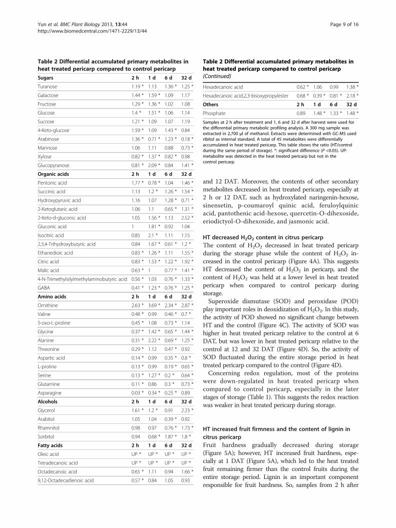
Table 2 Differential accumulated primary metabolites inheat treated pericarp compared to control pericarp
Sugars 2 h 1 d 6 d 32 d
Turanose 1.19 * 1.13 1.36 * 1.25 *
Galactose 1.44 * 1.59 * 1.09 1.17
Fructose 1.29 * 1.36 * 1.02 1.08
Glucose 1.4 * 1.51 * 1.06 1.14
Sucrose 1.21 * 1.09 1.07 1.19
4-Keto-glucose 1.59 * 1.09 1.43 * 0.84
Arabinose 1.36 * 0.71 * 1.23 * 0.18 *
Mannose 1.06 1.11 0.88 0.73 *
Xylose 0.82 * 1.37 * 0.82 * 0.98
Glucopyranose 0.81 * 2.09 * 0.84 1.41 *
Organic acids 2 h 1 d 6 d 32 d
Pentonic acid 1.77 * 0.78 * 1.04 1.46 *
Succinic acid 1.13 1.2 * 1.26 * 1.54 *
Hydroxypyruvic acid 1.16 1.07 1.28 * 0.71 *
2-Ketoglutaric acid 1.06 1.1 0.65 * 1.31 *
2-Keto-d-gluconic acid 1.05 1.56 * 1.13 2.52 *
Gluconic acid 1 1.81 * 0.92 1.04
Isocitric acid 0.85 2.1 * 1.11 1.15
2,3,4-Trihydroxybutyric acid 0.84 1.67 * 0.61 * 1.2 *
Ethanedioic acid 0.83 * 1.26 * 1.11 1.55 *
Citric acid 0.83 * 1.53 * 1.22 * 1.92 *
Malic acid 0.63 * 1 0.77 * 1.41 *
4-N-Trimethylsilylmethylaminobutyric acid 0.56 * 1.03 0.76 * 1.33 *
GABA 0.41 * 1.23 * 0.76 * 1.25 *
Amino acids 2 h 1 d 6 d 32 d
Ornithine 2.63 * 3.69 * 2.34 * 2.87 *
Valine 0.48 * 0.99 0.46 * 0.7 *
5-oxo-L-proline 0.45 * 1.08 0.73 * 1.14
Glycine 0.37 * 1.42 * 0.65 * 1.44 *
Alanine 0.31 * 2.22 * 0.69 * 1.25 *
Threonine 0.29 * 1.12 0.47 * 0.92
Aspartic acid 0.14 * 0.99 0.35 * 0.8 *
L-proline 0.13 * 0.99 0.19 * 0.65 *
Serine 0.13 * 1.27 * 0.2 * 0.64 *
Glutamine 0.11 * 0.86 0.3 * 0.73 *
Asparagine 0.03 * 0.34 * 0.25 * 0.89
Alcohols 2 h 1 d 6 d 32 d
Glycerol 1.61 * 1.2 * 0.91 2.23 *
Arabitol 1.05 1.04 0.39 * 0.92
Rhamnitol 0.98 0.97 0.76 * 1.73 *
Sorbitol 0.94 0.68 * 1.87 * 1.8 *
Fatty acids 2 h 1 d 6 d 32 d
Oleic acid UP * UP * UP * UP *
Tetradecanoic acid UP * UP * UP * UP *
Octadecanoic acid 0.65 * 1.11 0.94 1.66 *
9,12-Octadecadienoic acid 0.57 * 0.84 1.05 0.93
Table 2 Differential accumulated primary metabolites inheat treated pericarp compared to control pericarp(Continued)
Hexadecanoic acid 0.62 * 1.06 0.99 1.38 *
Hexadecanoic acid,2,3-bisoxypropylester 0.68 * 0.39 * 0.81 * 2.18 *
Others 2 h 1 d 6 d 32 d
Phosphate 0.89 1.48 * 1.33 * 1.48 *
Samples at 2 h after treatment and 1, 6 and 32 d after harvest were used forthe differential primary metabolic profiling analysis. A 300 mg sample wasextracted in 2,700 μl of methanol. Extracts were determined with GC-MS usedribitol as internal standard. A total of 45 metabolites were differentiallyaccumulated in heat treated pericarp. This table shows the ratio (HT/controlduring the same period of storage). *: significant difference (P <0.05). UP:metabolite was detected in the heat treated pericarp but not in thecontrol pericarp.
Yun et al. BMC Plant Biology 2013, 13:44 Page 9 of 16http://www.biomedcentral.com/1471-2229/13/44
and 12 DAT. Moreover, the contents of other secondarymetabolites decreased in heat treated pericarp, especially at2 h or 12 DAT, such as hydroxylated naringenin-hexose,sinensetin, p-coumaroyl quinic acid, feruloylquinicacid, pantothenic acid-hexose, quercetin-O-dihexoside,eriodictyol-O-dihexoside, and jasmonic acid.
HT decreased H2O2 content in citrus pericarpThe content of H2O2 decreased in heat treated pericarpduring the storage phase while the content of H2O2 in-creased in the control pericarp (Figure 4A). This suggestsHT decreased the content of H2O2 in pericarp, and thecontent of H2O2 was held at a lower level in heat treatedpericarp when compared to control pericarp duringstorage.Superoxide dismutase (SOD) and peroxidase (POD)
play important roles in deoxidization of H2O2. In this study,the activity of POD showed no significant change betweenHT and the control (Figure 4C). The activity of SOD washigher in heat treated pericarp relative to the control at 6DAT, but was lower in heat treated pericarp relative to thecontrol at 12 and 32 DAT (Figure 4D). So, the activity ofSOD fluctuated during the entire storage period in heattreated pericarp compared to the control (Figure 4D).Concerning redox regulation, most of the proteins
were down-regulated in heat treated pericarp whencompared to control pericarp, especially in the laterstages of storage (Table 1). This suggests the redox reactionwas weaker in heat treated pericarp during storage.
HT increased fruit firmness and the content of lignin incitrus pericarpFruit hardness gradually decreased during storage(Figure 5A); however, HT increased fruit hardness, espe-cially at 1 DAT (Figure 5A), which led to the heat treatedfruit remaining firmer than the control fruits during theentire storage period. Lignin is an important componentresponsible for fruit hardness. So, samples from 2 h after

Table 3 Secondary metabolites specifically accumulatedin heat treated pericarp
Secondary metabolites 2 h 1 d 6 d 32 d
Quercetin-dihexose-deoxyhexose 16.71 14.81 15.42 n.s.
Hesperetin 12.32 2.08 n.s. n.s.
Naringenin chalcone-hexose 15.04 n.s. 4.77 n.s.
Vanillic acid 14.73 n.s. n.s. 14.37
Hydroxybenzoic acid-hexose 14.99 n.s. n.s. n.s.
Diosmin 4.06 n.s. n.s. n.s.
Rutin 3.69 n.s. n.s. n.s.
Phloretin-C-diglycoside 14.97 n.s. 0.06 n.s.
Chlorogenic acid 15.96 0.06 0.12 n.s.
Isosakuranetin n.s. 16.42 n.s. n.s.
Qucercetin n.s. 16.03 0.14 0.12
Neohesperidin n.s. 1.31 0.14 n.s.
Protocatechuic acid n.s. 0.09 n.s. n.s.
Caffeic acid hexose n.s. 0.06 0.20 15.90
Naringenin n.s. n.s. 15.31 n.s.
Caffeic acid n.s. n.s. 0.09 n.s.
Hydroxylated naringenin-hexose n.s. n.s. 0.10 n.s.
Sinensetin n.s. n.s. 0.08 n.s.
p-Coumaroyl quinic acid n.s. n.s. 0.07 n.s.
Feruloylquinic acid n.s. n.s. 0.06 n.s.
Pantothenic acid -hexose n.s. n.s. 0.06 n.s.
Quercetin-O-dihexoside 0.08 n.s. 1.13 0.46
Cinnamic acid 0.11 n.s. n.s. n.s.
Eriodictyol-O-dihexoside 0.06 n.s. n.s. 0.06
Ferulic acid 0.42 n.s. 0.09 n.s.
Sinapic acid 0.05 n.s. 0.07 n.s.
Jasmonic acid 0.07 n.s. 0.06 n.s.
Samples at 2 h and 1, 6 and 32 DAT were used for the differential secondarymetabolic profiling analysis using the HPLC-QTOF-MS instrument. Fourreplicates were compared and statistically analysed per sample using Student’st-test; P < 0.05. After MS/MS analysis, a total of 58 metabolites were found tobe significantly regulated (2-fold) in heat treated pericarp when compared tocontrol pericarp (during the same period of storage), and 27 of thesemetabolites were identified. This table shows the ratio (HT/control during thesame period of storage). n.s.: not significant.
Yun et al. BMC Plant Biology 2013, 13:44 Page 10 of 16http://www.biomedcentral.com/1471-2229/13/44
treatment and 1, 6 and 32 DAT were used to detect lignincontent changes over time. Results show HT significantlyinduced lignin accumulation in pericarp (Figure 5B).Also, the activity of phenylalanine ammonia-lyase(PAL), a critical enzyme in lignin synthesis, initially in-creased in heat treated pericarp at 1 DAT but subse-quently fell into the same level as in the control(Figure 4B). Furthermore, levels of ferulic acid, sinapicacid, cinnamic acid and caffeic acid, the precursors oflignin synthesis, declined in heat treated pericarp whencompared to control pericarp (Table 3). This suggests arapid transformation occurred from ferulic acid, sinapicacid, cinnamic acid and caffeic acid to lignin in heattreated pericarp after HT.
DiscussionHT improved fruit pathogen resistance without negativelyimpacting fruit qualityPhysical treatments are used frequently to keep fruit fresh,including microporous membranes, pre-storage treatment(sweating), ultraviolet illumination, radiofrequency treat-ment, heat treatments (heat therapy), and other storagetechniques. Postharvest heat treatments such as hot watertreatment and hot air treatment provide a type of quaran-tine, inhibit the development of pathogens, enhance fruitresistance to chilling injury in cold-sensitive cultivars, andhelp preserve fruit quality during storage and extend itsshelf life [7]. In the present study, HT effectively inhibitedthe development of pathogens. Also, HT (52°C, 2 min)caused a statistically significant reduction the incidence ofchilling injury [9,11].Also, postharvest HT had no obviousnegative effects on fruit flesh quality during subsequentstorage. This suggests HT increased the ability of fruit totolerate storage stress without affecting their commercialqualities, and shows strong potential application in thecitrus industry.
HT induced stress response proteinsHT can effectively inhibit fruit chilling injury during coldstorage [12]. After HT, some proteins were up-regulated,while others were down-regulated. The up-regulatedproteins might be responsible for heat induced fruitresistance. Interesting, some proteins were up-regulated at1 d after HT, and decreased to the previous level of controlduring the storage period. Those kinds of proteinsmight be just involved in response to HT, but not play avital role in fruit disease resistance. The proteins, whichare up-regulated during whole storage period might bedirectly involved in fruit disease resistance.In the present study, heat shock protein (HSP) 17.7,
putative HSP 70 and low molecular weight HSP wereup-regulated in heat treated pericarp compared to thecontrol during entire storage period (Table 1). HSPs areencoded by a multigene family and are located in dif-ferent subcellular compartments [13]. These molecularchaperones play a crucial role in protecting plantsagainst stress by re-establishing normal protein configur-ation and thus helping to maintain cellular homeostasis[14]. Abiotic stresses usually causes protein dysfunction;so maintaining proteins in their functional configurationsand preventing the aggregation of non-native proteinsis particularly important for cell survival under stress[14]. HSPs are responsible for protein folding, assem-bly, translocation and degradation during cellular pro-cesses. HSPs also stabilize proteins and membranes,and can assist in protein refolding under stressful con-ditions [15]. Our results suggest HSPs play a crucialrole in reducing the susceptibility of HT fruit to bioticand abiotic stress.
Figure 4 Changes of H2O2 content and enzyme activity in HT and control pericarp. A: H2O2 content. Samples at 2 h and 1, 6 and 32 DATwere used for H2O2 content determined in HT and control pericarp. B-D: Enzyme activity. Samples at 1, 6, 12 and 32 DAT were used for enzymeactivity determination. PAL: phenylalanine ammonia-lyase; SOD: superoxide dismutase; POD: peroxidase. Four replicates were completed andstatistically significant differences analysis was conducted between HT and control pericarp during the same period using Student’s t-test.*: significant difference (P < 0.05). Mean values and SE bars are provided.
Figure 5 Changes of fruit hardness (A) and lignin content (B) inHT and control pericarp. Samples at 2 h and 1, 6 and 32 DAT wereused for fruit hardness and lignin content determination. An analysis ofstatistically significant differences was conducted between HT andcontrol pericarp during the same period using Student’s t-test.*: significant difference (P < 0.05). Mean values and SE bars are provided.
Yun et al. BMC Plant Biology 2013, 13:44 Page 11 of 16http://www.biomedcentral.com/1471-2229/13/44
HT significantly inhibited pathogen developmentand fruit decay during storage [16]. Chitinase and β-1,3-glucanase were up-regulated in heat treated fruit inthis study; these enzymes have long been thought tobe active in fruit antifungal defences. Pathogenesis-related(PR) proteins have been classified into five groups and twoof these groups contain proteins with β-1, 3-glucanaseand chitinase [17]. Chitinase catalyses the hydrolysis ofchitin, a β-1, 4-linked polymer of N-acetyl D-glucosamineand is a major component of cell walls of most phytopath-ogenic fungi [18]. Crude protein extracts of chitinase andβ-1, 3-glucanase were observed to effectively inhibit fungalactivity [19]. Also, β-1, 3-glucanase and chitinase arethought to contribute to the level of resistance providedand are known to inhibit mycelial growth of a wide rangeof fungal pathogens [20]. This suggests β-1, 3-glucanaseand chitinase were directly involved in HT inducedpathogen resistance in fruits.
HT increased the content of stress response metabolitesPrimary metabolites are directly involved in normalgrowth, development, and reproduction. In the presentstudy, the content of ornithine, oleic acid and tetradecanoicacid increased in heat treated pericarp compared to thecontrol during storage, and levels of seven types of sugarsincreased in heat treated pericarp compared to the controlat 2 h after HT (Table 2). In plants, ornithine is requiredfor the synthesis of polyamines and alkaloids, which

Yun et al. BMC Plant Biology 2013, 13:44 Page 12 of 16http://www.biomedcentral.com/1471-2229/13/44
contribute to oxidative stress tolerance in plants subjectedto severe water stress [21].Flavonoids are important defensive compounds which
protect fruit against the development of pathogens, actingeither as phytoanticipins or phytoalexins [22]. Flavonoidswere observed to be up-regulated in heat treated pericarp(compared to the control), including quercetin-dihexose-deoxyhexose, hesperetin, naringenin, naringenin chalcone-hexose, hydroxybenzoic acid-hexose, isosakuranetin,diosmin, and rutin, which may play a vital role in responseto stress [23]. Rutin is a bioflavonoid with strong antioxi-dant activity, which had an antimicrobial effect on patho-gens, as evidenced by reduced conidial germination andappressorium formation of the pathogen [22]. Quercetinprotected cells from oxidative stress-induced cell deathby blocking both the cyclooxygenase and lipooxygenasepathways [24]. Quercetin has a protective function againstoxidative stress by reducing oxidative injury to the cellsduring H2O2 treatment [25]. Also, plant extracts rich inquercetin have been applied as an interesting resource forfunctional food products and other foods used to reducethe risks of age-related macular degeneration [25]. Hesperi-din acts as an antioxidant, based on in vitro studies, andhesperidin also provides strong cellular antioxidant pro-tection against the damaging effects induced by stress[26]. Diosmin, hesperidin and naringin are flavonoidglycosides that occur naturally in citrus fruits; theyexert a variety of properties such as antioxidant and freeradical scavenging [27]. Interestingly, naringenin andrutin are not only involved in response to stress, butalso were assumed to be signalling molecules [23].
HT induced fruit pathogen resistance and increased lignincontentThe functions of phenylpropanoid compounds in plantdefence range from preformed or inducible physical andchemical barriers against infection [10]. Defensive functionsare not restricted to a particular class of phenylpropanoidcompounds, but are found in the lignification ofmonolignols to lignin [28]. In the present study, HTdecreased the level of precursors needed in lignin synthesis(ferulic acid, sinapic acid, cinnamic acid and caffeic acid),but increased lignin content (Table 3, Figure 5). Ligninis one of the major components of secondary cell walls,providing cells with mechanical support and isolation fromthe environment. In plants, cells can sense cell wall polymerstructure and this activates trans-plasma membrane sensorproteins which might trigger a cascade of signal transduc-tion events interwoven with other pathways that mightmodulate cellular functions by the activation or inhibitionof specific transcription factors as well as by affectingposttranslational control of gene expression and proteinfunction [29]. If a cell wall is damaged, cells activate aseries of responses to the invading disease organisms, which
might negatively impact fruit quality. The accumulation oflignin induced by HT creates a thickening of cell wallswhich forms an effective physical barrier to pathogens,delaying the invasion of disease organisms. Also, HTincreased fruit firmness; this might be a result of HTinduced lignin thickened fruit cell walls.
HT reduced the content of H2O2
HT is an effective method for improving citrus storability.In storage, fruits suffer a variety of biotic and abioticenvironmental stresses. All these stresses could leadto the generation of reactive oxygen species (ROS).To avoid the accumulation of ROS, which may leadto cell death, plants have up-regulated enzymatic andnon-enzymatic antioxidants to scavenge ROS and al-leviate their negative effects [30]. In this study, HTdown-regulated H2O2 content in pericarp, which wasconsistent with the effect of HT on yeast cells. HTyeast cells accumulated less ROS than untreated cellsin response to stresses [31].Also, isoflavone reductase (IFR) might have an important
role as an antioxidant [32]. IFR is an enzyme involvedin the production of isoflavone phytoalexins and itaccumulates in response to pathogenic attack, fungalelicitor and biotic stress [33]. Isoflavonoids are import-ant secondary metabolites for essential physiologicalprocesses. Kim et al. [32] reported the isoflavonereductase-like gene may act as a down-regulator byproducing antioxidant chemicals to prevent the elevationof ROS. In this study, isoflavone reductase, a related pro-tein (H10), was up-regulated in heat treated pericarp whencompared to control pericarp (Table 1), which might beanother factor leading to lower H2O2 levels and strongerresistance to pathogens in heat treated pericarp whencompared to control pericarp.Ascorbate peroxidase scavenges H2O2 in specific organ-
elles of the cell [34]. Also, Cu/Zn-SOD is the main cellularisoform of SODs which play a key role in the antioxidantdefence system through the disproportionation of H2O2
[34]. Both of these can prevent ROS caused damages tothe cellular membrane and act as a primary defencemechanism when organisms were exposed to oxidativestress. These proteins are considered the main enzymaticand non-enzymatic systems for protecting cells againstoxidative damage, and are responsible for disease resistance.In response to biotic stress, these proteins also act asthe primary defence mechanism against oxidative stressescaused by pathogens, therefore preventing damage of ROSto cellular membranes. However, the activities of POD andSOD were not elevated in Heat treated pericarp whencompared to control pericarp, which might be causedby the lower H2O2 content in heat treated pericarp.Also, other proteins involved in scavenging ROS weredown-regulated in heat treated pericarp when compared
Yun et al. BMC Plant Biology 2013, 13:44 Page 13 of 16http://www.biomedcentral.com/1471-2229/13/44
to control pericarp, including oxidoreductase (H11),oxidoreductase (H12, 17), aldo/keto reductase (H13), 2-oxoacid dehydrogenase (H14), superoxide dismutase(H16). This suggests HT induced a level of resistance topathogens attacking fruits by causing a decline in fruitH2O2 content and by down-regulated scavenging ROSenzymes.
Role of sugars, organic acids and amino acids inimproving storabilityIn this study, the profiling of primary metabolics showedHT induced the accumulation of sugars at early stagesof storage (Table 2). Soluble sugars, especially sucrose,glucose, and fructose, accumulated when fruits weresubjected to stress, and accumulated soluble sugars playan obviously vital role in the response of fruit to a numberof stressors [35]. Soluble sugars do not only act as nutrientsand metabolites, but also act as signal substance inthe modification of redox homeostasis [36]. However,less information was available on how other sugars areinvolved in the stress response, especially arabinose,mannose, galactose, and ribose.Moreover, primary metabolic data shows HT reduced
organic acids and amino acids content especially at 2 hafter treatment in this study. Although organic acids andamino acids play an obvious central role in energy andmetabolism at the cellular and whole-organism levels, thereseems to be a rapid conversion from organic acids andamino acids to sugars in heat treated pericarp. However,little is known about the decreased levels of organic acidsand amino acids seen during a stress response, and furtherstudy will be required to validate this response.
ConclusionsThis study provides a broad picture of differential accumu-lation of proteins and metabolites, and gives a new insightinto how HT increases fruit stress resistance during subse-quent storage of citrus fruit. In metabolic substrates, theup-accumulation of secondary metabolites played vital rolesin increasing the ability of fruit to deal with stress.Flavonoids are directly involved in the response toexternal stress. The increased lignin and decreasedH2O2 contents involved in HT probably increasedfruit resistibility in response to external stress.
MethodsSample collection‘Kamei’ Satsuma mandarin (Citrus unshiu Marc.) fruitswere harvested at commercial maturity (fruit colour hasturned completely orange and the level of soluble solidsapproaches 9%) from an orchard in the city of Yichang,Hubei Province, China, in October 2009. Fruits of auniform size and colour, free of visible injury or blem-ishes, were selected for the experiments. Three hundred
kilograms of fruit were divided into two equal parts,150 kg were used for the HT and the other 150 kg usedfor a control. After treatments, fruits were stored in aventilated warehouse (storage temperature: 12–16°C;relative humidity: 90–95%). Samples were collected at2 h, and 1, 2, 3, 4, 6, 9, 12, 16, 20, 24, 28, 32, 38, 44, 50and 60 DAT. The pericarp was sampled along theequatorial plane of each fruit; 30 fruits were selected asone sample and ground to powder in liquid nitrogen,then stored at –80°C for further analysis.
Heat treatment (HT)Heat treated fruits were dipped in a 52°C warm water bathfor 2 min, and control fruits were dipped in a 25°C waterbath for 2 min. After treatments, fruits were air dried andstored in a ventilated warehouse prior to sampling.
Fruit quality determinationFruits were checked for weight loss (%), respiration rate(mg kg–1 h–1) and total soluble solids (TSS,%) at 1, 2, 3,4, 6, 9, 12, 16, 20, 24, 28, 32, 38, 44, 50 and 60 DAT. Theweight was measured using an electronic hydrostaticbalance (Model: MP31001, Shanghai Selon ScientificInstrument, Co., Ltd, Shanghai, China) with an accuracyof ± 0.01 g. The respiration rate of fruit was measuredwith an infrared gas analyser (Model: GXH-305H, JunfangScience & Technology Institute of Physical and ChemicalResearch, Beijing, China). TSS were determined with arefractometer (Model: Pocket PAL-1, Atago Inc., Toyko,Japan) according to the manufacturer’s instructions.
Antifungal assay of HT in wounded citrus fruitsBlue mould (Penicillium italicum) was chosen as an indi-cator to evaluate the resistance of treated fruit to fungalinfections. 150 fruits from each treatment were used forfungal inoculation. A uniform lesion (3 mm deep, 4 mmwide) was made at the equator of the fruit using a sterilenail. Aliquots of 10 μl suspension of P. italicum at 1×10⁵
spore ml–1 were inoculated into each wound site. Afterfungal inoculation, fruits were stored in a storage chamber(relative humidity, 95%; temperature, 25°C), and diseaseincidence was detected at 1, 2, 3, 4, 5, 6 and 7 d after in-oculation. Disease incidence rates and lesion diameterswere recorded based on the following equations:
Disease incidence rate %ð Þ¼ ∑ Number of diseased=decaying citrus
=total number of fruit in the treatment� 100;
Lesion diameter cmð Þ¼ ∑ Lesion diameter in diseased=decayingcitrusfruit
=total number of fruit in the treatment

Yun et al. BMC Plant Biology 2013, 13:44 Page 14 of 16http://www.biomedcentral.com/1471-2229/13/44
Two-dimensional gel electrophoresis and MAILDI-TOF/TOFSamples at 1, 6, 12 and 32 DAT were used for total proteinextraction based on the method described by Isaacson et al.[37]. The precipitate was then nitrogen gas-dried andsolubilized in the lysis buffer containing 8 M urea,0.2% (w/v) Bio-Lyte, 4% CHAPS, 65 mM DTT. Theprotein concentration was determined with a Bio-Radprotein assay kit (Bio-Rad Laboratories Inc., Hercules,CA, USA) based on the Bradford method using BSA asthe standard. Two-dimensional gel electrophoresis andgel staining was carried out based on the method ofYun et al. [38]. The stained gels were imaged with a UVPimager (Bio-Rad) in the ‘trans white’ mode using PDQuest2-D analysis software version 7.4 (Bio-Rad). Protein spotsmatching and differential protein spots analysis werecompleted based on the method of Yun et al. [38].Isoelectric points and molecular weights of the proteinswere determined by comparison with the markers.In-gel digestion and protein identification were performed
using a 4800 Proteomics Analyzer MALDI TOF/TOF(Applied Biosystems, Foster City, CA) as described byYun et al. [38]. MS/MS data were then submitted to theMASCOT program for protein identification against thegreen plants database. The following are the searchparameters: trypsin enzyme, one missed cleavage, fixedmodifications of carbamidomethyl (C), fragment masstolerance of ± 0.5 Da, variable modifications of oxidation(Met), peptide tolerance of 100 ppm, and peptide chargeof 1+. Only results of peptides with MS/MS P < 0.05confidence were accepted.
The primary metabolic profilingSamples collected at 2 h and 1, 6 and 32 DAT were usedfor the differential primary metabolic profiling analysis.A 300 mg sample was extracted in 2,700 μl of methanolas described by Roessner-Tunali et al. [39]. 300 μl of0.2 mg ml–1 ribitol in water was added as a quantifica-tion internal standard. A derivatization reaction wasperformed based on the protocol of Zhang et al. [40]with little modification. Extracts were incubated in50 μl of 20 mg ml–1 methoxyamine hydrochloride inpyridine for 30 min at 50°C, followed by a 40 min treatmentat 60°C using 50 μl BSTFA (containing 1% TMCS). Eachsample (1 μl) was injected into the gas chromatographsystem through a fused-silica capillary column (30 m ×0.25 mm i.d., 0.25 μm) DB-5 MS stationary phase. Theinjector temperature was 250°C, with a carrier gas flowrate of 1.0 ml min–1. The column temperature washeld at 100°C for 1 min; increased to 184°C at a rate of3°C min–1, increased to 190°C at a rate of 0.5°C min–1,increased to 280°C at 15°C min–1. The flow rate of thecarrier helium (99.999%) gas was 1 ml min–1. The followingwere MS operating parameters: ionization voltage, 70 eV(electron impact ionization); ion source temperature,
200°C; interface temperature, 250°C. TIC (total ioncurrent)spectra were recorded in the mass range of 45–600 atomicmass units in scanning mode.
The secondary metabolic profilingSamples at 2 h and 1, 6 and 32 DAT were used for dif-ferential secondary metabolic profiling analysis usingHPLC-MS. 300 mg dried powder was extracted withmethanol, the mixture was filtered through a 0.22–mmpolytetrafluoroethylene membrane filter.Metabolite profiling was performed using a QTOF 6520
mass spectrometer (Agilent Technologies, Palo Alto, CA,USA) coupled to a 1200 series Rapid Resolution HPLCsystem as described by Page et al. [41]. 2 μl of sampleextract was loaded onto a Zorbax Eclipse Plus C181.8 μm, 2.1 × 100 mm reverse-phase analytical column(Agilent Technologies). Mobile phase A comprised 0.1%formic acid in water and mobile phase B was acetonitrile.The following gradient was used: 0 min–10% B;20 min–95% B; 22 min–95% B; 22.1 min–10% B;30 min–10% B. The flow rate was 0.3 ml min–1 and thecolumn temperature was held at 35°C for the duration.The source conditions for electrospray ionization wereas follows: gas temperature was 350°C with a dryinggas flow rate of 10 l min–1 and a nebulizer pressure of40 psig. The capillary voltage was 3.5 kV in both positiveand negative ion mode. The fragmentor voltage was135 V and the skimmer was 65 V.
Assay of phenylalanine ammonia-lyase (PAL), superoxidedismutase (SOD) and peroxidase (POD) activitiesSamples collected at 1, 6, 12 and 32 DAT were used for en-zyme activity determination. A 5 g sample was homogenizedin 25 ml of 0.05 M sodium borate buffer (pH 8.8 for PAL,containing 5 mM β-mercaptoethanol) and 25 ml of ice-cold100 mM sodium phosphate (pH 7.8 for POD and SOD,containing 0.5 g polyvinyl polypyrrolidone). PAL activitywas determined by measuring the absorbance of cinnamicacid at 290 nm over a period of 30 min at 30°C [42]. PODactivity was analysed using guaiacol as the substrate [43].SOD activity was determined using the xanthine-xanthineoxidase method, and nitro blue tetrazolium was used as theindicator of superoxide radical production [44].
Determination the content of H2O2 in the peelSamples collected at 2 h and 1, 6 and 32 DAT were used tomeasure hydrogen peroxide levels based on the methods ofPatrick and Wagner [45] with some modifications. Briefly,peel tissues (0.5 g) were ground in liquid nitrogen andhomogenized in 5 ml of physiological saline. Samples werethen subjected to an ultrasonic bath for 15 min andcentrifuged at 6,000 g for 20 min. The supernatants (50 μl)were subsequently added into 0.5 ml of solution I andwarmed in a 37°C water bath for 10 min before 0.5 ml of

Yun et al. BMC Plant Biology 2013, 13:44 Page 15 of 16http://www.biomedcentral.com/1471-2229/13/44
solution II was added. The absorbance of the mixture wasmeasured at 405 nm and the content of H2O2 in eachsample was calculated by comparison with a standardcalibration curve.
Determination of fruit firmnessSamples collected at 2 h and 1, 6 and 32 DAT were usedfor the determination of fruit firmness. Eighteen fruitswere measured and every fruit was measured on threedifferent spots around the equatorial plane. Firmness wasmeasured with a fruit sclerometer (GY-B, Jilin, China)with 4 mm diameter and expressed as N cm–2 and theaverage value per fruit was used for further analysis, afterremoving three largest and three smallest measurements;a total twelve replicates were comparative statisticallyanalysed per sample using Student’s t-test (P < 0.05).
Determination of lignin in pericarpSamples collected at 2 h and 1, 6 and 32 DAT were used tomeasure fruit lignin content. Lignin levels were determinedusing the method described by Lei et al. [46].
Statistical analysisThe experimental design was completely randomizedwith more than three replications. Four individual replicateswere completed in 2-DE, GC-MS, HPLC-MS, H2O2 con-tent, lignin content and enzyme activity determination.Twelve replicates were completed to measure weight loss,respiration rate, total soluble solids and fruit firmness. Datafor each sample was statistically analysed using Student’st-test (P < 0.05).
Abbreviations2-DE: Two-dimensional gel electrophoresis; GC-MS: Gas chromatographycoupled to mass spectrometry; HSP: Heat shock protein; HT: Heat treatment;IFR: Isoflavone reductase; LC-QTOF-MS: Liquid chromatography quadrupoletime-of-flight mass spectrometry; MALDI-TOF MS: Matrix-assisted laserdesorption/ionization-time-of-flight tandem mass spectrometry;NCBI: National center for biotechnology information; PAL: Phenylalanineammonia-lyase; POD: Peroxidise; PR: Pathogenesis-related; ROS: Reactiveoxygen species; SOD: Superoxide dismutase; TSS: Total soluble solids.
Competing interestsThe authors declare that they have no competing interests.
Authors’ contributionsZY, HJG and YJC conceived the study, designed all the experiments andperformed silicon and biochemical analyses. SZL carried out primarymetabolite detection and statistical analysis. SJ carried out secondarymetabolite detection and statistical analysis. ZY, HJG, TL, QX, JX, YJC and XXDinterpreted the experimental dada and participated in writing themanuscript. All the authors read and approved the final manuscript.
AcknowledgementsWe thank Prof. Hanhui Kuang of Huazhong Agricultural University for help inrevising the manuscript. This work was supported by Huazhong AgriculturalUniversity Scientific & Technological Self-innovation Foundation, the NaturalScience Foundation of China (NSFC No. 31271968), The Important Project ofMinistry of Education (311029), and National Modern Agriculture (Citrus)Technology Systems of China (No. CARS-27).
Received: 4 September 2012 Accepted: 5 March 2013Published: 16 March 2013
References1. Gonzalez-Candelas L, Alamar S, Sanchez-Torres P, Zacarias L, Marcos JF: A
transcriptomic approach highlights induction of secondary metabolismin citrus fruit in response to Penicillium digitatum infection. BMC PlantBiol 2010, 10:194.
2. Tu K, Shao XF, Tu SC, Su J, Zhao Y: Effects of heat treatment on woundhealing in gala and Red fuji apple fruits. J Agr Food Chem 2010, 58:4303–4309.
3. Lara MV, Borsani J, Budde CO, Lauxmann MA, Lombardo VA, Murray R,Andreo CS, Drincovich MF: Biochemical and proteomic analysis of‘Dixiland’ peach fruit (Prunus persica) upon heat treatment. J Exp Bot2009, 60:4315–4333.
4. Gould GW: History of science - spores - lewis B perry memorial lecture2005. J Appl Microbiol 2006, 101:507–513.
5. Hong SI, Lee HH, Kim D: Effects of hot water treatment on the storagestability of satsuma mandarin as a postharvest decay control. PostharvestBiol Tec 2007, 43:271–279.
6. Zhang ZK, Bi Y, Ge YH, Wang JJ, Deng JJ, Xie DF, Wang Y: Multiple pre-harvest treatments with acibenzolar-S-methyl reduce latent infectionand induce resistance in muskmelon fruit. Sci Hortic-Amsterdam 2011,130:126–132.
7. Schirra M, D’Aquino S, Cabras P, Angioni A: Control of postharvestdiseases of fruit by heat and fungicides: efficacy, residue levels, andresidue persistence. A Review. J Agr Food Chem 2011, 59:8531–8542.
8. Perotti VE, Del Vecchio HA, Sansevich A, Meier G, Bello F, Cocco M, GarránSM, Anderson C, Vázquez D, Podestá FE: Proteomic, metabalomic, andbiochemical analysis of heat treated Valencia oranges during storage.Postharvest Biol Tec 2011, 62:97–114.
9. Sapitnitskaya M, Maul P, McCollum GT, Guy CL, Weiss B, Samach A, Porat R:Postharvest heat and conditioning treatments activate differentmolecular responses and reduce chilling injuries in grapefruit. J Exp Bot2006, 57:2943–2953.
10. Dixon RA, Achnine L, Kota P, Liu CJ, Reddy MSS, Wang LJ: Thephenylpropanoid pathway and plant defence - a genomics perspective.Mol Plant Pathol 2002, 3:371–390.
11. Ghasernnezhad M, Marsh K, Shilton R, Babalar M, Woolf A: Effect of hotwater treatments on chilling injury and heat damage in ‘satsuma’mandarins: Antioxidant enzymes and vacuolar ATPase, andpyrophosphatase. Postharvest Biol Tec 2008, 48:364–371.
12. Vigneault C, Lu J, Charles MT, Goyette B, Raghavan GSV: Effect of heattreatment uniformity on tomato ripening and chilling injury. PostharvestBiol Tec 2010, 56:155–162.
13. Vierling E: The roles of heat shock proteins in plants. Annu Rev Plant Biol1991, 42:579–620.
14. Wang WX, Vinocur B, Shoseyov O, Altman A: Role of plant heat-shockproteins and molecular chaperones in the abiotic stress response. TrendsPlant Sci 2004, 9:244–252.
15. Basha E, Jones C, Wysocki V, Vierling E: Mechanistic differences betweentwo conserved classes of small heat shock proteins found in the plantcytosol. J Biol Chem 2010, 285:11489–11497.
16. Hu WZ, Jiang AL, Jin LM, Liu CH, Tian MX, Wang YY: Effect of heattreatment on quality, thermal and pasting properties of sweet potatostarch during yearlong storage. J Sci Food Agr 2011, 91:1499–1504.
17. Schraudner M, Ernst D, Langebartels C, Sandermann H Jr: Biochemicalplant responses to ozone: III. Activation of the defence-relatedproteins β-1, 3-glucanase and chitinase in tobacco leaves. PlantPhysiol 1992, 99:1321.
18. Chalavi V, Tabaeizadeh Z, Thibodeau P: Enhanced resistance to Verticilliumdahliae in transgenic strawberry plants expressing a Lycopersiconchilense chitinase gene. J Am Soc Hortic Sci 2003, 128:747–753.
19. Mauch F, Mauch-Mani B, Boller T: Antifungal hydrolases in pea tissue: II.Inhibition of fungal growth by combinations of chitinase and β-1,3-glucanase. Plant Physiol 1988, 88:936–942.
20. Magnin-Robert M, Trotel-Aziz P, Quantinet D, Biagianti S, Aziz A: Biologicalcontrol of Botrytis cinerea by selected grapevine-associated bacteria andstimulation of chitinase and beta-1,3 glucanase activities under fieldconditions. Eur J Plant Pathol 2007, 118:43–57.
21. Kalamaki MS, Alexandrou D, Lazari D, Merkouropoulos G, Fotopoulos V,Pateraki I, Aggelis A, Carrillo-Lopez A, Rubio-Cabetas MJ, Kanellis AK:
Yun et al. BMC Plant Biology 2013, 13:44 Page 16 of 16http://www.biomedcentral.com/1471-2229/13/44
Over-expression of a tomato N-acetyl-L-glutamate synthase gene(SlNAGS1) in Arabidopsis thaliana results in high ornithine levels andincreased tolerance in salt and drought stresses. J Exp Bot 2009,60:1859–1871.
22. Radhakrishna Shetty R, Jensen JD: Silicon-induced changes in antifungalphenolic acids, flavonoids and Key phenylpropanoid pathway genesduring the interaction between miniature roses and the biotrophicpathogen podosphaera pannosa. Plant Physiol 2011, 157:2194–2205.
23. Treutter D: Significance of flavonoids in plant resistance: a review.Environ Chem Lett 2006, 4:147–157.
24. Nijveldt RJ, van Nood E, van Hoorn DEC, Boelens PG, van Norren K, vanLeeuwen PAM: Flavonoids: a review of probable mechanisms of actionand potential applications. Am J Clin Nutr 2001, 74:418–425.
25. Saviranta NMM, Veeroos L, Granlund LJ, Hassinen VH, Kaarniranta K,Karjalainen RO: Plant flavonol quercetin and isoflavone biochanin Adifferentially induce protection against oxidative stress andinflammation in ARPE-19 cells. Food Res Int 2011, 44:109–113.
26. Wilmsen PK, Spada DS, Salvador M: Antioxidant activity of the flavonoidhesperidin in chemical and biological systems. J Agr Food Chem 2005,53:4757–4761.
27. Kanaze FI, Gabrieli C, Kokkalou E, Georgarakis M, Niopas I: Simultaneousreversed-phase high-performance liquid chromatographic method forthe determination of diosmin, hesperidin and naringin in different citrusfruit juices and pharmaceutical formulations. J Pharmaceut Biomed 2003,33:243–249.
28. Denness L, McKenna JF, Segonzac C, Wormit A, Madhou P, Bennett M,Mansfield J, Zipfel C, Hamann T: Cell wall damage-induced ligninbiosynthesis is regulated by a reactive oxygen species- andjasmonic acid-dependent process in arabidopsis. Plant Physiol 2011,156:1364–1374.
29. Seifert GJ, Blaukopf C: Irritable walls: the plant extracellular matrix andsignaling. Plant Physiol 2010, 153:467–478.
30. Apel K, Hirt H: Reactive oxygen species: metabolism, oxidative stress, andsignal transduction. Annu Rev Plant Biol 2004, 55:373–399.
31. Liu J, Wisniewski M, Droby S, Tian SP, Hershkovitz V, Tworkoski T: Effect ofheat shock treatment on stress tolerance and biocontrol efficacy ofMetschnikowia fructicola. FEMS Microbiol Ecol 2011, 76:145–155.
32. Kim SG, Kim ST, Wang YM, Kim SK, Lee CH, Kim KK, Kim JK, Lee SY, Kang KY:Overexpression of rice isoflavone reductase-like gene (OsIRL) conferstolerance to reactive oxygen species. Physiol Plantarum 2010, 138:1–9.
33. Kim ST, Cho KS, Yu S, Kim SG, Hong JC, Han CD, Bae DW, Myung AE, KangKY: Proteomic analysis of differentially expressed proteins induced byrice blast fungus and elicitor in suspension-cultured rice cells. Proteomics2003, 3:2368–2378.
34. Zhang HX, Xia Y, Wang GP, Shen ZG: Excess copper induces accumulationof hydrogen peroxide and increases lipid peroxidation and total activityof copper-zinc superoxide dismutase in roots of Elsholtzia haichowensis.Planta 2008, 227:465–475.
35. Rosa M, Prado C, Podazza G, Interdonato R, González JA, Hilal M, Prado FE:Soluble sugars—metabolism, sensing and abiotic stress: a complexnetwork in the life of plants. Plant Signal Behav 2009, 4:388.
36. Rosenwasser S, Rot I, Sollner E, Meyer AJ, Smith Y, Leviatan N, Fluhr R,Friedman H: Organelles contribute differentially to reactive oxygenspecies-related events during extended darkness. Plant Physiol 2011,156:185.
37. Isaacson T, Damasceno CMB, Saravanan RS, He Y, Catala C, Saladie M, RoseJKC: Sample extraction techniques for enhanced proteomic analysis ofplant tissues. Nat Protoc 2006, 1:769–774.
38. Yun Z, Li WY, Pan ZY, Xu J, Cheng YJ, Deng XX: Comparative proteomicsanalysis of differentially accumulated proteins in juice sacs of ponkan(Citrus reticulata) fruit during postharvest cold storage. PostharvestBiology and Technology 2010, 56:189–201.
39. Roessner-Tunali U, Hegemann B, Lytovchenko A, Carrari F, Bruedigam C,Granot D, Fernie AR: Metabolic profiling of transgenic tomato plantsoverexpressing hexokinase reveals that the influence of hexosephosphorylation diminishes during fruit development. Plant Physiol 2003,133:84–99.
40. Zhang JJ, Wang X, Yu O, Tang JJ, Gu XG, Wan XC, Fang CB: Metabolicprofiling of strawberry (Fragariaxananassa Duch.) during fruitdevelopment and maturation. J Exp Bot 2011, 62:1103–1118.
41. Page M, Sultana N, Paszkiewicz K, Florance H, Smirnoff N: The influence ofascorbate on anthocyanin accumulation during high light acclimation inArabidopsis thaliana: further evidence for redox control of anthocyaninsynthesis. Cell & Environment: Plant; 2011.
42. Ballester AR, Lafuente MT, Gonzalez-Candelas L: Spatial study ofantioxidant enzymes, peroxidase and phenylalanine ammonia-lyase inthe citrus fruit-Penicillium digitatum interaction. Postharvest Biol Tec 2006,39:115–124.
43. Meng XH, Han J, Wang Q, Tian SP: Changes in physiology and quality ofpeach fruits treated by methyl jasmonate under low temperature stress.Food Chem 2009, 114:1028–1035.
44. Sala JM, Lafuente MT: Antioxidant enzymes activities and rindstaining in‘Navelina’ oranges as affected by storage relative humidity and ethyleneconditioning. Postharvest Biol Tec 2004, 31:277–285.
45. Patrick WA, Wagner HB: Determination of hydrogen peroxide in smallconcentrations. Anal Chem 1949, 21:1279–1280.
46. Lei Y, Liu Y-Z, Gu Q-Q, Yang X-Y, Deng X-X, Chen J-Y: Comparison of cellwall metabolism in the pulp of three cultivars of ‘Nanfeng’ tangerinediffering in mastication trait. J Sci Food Agr 2011, 92:496–502.
doi:10.1186/1471-2229-13-44Cite this article as: Yun et al.: Comparative proteomic and metabolomicprofiling of citrus fruit with enhancement of disease resistance bypostharvest heat treatment. BMC Plant Biology 2013 13:44.
Submit your next manuscript to BioMed Centraland take full advantage of:
• Convenient online submission
• Thorough peer review
• No space constraints or color figure charges
• Immediate publication on acceptance
• Inclusion in PubMed, CAS, Scopus and Google Scholar
• Research which is freely available for redistribution
Submit your manuscript at www.biomedcentral.com/submit