Research Article ... · (MMC absorbed at 90 minutes = 67 8) ... 015 30 45 60 90120 Abdominal...
8
Hindawi Publishing Corporation Gastroenterology Research and Practice Volume 2012, Article ID 623417, 7 pages doi:10.1155/2012/623417 Research Article Hyperthermic Intraoperative Thoracoabdominal Chemotherapy Paul H. Sugarbaker, 1 David Chang, 2 and O. Anthony Stuart 1 1 Washington Hospital Center, Washington Cancer Institute, 106 Irving Street, NW, Suite 3900, Washington, DC 20010, USA 2 Westat, 1600 Research Boulevard, Rockville, MD 20850-3129, USA Correspondence should be addressed to Paul H. Sugarbaker, [email protected] Received 6 February 2012; Accepted 1 March 2012 Academic Editor: Pompiliu Piso Copyright © 2012 Paul H. Sugarbaker et al. This is an open access article distributed under the Creative Commons Attribution License, which permits unrestricted use, distribution, and reproduction in any medium, provided the original work is properly cited. Cytoreductive surgery combined with hyperthermic intraperitoneal chemotherapy (HIPEC) is a treatment option for selected patients with pseudomyxoma peritonei (PMP) and diffuse malignant peritoneal mesothelioma (DMPM). Tumor infiltration of the hemidiaphragm requiring partial resection occurs as a result of large volume and/or invasive disease at this anatomic site. Transmission of disease from abdomen to chest is a great danger in this group of patients. From a prospective database, patients who had diaphragm resection and then hyperthermic thoracoabdominal chemotherapy (HITAC) as a component of a cytoreductive surgical procedure were identified. Data from control patients receiving HIPEC or hyperthermic intrathoracic chemotherapy (HITOC) were analyzed for comparison. The morbidity, mortality, survival, and recurrence rate within the thoracic space were presented. Thirty patients had partial resection of a hemidiaphragm as part of a cytoreductive surgical procedure that utilized HITAC. The pharmacologic benefit of intracavitary chemotherapy administration was documented with an area under the curve ratio of intracavitary concentration times time to plasma concentration times time of 27 ± 10 for mitomycin C and 75 ± 26 for doxorubicin. Comparing percent chemotherapy absorbed for a ninety-minute treatment showed the largest for HIPEC, then for HITAC, and lowest for HITOC. The incidence of grade 3 and 4 adverse events was 43%. There was no mortality. Adjustments in the chemotherapy dose are not necessary with HITAC. The morbidity was high, the survival was acceptable, and intrathoracic recurrence was low. 1. Introduction Increasing interest in the surgical management of peritone- al metastases from gastrointestinal cancer is evident from the many recent publications on this subject [1–5]. Improve- ments in surgical technology by using cytoreductive surgery with peritonectomy are a necessary part of these new man- agement strategies [6]. Also, perioperative chemotherapy, es- pecially hyperthermic intraperitoneal chemotherapy, has routinely been added to the surgical intervention [7]. Gastro- intestinal cancer with peritoneal metastases may accumulate in large volume on the undersurfaces of the hemidiaphra- gms. Invasion of the hemidiaphragm, especially its tendinous midportion, may be required in order to achieve optimal cy- toreduction. The perioperative chemotherapy and clinical management strategy for patients whose cytoreduction re- quired partial excision of a hemidiaphragm are the subject of this paper. 2. Materials and Methods Permission to accumulate and analyze these data was ob- tained from the ethics committee of our institution. From a prospective database of patients with appendiceal mucinous neoplasms with peritoneal metastases, colon cancer patients with peritoneal metastases, gastric cancer with peritoneal metastases, and peritoneal mesothelioma patients; we iden- tified those in whom a diaphragm resection was required at the time of cytoreductive surgery. The clinical features of these patients including their diagnosis, age, gender, and dia- phragm resected (right versus left versus both right and left) were tabulated. All of these patients had an attempt at complete removal of their peritoneal surface malignancy prior to the initiation of the hyperthermic intraoperative chemotherapy [8]. In all of these patients disease infiltration of the diaphragm required resection of a portion of the diaphragm in order to
Transcript of Research Article ... · (MMC absorbed at 90 minutes = 67 8) ... 015 30 45 60 90120 Abdominal...
Hindawi Publishing CorporationGastroenterology Research and PracticeVolume 2012, Article ID 623417, 7 pagesdoi:10.1155/2012/623417
Research Article
Hyperthermic Intraoperative Thoracoabdominal Chemotherapy
Paul H. Sugarbaker,1 David Chang,2 and O. Anthony Stuart1
1 Washington Hospital Center, Washington Cancer Institute, 106 Irving Street, NW, Suite 3900, Washington, DC 20010, USA2 Westat, 1600 Research Boulevard, Rockville, MD 20850-3129, USA
Correspondence should be addressed to Paul H. Sugarbaker, [email protected]
Received 6 February 2012; Accepted 1 March 2012
Academic Editor: Pompiliu Piso
Copyright © 2012 Paul H. Sugarbaker et al. This is an open access article distributed under the Creative Commons AttributionLicense, which permits unrestricted use, distribution, and reproduction in any medium, provided the original work is properlycited.
Cytoreductive surgery combined with hyperthermic intraperitoneal chemotherapy (HIPEC) is a treatment option for selectedpatients with pseudomyxoma peritonei (PMP) and diffuse malignant peritoneal mesothelioma (DMPM). Tumor infiltrationof the hemidiaphragm requiring partial resection occurs as a result of large volume and/or invasive disease at this anatomicsite. Transmission of disease from abdomen to chest is a great danger in this group of patients. From a prospective database,patients who had diaphragm resection and then hyperthermic thoracoabdominal chemotherapy (HITAC) as a component ofa cytoreductive surgical procedure were identified. Data from control patients receiving HIPEC or hyperthermic intrathoracicchemotherapy (HITOC) were analyzed for comparison. The morbidity, mortality, survival, and recurrence rate within the thoracicspace were presented. Thirty patients had partial resection of a hemidiaphragm as part of a cytoreductive surgical procedure thatutilized HITAC. The pharmacologic benefit of intracavitary chemotherapy administration was documented with an area under thecurve ratio of intracavitary concentration times time to plasma concentration times time of 27 ± 10 for mitomycin C and 75 ± 26for doxorubicin. Comparing percent chemotherapy absorbed for a ninety-minute treatment showed the largest for HIPEC, thenfor HITAC, and lowest for HITOC. The incidence of grade 3 and 4 adverse events was 43%. There was no mortality. Adjustmentsin the chemotherapy dose are not necessary with HITAC. The morbidity was high, the survival was acceptable, and intrathoracicrecurrence was low.
1. Introduction
Increasing interest in the surgical management of peritone-al metastases from gastrointestinal cancer is evident fromthe many recent publications on this subject [1–5]. Improve-ments in surgical technology by using cytoreductive surgerywith peritonectomy are a necessary part of these new man-agement strategies [6]. Also, perioperative chemotherapy, es-pecially hyperthermic intraperitoneal chemotherapy, hasroutinely been added to the surgical intervention [7]. Gastro-intestinal cancer with peritoneal metastases may accumulatein large volume on the undersurfaces of the hemidiaphra-gms. Invasion of the hemidiaphragm, especially its tendinousmidportion, may be required in order to achieve optimal cy-toreduction. The perioperative chemotherapy and clinicalmanagement strategy for patients whose cytoreduction re-quired partial excision of a hemidiaphragm are the subject ofthis paper.
2. Materials and Methods
Permission to accumulate and analyze these data was ob-tained from the ethics committee of our institution. From aprospective database of patients with appendiceal mucinousneoplasms with peritoneal metastases, colon cancer patientswith peritoneal metastases, gastric cancer with peritonealmetastases, and peritoneal mesothelioma patients; we iden-tified those in whom a diaphragm resection was requiredat the time of cytoreductive surgery. The clinical features ofthese patients including their diagnosis, age, gender, and dia-phragm resected (right versus left versus both right and left)were tabulated.
All of these patients had an attempt at complete removalof their peritoneal surface malignancy prior to the initiationof the hyperthermic intraoperative chemotherapy [8]. Inall of these patients disease infiltration of the diaphragmrequired resection of a portion of the diaphragm in order to
2 Gastroenterology Research and Practice
try and achieve the goal of complete cytoreduction. If even asmall transection of the hemidiaphragm occurred, the entirecentral tendinous portion of the diaphragm was resected.The pleural space was visualized as possible through theopening in the hemidiaphragm. If cancer nodules within thethoracic space were visualized, one or several were biopsied.An attempt at pleurectomy was not added to the treatmentstrategy. This was to allow the free flow of hyperthermicintraoperative chemotherapy into the thoracic cavity fromthe abdominal cavity. To deliver the chemotherapy therewas a single inflow catheter that was periodically movedaround the abdominal space. There were three closed suctiondrains within the abdomen and a thoracostomy tube withinthe chest. The chest tube was 28 French diameter andmany times larger than the intraabdominal drainage tu-bes thereby favoring heated chemotherapy flow from ab-domen, through the diaphragm, and into the thoraciccavity.
During the chemotherapy treatment specimens fromblood and the thoracoabdominal fluid were obtained at 15-minute intervals for 60 minutes and then a single sample at90 minutes. These samples were centrifuged to remove debrisor red blood cells. The cell-free solutions were frozen andstored for high-performance liquid chromatography (HPLC)analysis which was performed within 1 week.
The dose of chemotherapy was 15 mg/m2 for mitomycinC and 15 mg/m2 for doxorubicin. The volume of chemother-apy solution was always 1.5 liters/m2 of body surface. Thiswas a volume of chemotherapy solution that would fill theperitoneal space and the thoracic space at the initiation ofthe combined thoracoabdominal chemotherapy lavage.
Following the hyperthermic thoracoabdominal chemo-therapy treatments, all chemotherapy fluid was removedfrom the abdomen and pelvis. The volume of chemotherapysolution was carefully measured so that the total milligramsof chemotherapy that left the thoracoabdominal space intothe body compartment could be calculated.
Following the chemotherapy treatments, the diaphragmwas closed in a routine fashion with interrupted and continu-ous sutures. Thereafter, reconstruction of the gastrointestinaltract and closure of the abdomen occurred.
Pharmacokinetic studies of mitomycin C and doxoru-bicin were performed on peritoneal fluid and plasma inorder to determine the relative exposure (area under thecurve ratio) of chemotherapy in the peritoneal and pleuralfluid versus chemotherapy in the plasma. The drug concen-trations in these fluids were determined by HPLC assay aspreviously described [9, 10]. Also, 25 patients in whom theregional chemotherapy was confined to the peritoneal cavity(HIPEC) matched for age, diagnosis, and extent of disease(except for diaphragm resection) who had mitomycin C anddoxorubicin pharmacokinetics determined were used as acomparison for data from patients who had both thoracicand abdominal chemotherapy lavage (HITAC). In a thirdgroup of patients, the hyperthermic chemotherapy treatmentwas limited to the thoracic cavity after pleurectomy anddecortication (HITOC). The percent of the chemothera-py absorbed in these three groups of patients was com-pared.
Table 1: Clinical and demographic information on patients hav-ing hyperthermic intraoperative thoracoabdominal chemotherapy(HITAC), patients having hyperthermic perioperative chemother-apy (HIPEC), and patients having hyperthermic intrathoracicchemotherapy (HITOC).
DEMOGRAPHICS HITAC HIPEC HITOC
Total patients 30 25 5
Male 14 10 1
Female 16 15 4
Diagnosis
Appendix 16 23 4
Colon 5 2
Peritoneal mesothelioma 8 1
Gastric 1
Right 23 Not applicable 3
Left 6 Not applicable 2
Right and left 1 Not applicable 0
Complete cytoreduction
CC-0/CC-1 18 21 5
Incomplete cytoreduction
CC-2/CC-3 12 4 0
Median age 50 48 50
Range 33–68 33–67 43–58
2.1. Statistics. A Student’s t-test (Microsoft Excel 2007) wasused to compare the percent of total intracavitary chemo-therapy absorbed over 90 minutes from the thoracoabdom-inal space versus the thoracic space. Survival analyses wereprepared using the Kaplan-Meier method.
A postoperative prospective morbidity/mortality data-base was maintained on these patients. There were 8 cate-gories of events and 47 items scored in the eight categories aspreviously described [11]. Also, followup on these patients interms of long-term survival and recurrence of disease withinthe hemithorax was determined.
3. Results
There were a total of 30 patients with peritoneal metastasesbetween January 2000 and September 2011 whose cytoreduc-tive surgery required a diaphragm excision. Sixteen patientshad appendiceal mucinous neoplasm, 5 colon cancer, 8peritoneal mesothelioma, and 1 gastric cancer. The medianage of these patients was 50 with a range of 33 to 68 years.There were 16 female patients and 14 male patients. Theright pleural space was entered in 23 patients, the left pleuralspace was entered in 6 patients, and both right and lefthemidiaphragms partially resection in a single patient. At theclose of the cytoreductive surgery, a CC-0/CC-1 (complete)cytoreduction was recorded in 18 patients and a CC-2/CC-3(incomplete) cytoreduction in 12 (Table 1). In 6 patients thecytoreduction was scored as incomplete because of residualdisease within the thorax.
In the 25 control patients with hyperthermic chemother-apy limited to the peritoneal space, 23 had a diagnosis ofappendiceal mucinous neoplasm, and two a diagnosis of
Gastroenterology Research and Practice 3
100
10
0.1
1
0.01
Plasma
AUC ratio (PF/PL) = 27 10
0 15 30 45 60 90 120
Time (minutes)
(MMC absorbed at 90 minutes = 67 8)
Thoracoabdominal
±
±
Mit
omyc
in C
(µ
g/m
L)
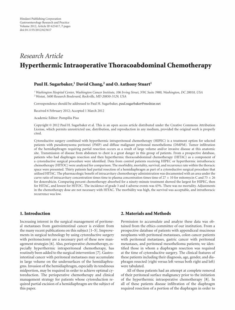
Figure 1: Pharmacokinetics of mitomycin C in 12 patients withsimultaneous intraabdominal and intrathoracic chemotherapy usedin conjunction with cytoreductive surgery.
colon cancer. In the three patients who received intrathoracicmitomycin C, all had an appendiceal mucinous neoplasm.In the four patients who received intrathoracic doxorubicin,three had appendiceal mucinous neoplasm and one hadperitoneal mesothelioma (Table 1).
3.1. Pharmacokinetics of Hyperthermic Chemotherapy inThoracic and Abdominal Cavity (HITAC). In 12 patientscomplete pharmacokinetic data was available so that themitomycin C concentrations in fluid from the thoracic andabdominal space could be determined along with plasmaconcentrations of this drug. Figure 1 shows the area underthe curve for thoracoabdominal mitomycin C as comparedto the area under curve for plasma mitomycin C. The areaunder the curve ratio was 27 ± 10. Nine of these patientshad appendiceal neoplasms, 2 colon cancer, and 1 peritonealmesothelioma.
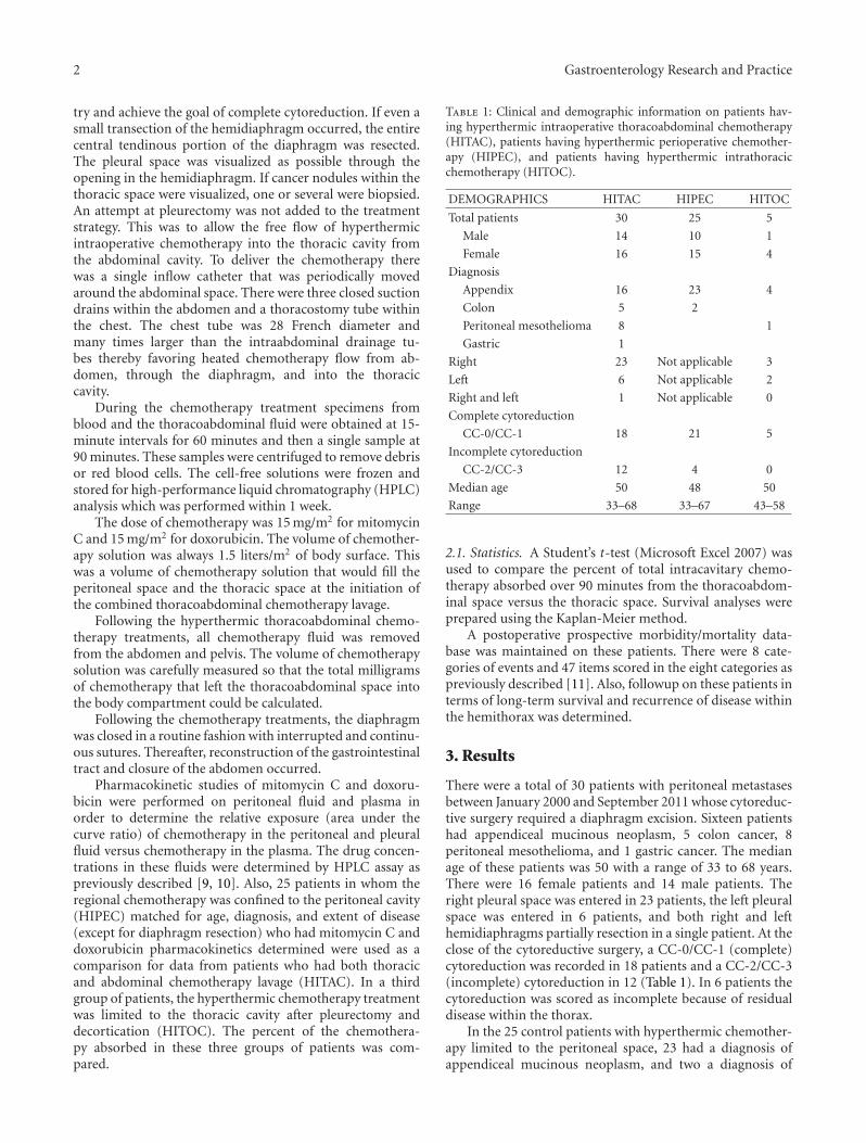
Similar data was obtained from the same 12 patients whoreceived intrathoracic and abdominal doxorubicin. The areaunder the curve for thoracoabdominal fluid as compared tothe area under the curve for plasma is shown in Figure 2. Thearea under the curve ratio was 75 ± 26.
3.2. Comparison of Thoracic and Abdominal ChemotherapyTreatment (HITAC) to a Chemotherapy Lavage Limited to theAbdominal Space (HIPEC) and to a Chemotherapy LavageLimited to the Thoracic Space (HITOC). In 25 controlpatients in whom hyperthermic intraoperative chemother-apy treatment was limited to the abdominal and pelvic space(HIPEC), the percent of drug absorbed from the peritonealcavity over the 90 minutes of treatment was determined. Alsoin three patients the hyperthermic mitomycin C treatmentswere limited to the thoracic cavity (HITOC). These resultswere compared to the percent of chemotherapy absorbed inpatients with thoracic and abdominal chemotherapy lavage
Plasma
0 15 30 45 60 90 120
Time (minutes)
(DOX absorbed at 90 minutes = 90 3)
Thoracoabdominal
100
10
0.1
0.01
AUC ratio (PF/PL) = 75 26
1
±
±
Dox
oru
bici
n (µ
g/m
L)
Figure 2: Pharmacokinetics of doxorubicin in 12 patients withsimultaneous intraabdominal and intrathoracic chemotherapyadministration in conjunction with cytoreductive surgery.
90
80
100
70
60
50
40
30
20
10
00 15 30 45 60 90 120
Mit
omyc
in C
abs
orbe
d (%
)
P= 0.0015
AbdominalThoracoabdominalThoracic
Time (minutes)
Approx 75% absorbed at 90 minutes (abdominal)
Approx 67% absorbed at 90 minutes (combined)
Approx 41% absorbed at 90 minutes (thoracic)
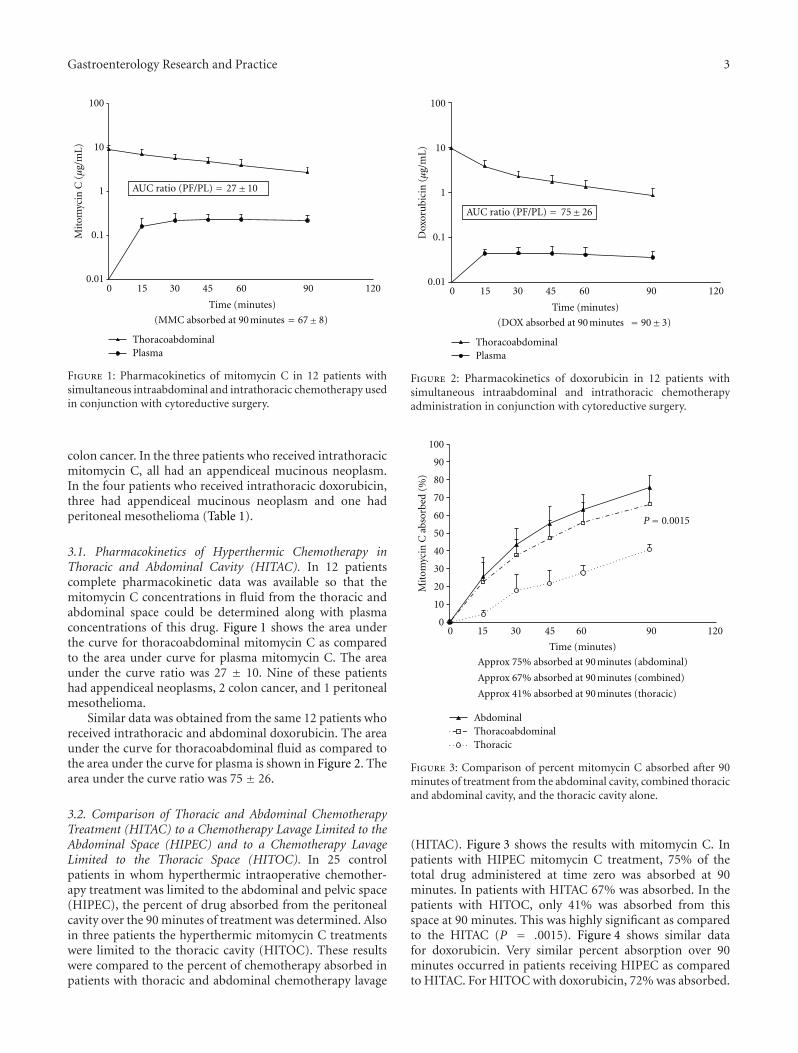
Figure 3: Comparison of percent mitomycin C absorbed after 90minutes of treatment from the abdominal cavity, combined thoracicand abdominal cavity, and the thoracic cavity alone.
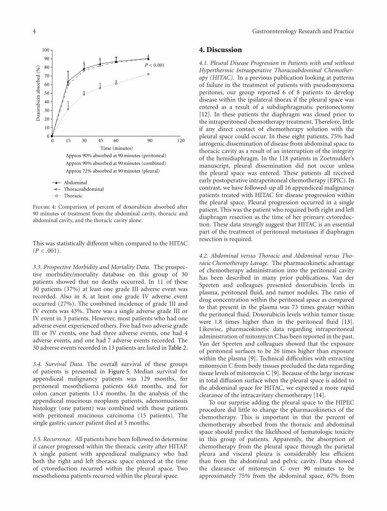
(HITAC). Figure 3 shows the results with mitomycin C. Inpatients with HIPEC mitomycin C treatment, 75% of thetotal drug administered at time zero was absorbed at 90minutes. In patients with HITAC 67% was absorbed. In thepatients with HITOC, only 41% was absorbed from thisspace at 90 minutes. This was highly significant as comparedto the HITAC (P = .0015). Figure 4 shows similar datafor doxorubicin. Very similar percent absorption over 90minutes occurred in patients receiving HIPEC as comparedto HITAC. For HITOC with doxorubicin, 72% was absorbed.
4 Gastroenterology Research and Practice
90
80
100
70
60
50
40
30
20
10
00 15 30 45 60 90 120
AbdominalThoracoabdominalThoracic
Time (minutes)
Approx 90% absorbed at 90 minutes (peritoneal)
Approx 90% absorbed at 90 minutes (combined)
Approx 72% absorbed at 90 minutes (pleural)
Dox
oru
bici
n a
bsor
bed
(%) P < 0.001
Figure 4: Comparison of percent of doxorubicin absorbed after90 minutes of treatment from the abdominal cavity, thoracic andabdominal cavity, and the thoracic cavity alone.
This was statistically different when compared to the HITAC(P < .001).
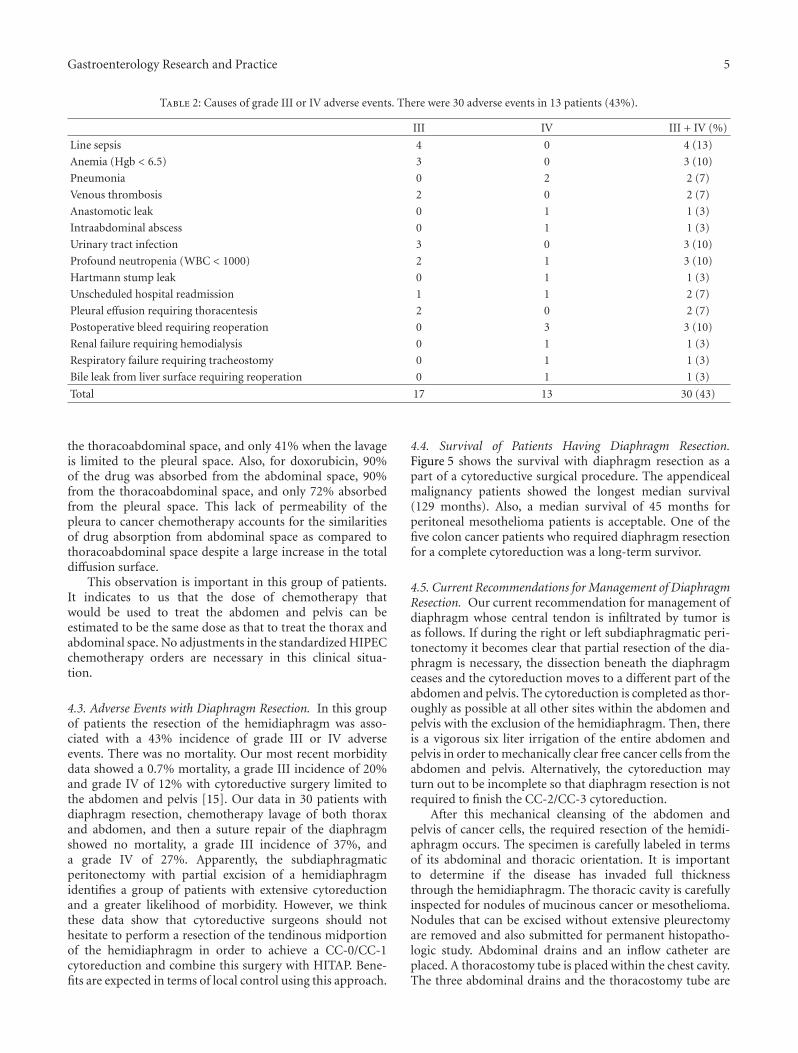
3.3. Prospective Morbidity and Mortality Data. The prospec-tive morbidity/mortality database on this group of 30patients showed that no deaths occurred. In 11 of these30 patients (37%) at least one grade III adverse event wasrecorded. Also in 8, at least one grade IV adverse eventoccurred (27%). The combined incidence of grade III andIV events was 43%. There was a single adverse grade III orIV event in 3 patients. However, most patients who had oneadverse event experienced others. Five had two adverse gradeIII or IV events, one had three adverse events, one had 4adverse events, and one had 7 adverse events recorded. The30 adverse events recorded in 13 patients are listed in Table 2.
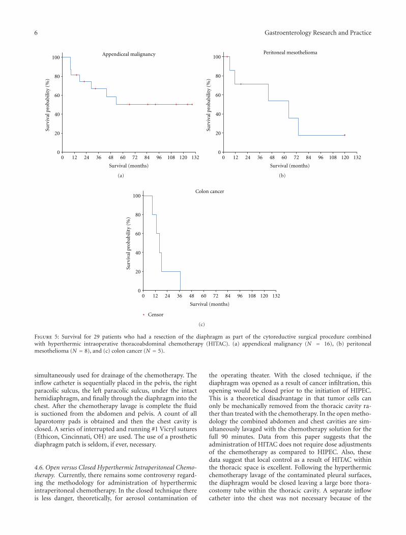
3.4. Survival Data. The overall survival of these groupsof patients is presented in Figure 5. Median survival forappendiceal malignancy patients was 129 months, forperitoneal mesothelioma patients 44.6 months, and forcolon cancer patients 13.4 months. In the analysis of theappendiceal mucinous neoplasm patients, adenomucinosishistology (one patient) was combined with those patientswith peritoneal mucinous carcinoma (15 patients). Thesingle gastric cancer patient died at 5 months.
3.5. Recurrence. All patients have been followed to determineif cancer progressed within the thoracic cavity after HITAP.A single patient with appendiceal malignancy who hadboth the right and left thoracic space entered at the timeof cytoreduction recurred within the pleural space. Twomesothelioma patients recurred within the pleural space.
4. Discussion
4.1. Pleural Disease Progression in Patients with and withoutHyperthermic Intraoperative Thoracoabdominal Chemother-apy (HITAC). In a previous publication looking at patternsof failure in the treatment of patients with pseudomyxomaperitonei, our group reported 6 of 8 patients to developdisease within the ipsilateral thorax if the pleural space wasentered as a result of a subdiaphragmatic peritonectomy[12]. In these patients the diaphragm was closed prior tothe intraperitoneal chemotherapy treatment. Therefore, littleif any direct contact of chemotherapy solution with thepleural space could occur. In these eight patients, 75% hadiatrogenic dissemination of disease from abdominal space tothoracic cavity as a result of an interruption of the integrityof the hemidiaphragm. In the 118 patients in Zoetmulder’smanuscript, pleural dissemination did not occur unlessthe pleural space was entered. These patients all receivedearly postoperative intraperitoneal chemotherapy (EPIC). Incontrast, we have followed-up all 16 appendiceal malignancypatients treated with HITAC for disease progression withinthe pleural space. Pleural progression occurred in a singlepatient. This was the patient who required both right and leftdiaphragm resection as the time of her primary cytoreduc-tion. These data strongly suggest that HITAC is an essentialpart of the treatment of peritoneal metastases if diaphragmresection is required.
4.2. Abdominal versus Thoracic and Abdominal versus Tho-racic Chemotherapy Lavage. The pharmacokinetic advantageof chemotherapy administration into the peritoneal cavityhas been described in many prior publications. Van derSpeeten and colleagues presented doxorubicin levels inplasma, peritoneal fluid, and tumor nodules. The ratio ofdrug concentration within the peritoneal space as comparedto that present in the plasma was 73 times greater withinthe peritoneal fluid. Doxorubicin levels within tumor tissuewere 1.8 times higher than in the peritoneal fluid [13].Likewise, pharmacokinetic data regarding intraperitonealadministration of mitomycin C has been reported in the past.Van der Speeten and colleagues showed that the exposureof peritoneal surfaces to be 26 times higher than exposurewithin the plasma [9]. Technical difficulties with extractingmitomycin C from body tissues precluded the data regardingtissue levels of mitomycin C [9]. Because of the large increasein total diffusion surface when the pleural space is added tothe abdominal space for HITAC, we expected a more rapidclearance of the intracavitary chemotherapy [14].
To our surprise adding the pleural space to the HIPECprocedure did little to change the pharmacokinetics of thechemotherapy. This is important in that the percent ofchemotherapy absorbed from the thoracic and abdominalspace should predict the likelihood of hematologic toxicityin this group of patients. Apparently, the absorption ofchemotherapy from the pleural space through the parietalpleura and visceral pleura is considerably less efficientthan from the abdominal and pelvic cavity. Data showedthe clearance of mitomycin C over 90 minutes to beapproximately 75% from the abdominal space, 67% from
Gastroenterology Research and Practice 5
Table 2: Causes of grade III or IV adverse events. There were 30 adverse events in 13 patients (43%).
III IV III + IV (%)
Line sepsis 4 0 4 (13)
Anemia (Hgb < 6.5) 3 0 3 (10)
Pneumonia 0 2 2 (7)
Venous thrombosis 2 0 2 (7)
Anastomotic leak 0 1 1 (3)
Intraabdominal abscess 0 1 1 (3)
Urinary tract infection 3 0 3 (10)
Profound neutropenia (WBC < 1000) 2 1 3 (10)
Hartmann stump leak 0 1 1 (3)
Unscheduled hospital readmission 1 1 2 (7)
Pleural effusion requiring thoracentesis 2 0 2 (7)
Postoperative bleed requiring reoperation 0 3 3 (10)
Renal failure requiring hemodialysis 0 1 1 (3)
Respiratory failure requiring tracheostomy 0 1 1 (3)
Bile leak from liver surface requiring reoperation 0 1 1 (3)
Total 17 13 30 (43)
the thoracoabdominal space, and only 41% when the lavageis limited to the pleural space. Also, for doxorubicin, 90%of the drug was absorbed from the abdominal space, 90%from the thoracoabdominal space, and only 72% absorbedfrom the pleural space. This lack of permeability of thepleura to cancer chemotherapy accounts for the similaritiesof drug absorption from abdominal space as compared tothoracoabdominal space despite a large increase in the totaldiffusion surface.
This observation is important in this group of patients.It indicates to us that the dose of chemotherapy thatwould be used to treat the abdomen and pelvis can beestimated to be the same dose as that to treat the thorax andabdominal space. No adjustments in the standardized HIPECchemotherapy orders are necessary in this clinical situa-tion.
4.3. Adverse Events with Diaphragm Resection. In this groupof patients the resection of the hemidiaphragm was asso-ciated with a 43% incidence of grade III or IV adverseevents. There was no mortality. Our most recent morbiditydata showed a 0.7% mortality, a grade III incidence of 20%and grade IV of 12% with cytoreductive surgery limited tothe abdomen and pelvis [15]. Our data in 30 patients withdiaphragm resection, chemotherapy lavage of both thoraxand abdomen, and then a suture repair of the diaphragmshowed no mortality, a grade III incidence of 37%, anda grade IV of 27%. Apparently, the subdiaphragmaticperitonectomy with partial excision of a hemidiaphragmidentifies a group of patients with extensive cytoreductionand a greater likelihood of morbidity. However, we thinkthese data show that cytoreductive surgeons should nothesitate to perform a resection of the tendinous midportionof the hemidiaphragm in order to achieve a CC-0/CC-1cytoreduction and combine this surgery with HITAP. Bene-fits are expected in terms of local control using this approach.
4.4. Survival of Patients Having Diaphragm Resection.Figure 5 shows the survival with diaphragm resection as apart of a cytoreductive surgical procedure. The appendicealmalignancy patients showed the longest median survival(129 months). Also, a median survival of 45 months forperitoneal mesothelioma patients is acceptable. One of thefive colon cancer patients who required diaphragm resectionfor a complete cytoreduction was a long-term survivor.
4.5. Current Recommendations for Management of DiaphragmResection. Our current recommendation for management ofdiaphragm whose central tendon is infiltrated by tumor isas follows. If during the right or left subdiaphragmatic peri-tonectomy it becomes clear that partial resection of the dia-phragm is necessary, the dissection beneath the diaphragmceases and the cytoreduction moves to a different part of theabdomen and pelvis. The cytoreduction is completed as thor-oughly as possible at all other sites within the abdomen andpelvis with the exclusion of the hemidiaphragm. Then, thereis a vigorous six liter irrigation of the entire abdomen andpelvis in order to mechanically clear free cancer cells from theabdomen and pelvis. Alternatively, the cytoreduction mayturn out to be incomplete so that diaphragm resection is notrequired to finish the CC-2/CC-3 cytoreduction.
After this mechanical cleansing of the abdomen andpelvis of cancer cells, the required resection of the hemidi-aphragm occurs. The specimen is carefully labeled in termsof its abdominal and thoracic orientation. It is importantto determine if the disease has invaded full thicknessthrough the hemidiaphragm. The thoracic cavity is carefullyinspected for nodules of mucinous cancer or mesothelioma.Nodules that can be excised without extensive pleurectomyare removed and also submitted for permanent histopatho-logic study. Abdominal drains and an inflow catheter areplaced. A thoracostomy tube is placed within the chest cavity.The three abdominal drains and the thoracostomy tube are
6 Gastroenterology Research and Practice
0
20
40
60
80
100
0 12 24 36 48 60 72 84 96 108 120 132
Survival (months)
Appendiceal malignancySu
rviv
al p
roba
bilit
y (%
)
(a)
0
20
40
60
80
100
0 12 24 36 48 60 72 84 96 108 120 132
Survival (months)
Surv
ival
pro
babi
lity
(%)
Peritoneal mesothelioma
(b)
Censor
0
20
40
60
80
100
0 12 24 36 48 60 72 84 96 108 120 132
Survival (months)
Colon cancer
Surv
ival
pro
babi
lity
(%)
(c)
Figure 5: Survival for 29 patients who had a resection of the diaphragm as part of the cytoreductive surgical procedure combinedwith hyperthermic intraoperative thoracoabdominal chemotherapy (HITAC). (a) appendiceal malignancy (N = 16), (b) peritonealmesothelioma (N = 8), and (c) colon cancer (N = 5).
simultaneously used for drainage of the chemotherapy. Theinflow catheter is sequentially placed in the pelvis, the rightparacolic sulcus, the left paracolic sulcus, under the intacthemidiaphragm, and finally through the diaphragm into thechest. After the chemotherapy lavage is complete the fluidis suctioned from the abdomen and pelvis. A count of alllaparotomy pads is obtained and then the chest cavity isclosed. A series of interrupted and running #1 Vicryl sutures(Ethicon, Cincinnati, OH) are used. The use of a prostheticdiaphragm patch is seldom, if ever, necessary.
4.6. Open versus Closed Hyperthermic Intraperitoneal Chemo-therapy. Currently, there remains some controversy regard-ing the methodology for administration of hyperthermicintraperitoneal chemotherapy. In the closed technique thereis less danger, theoretically, for aerosol contamination of
the operating theater. With the closed technique, if thediaphragm was opened as a result of cancer infiltration, thisopening would be closed prior to the initiation of HIPEC.This is a theoretical disadvantage in that tumor cells canonly be mechanically removed from the thoracic cavity ra-ther than treated with the chemotherapy. In the open metho-dology the combined abdomen and chest cavities are sim-ultaneously lavaged with the chemotherapy solution for thefull 90 minutes. Data from this paper suggests that theadministration of HITAC does not require dose adjustmentsof the chemotherapy as compared to HIPEC. Also, thesedata suggest that local control as a result of HITAC withinthe thoracic space is excellent. Following the hyperthermicchemotherapy lavage of the contaminated pleural surfaces,the diaphragm would be closed leaving a large bore thora-costomy tube within the thoracic cavity. A separate inflowcatheter into the chest was not necessary because of the
Gastroenterology Research and Practice 7
large volume of chemotherapy outflow through the opendiaphragm and out through the thoracostomy tube. A theo-retical and probably actual clinical advantage of the openmethod over the closed method occurs in patients requiredto have diaphragm resection.
4.7. Rationale for HITAC in Patients with CC-2/CC-3 Cytore-duction. The data regarding palliative benefit of HIPECin patients with CC-2/CC-3 cytoreduction has never beenrigorously studied. There is no doubt that incompletecytoreduction is associated with a poor prognosis in appen-diceal cancer, colorectal cancer, and peritoneal mesotheliomapatients. However, when added to an extensive debulkingprocedure, the hyperthermic intracavitary chemotherapymay achieve a partial response and prolong the patient’slife. This may be most likely if all adhesions on bowel loopsare separated so the HIPEC is in contact with all peritonealsurfaces. Also, any patients who have ascites as a componentof their peritoneal surface malignancy should receive intra-cavitary chemotherapy in order to guard against debilitatingascites occurring as the disease progresses. Garofalo andValle showed that HIPEC is an excellent treatment for themanagement of cancerous ascites [16].
5. Conclusions
HITAC is a treatment option for cytoreductive surgery inappendiceal and DMPM patients when the diaphragm mustbe partially resected as part of a cytoreductive surgical proce-dure. The pharmacokinetic advantage of direct intracavitaryadministration is preserved when HITAC is utilized. Also,judgments in the chemotherapy dose were not found to benecessary with HITAC as compared to HIPEC. An assess-ment of adverse events showed a 43% incidence of grade IIIor grade IV adverse events which is higher than reported formost groups of patients undergoing cytoreductive surgeryand HIPEC. However, the survival was acceptable and theincidence of intrathoracic recurrence was low.
References
[1] P. H. Sugarbaker, “Comprehensive management of peritonealsurface malignancy using cytoreductive surgery and perioper-ative intraperitoneal chemotherapy,” Cancer Research in Can-cer, vol. 3, pp. 179–203, 2009.
[2] O. Glehen, F. N. Gilly, F. Boutitie et al., “Toward curativetreatment of peritoneal carcinomatosis from nonovarian ori-gin by cytoreductive surgery combined with perioperative in-traperitoneal chemotherapy: a multi-institutional study of1290 patients,” Cancer, vol. 116, no. 24, pp. 5608–5618, 2010.
[3] H. Youssef, C. Newman, K. Chandrakumaran, F. Mohamed,T. D. Cecil, and B. J. Moran, “Operative findings, early com-plications, and long-term survival in 456 patients with pseu-domyxoma peritonei syndrome of appendiceal origin,” Dis-eases of the Colon and Rectum, vol. 54, no. 3, pp. 293–299, 2011.
[4] D. Elias, J. H. Lefevre, J. Chevalier et al., “Complete cytore-ductive surgery plus intraperitoneal chemohyperthermia withoxaliplatin for peritoneal carcinomatosis of colorectal origin,”Journal of Clinical Oncology, vol. 27, no. 5, pp. 681–685, 2009.
[5] T. D. Yan, D. Black, P. H. Sugarbaker et al., “A systematic reviewand meta-analysis of the randomized controlled trials onadjuvant intraperitoneal chemotherapy for resectable gastriccancer,” Annals of Surgical Oncology, vol. 14, no. 10, pp. 2702–2713, 2007.
[6] P. H. Sugarbaker, “Peritonectomy procedures,” Annals of Sur-gery, vol. 221, no. 1, pp. 29–42, 1995.
[7] K. Van der Speeten, O. A. Stuart, and P. H. Sugarbaker,“Pharmacokinetics and pharmacodynamics of perioperativecancer chemotherapy in peritoneal surface malignancy,” Can-cer Journal, vol. 15, no. 3, pp. 216–224, 2009.
[8] P. H. Sugarbaker, “Peritonectomy procedures,” Surgical Oncol-ogy Clinics of North America, vol. 12, no. 3, pp. 703–727, 2003.
[9] K. Van der Speeten, O. A. Stuart, D. Chang, H. Mahteme, andP. H. Sugarbaker, “Changes induced by surgical and clinicalfactors in the pharmacology of intraperitoneal mitomycin C in145 patients with peritoneal carcinomatosis,” Cancer Chemo-therapy and Pharmacology, vol. 68, no. 1, pp. 147–156, 2011.
[10] P. H. Sugarbaker, K. Van der Speeten, O. Anthony Stuart,and D. Chang, “Impact of surgical and clinical factors on thepharmacology of intraperitoneal doxorubicin in 145 patientswith peritoneal carcinomatosis,” European Journal of SurgicalOncology, vol. 37, no. 8, pp. 719–726, 2011.
[11] P. H. Sugarbaker, R. Alderman, G. Edwards et al., “Prospectivemorbidity and mortality assessment of cytoreductive surgeryplus perioperative intraperitoneal chemotherapy to treat peri-toneal dissemination of appendiceal mucinous malignancy,”Annals of Surgical Oncology, vol. 13, no. 5, pp. 635–644, 2006.
[12] F. A. N. Zoetmulder and P. H. Sugarbaker, “Patterns of failurefollowing treatment of pseudomyxoma peritonei of appendi-ceal origin,” European Journal of Cancer Part A, vol. 32, no. 10,pp. 1727–1733, 1996.
[13] K. Van der Speeten, O. A. Stuart, H. Mahteme, and P.H. Sugarbaker, “A pharmacologic analysis of intraoperativeintracavitary cancer chemotherapy with doxorubicin,” CancerChemotherapy and Pharmacology, vol. 63, no. 5, pp. 799–805,2009.
[14] R. L. Dedrick and M. F. Flessner, “Pharmacokinetic problemsin peritoneal drug administration: tissue penetration and sur-face exposure,” Journal of the National Cancer Institute, vol. 89,no. 7, pp. 480–487, 1997.
[15] P. H. Sugarbaker, K. Van der Speeten, O. A. Stuart, D. Chang,and H. Mahteme, “Patient- and treatment-related variables,adverse events and their statistical relationship for treatmentof peritoneal metastases,” in Cytoreductive Surgery and Peri-operative Chemotherapy for Peritoneal Surface Malignancy:Textbook and Video Atlas, P. H. Sugarbaker, Ed.
[16] A. Garofalo and M. Valle, “Laparoscopy in the management ofperitoneal carcinomatosis,” Cancer Journal, vol. 15, no. 3, pp.190–195, 2009.

Submit your manuscripts athttp://www.hindawi.com
Stem CellsInternational
Hindawi Publishing Corporationhttp://www.hindawi.com Volume 2014
Hindawi Publishing Corporationhttp://www.hindawi.com Volume 2014
MEDIATORSINFLAMMATION
of
Hindawi Publishing Corporationhttp://www.hindawi.com Volume 2014
Behavioural Neurology
EndocrinologyInternational Journal of
Hindawi Publishing Corporationhttp://www.hindawi.com Volume 2014
Hindawi Publishing Corporationhttp://www.hindawi.com Volume 2014
Disease Markers
Hindawi Publishing Corporationhttp://www.hindawi.com Volume 2014
BioMed Research International
OncologyJournal of
Hindawi Publishing Corporationhttp://www.hindawi.com Volume 2014
Hindawi Publishing Corporationhttp://www.hindawi.com Volume 2014
Oxidative Medicine and Cellular Longevity
Hindawi Publishing Corporationhttp://www.hindawi.com Volume 2014
PPAR Research
The Scientific World JournalHindawi Publishing Corporation http://www.hindawi.com Volume 2014
Immunology ResearchHindawi Publishing Corporationhttp://www.hindawi.com Volume 2014
Journal of
ObesityJournal of
Hindawi Publishing Corporationhttp://www.hindawi.com Volume 2014
Hindawi Publishing Corporationhttp://www.hindawi.com Volume 2014
Computational and Mathematical Methods in Medicine
OphthalmologyJournal of
Hindawi Publishing Corporationhttp://www.hindawi.com Volume 2014
Diabetes ResearchJournal of
Hindawi Publishing Corporationhttp://www.hindawi.com Volume 2014
Hindawi Publishing Corporationhttp://www.hindawi.com Volume 2014
Research and TreatmentAIDS
Hindawi Publishing Corporationhttp://www.hindawi.com Volume 2014
Gastroenterology Research and Practice
Hindawi Publishing Corporationhttp://www.hindawi.com Volume 2014
Parkinson’s Disease
Evidence-Based Complementary and Alternative Medicine
Volume 2014Hindawi Publishing Corporationhttp://www.hindawi.com