
Research Article - Hindawi Publishing...
11
Hindawi Publishing Corporation Journal of Biomedicine and Biotechnology Volume 2012, Article ID 875383, 10 pages doi:10.1155/2012/875383 Research Article Generation of a Chinese Hamster Ovary Cell Line Producing Recombinant Human Glucocerebrosidase Juliana Branco Novo, 1, 2 Ligia Morganti, 3 Ana Maria Moro, 4 Adriana Franco Paes Leme, 5 Solange Maria de Toledo Serrano, 5 Isaias Raw, 1 and Paulo Lee Ho 1, 2 1 Centro de Biotecnologia, Instituto Butantan, Avenue Vital Brasil, 1500, 05503-900 S˜ ao Paulo, SP, Brazil 2 Interunidades em Biotecnologia, Instituto de Ciˆ encias Biom´ edicas, Universidade de S˜ ao Paulo, 05508-000 S˜ ao Paulo, SP, Brazil 3 Centro de Biotecnologia, Instituto de Pesquisas Energ´ eticas e Nucleares, 055508-000 S˜ ao Paulo, SP, Brazil 4 Laborat´ orio de Biof´ armacos em C´ elulas Animais, Instituto Butantan, Avenue Vital Brasil, 1500, 05503-900 S˜ ao Paulo, SP, Brazil 5 Laborat´ orio Especial de Toxinologia Aplicada, CAT-cepid, Instituto Butantan, Avenue Vital Brasil, 1500, 05503-900 S˜ ao Paulo, SP, Brazil Correspondence should be addressed to Paulo Lee Ho, [email protected] Received 9 March 2012; Revised 30 June 2012; Accepted 19 July 2012 Academic Editor: D. M. Clarke Copyright © 2012 Juliana Branco Novo et al. This is an open access article distributed under the Creative Commons Attribution License, which permits unrestricted use, distribution, and reproduction in any medium, provided the original work is properly cited. Impaired activity of the lysosomal enzyme glucocerebrosidase (GCR) results in the inherited metabolic disorder known as Gaucher disease. Current treatment consists of enzyme replacement therapy by administration of exogenous GCR. Although effective, it is exceptionally expensive, and patients worldwide have a limited access to this medicine. In Brazil, the public healthcare system provides the drug free of charge for all Gaucher’s patients, which reaches the order of $ 84 million per year. However, the production of GCR by public institutions in Brazil would reduce significantly the therapy costs. Here, we describe a robust protocol for the generation of a cell line producing recombinant human GCR. The protein was expressed in CHO-DXB11 (dhfr − ) cells after stable transfection and gene amplification with methotrexate. As expected, glycosylated GCR was detected by immunoblotting assay both as cell-associated (∼64 and 59 kDa) and secreted (63–69 kDa) form. Analysis of subclones allowed the selection of stable CHO cells producing a secreted functional enzyme, with a calculated productivity of 5.14pg/cell/day for the highest producer. Although being laborious, traditional methods of screening high-producing recombinant cells may represent a valuable alternative to generate expensive biopharmaceuticals in countries with limited resources. 1. Introduction Glucocerebrosidase (GCR; EC 3.2.1.45) is a membrane- associated lysosomal enzyme [1] responsible for the hydrol- ysis of glucocerebroside to glucose and ceramide [2, 3]. Deficiency in the GCR activity results in Gaucher disease, the most common lysosomal storage disorder [4] characterized by an accumulation of the glycolipid in the monocyte- macrophage system [5, 6], mainly in liver, spleen and bone marrow. Gaucher disease is an autosomal recessive disorder [7], caused by more than 300 mutations in the GCR gene [5, 8], which results in a highly variable clinical presentation. According to the symptoms, Gaucher disease is historically classified into types I, II, and III [4, 6]. Type I (nonneuronopathic) is the most prevalent form [9], usually treated by enzyme replacement therapy (ERT) [10] via intravenous infusions of recombinant GCR produced in Chinese hamster ovary (CHO) cells [11]. ERT has proven to be safe and effective [8], reversing most symptoms of the disease in more than 4,500 patients worldwide [12]. This therapy was first introduced in 1991, using human placental- derived GCR (aglucerase, Ceredase) that was then replaced in 1994 by the recombinant form (imiglucerase, Cerezyme) [5, 12, 13], both manufactured by Genzyme Corporation (Cambridge, MA, USA), which was latterly acquired by Sanofi-Aventis (Montpellier, France). Currently, two new recombinant GCRs, regarded as biosimilar (or follow-on) preparations of imiglucerase [14], have received market
Transcript of Research Article - Hindawi Publishing...

Hindawi Publishing CorporationJournal of Biomedicine and BiotechnologyVolume 2012, Article ID 875383, 10 pagesdoi:10.1155/2012/875383
Research Article
Generation of a Chinese Hamster Ovary Cell Line ProducingRecombinant Human Glucocerebrosidase
Juliana Branco Novo,1, 2 Ligia Morganti,3 Ana Maria Moro,4 Adriana Franco Paes Leme,5
Solange Maria de Toledo Serrano,5 Isaias Raw,1 and Paulo Lee Ho1, 2
1 Centro de Biotecnologia, Instituto Butantan, Avenue Vital Brasil, 1500, 05503-900 Sao Paulo, SP, Brazil2 Interunidades em Biotecnologia, Instituto de Ciencias Biomedicas, Universidade de Sao Paulo, 05508-000 Sao Paulo, SP, Brazil3 Centro de Biotecnologia, Instituto de Pesquisas Energeticas e Nucleares, 055508-000 Sao Paulo, SP, Brazil4 Laboratorio de Biofarmacos em Celulas Animais, Instituto Butantan, Avenue Vital Brasil, 1500, 05503-900 Sao Paulo, SP, Brazil5 Laboratorio Especial de Toxinologia Aplicada, CAT-cepid, Instituto Butantan, Avenue Vital Brasil, 1500,05503-900 Sao Paulo, SP, Brazil
Correspondence should be addressed to Paulo Lee Ho, [email protected]
Received 9 March 2012; Revised 30 June 2012; Accepted 19 July 2012
Academic Editor: D. M. Clarke
Copyright © 2012 Juliana Branco Novo et al. This is an open access article distributed under the Creative Commons AttributionLicense, which permits unrestricted use, distribution, and reproduction in any medium, provided the original work is properlycited.
Impaired activity of the lysosomal enzyme glucocerebrosidase (GCR) results in the inherited metabolic disorder known as Gaucherdisease. Current treatment consists of enzyme replacement therapy by administration of exogenous GCR. Although effective, itis exceptionally expensive, and patients worldwide have a limited access to this medicine. In Brazil, the public healthcare systemprovides the drug free of charge for all Gaucher’s patients, which reaches the order of $ 84 million per year. However, the productionof GCR by public institutions in Brazil would reduce significantly the therapy costs. Here, we describe a robust protocol for thegeneration of a cell line producing recombinant human GCR. The protein was expressed in CHO-DXB11 (dhfr−) cells after stabletransfection and gene amplification with methotrexate. As expected, glycosylated GCR was detected by immunoblotting assayboth as cell-associated (∼64 and 59 kDa) and secreted (63–69 kDa) form. Analysis of subclones allowed the selection of stableCHO cells producing a secreted functional enzyme, with a calculated productivity of 5.14 pg/cell/day for the highest producer.Although being laborious, traditional methods of screening high-producing recombinant cells may represent a valuable alternativeto generate expensive biopharmaceuticals in countries with limited resources.
1. Introduction
Glucocerebrosidase (GCR; EC 3.2.1.45) is a membrane-associated lysosomal enzyme [1] responsible for the hydrol-ysis of glucocerebroside to glucose and ceramide [2, 3].Deficiency in the GCR activity results in Gaucher disease, themost common lysosomal storage disorder [4] characterizedby an accumulation of the glycolipid in the monocyte-macrophage system [5, 6], mainly in liver, spleen andbone marrow. Gaucher disease is an autosomal recessivedisorder [7], caused by more than 300 mutations in theGCR gene [5, 8], which results in a highly variable clinicalpresentation. According to the symptoms, Gaucher diseaseis historically classified into types I, II, and III [4, 6].
Type I (nonneuronopathic) is the most prevalent form [9],usually treated by enzyme replacement therapy (ERT) [10]via intravenous infusions of recombinant GCR produced inChinese hamster ovary (CHO) cells [11]. ERT has proven tobe safe and effective [8], reversing most symptoms of thedisease in more than 4,500 patients worldwide [12]. Thistherapy was first introduced in 1991, using human placental-derived GCR (aglucerase, Ceredase) that was then replacedin 1994 by the recombinant form (imiglucerase, Cerezyme)[5, 12, 13], both manufactured by Genzyme Corporation(Cambridge, MA, USA), which was latterly acquired bySanofi-Aventis (Montpellier, France). Currently, two newrecombinant GCRs, regarded as biosimilar (or follow-on)preparations of imiglucerase [14], have received market
2 Journal of Biomedicine and Biotechnology
approval [15, 16]: a gene-activated GCR (velaglucerasealfa, VPRIV, Shire Human Genetic Therapies, Cambridge,MA, USA) produced in a human fibroblast cell line [17–19] and a plant-derived human GCR (taliglucerase alfa,Elelyso, Protalix Biotherapeutics, Carmiel, Israel) [20, 21].Recently, a viral contamination occurred in the Genzyme’sbioreactor resulting in a global supply shortage of Cerezyme.As a consequence, both enzymes velaglucerase alfa andtaliglucerase alfa had an accelerated market entry [22]. Thereis a great expectation that these and possibly other GCRformulations emerging in the near future could lead toa reduction in the price of the drug, which is extremelyexpensive [8] and hampers access to treatment [2, 14],especially in countries with limited resources [23–25]. Here,we propose the cloning and expression of human GCR inCHO cells, in order to obtain high-expressing clones suitablefor future GCR production at a lower cost by InstituteButantan (Sao Paulo, Brazil). The Institute Butantan hasthe public mission of producing sera, vaccines, and bio-pharmaceuticals at reduced costs for the Brazilian publichealthcare system. The GCR cDNA was inserted into thepED dicistronic expression vector [26], and the recombinantprotein was expressed in CHO-DXB11 cells, an auxotrophicline deficient in dihydrofolate reductase (DHFR) activity[27]. This is a well-characterized expression system [28, 29],commonly used in the biopharmaceutical industry for theproduction of glycoproteins [30, 31]. Cells were submittedto gene amplification with increasing methotrexate (MTX),a competitive inhibitor of DHFR, and clones were selectedby traditional methods to screen high producers sincecurrent high-throughput techniques are not available tomost undeveloped or emerging countries. The properlyglycosylated GCR was detected in the culture supernatantsand cellular extracts. Subcloning of the best producer clonefollowed by long-term culture in the MTX-free mediumallowed the selection of stable CHO cell lines producing afunctional enzyme. In addition to a successful protocol forthe generation of stable cells producing recombinant humanGCR, this study shows that traditional screening methodsto search for high-producing cell lines for recombinantproteins can be a useful alternative for the production ofbiopharmaceuticals in emerging countries.
2. Materials and Methods
2.1. Recombinant DNA Procedures. Human GCR cDNA wasobtained by purification of total RNA from ECV 304 cells(ATCC CRL 1998), followed by cDNA synthesis throughRT-PCR (results not shown). The 1,565 bp cDNA encodinghuman GCR with its own signal peptide was amplified byPCR using the forward primer (5′ TCT AGA CCA TGGCTG GCA GCC TCA CA 3′) containing a XbaI restrictionsite and the Kozak sequence (sequence in bold), and thereverse primer (5′ GAA TTC TCA CTG GCG ATG CCACAG 3′) containing an EcoRI restriction site. PCR wasperformed using Pfx DNA polymerase (Invitrogen), 0.2 mMof each deoxynucleoside triphosphate and 20 pmol of eachprimer. PCR amplification conditions were as follows: 94◦C,
5 min; 30 cycles of 94◦C, 1 min; 52◦C, 45 s; 68◦C, 3 min;a single step of 68◦C, 7 min for final extension. PCRproducts were cloned into the pGEM-T vector (Promega).DH5α E. coli competent cells were transformed with theligation product for propagation and amplification of therecombinant DNA. Positive clones were confirmed by DNAsequencing using an automated DNA sequencer (ABI 3100)based on the dideoxytermination method [32]. The GCRcDNA was mutated to produce an enzyme with His inplace of Arg at position 495 (procedures not shown)(Figure 1). The resulted plasmid pGEM-T-GCR was digestedand the insert was subcloned into the XbaI and EcoRIsites of the pED expression vector [26], kindly providedby Dr. R. J. Kaufman (Howard Hughes Medical Institute,University of Michigan Medical Center), which provideshigh-expression levels of heterologous proteins in mam-malian cells based on a dicistronic expression system. ThepED vector carriers the encephalomyocarditis virus leadersequence, a putative internal ribossomal entry site, insertedupstream of the selectable and amplifiable gene marker dhfr(Figure 1).
2.2. Cell Culture, Stable Transfection and Gene Amplification.Cells of the lineage CHO-DXB11 (dhfr−) [27] kindlyprovided by Dr. L. A. Chasin (Department of BiologicalSciences, Columbia University), were maintained in Min-imum Essential Medium Alfa Medium (α-MEM; Invit-rogen) supplemented with 2 mM L-glutamine, antibiotics(100 U/mL penicillin, 100 μg/mL streptomycin, 0.25 μg/mLamphotericin B; Invitrogen) and 10% fetal bovine serum(FBS; Cultilab) (nonselective medium). Transfection of theplasmid pED-GCR (20 μg) was carried out in 100 mmtissue culture plates containing 106 cells by the calciumphosphate precipitation method [33]. The medium wasreplaced 48 h after transfection by α-MEM without nucleo-sides and supplemented with 10% dialyzed FBS (Invitrogen),antibiotics and L-glutamine (selective medium). After 2weeks, 48 DHFR-positive clones (named as C 1 to C 48)were transferred into 12-well tissue culture plates usingplastic inoculating loops and submitted to gene amplificationwith increasing MTX (Sigma) in the selective medium atconcentrations of 0, 20, 50, 150, 350, 700, and 1200 nM.Cells were fed with fresh medium every 2-3 days andwere passaged upon reaching confluence by trypsinization.In each selection step, cells were cultivated for at least15 days before the MTX concentration was increased.To generate conditioned media and cell lysates for GCRexpression analysis, cell clones were seeded into 6-well tissueculture plates containing selective medium and MTX. Whensemiconfluence was reached, cells were washed 3 times withPBS and the culture medium was replaced by selectivemedium lacking FBS. After incubation for 24 h, both theculture medium (conditioned medium) and the cells werecollected, centrifuged at 300×g for 10 min, and stored at−80◦C until use. Cell concentration was determined for eachwell with a Neubauer hemocytometer. For some analyses,cell culture supernatants were precipitated with 10% (v/v)trichloroacetic acid (TCA) and washed with cold acetone.

Journal of Biomedicine and Biotechnology 3
SV40-pA
DHFR
IVS
SV40 AdMLP AdTPL EM
C-L
Am
pR
pED
5.3 kb
SMC
PstISalIXbaISmaIEcoRI
(a)
YCDSFDPPTF PALGTFSRYE STRSGRRMEL SMGPIQANHT
LQAVSWASGA RPCIPKSFGY SSVVCVCNAT
GTGLLLTLQP EQKFQKVKGF GGAMTDAAAL NILALSPPAQ
NLLLKSYFSE EGIGYNIIRV PMASCDFSIR TYTYADTPDD
FQLHNFSLPE EDTKLKIPLI HRALQLAQRP VSLLASPWTS
PTWLKTNGAV NGKGSLKGQP GDIYHQTWAR YFVKFLDAYA
EHKLQFWAVT AENEPSAGLL SGYPFQCLGF TPEHQRDFIA
RDLGPTLANS THHNVRLLML DDQRLLLPHW AKVVLTDPEA
AKYVHGIAVH WYLDFLAPAK ATLGETHRLF PNTMLFASEA
CVGSKFWEQS VRLGSWDRGM QYSHSIITNL LYHVVGWTDW
NLALNPEGGP NWVRNFVDSP IIVDITKDTF YKQPMFYHLG
HFSKFIPEGS QRVGLVASQK NDLDAVALMH PDGSAVVVVL
NRSSKDVPLT IKDPAVGFLE TISPGYSIHT YLWHRQ
MAGSLTGLLL 21
61
101
141
181
221
261
301
341
381
421
461
497
(b)
Figure 1: Strategy of cloning of GCR cDNA in the mammalian expression vector pED. (a) Map of dicistronic vector pED, a pUC 18derivative, containing the simian virus 40 (SV40) origin of replication and enhancer element, the adenovirus major late promoter (AdMLP),the tripartite leader from adenovirus late mRNA (AdTPL), a hybrid intron composed of the 5′ splice site from the first leader of adenovirusmajor late mRNAs and a 3′ splice site from an immunoglobulin gene (IVS), a multiple cloning site (SMC), the 5′ untranslated leaderfrom encephalomyocarditis (EMC-L) virus, a murine DHFR-coding region, the simian virus 40 late polyadenylation signal (SV40-pA), andampicillin resistance gene for propagation and selection in E. coli (AmpR) [26]. GCR cDNA was cloned into XbaI and EcoRI sites. (b) Aminoacid sequence of cloned GCR cDNA. The signal peptide of 19 amino acids is underlined. Potential carbohydrate binding asparagine residuesare shown by diamonds. The arginine at position 495 was replaced by histidine (mutation R495H) (boxed).
All cell cultures were carried out at 37◦C in a humidifiedincubator with 5% CO2.
2.3. Single-Cell Subcloning Procedures. To isolate a cell linewith the highest GCR productivity, cells from the best cloneobtained at 700 nM MTX were subcloned by seeding 50,100, and 200 cells into 3 separate 100 mm tissue cultureplates and maintained in selective medium supplementedwith MTX. After 15 days, 24 single-cell subclones (named asSC 12.1 to SC 12.24) were randomly selected, transferred to12-well tissue-culture plates using plastic inoculating loopsand expanded in the same medium for storage.
2.4. Clonal Stability Studies. The highest producer cloneand the isolated subclones were cultured into 12-well tissueculture plates for 45 days in the absence of MTX selectionpressure in nonselective medium. Cells were seeded into 6-well tissue culture plates to obtain conditioned media, aspreviously described. Samples were collected on days 15, 30,and 45 of the culture, and analyzed for secreted GCR. Cellconcentration was determined by counting each well aftercollection of the supernatant.
2.5. Western Blotting Analysis. CHO-DXB11 cell super-natants (conditioned media) in 1-fold SDS-PAGE samplebuffer and CHO cell pellets lysed in 2-fold SDS-PAGEsample buffer, containing 8 M urea, 1.5 mM EDTA and aprotease inhibitor cocktail (Sigma) were fractionated on a10% SDS-PAGE and electrotransferred to nitrocellulose orPVDF membranes (GE Healthcare). The membranes were
blocked with 5% (w/v) nonfat dried milk in 0.1% (v/v)Tween 20, 150 mM NaCl, and 20 mM Tris-HCl pH 8.0 (TBS-T) for 16 h. Membranes were washed three times for 10 minwith TBS-T, and further incubated with a 1 : 4,000 dilutionof the murine anti-human GCR polyclonal antibodies [34],in 1% (w/v) bovine serum albumin (BSA)/TBS-T, for 1.5 h.The membranes were washed three times with TBS-T andincubated for 1 h with a proper dilution of anti-mouseIgG peroxidase conjugate (Sigma) in TBS-T, washed, andrevealed with ECL or ECL Plus detection reagent (GEHealthcare), following manufacturer’s instructions.
2.6. Enzymatic Activity Assay. Hydrolytic activity of secretedGCR was determined fluorometrically with synthetic sub-strate 4-methylumbelliferyl-β-D-glucopyranoside (4-MUG;Sigma). The enzymatic reactions were performed in 0.2 mLof 0.2 M citrate phosphate buffer pH 5.5, containing 0.15%(v/v) Triton X-100 (Sigma), 0.125% (w/v) sodium tauro-cholate (Sigma), 5 mM 4-MUG, and conditioned media(collected as described above). After incubation for 1 h at37◦C, the reactions were stopped by adding 1 mL of 0.1 Mglycine, 0.1 M NaOH pH 10.3. The product of enzymaticreactions 4-methylumbelliferone (4-MU) was measured witha spectrometer SLM-AMINCO Bowman Series II (Spec-tronic Instruments) excitation wavelength 365 nm, emissionwavelength 445 nm. A standard curve was constructedusing well-known concentrations of commercial enzymeCerezyme and specific activity data (40 U/mg) presentedby the manufacturer. Total protein concentrations weredetermined by absorbance at 205 nm [35].

4 Journal of Biomedicine and Biotechnology
2.7. GCR Quantification. Quantification of recombinanthuman GCR secreted from CHO-DXB11 cell subcloneswas estimated by a standard curve containing well-knownconcentrations of nonglycosylated recombinant GCR puri-fied from E. coli [34] on a 10% SDS-PAGE, followed bywestern blotting and densitometry analysis. Conditionedmedia used for GCR quantification were collected as previ-ously described. Densitometry analysis was performed usingsoftware from Eagle Eye still video system (Stratagene).
2.8. Endoglycosidase Digestion. Endo H and PNGase F diges-tion of recombinant GCR were performed in conditionedmedium of the best producer subclone previously precipi-tated with 10% (v/v) TCA and washed with cold acetone.Samples were heated for 10 min at 100◦C in glycoproteindenaturing buffer, and reactions were performed accordingto the manufacturer’s instructions (New England Biolabs).After incubation at 37◦C for 16 h, reaction products weresubjected to 10% SDS-PAGE followed by western blottinganalysis, as described above.
3. Results
3.1. GCR Expression in CHO-DXB11 Cells. The human GCRcDNA containing its signal peptide sequence (1,565 bp)and the mutation R495H was cloned into the pED vector(Figure 1) for stable expression of GCR in CHO-DXB11cells, with the aim of obtaining high-producing clones.Transfection efficiency using calcium phosphate was about0,1%. Transfected clones were treated with increasing MTXconcentrations to amplify the copy number of integratedcDNA. After the third round of amplification with 150 nMMTX, protein bands corresponding to glycosylated recombi-nant GCR were detected by western blotting analysis, usingan anti-human GCR polyclonal antibody (Figure 2). Twoto three GCR bands of 63–69 kDa were observed in theconditioned media (Figure 2(a)), while bands around 64 and59 kDa were detected in cell lysates (Figure 2(b)). A 56 kDaband was detected (Figures 2(a) and 2(b)) corresponding tononglycosylated recombinant GCR purified from E. coli [34](positive control).
The amplification process was followed using increasingconcentrations of MTX up to 700 nM in the culture medium.Utilization of MTX at higher concentration (1200 nM) wasdiscontinued because it induced a high rate of cell death.At each increase step of MTX concentration, conditionedmedia were collected to evaluate the GCR expression bythe cells. The western blotting analysis, performed with theanti-human GCR polyclonal antibody, showed that GCRexpression levels were higher with increasing MTX concen-tration, which was evident in the last step with 700 nMMTX in the culture medium (Figure 3). The nonglycosylatedrecombinant GCR of 56 kDa purified from E. coli was used aspositive control. These results suggested that 700 nM was thehighest concentration of MTX suitable for the amplificationof the cDNA and consequently the generation of high-producer clones for GCR. With this purpose, clones selectedat 700 nM MTX were evaluated for GCR expression by
western blotting analysis, and the clone 12 was chosen as thebest expressing clone for GCR enzyme (data not shown).
3.2. Subcloning and Cell Culture in MTX-Free Medium. Inorder to evaluate the clonal stability for GCR expression,the high-producer clone (clone 12) was cultivated for 45days in the absence of MTX selective pressure, and samplesof conditioned medium were collected on days 15, 30, and45. The western blotting assay showed a decreasing in theGCR expression over time (Figure 4(a)). After 45 days,the expression level of GCR by clone 12 was similar tothat of nontransfected CHO-DXB11 cells (negative control),indicating absence of the recombinant protein expression.Since this analysis was carried out using a pool of cells, wehypothesized that most of the cells were unstable and only afew stably GCR expressing cells were present in the culture.
To identify stable high-producer cells from the amplifiedpool of cells derived from clone 12, a single-cell subcloningprocess was performed. The western blotting analysis of theconditioned culture medium of subclones cultivated for 45days in the absence of MTX showed evident bands around66 kDa corresponding to recombinant GCR in 5 subclonesanalyzed (Figure 4(b)). The GCR expression level observedfor these subclones was similar to that detected for theclone 12 maintained with 700 nM MTX, and used as positivecontrol. For 13 additional subclones analyzed in the absenceof MTX, the GCR expression was not detected (data notshown).
3.3. Hydrolytic Activity of Recombinant GCR. The hydrolyticactivity of secreted recombinant GCR was evaluated usingthe synthetic substrate 4-MUG for the 5 stable producersubclones identified in the western blotting analysis (Fig-ure 4(b)). The presence of a functional GCR was observedfor all subclones analyzed (Figure 5). Specific activities werecalculated based on a standard curve containing well-knownunits of Cerezyme. GCR produced by subclones showedsimilar specific activities to commercial enzyme, taking intoaccount that the protein had not been purified from theculture medium. The highest activity for GCR was detectedin subclone 12.9 (28.54± 2.75μmol 4-MU/min/mg).
3.4. GCR Expression Levels. Recombinant GCR secretedfrom the best CHO-DXB11 cell subclones was quantifiedby western blotting and densitometry analysis, based ona standard curve of nonglycosylated recombinant GCRpurified from E. coli. As observed in Table 1, subclone 12.9showed the highest specific productivity of recombinantGCR (5.14 pg/cell/day), followed by subclones 12.19 and12.8 (3.22 and 1.84 pg/cell/day, resp.). These results are inagreement with the enzymatic activity assay that showed thesame order of potency to hydrolyze the synthetic substrate4-MUG by these subclones. The subclone 12.9 was thenselected as the best producer of recombinant GCR in secretedform.
3.5. Glycosylation Analyses of Recombinant GCR. Recom-binant GCR secreted from the highest producer subclone

Journal of Biomedicine and Biotechnology 5
DX
B11
66
45
C 1
2
C 2
0
C 2
2
C 3
7
C 4
5
97
rGC
RE
.col
i
(kD
a)
(a)
DX
B11
66
45
C 1
2
C 2
0
C 2
2
C 3
7
C 4
5
66
45
9797
rGC
RE
.col
i
DX
B11
C 1
2
C 2
0
C 2
2
C 3
7
C 4
5
rGC
RE
.col
i
(kD
a)
(kD
a)
(b)
Figure 2: Western blotting analysis of the expression of recombinant human GCR in stably transfected CHO-DXB11 cells, after the thirdround of amplification with 150 nM MTX. (a) Conditioned media of 5 selected clones (C 12, C 20, C 22, C 37, and C 45). (b) Cellular extractsfrom the selected clones and the corresponding membrane stained with Ponceau S after proteins transfer. The total protein from conditionedmedia was previously concentrated by precipitation with 10% TCA. Nonglycosylated recombinant human GCR of 56 kDa purified from E.coli was used as positive control. Conditioned medium and cellular extract of nontransfected CHO-DXB11 cells were the negative control.Glycosylated recombinant GCR bands of 59–69 kDa are indicated by the arrows. The volume of sample applied to the gel corresponded tothe supernatant (a) or extract (b) of 150,000 cells.
Table 1: Expression levels of recombinant GCR secreted from thebest CHO-DXB11 cell subclones.
Subclone Productivity (pg/cell/day)∗
12.8 1.84
12.9 5.14
12.19 3.22∗
The productivities were calculated by comparing the GCR bands ofsubclones with a standard curve of non-glycosylated recombinant GCRpurified from E. coli, in a western blotting assay followed by densitometricanalysis. The experiment was conducted in triplicate. The volume ofconditioned medium applied to the gel corresponded to the supernatant of70,000 cells.
(SC 12.9) was characterized by digestion with Endo H andPNGase F. The western blotting (Figure 6(a)) showed a shift
in the molecular mass of the protein from 66–69 kDa to56 kDa after PNGase F digestion, which is in agreementwith the expected size for nonglycosylated GCR. A 56 kDaband was also observed for recombinant GCR purified fromE. coli (positive control). In the treatment with Endo H,GCR was partially resistant. A decrease of ∼3 kDa in the69 kDa protein band corresponding to the loss of N-linkedhigh-mannose-type oligosaccharide was observed, with theprobable retention of complex-type oligosaccharides termi-nating in galactose or sialic acid, which are not digestedby Endo H. No recombinant GCR bands were detectedin the culture medium of nontransfected CHO-DXB11cells (negative control). Analysis using purified commercialenzyme (Cerezyme) of 60 kDa containing remodeled glycans(Figure 6(b)) confirmed the glycosylation status of GCRproduced by CHO cells with a pattern of two bands
6 Journal of Biomedicine and Biotechnology
66
45
[MTX]
0 n
M
20 n
M
50 n
M
150
nM
350
nM
700
nM
97
rGC
RE
.col
i
(kD
a)
(a)
66
45
[MTX]
0 n
M
20 n
M
50 n
M
150
nM
350
nM
700
nM
97
rGC
RE
.col
i
(kD
a)
(b)
66
45
[MTX]
0 n
M
20 n
M
50 n
M
150
nM
350
nM
700
nM
97rG
CR
E.c
oli
(kD
a)
(c)
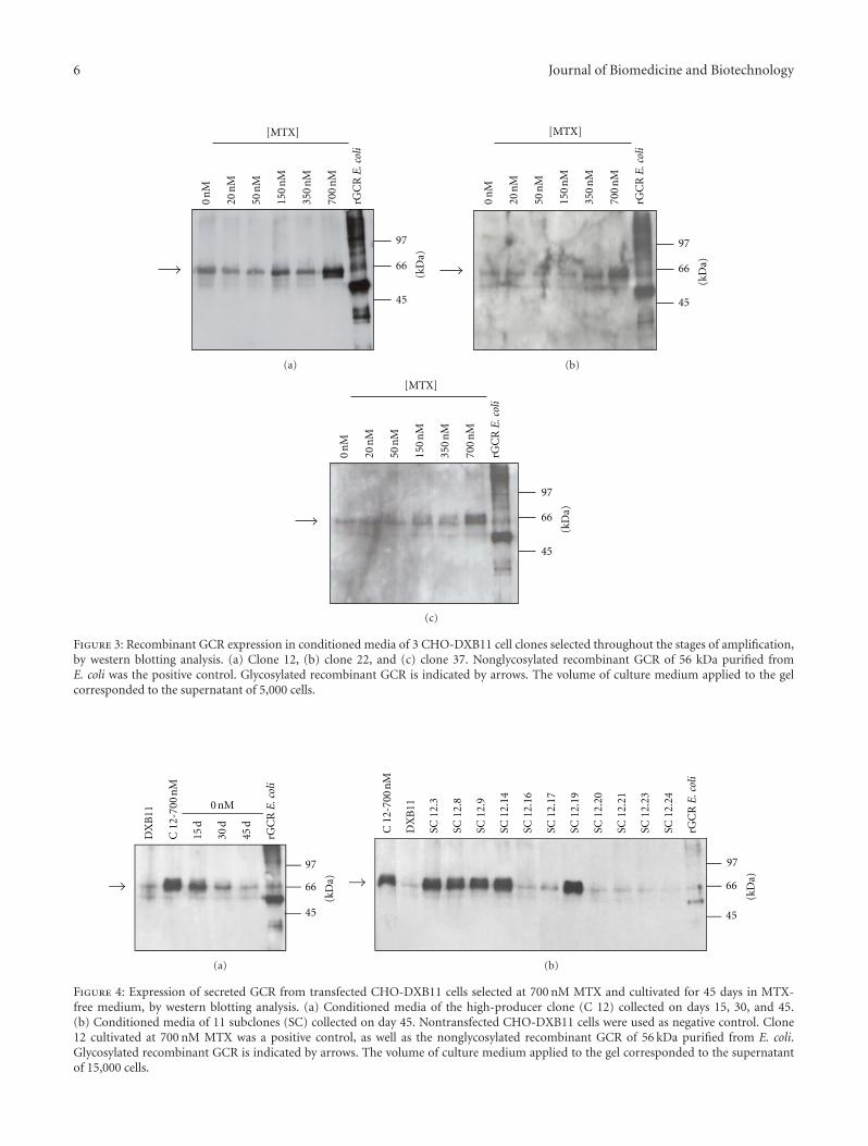
Figure 3: Recombinant GCR expression in conditioned media of 3 CHO-DXB11 cell clones selected throughout the stages of amplification,by western blotting analysis. (a) Clone 12, (b) clone 22, and (c) clone 37. Nonglycosylated recombinant GCR of 56 kDa purified fromE. coli was the positive control. Glycosylated recombinant GCR is indicated by arrows. The volume of culture medium applied to the gelcorresponded to the supernatant of 5,000 cells.
66
45
0 nM
DX
B11
C 1
2-70
0 n
M
15 d
30 d
45 d
97
rGC
RE
.col
i
(kD
a)
(a)
66
45
C 1
2-70
0 n
M
SC 1
2.3
SC 1
2.8
SC 1
2.9
SC 1
2.14
SC 1
2.16
SC 1
2.17
SC 1
2.19
SC 1
2.20
SC 1
2.21
SC 1
2.23
SC 1
2.24
DX
B11
97
rGC
RE
.col
i
(kD
a)
(b)
Figure 4: Expression of secreted GCR from transfected CHO-DXB11 cells selected at 700 nM MTX and cultivated for 45 days in MTX-free medium, by western blotting analysis. (a) Conditioned media of the high-producer clone (C 12) collected on days 15, 30, and 45.(b) Conditioned media of 11 subclones (SC) collected on day 45. Nontransfected CHO-DXB11 cells were used as negative control. Clone12 cultivated at 700 nM MTX was a positive control, as well as the nonglycosylated recombinant GCR of 56 kDa purified from E. coli.Glycosylated recombinant GCR is indicated by arrows. The volume of culture medium applied to the gel corresponded to the supernatantof 15,000 cells.

Journal of Biomedicine and Biotechnology 7
5.98± 1.50
22.36± 2.58
28.54± 2.75
7.81± 0.42
24.73± 1.9
40
SC 12.3 SC 12.8 SC 12.9 SC 12.14 SC 12.19 Cerezyme
Specific activity (µmol 4-MU/min/mg)
Figure 5: Enzymatic activity of recombinant GCR secreted fromCHO-DXB11 cell subclones (SC). The reactions were carried outwith the synthetic fluorogenic substrate 4-MUG. The product ofenzymatic reactions 4-MU was measured at wavelength of 445 nmafter excitation at 365 nm. A standard curve (units per fluorescence)was constructed using specific activity data of Cerezyme. Resultsare specific activities obtained for subclones relative to Cerezyme,presented as means ± SEM, measured in triplicate. The volume ofconditioned medium used in this assay corresponded to the super-natant of 70,000 cells.
(66 and 69 kDa), as observed by endoglycosidase treatmentand described elsewhere [36, 37].
4. Discussion
Although ERT has become the standard of care for type IGaucher disease [8, 38], its extremely high cost prevents itfrom being available to many patients in several countries[14, 23–25]. In Brazil, the drug is imported and providedby the public healthcare system free of charge and thecost for just 610 Gaucher’s patients reaches the order of$ 84 million per year (Department of Science, Technologyand Strategic Inputs, personal communication, 2010), a valuenot comparable with the expenses to treat other prioritydiseases. However, the high cost of treatment could bereduced with the production of GCR by public institutions.GCR is a glycoprotein of 497 amino acids with four ofthe five putative N-glycosylation sites usually occupied [39].The glycosylation process was proven to be essential toproduce a catalytically active enzyme [39, 40]. For thisreason, GCR has been produced in eukaryotic cells that havethe machinery required to perform the posttranslationalmodifications [41–43]: Genzyme has used CHO-DG44 cellsto produce imiglucerase [44], based on the dhfr amplifiablegene marker, while Shire and Protalix have developed a gene-activated expression system in a human fibroblast cell lineand a plant (carrot) cell-based expression system to producevelaglucerase alfa and taliglucerase alfa [19, 21], respectively.In contrast to velaglucerase that have an amino acid sequenceidentical to that of the natural enzyme, imiglucerase, andtaliglucerase contain a mutation at position 495 (an Argto His substitution). In addition, taliglucerase sequence ismodified at the N- and C-termini to add short tags of 2
and 7 amino acids, respectively. In this work, we describe thecloning of GCR cDNA containing the mutation R495H andthe protein expression in CHO-DXB11, a cell line deficientin the DHFR synthesis, similar to CHO-DG44 cells.
The GCR cDNA was cloned into the pED dicistronicexpression vector, which is commonly used to obtain high-expression levels of heterologous proteins in mammaliancells [45–47], and positive clones were isolated and amplifiedwith increasing MTX concentration up to 700 nM MTX.Probably due to the inability of the transfected cells tofurther amplify the dhfr gene [28], higher concentrationof MTX (1200 nM) caused cell death. MTX selection ofamplificants has been considered a major bottleneck inthe production of biopharmaceuticals because many roundsare required to obtain cells with high-gene copy numbers[28, 48]. Currently, high-throughput methods to screenhigh-producer cells have been developed [48, 49]; includingautomated systems such as colony picking and the Cellosystems [50]. However, many of these techniques are veryexpensive and nonaccessible to most undeveloped or emerg-ing countries. In this regard, although time consumingand labor intensive, traditional methods of screening stillrepresent an alternative for the production of recombinantglycoproteins at lower cost. Some significant problems ofusing traditional techniques are the relative low number ofclones that can be characterized, and the need of downstreamanalysis of the product levels, since protein secretion cannotbe measured on an individual cell basis [50].
Using traditional methods of screening for recombi-nant production clones, we demonstrated by immunoassaysthe expression of recombinant GCR either cell-associated(∼64 and 59 kDa) or secreted into the culture medium(63–69 kDa), using a murine anti-human GCR polyclonalantibody [34]. GCR has a molecular mass ranging from59 to 69 kDa [36], depending on the complexity of itsglycan chains [1, 51], while the nonglycosylated GCR is anonfunctional enzyme of 56 kDa [39]. Thus, the molecularmasses detected here for recombinant human GCR areconsistent with a properly glycosylated and active enzyme.However, to use in ERT, GCR should be modified in theoligosaccharide moieties to produce mannose-terminal car-bohydrates, facilitating mannose receptor-mediated uptakeinto macrophages [52]. GCR produced in CHO cells andcommercialized by Genzyme is sequentially treated withthree exoglycosidases during purification process to removesialic acid, galactose, and N-acetylglucosamine residues [53,54]. These steps have to be further included in our futureproduction process of a therapeutically functional enzyme inCHO cells in order to obtain a biosimilar GCR.
To evaluate the stability of the producer clone, MTX waswithdrawn from the culture medium and an unexpecteddecreased in the GCR expression was observed. Long-term culture in the absence of selective pressure mayresult in the loss of transgene copy number [55, 56], andreduction or elimination in the gene-specific transcription(gene silencing) [49, 57], according to the site of transgeneintegration. Both processes may result in the prevalence ofcells expressing lower levels of the recombinant protein dueto the higher growth rate presented by these cells when

8 Journal of Biomedicine and Biotechnology
66
45
SC 12.9
DX
B11
Non
dige
sted
En
do H
dig
este
d
PN
Gas
e F
dige
sted
97
rGC
RE
.col
i
(kD
a)
(a)
66
45
Cer
ezym
e
SC 1
2.9
non
dige
sted
97
(kD
a)
(b)
Figure 6: Glycosylation analysis of recombinant GCR secreted from high-producer subclone (SC 12.9) by western blotting assays. (a)Endoglycosidase digestion with Endo H and PNGase F. Samples of conditioned medium (supernatant of 150,000 cells corresponding toabout 750 ng of GCR) were precipitated with 10% TCA previously to digestion reactions. Non-digested sample of nontransfected CHO-DXB11 cells was used as negative control. Nonglycosylated recombinant GCR purified from E. coli was a positive control. Glycosylated GCRbands of 66–69 kDa and nonglycosylated GCR of 56 kDa are indicated by solid and dashed arrows, respectively. (b) Purified commercialenzyme Cerezyme (150 ng) of 60 kDa containing remodeled glycosylation moieties was compared to GCR secreted from subclone 12.9(supernatant of 30,000 cells corresponding to about 150 ng) with complex- and hybrid-type glycosylation pattern (66–69 kDa).
compared with the high producers [58], despite the fact thatboth have potentially been derived from the same single cell[56, 57].
Recloning was then performed in order to evaluate theheterogeneity of the producer clone, and stable subclonesexpressing a functional GCR with level of biological activitysimilar to that of Cerezyme were identified. Althoughthe highest productivity achieved for secreted GCR wascalculated as 5.14 pg/cell/day, higher expression levels havebeen reported for other recombinant proteins in CHO cells[45, 47]. Here, it should be taken into account the factthat GCR is a membrane-associated glycoprotein secretedat low level or rather not secreted under natural conditions[37]. Despite the importance of the production of GCRfor the treatment of ERT, there is a lack of data in theliterature describing the expression of recombinant GCRin CHO cells. Leonova and Grabowski [37] reported theintracellular degradation and secretion processes of GCR instably transfected CHO cells, and Genzyme’s data concerningthe production of imiglucerase in CHO-DG44 cells wereonly summarily described by Hoppe [59]. Additionally, someother information can be found in the patent files [44, 53].Here, we report a robust protocol for the production ofrecombinant GCR in CHO cells along with the all the stepsto obtain a stable cell line expressing the enzyme.
In conclusion, we showed the generation of stable CHOcells producing a functional recombinant human GCR,properly glycosylated and secreted, using low-throughputmethods for selection of high-producing clones. Effortsto develop a process for producing GCR by nonprofitinstitution may possibly result in reduced cost of treating
Gaucher disease in the future thus supplying the needs ofthe Brazilian public healthcare system. Furthermore, thetraditional screening strategies shown here can be usefulfor other emerging countries to produce biopharmaceuticalsand other biological products of high cost-effectiveness andpublic health value.
Acknowledgments
The authors are grateful to Dr. Geraldo Santana Magalhaes(Laboratorio de Imunopatologia, Instituto Butantan) andDr. Eneas de Carvalho (Centro de Biotecnologia, InstitutoButantan) for helpful discussions, to Dr. Rosa Maria Chura-Chambi (Centro de Biotecnologia, Instituto de PesquisasEnergeticas e Nucleares) for helping them with the transfec-tion and gene amplification procedures, and to Dr. HenriqueRoman Ramos (Centro de Biotecnologia, Instituto Butan-tan) for assistance in the fluorometric analysis. This work wassupported by grants from FAPESP (Fundacao de Amparoa Pesquisa do Estado de Sao Paulo), CNPq (ConselhoNacional de Desenvolvimento Cientıfico e Tecnologico), andFundacao Butantan.
References
[1] A. H. Erickson, E. I. Ginns, and J. A. Barranger, “Biosynthesisof the lysosomal enzyme glucocerebrosidase,” The Journal ofBiological Chemistry, vol. 260, no. 26, pp. 14319–14324, 1985.
[2] E. Beutler, “Gaucher disease: new molecular approaches todiagnosis and treatment,” Science, vol. 256, no. 5058, pp. 794–799, 1992.

Journal of Biomedicine and Biotechnology 9
[3] R. O. Brady, J. N. Kanfer, and D. Shapiro, “Metabolism ofglucocerebrosides II. Evidence of an enzymatic deficiencyin Gaucher’s disease,” Biochemical and Biophysical ResearchCommunications, vol. 18, no. 2, pp. 221–225, 1965.
[4] M. Jmoudiak and A. H. Futerman, “Gaucher disease: patho-logical mechanisms and modern management,” British Journalof Haematology, vol. 129, no. 2, pp. 178–188, 2005.
[5] D. Elstein and A. Zimran, “Review of the safety and efficacy ofimiglucerase treatment of Gaucher disease,” Biologics, vol. 3,pp. 407–417, 2009.
[6] G. A. Grabowski, “Recent clinical progress in Gaucher dis-ease,” Current Opinion in Pediatrics, vol. 17, no. 4, pp. 519–524,2005.
[7] J. A. Barranger and E. O’Rourke, “Lessons learned from thedevelopment of enzyme therapy for Gaucher disease,” Journalof Inherited Metabolic Disease, vol. 24, no. 2, supplement, pp.89–96, 2001.
[8] G. A. Grabowski, “Phenotype, diagnosis, and treatment ofGaucher’s disease,” The Lancet, vol. 372, no. 9645, pp. 1263–1271, 2008.
[9] E. Beutler, “Enzyme replacement in Gaucher disease,” PLoSMedicine, vol. 1, article e21, pp. 118–121, 2004.
[10] R. O. Brady, P. G. Pentchev, and A. E. Gal, “Replacementtherapy for inherited enzyme deficiency. Use of purifiedglucocerebrosidase in Gaucher’s disease,” The New EnglandJournal of Medicine, vol. 291, no. 19, pp. 989–993, 1974.
[11] Y. Kacher, B. Brumshtein, S. Boldin-Adamsky et al., “Acid β-glucosidase: onsights from structural analysis and relevance toGaucher disease therapy,” Biological Chemistry, vol. 389, no.11, pp. 1361–1369, 2008.
[12] K. Starzyk, S. Richards, J. Yee, S. E. Smith, and W. Kingma,“The long-term international safety experience of imiglu-cerase therapy for Gaucher disease,” Molecular Genetics andMetabolism, vol. 90, no. 2, pp. 157–163, 2007.
[13] P. B. Deegan and T. M. Cox, “Imiglucerase in the treatmentof Gaucher disease: a history and perspective,” Drug Design,Development and Therapy, vol. 6, pp. 81–106, 2012.
[14] T. M. Cox, “Gaucher disease: clinical profile and therapeuticdevelopments,” Biologics, vol. 4, pp. 299–313, 2011.
[15] J. M. F. G. Aerts, U. Yasothan, and P. Kirkpatrick, “Velaglu-cerase alfa,” Nature Reviews Drug Discovery, vol. 9, no. 11, pp.837–838, 2010.
[16] K. Traynor, “Taliglucerase alfa approved for Gaucher disease,”American Journal of Health-System Pharmacy, vol. 69, no. 12,p. 1009, 2012.
[17] B. Brumshtein, P. Salinas, B. Peterson et al., “Characterizationof gene-activated human acid-β-glucosidase: crystal structure,glycan composition, and internalization into macrophages,”Glycobiology, vol. 20, no. 1, pp. 24–32, 2010.
[18] J. L. Morris, “Velaglucerase alfa for the management of type 1Gaucher disease,” Clinical Therapeutics, vol. 34, pp. 259–271,2012.
[19] A. Zimran, K. Loveday, C. Fratazzi, and D. Elstein, “A phar-macokinetic analysis of a novel enzyme replacement therapywith Gene-Activated human glucocerebrosidase (GA-GCB) inpatients with type 1 Gaucher disease,” Blood Cells, Molecules,and Diseases, vol. 39, no. 1, pp. 115–118, 2007.
[20] C. E. Hollak, “An evidence-based review of the potentialbenefits of taliglucerase alfa in the treatment of patients withGaucher disease,” Core Evidence, vol. 7, pp. 15–20, 2012.
[21] Y. Shaaltiel, D. Bartfeld, S. Hashmueli et al., “Production ofglucocerebrosidase with terminal mannose glycans for enzyme
replacement therapy of Gaucher’s disease using a plant cellsystem,” Plant Biotechnology Journal, vol. 5, no. 5, pp. 579–590,2007.
[22] C. E. M. Hollak, S. vom Dahl, J. M. F. G. Aerts et al.,“Force Majeure: therapeutic measures in response to restrictedsupply of imiglucerase (Cerezyme) for patients with Gaucherdisease,” Blood Cells, Molecules, and Diseases, vol. 44, no. 1, pp.41–47, 2010.
[23] E. Beutler, “Gaucher disease as a paradigm of current issuesregarding single gene mutations of humans,” Proceedings of theNational Academy of Sciences of the United States of America,vol. 90, no. 12, pp. 5384–5390, 1993.
[24] E. Beutler, “Lysosomal storage diseases: natural history andethical and economic aspects,” Molecular Genetics and Metab-olism, vol. 88, no. 3, pp. 208–215, 2006.
[25] A. H. Futerman, J. L. Sussman, M. Horowitz, I. Silman, and A.Zimran, “New directions in the treatment of Gaucher disease,”Trends in Pharmacological Sciences, vol. 25, no. 3, pp. 147–151,2004.
[26] R. J. Kaufman, M. V. Davies, L. C. Wasley, and D. Michnick,“Improved vectors for stable expression of foreign genes inmammalian cells by use of the untranslated leader sequencefrom EMC virus,” Nucleic Acids Research, vol. 19, no. 16, pp.4485–4490, 1991.
[27] G. Urlaub and L. A. Chasin, “Isolation of Chinese hamstercell mutants deficient in dihydrofolate reductase activity,”Proceedings of the National Academy of Sciences of the UnitedStates of America, vol. 77, no. 7, pp. 4216–4220, 1980.
[28] J. J. Cacciatore, L. A. Chasin, and E. F. Leonard, “Gene amplifi-cation and vector engineering to achieve rapid and high-leveltherapeutic protein production using the Dhfr-based CHOcell selection system,” Biotechnology Advances, vol. 28, no. 6,pp. 673–681, 2010.
[29] R. J. Kaufman, “Selection and coamplification of heterologousgenes in mammalian cells,” Methods in Enzymology, vol. 185,pp. 537–566, 1990.
[30] K. P. Jayapal, K. F. Wlaschin, W. S. Hu, and M. G. S.Yap, “Recombinant protein therapeutics from CHO Cells—20years and counting,” Chemical Engineering Progress, vol. 103,no. 10, pp. 40–47, 2007.
[31] F. M. Wurm, “Production of recombinant protein therapeuticsin cultivated mammalian cells,” Nature Biotechnology, vol. 22,no. 11, pp. 1393–1398, 2004.
[32] F. Sanger, S. Nicklen, and A. R. Coulson, “DNA sequencingwith chain-terminating inhibitors,” Proceedings of the NationalAcademy of Sciences of the United States of America, vol. 74, no.12, pp. 5463–5467, 1977.
[33] F. L. Graham and A. J. van der Eb, “A new technique for theassay of infectivity of human adenovirus 5 DNA,” Virology, vol.52, no. 2, pp. 456–467, 1973.
[34] J. B. Novo, M. L. S. Oliveira, G. S. Magalhaes, L. Morganti,I. Raw, and P. L. Ho, “Generation of polyclonal antibodiesagainst recombinant human glucocerebrosidase produced inescherichia coli,” Molecular Biotechnology, vol. 46, no. 3, pp.279–286, 2010.
[35] C. M. Stoscheck, “Quantitation of protein,” Methods in Enzy-mology, vol. 182, pp. 50–68, 1990.
[36] J. E. Bergmann and G. A. Grabowski, “Posttranslational pro-cessing of human lysosomal acid β-glucosidase: a continuumof defects in Gaucher disease type 1 and type 2 fibroblasts,”American Journal of Human Genetics, vol. 44, no. 5, pp. 741–750, 1989.

10 Journal of Biomedicine and Biotechnology
[37] T. Leonova and G. A. Grabowski, “Fate and sorting of acidβ-glucosidase in transgenic mammalian cells,” MolecularGenetics and Metabolism, vol. 70, no. 4, pp. 281–294, 2000.
[38] M. Beck, “Therapy for lysosomal storage disorders,” IUBMBLife, vol. 62, no. 1, pp. 33–40, 2010.
[39] A. Berg-Fussman, M. E. Grace, Y. Ioannou, and G. A.Grabowski, “Human acid β-glucosidase. N-glycosylation siteoccupancy and the effect of glycosylation on enzymaticactivity,” The Journal of Biological Chemistry, vol. 268, no. 20,pp. 14861–14866, 1993.
[40] M. E. Grace and G. A. Grabowski, “Human acid β-glucosidase:glycosylation is required for catalytic activity,” Biochemical andBiophysical Research Communications, vol. 168, no. 2, pp. 771–777, 1990.
[41] S. A. Brooks, “Appropriate glycosylation of recombinantproteins for human use: implications of choice of expressionsystem,” Applied Biochemistry and Biotechnology—Part B, vol.28, no. 3, pp. 241–256, 2004.
[42] Y. Durocher and M. Butler, “Expression systems for therapeu-tic glycoprotein production,” Current Opinion in Biotechnol-ogy, vol. 20, no. 6, pp. 700–707, 2009.
[43] G. Walsh, “Post-translational modifications of protein bio-pharmaceuticals,” Drug Discovery Today, vol. 15, no. 17-18, pp.773–780, 2010.
[44] J. Rasmussen, G. Barsomian, and M. Bergh, “Enzymaticallyactive recombinant glucocerebrosidase,” US Patent, 5, 236,838, 1993.
[45] R. M. Chura-Chambi, P. H. Tornieri, P. J. Spencer, P. A. Nasci-mento, M. B. Mathor, and L. Morganti, “High-level synthesisof recombinant murine endostatin in Chinese hamster ovarycells,” Protein Expression and Purification, vol. 35, no. 1, pp.11–16, 2004.
[46] C. N. Peroni, C. R. J. Soares, E. Gimbo, L. Morganti, M. T. C.P. Ribela, and P. Bartolini, “High-level expression of humanthyroid-stimulating hormone in Chinese hamster ovary cellsby co-transfection of dicistronic expression vectors followedby a dual-marker amplification strategy,” Biotechnology andApplied Biochemistry, vol. 35, no. 1, pp. 19–26, 2002.
[47] C. R. J. Soares, L. Morganti, B. Miloux, J. H. Lupker, P. Ferrara,and P. Bartolini, “High-level synthesis of human prolactinin Chinese-hamster ovary cells,” Biotechnology and AppliedBiochemistry, vol. 32, no. 2, pp. 127–135, 2000.
[48] D. Kuystermans, B. Krampe, H. Swiderek, and M. Al-Rubeai,“Using cell engineering and omic tools for the improvementof cell culture processes,” Cytotechnology, vol. 53, no. 1–3, pp.3–22, 2007.
[49] D. L. Hacker, M. De Jesus, and F. M. Wurm, “25 years ofrecombinant proteins from reactor-grown cells—where do wego from here?” Biotechnology Advances, vol. 27, no. 6, pp.1023–1027, 2009.
[50] S. M. Browne and M. Al-Rubeai, “Selection methods for high-producing mammalian cell lines,” Trends in Biotechnology, vol.25, no. 9, pp. 425–432, 2007.
[51] S. Takasaki, G. J. Murray, and F. S. Furbish, “Structure of theN-asparagine-linked oligosaccharide units of human placentalβ-glucocerebrosidase,” The Journal of Biological Chemistry, vol.259, no. 16, pp. 10112–10117, 1984.
[52] S. M. Van Patten, H. Hughes, M. R. Huff et al., “Effect ofmannose chain length on targeting of glucocerebrosidase forenzyme replacement therapy of Gaucher disease,” Glycobiol-ogy, vol. 17, no. 5, pp. 467–478, 2007.
[53] B. Friedman and M. Hayes, “Enhanced in vivo uptake ofglucocerebrosidase,” US Patent, 5, 549, 892, 1996.
[54] F. S. Furbish, C. J. Steer, N. L. Krett, and J. A. Barranger,“Uptake and distribution of placental glucocerebrosidase inrat hepatic cells and effects of sequential deglycosylation,”Biochimica et Biophysica Acta, vol. 673, no. 4, pp. 425–434,1981.
[55] C. H. Fann, F. Guirgis, G. Chen, M. S. Lao, and J. M. Piret,“Limitations to the amplification and stability of humantissue-type plasminogen activator expression by Chinese ham-ster ovary cells,” Biotechnology and Bioengineering, vol. 69, no.2, pp. 204–212, 2000.
[56] N. S. Kim, S. J. Kim, and G. M. Lee, “Clonal variability withindihydrofolate reductase-mediated gene amplified Chinesehamster ovary cells: stability in the absence of selective pres-sure,” Biotechnology and Bioengineering, vol. 60, pp. 679–688,1998.
[57] J. Chusainow, Y. S. Yang, J. H. M. Yeo et al., “A study ofmonoclonal antibody-producing CHO cell lines: what makesa stable high producer?” Biotechnology and Bioengineering, vol.102, no. 4, pp. 1182–1196, 2009.
[58] M. B. Gu, J. A. Kern, P. Todd, and D. S. Kompala, “Effect ofamplification of dhfr and lac Z genes on growth and β-gal-actosidase expression in suspension cultures of recombinantCHO cells,” Cytotechnology, vol. 9, no. 1–3, pp. 237–245, 1992.
[59] H. Hoppe, “Cerezyme—recombinant protein treatment forGaucher’s disease,” Journal of Biotechnology, vol. 76, no. 2-3,pp. 259–261, 2000.

Submit your manuscripts athttp://www.hindawi.com
Hindawi Publishing Corporationhttp://www.hindawi.com Volume 2014
Anatomy Research International
PeptidesInternational Journal of
Hindawi Publishing Corporationhttp://www.hindawi.com Volume 2014
Hindawi Publishing Corporation http://www.hindawi.com
International Journal of
Volume 2014
Zoology
Hindawi Publishing Corporationhttp://www.hindawi.com Volume 2014
Molecular Biology International
GenomicsInternational Journal of
Hindawi Publishing Corporationhttp://www.hindawi.com Volume 2014
The Scientific World JournalHindawi Publishing Corporation http://www.hindawi.com Volume 2014
Hindawi Publishing Corporationhttp://www.hindawi.com Volume 2014
BioinformaticsAdvances in
Marine BiologyJournal of
Hindawi Publishing Corporationhttp://www.hindawi.com Volume 2014
Hindawi Publishing Corporationhttp://www.hindawi.com Volume 2014
Signal TransductionJournal of
Hindawi Publishing Corporationhttp://www.hindawi.com Volume 2014
BioMed Research International
Evolutionary BiologyInternational Journal of
Hindawi Publishing Corporationhttp://www.hindawi.com Volume 2014
Hindawi Publishing Corporationhttp://www.hindawi.com Volume 2014
Biochemistry Research International
ArchaeaHindawi Publishing Corporationhttp://www.hindawi.com Volume 2014
Hindawi Publishing Corporationhttp://www.hindawi.com Volume 2014
Genetics Research International
Hindawi Publishing Corporationhttp://www.hindawi.com Volume 2014
Advances in
Virolog y
Hindawi Publishing Corporationhttp://www.hindawi.com
Nucleic AcidsJournal of
Volume 2014
Stem CellsInternational
Hindawi Publishing Corporationhttp://www.hindawi.com Volume 2014
Hindawi Publishing Corporationhttp://www.hindawi.com Volume 2014
Enzyme Research
Hindawi Publishing Corporationhttp://www.hindawi.com Volume 2014
International Journal of
Microbiology


![Neurons: A Numerical Approach - univie.ac.at · c¶elulas cerebrales, basado en un modelo introducido en [8]. Este mo-delo b¶asicamente hace analog¶‡as entre circuitos el¶ectricos](https://static.fdocuments.us/doc/165x107/5c03b36a09d3f2ab198d778e/neurons-a-numerical-approach-celulas-cerebrales-basado-en-un-modelo-introducido.jpg)