Research Article -Estradiol Attenuates Poststroke...
11
Hindawi Publishing Corporation BioMed Research International Volume 2013, Article ID 392434, 10 pages http://dx.doi.org/10.1155/2013/392434 Research Article 17-Estradiol Attenuates Poststroke Depression and Increases Neurogenesis in Female Ovariectomized Rats Yifan Cheng, 1 Qiaoer Su, 1 Bei Shao, 1,2 Jianhua Cheng, 1 Hong Wang, 1 Liuqing Wang, 1 Zhenzhen Lin, 1 Linhui Ruan, 1 Qichuan ZhuGe, 1 and Kunlin Jin 1,3 1 Zhejiang Provincial Key Laboratory of Aging and Neurological Disorder Research, First Affiliated Hospital, Wenzhou Medical University, Wenzhou 35000, China 2 Department of Neurology, First Affiliated Hospital, Wenzhou Medical University, Wenzhou 325000, China 3 Department of Pharmacology and Neuroscience, Institute for Aging and Alzheimer’s Disease Research, University of North Texas Health Science Center at Fort Worth, 3500 Camp Bowie Boulevard, Fort Worth, TX 76107, USA Correspondence should be addressed to Bei Shao; shao [email protected] and Kunlin Jin; [email protected] Received 14 July 2013; Revised 11 September 2013; Accepted 18 September 2013 Academic Editor: Monica Fedele Copyright © 2013 Yifan Cheng et al. is is an open access article distributed under the Creative Commons Attribution License, which permits unrestricted use, distribution, and reproduction in any medium, provided the original work is properly cited. Studies have linked neurogenesis to the beneficial actions of specific antidepressants. However, whether 17-estradiol (E 2 ), an antidepressant, can ameliorate poststroke depression (PSD) and whether E 2 -mediated improvement of PSD is associated with neurogenesis are largely unexplored. In the present study, we found that depressive-like behaviors were observed at the first week aſter focal ischemic stroke in female ovariectomized (OVX) rats, as measured by sucrose preference and open field test, suggesting that focal cerebral ischemia could induce PSD. ree weeks aſter middle cerebral artery occlusion (MCAO), rats were treated with E 2 for consecutive 14 days. We found that E 2 -treated rats had significantly improving ischemia-induced depression-like behaviors in the forced-swimming test and sucrose preference test, compared to vehicle-treated group. In addition, we also found that BrdU- and doublecortin (DCX)-positive cells in the dentate gyrus of the hippocampus and the subventricular zone (SVZ) were significantly increased in ischemic rats aſter E 2 treatment, compared to vehicle-treated group. Our data suggest that focal cerebral ischemia can induce PSD, and E 2 can ameliorate PSD. In addition, newborn neurons in the hippocampus may play an important role in E 2 -mediated antidepressant like effect aſter ischemic stroke. 1. Introduction Poststroke depression (PSD) is the most frequent and impor- tant neuropsychiatric consequence of stroke, which occurs about 33% of all stroke survivors [1, 2]. Compared to stroke patients without depression, patients with PSD were found to be associated with increases in physical disability, cognitive impairment, mortality, and risk of falling, as well as with worsened rehabilitation outcome [3]. Although there have been abundant papers focused on PSD regarding epidemio- logical features and impact of PSD both on functional out- come, the evidence for effective treatments for PSD remains largely under developed [2]. Estrogens are a group of steroid compounds that func- tions in the reproductive system, as well as in nonreproduc- tive tissue such as the skeletal and cardiovascular systems. Estrogen treatment to ovariectomized (OXV) female rats significantly reduced the infarct volume [4] and improved sensorimotor dysfunction aſter focal ischemia [5–7]. In addi- tion to its action on neuroprotection, estrogen is also demon- strated to be beneficial for improving depressive mood in women with reproductive-related mood disorders, including postpartum depression [8] and perimenopausal depressive disorders [9]. Animal studies suggest that estrogen admin- istration can reduce immobility time in the forced swimming test, a paradigm used to test the efficacy of antidepressants and immobility means of depression. Interestingly, recent studies also reveal that estrogen may play a significant role in modulating adult neurogenesis [10, 11]. Administration of 17-estradiol aſter ischemic stroke profoundly enhanced neurogenesis by increasing the number of newborn neurons in the subventricular zone (SVZ) and facilitating migration of newborn neurons to ischemic regions [12].
Transcript of Research Article -Estradiol Attenuates Poststroke...
Hindawi Publishing CorporationBioMed Research InternationalVolume 2013, Article ID 392434, 10 pageshttp://dx.doi.org/10.1155/2013/392434
Research Article17𝛽-Estradiol Attenuates Poststroke Depression and IncreasesNeurogenesis in Female Ovariectomized Rats
Yifan Cheng,1 Qiaoer Su,1 Bei Shao,1,2 Jianhua Cheng,1 Hong Wang,1 Liuqing Wang,1
Zhenzhen Lin,1 Linhui Ruan,1 Qichuan ZhuGe,1 and Kunlin Jin1,3
1 Zhejiang Provincial Key Laboratory of Aging and Neurological Disorder Research, First Affiliated Hospital,Wenzhou Medical University, Wenzhou 35000, China
2Department of Neurology, First Affiliated Hospital, Wenzhou Medical University, Wenzhou 325000, China3Department of Pharmacology and Neuroscience, Institute for Aging and Alzheimer’s Disease Research,University of North Texas Health Science Center at Fort Worth, 3500 Camp Bowie Boulevard, Fort Worth, TX 76107, USA
Correspondence should be addressed to Bei Shao; shao [email protected] and Kunlin Jin; [email protected]
Received 14 July 2013; Revised 11 September 2013; Accepted 18 September 2013
Academic Editor: Monica Fedele
Copyright © 2013 Yifan Cheng et al. This is an open access article distributed under the Creative Commons Attribution License,which permits unrestricted use, distribution, and reproduction in any medium, provided the original work is properly cited.
Studies have linked neurogenesis to the beneficial actions of specific antidepressants. However, whether 17𝛽-estradiol (E2), an
antidepressant, can ameliorate poststroke depression (PSD) and whether E2-mediated improvement of PSD is associated with
neurogenesis are largely unexplored. In the present study, we found that depressive-like behaviors were observed at the first weekafter focal ischemic stroke in female ovariectomized (OVX) rats, as measured by sucrose preference and open field test, suggestingthat focal cerebral ischemia could induce PSD.Three weeks after middle cerebral artery occlusion (MCAO), rats were treated withE2for consecutive 14 days.We found that E
2-treated rats had significantly improving ischemia-induced depression-like behaviors in
the forced-swimming test and sucrose preference test, compared to vehicle-treated group. In addition, we also found that BrdU- anddoublecortin (DCX)-positive cells in the dentate gyrus of the hippocampus and the subventricular zone (SVZ) were significantlyincreased in ischemic rats after E
2treatment, compared to vehicle-treated group. Our data suggest that focal cerebral ischemia
can induce PSD, and E2can ameliorate PSD. In addition, newborn neurons in the hippocampus may play an important role in
E2-mediated antidepressant like effect after ischemic stroke.
1. Introduction
Poststroke depression (PSD) is the most frequent and impor-tant neuropsychiatric consequence of stroke, which occursabout 33% of all stroke survivors [1, 2]. Compared to strokepatients without depression, patients with PSDwere found tobe associated with increases in physical disability, cognitiveimpairment, mortality, and risk of falling, as well as withworsened rehabilitation outcome [3]. Although there havebeen abundant papers focused on PSD regarding epidemio-logical features and impact of PSD both on functional out-come, the evidence for effective treatments for PSD remainslargely under developed [2].
Estrogens are a group of steroid compounds that func-tions in the reproductive system, as well as in nonreproduc-tive tissue such as the skeletal and cardiovascular systems.Estrogen treatment to ovariectomized (OXV) female rats
significantly reduced the infarct volume [4] and improvedsensorimotor dysfunction after focal ischemia [5–7]. In addi-tion to its action on neuroprotection, estrogen is also demon-strated to be beneficial for improving depressive mood inwomen with reproductive-related mood disorders, includingpostpartum depression [8] and perimenopausal depressivedisorders [9]. Animal studies suggest that estrogen admin-istration can reduce immobility time in the forced swimmingtest, a paradigm used to test the efficacy of antidepressantsand immobility means of depression. Interestingly, recentstudies also reveal that estrogen may play a significant rolein modulating adult neurogenesis [10, 11]. Administrationof 17𝛽-estradiol after ischemic stroke profoundly enhancedneurogenesis by increasing the number of newborn neuronsin the subventricular zone (SVZ) and facilitating migrationof newborn neurons to ischemic regions [12].
2 BioMed Research International
Neurogenesis is a continuous process of the generation ofnew neurons, which occurs throughout adulthood primarilyin the dentate gyrus (DG) of the hippocampus and the SVZ.Current evidence indicate that there is a link between adulthippocampal neurogenesis and depression [13, 14]. Severalrisk factors for clinical depression, such as chronic stress[15], alcohol abuse [16], infection [17], and neurodegenera-tive disorders [18], also suppress neurogenesis in the adulthippocampus. Although it is controversial whether impairedneurogenesis is sufficient to cause depressive phenotype, therole of neurogenesis in mediating therapeutic efficacy ofantidepressants in depression is recognized [19]. It is wellaccepted that the behavioral effects of antidepressants ispartly mediated by the stimulation of hippocampal neuro-genesis. Most antidepressants that confer antidepressant-likebehavioral effects induce adult hippocampal neurogenesisby upregulating molecular pathways involving monoaminerelease [20], activation of serotonin 1A receptor [19], andneurotrophic factor expression [21]. These findings led us toinvestigate whether estrogen can improve the PSD symptomand its underlying mechanisms.
In this study, we examined the therapeutic potential of17𝛽-estradiol (E
2) in poststroke depression.We found that E
2
treatment reduced depressive-like behavior and significantlypromoted neurogenesis in the DG of the hippocampus andthe SVZ after focal cerebral ischemia. Our data suggest thatestrogen-induced neurogenesis may play a critical role inantidepressant therapy in PSD.
2. Material and Methods
2.1. Experimental Animals. Female Sprague-Dawley (SD) rats(250–300 g) were housed four per cage under conditionsof constant temperature (23 ± 1∘C) and humidity (50%) ina 12:12 hr light-dark cycle with ad libitum access to foodand water. Four groups of ovariectomized female SD ratswere used in our experiment: (1) sham-operated rats treatedwith vehicle (sham + vehicle); (2) sham-operated rats treatedwith 17𝛽-estradiol (sham + E
2); (3) left middle cerebral
artery occlusion (MCAO) rats treated with vehicle (MCAO +vehicle); (4)MCAO rats treated with 17𝛽-estradiol (MCAO +E2). All procedures were conducted in accordance with the
Guidelines of the Chinese Council on Animal Care andapproved beforehand by the Institutional Animal Care andUse Committee of Wenzhou Medical University.
2.2. Ovariectomy and 17𝛽-Estradiol Treatment. Female SDrats were bilaterally OVX under chloral hydrate anesthesiaand aseptic conditions. Briefly, a single midline incision wasmade in the low abdominal area to expose the ovary; oviductswere bilaterally ligated and ovaries removed. After suturingtheir muscles and skin, the animals were returned to theirhome cages to recover for one week. The hormone therapybegan 3 weeks after surgery. 17𝛽-Estradiol (E
2; sigma; 10 𝜇g)
was dissolved in 0.1mL of soybean oil and administeredsubcutaneously for consecutive 14 days.
2.3. Behavioral Testing
2.3.1. Sucrose Preference Test (SPT). The SPT was performedas described by Benelli et al. [22]. Briefly, before testing,rats were exposed to a solution of 1% sucrose for 24 hrwithout any food and get habituated to consuming sucrosesolution, during the subsequent 24 hr, one bottle containedthe sucrose solution, the other contained tap water. After23 hr of deprivation of food and water, each rat was providedwith two identical bottles, one with 1% sucrose solution andanother with tap water. The amount of water and 1% sucrosesolution intake was recorded after a 1 hr test. Data wereexpressed as percentage of 1% sucrose consumption fromtotal consumption.
2.3.2. Open Field Test (OFT). The OFT was performed toevaluate general locomotor and rearing activity of the rats asdescribed by Wang et al. [23]. The apparatus consisted of adark varnished wooden box (100 × 100 × 40 cm3) with thefloor divided into 25 equal squares. Rats were gently placedon the center square and left to explore the floor for 3min.The measurement parameters of this test include locomotoractivity registered as the number of times the animal crossessquares and the rearing activity, which was registered as thenumber of times the animal stands upright on its hind legs.Both locomotor activity and rearing activity were manuallyrecorded over a 3min period by trained observers who wereblind to the experimental design.
2.3.3. Forced Swimming Test (FST). The modified FST wasperformed essentially as described byDetke and his colleague[24]. On the first day, the rats were individually placed ina glass cylinder (45 cm height × 18 cm diameter) containing30 cm of water at 23–25∘C for 15min. The rats were thenremoved from the cylinder, dried with tissue paper, andreturned to their home cage. On the second day, the rats wereplaced in the cylinder for 5min again, and behaviors werescored by observers unaware of experimental groups. Threedifferent behaviors were scored: (1) climbing—presentingactive movements with the forepaws in and out of the water,usually directed against the wall of tank; (2) swimming—showing active movements using forepaws and hindpawswithin the tank that mimicked swimming motions; (3)Immobility—floating in the water without struggling anddoing only those movements necessary to keep the headabove the water.
Sucrose preference test and open field test were per-formed weekly to assess endogenous depressive-like behav-ior after focal ischemia for three weeks. Likewise, sucrosepreference test and forced swimming test were performedweekly to assess behavioral changes after E
2administration.
As the same subjects were used for the behavioral tests, weperformed sucrose preference test, and animals were allowedto recover for a day.
2.4. Transient Focal Cerebral Ischemia. Female SD rats wereanesthetized with 8% chloral hydrate. The rectal tempera-ture was maintained at 37.0–37.5∘C with a heating blanketthroughout the operation. Transient focal cerebral ischemia
BioMed Research International 3
was induced by occlusion of the left middle cerebral artery(MCA) as described previously [25]. After a midline incisionin the neck, the left external carotid artery was ligated anddissected distally, and the left internal carotid was isolatedfrom the vagus nerve. The embolus, made up of nylon suturewith rounded tip, was inserted into the left internal carotidthrough a small incision into the external carotid artery andwas gently advanced 20-21mm past the carotid bifurcationto occlude the left MCA. The embolus was left in placefor 90min and then removed to allow reperfusion. Sham-operated animals were treated identically except that theMCA was not occluded after the neck incisions.
2.5. Measurement of Infarct Volume. The rat brains wereremoved, and 50𝜇m coronal sections were cut and stainedwith cresyl violet. Contralateral and ipsilateral hemisphereareas were measured by a blinded observer using the NIHImage program, and areas were multiplied by the distancebetween sections to obtain the respective volumes. Volumeloss (mm3) was calculated as a percentage of the volumeof thestructures in the control hemispheres according the followingformula: (100 × (𝑉
𝐶− 𝑉𝐿)/𝑉𝐶(𝑉𝐶
= control hemispherevolume, 𝑉
𝐿= lesioned hemisphere volume)), as described
previously [25].
2.6. BrdU Administration. BrdU (50mg kg−1 in saline) wasadministered intraperitoneally twice daily for 3 consecutivedays before rats were euthanized. The rats were perfusedtranscardially with 4% PFA in PBS, and brains were postfixedovernight and embedded in paraffin.
2.7. Immunohistochemistry. Immunohistochemistry (5-6animals per group) was performed as described previously[25]. Primary antibodies were mouse monoclonal anti-BrdU(2 𝜇g/mL; Roche) and affinity-purified goat anti-DCX(1 : 200; Santa Cruz Biotechnology); secondary antibodieswere biotinylated donkey anti-goat or biotinylated horseanti-mouse IgG (both 1 : 200; Santa Cruz Biotechnology).Sections were examined with a Nikon E800 epifluorescencemicroscope. Controls included omitting the primary andsecondary antibodies.
2.8. Dual-Label Immunohistochemistry. Dual-label immuno-histochemistry (5-6 animals per group) was performed asdescribed elsewhere [26]. Primary antibodies were thoselisted above; secondary antibodies were Alexa Fluor 488-,594-, or 647-conjugated donkey anti-mouse or anti-goatIgG (1 : 200–500; Molecular Probes). Fluorescence signalswere detected using an LSM 510 NLO Confocal ScanningSystem mounted on an Axiovert 200 inverted microscope(Carl Zeiss) equipped with a two-photon Chameleon laser(Coherent), and images were acquired using LSM 510 Imag-ing Software (Carl Zeiss). Two- or three-color images werescanned using Ar, 543 HeNe, 633 HeNe, and Chameleonlasers. Selected images were viewed at high magnification.Controls included omitting either the primary or secondaryantibody or preabsorbing the primary antibody.
2.9. Cell Counting. BrdU- andDCX-positive cells in SVZ andDG were counted in five to seven 50 𝜇m coronal sectionsper animal (𝑛 = 6 per group), spaced 200𝜇m apart, byan observer blind to the experimental condition using aZeiss microscope in bright field mode and a 40X objective.Confocal microscopy was used to count double-labeled cells.In SVZ, DCX- or BrdU-labeled cells were counted along thelateral walls of the lateral ventricles for a total of five to sixsections per rat. For the DG, all DCX- or BrdU-labeled cellswithin two cell diameters from the inner edge of the granulecell layer (GCL) of the DG were included in the analysis.Results were expressed as the average number of BrdU- andDCX-positive cells in SVZ and DG per section.
2.10. Statistical Analysis. All quantitative data were expressedas mean ± SEM. Behavioral data were analyzed by a repeatedmeasurement analysis of variance (ANOVA). The neuro-genesis cell count data was analyzed by one way ANOVAfollowed by LSD post hoc test. P values< 0.05 were consideredstatistically significant.
3. Results
3.1. Depressive-Like Behaviors Were Observed in PoststrokeRats. To determine whether focal cerebral ischemia couldinduce depressive-like behaviors, behavioral tests were per-formed in rat after focal ischemia. Compared with the sham-operated group, the ischemic rats displayed a reduction insucrose preference (𝐹(1, 38) = 80.688, 𝑃 < 0.001), whichreached statistical significance at the first week and persistedat least over 3 weeks (Figure 1(a)). In addition, the poststrokerats also showed a reduction in locomotor activity (𝐹(1, 38) =10.695, 𝑃 < 0.05) and rearing activity (𝐹(1, 38) = 12.699, 𝑃 <0.05) at first week after MCAO, which continued to declinein the following sessions, compared with sham-operatedanimals, indicating that the depressive-like behaviors weredeveloped at 1 week in poststroke rats (Figures 1(b) and 1(c)).
3.2. Administration of E2Attenuated Poststroke Depressive-
Like Behaviors. To investigate whether E2has effects on
depressive-like behaviors in poststroke rats, we performedsucrose preference test and forced swimming test. Inthe sucrose preference test, we found that estradiol haveincreased sucrose preference index in E
2+ MCAO group
since the first week administration, compared to vehicle +MCAO group (1W, 𝐹(3, 36) = 7.715, 2W, 𝐹(3, 36) = 7.093,all ∗𝑃 < 0.05). In addition, sucrose preference indexes in bothvehicle + sham group (1W, 𝑃 < 0.001; 2W, 𝑃 < 0.05) andE2+ sham group (1W, 2W, all ∗∗𝑃 < 0.001) were higher
than the vehicle + MCAO group (Figure 2). In the forcedswimming test, the longest immobility time was observedin vehicle + MCAO group, compared to the E
2+ MCAO
group (1W, 𝐹(3, 36) = 11.127, 2W, 𝐹(3, 36) = 7.177, all∗𝑃 < 0.05), the vehicle + sham group (1W, ∗∗𝑃 < 0.001, 2W,∗𝑃 < 0.05), and the E
2+ sham (1W, 2W, all ∗𝑃 < 0.001)
after E2treatment for one week. However, these differences
disappear at 2weeks after E2treatment, whichwasmainly due
to an increase in swimming behavior (1W, 𝐹(3, 36) = 4.741,2W, 𝐹(3, 36) = 3.664 ∗𝑃 < 0.05) (Figure 2).

4 BioMed Research International
1w 2w 3w30
45
60
75
90Su
cros
e pre
fere
nce i
ndex
(%)
Time after MCAO−1w
∗∗
(a)
1w 2w 3w30
45
60
75
90
Loco
mot
or ac
tivity
Time after MCAO−1w
∗
(b)
1w 2w 3w5.0
7.5
10.0
12.5
15.0
Rear
ing
activ
ity
Time after MCAO
ShamMCAO
−1w
∗
(c)
Figure 1: PSD is observed in OVX rats after focal ischemia (𝑛 = 20). (a) The percentage of sucrose intake was significantly decreased inOVX rats after MCAO, compared to the controls. Locomotor activity (b) and rearing activity (c) were also reduced in the MCAO animals,compared with sham-operated animals. Data were presented as mean ± SEM. ∗𝑃 < 0.05, ∗∗𝑃 < 0.001.
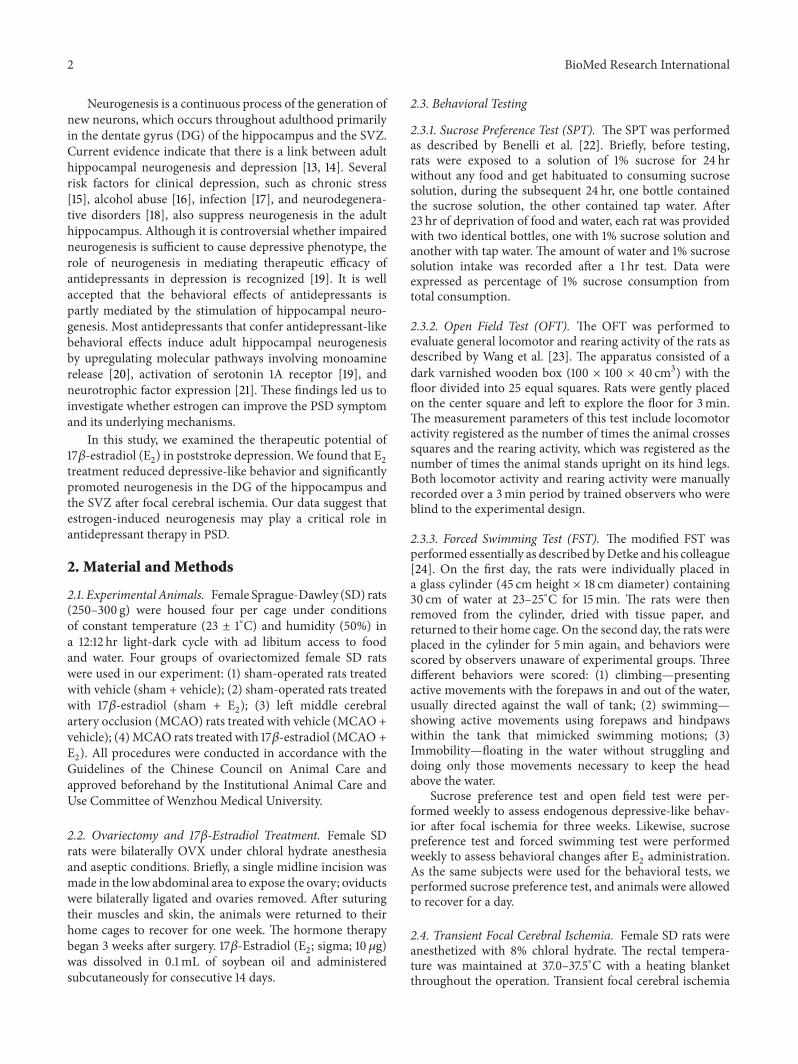
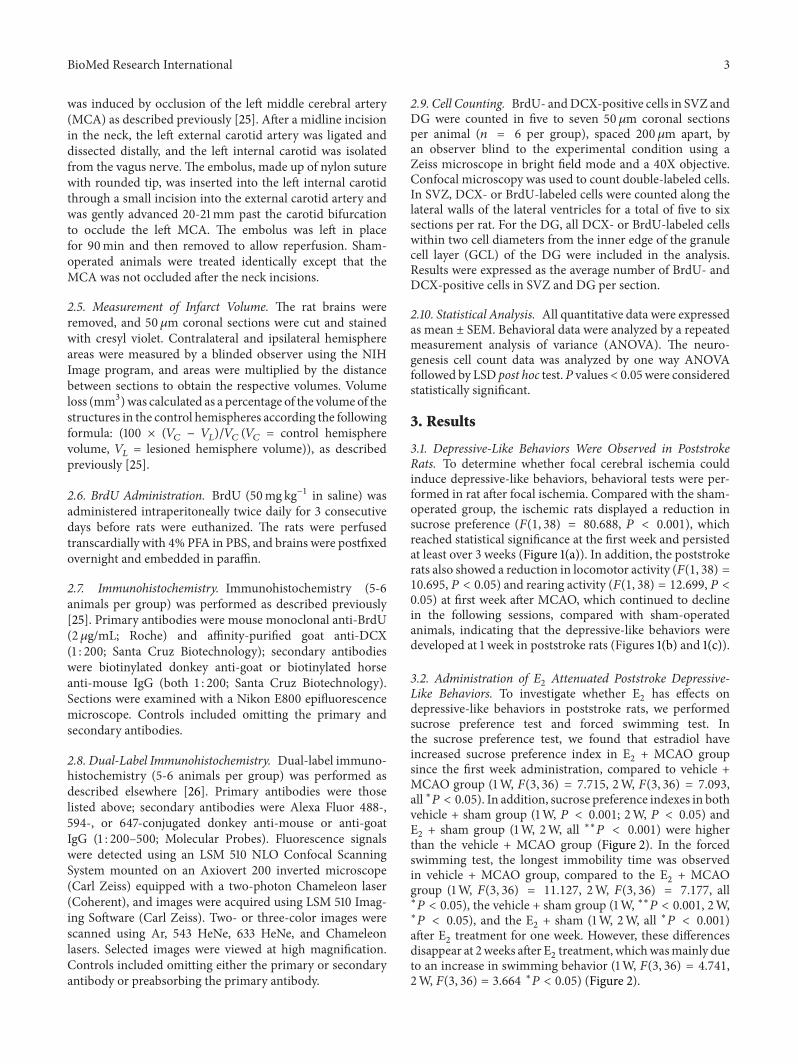
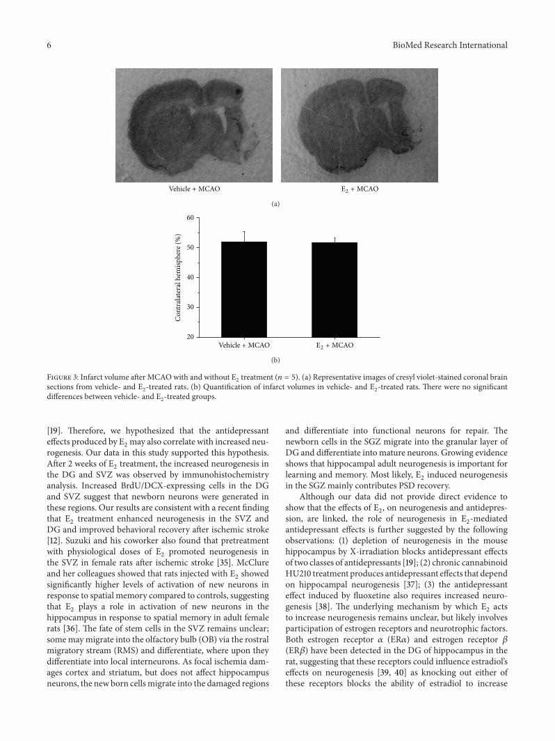
3.3. E2Treatment Did Not Affect Infarct Volumes after MCAO.
To investigate whether the ischemic infarct volumes could beattenuated by E
2administration, rats were sacrificed 2 weeks
after E2administration, and the brains were removed and
stained with cresyl violet. As shown in Figure 3, there wasno significant reduction in infarction volume of E
2-treated
ischemic rats, compared with vehicle-treated group.
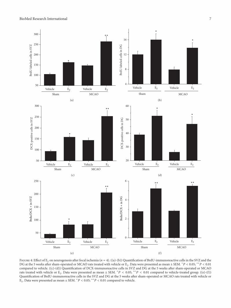
3.4. E2Increased Neurogenesis after Ischemic Stroke. To deter-
mine whether E2administration could enhance neurogenesis
in the SVZ and DG of ischemic brain, rats were treatedfor 3 days with BrdU, which labels cells that undergo DNAreplication in S-phase and therefore reflects the current rate ofcell division. As shown in Figure 4, BrdU- and DCX-positivecells in the SVZ and DG were significantly increased inE2-treated rats compared with control animals (∗𝑃 < 0.05).
An increase of BrdU- and DCX-positive cells in the SVZ wasalso observed in E
2+ MCAO group, compared to vehicle +
sham group. Interestingly, BrdU- and DCX-positive cells inthe DG were decreased after focal ischemia, compared tothe sham-operated rats (∗𝑃 < 0.05), which was reversedafter E
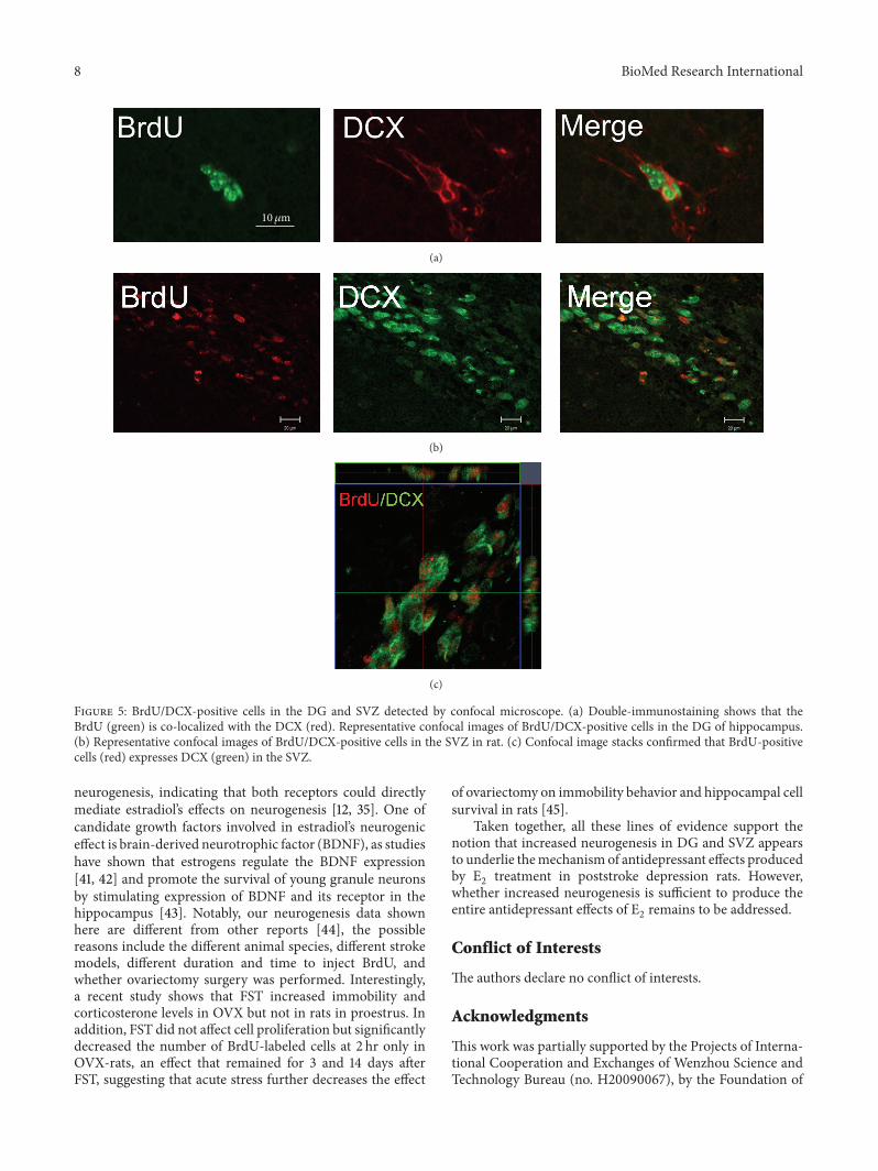
2administration (∗𝑃 < 0.05). Confocal images show
that BrdU-positive cells expressedDCX, suggesting that theseBrdU-positive cells were proliferative neuronal progenitorcells, and double-labeled cells in E
2-treated group were
significantly increased compared to vehicle-treated groupafter ischemia (∗∗𝑃 < 0.001) (Figure 5).
4. Discussion
In the present study, we developed a rat model of PSDusing left MCAO and found that E
2-treated PSD rats showed

BioMed Research International 5
30
45
60
75
90
21
Sucr
ose p
refe
renc
e ind
ex (%
) ∗
∗
∗∗∗
Time after E2 administration (weeks)
(a)
0
25
50
75
100
Clim
bing
(s)
21
∗
Time after E2 administration (weeks)
(b)
100
150
200
250
Swim
min
g (s
)
21
Vehicle + shamE2 + shamVehicle + MCAOE2 + MCAO
∗ ∗ ∗ ∗
Time after E2 administration (weeks)
(c)
15
30
45
60
75
*
Imm
obili
ty (s
)
21
Vehicle + shamE2 + shamVehicle + MCAOE2 + MCAO
∗
∗∗∗
∗∗
Time after E2 administration (weeks)
(d)
Figure 2: E2administration after ischemic stroke reverses depressive-like behavior (𝑛 = 10). (a) E
2-treated animals showed increased
percentage of sucrose consumption at 1 week and 2 weeks after E2administration, compared to vehicle-treated animals. (b) E
2-treated
animals showed no significant differences to vehicle-treated on climbing ability. E2-treated rats showed increased swimming behavior
(c) and decreased immobility (d) after focal ischemia, compared to vehicle-treated ischemic animals. Data were presented as mean ± SEM.∗𝑃 < 0.05, ∗∗𝑃 < 0.001.
significant improvement in their behavioral performance,as measured by the sucrose preference test and the forcedswimming test, suggesting that administration of E
2induces
antidepressant like effect in PSD rats. In addition, our resultsalso showed that the E
2-mediated depressive-like behav-
ioral improvements were concomitant with a significantlyincreased neurogenesis in the DG after focal ischemia,suggesting that neurogenesis may play a critical role in E
2-
mediated antidepressant effect after focal ischemia.Clinical evidence show that the susceptibility to develop
depression in women increases when the estrogen levels fluc-tuate during their life [27, 28]. Estrogen replacement therapyin these women may reduce depressive symptoms during thepremenopausal and postpartum periods [29]. E
2also induces
antidepressant like effects in animal models of depression[30–32]. Furthermore, it has been suggested that E
2can
enhance and shorten the antidepressant-like action of variousantidepressants, when combined with these antidepressants[33, 34]. However, whether E
2produces antidepressant effect
in animal model of poststroke depression remains unknown.Here, we applied two behavioral tests, including the sucrosepreference test and the force swimming test, which havebeen developed as straightforward tests for screening theefficacy of antidepressants, to investigate whether E
2induces
antidepressant effect.We observed that E2treatment reversed
depressive-like behavior in PSD rats, by increasing sucroseconsumption in the sucrose preference test, decreasing theimmobility time and increasing swimming time in theforce swim test, which is consistent with the suggestedantidepressant-like effect of E
2.
Previous studies have shown that neurogenesis plays acritical role in the antidepressant-mediated behavioral effects
6 BioMed Research International
Vehicle + MCAO E2 + MCAO
(a)
20
30
40
50
60
Con
tral
ater
al h
emisp
here
(%)
Vehicle + MCAO E2 + MCAO
(b)
Figure 3: Infarct volume after MCAOwith and without E2treatment (𝑛 = 5). (a) Representative images of cresyl violet-stained coronal brain
sections from vehicle- and E2-treated rats. (b) Quantification of infarct volumes in vehicle- and E
2-treated rats. There were no significant
differences between vehicle- and E2-treated groups.
[19]. Therefore, we hypothesized that the antidepressanteffects produced by E
2may also correlate with increased neu-
rogenesis. Our data in this study supported this hypothesis.After 2 weeks of E
2treatment, the increased neurogenesis in
the DG and SVZ was observed by immunohistochemistryanalysis. Increased BrdU/DCX-expressing cells in the DGand SVZ suggest that newborn neurons were generated inthese regions. Our results are consistent with a recent findingthat E
2treatment enhanced neurogenesis in the SVZ and
DG and improved behavioral recovery after ischemic stroke[12]. Suzuki and his coworker also found that pretreatmentwith physiological doses of E
2promoted neurogenesis in
the SVZ in female rats after ischemic stroke [35]. McClureand her colleagues showed that rats injected with E
2showed
significantly higher levels of activation of new neurons inresponse to spatial memory compared to controls, suggestingthat E
2plays a role in activation of new neurons in the
hippocampus in response to spatial memory in adult femalerats [36]. The fate of stem cells in the SVZ remains unclear;somemaymigrate into the olfactory bulb (OB) via the rostralmigratory stream (RMS) and differentiate, where upon theydifferentiate into local interneurons. As focal ischemia dam-ages cortex and striatum, but does not affect hippocampusneurons, the new born cellsmigrate into the damaged regions
and differentiate into functional neurons for repair. Thenewborn cells in the SGZ migrate into the granular layer ofDG and differentiate into mature neurons. Growing evidenceshows that hippocampal adult neurogenesis is important forlearning and memory. Most likely, E
2induced neurogenesis
in the SGZ mainly contributes PSD recovery.Although our data did not provide direct evidence to
show that the effects of E2, on neurogenesis and antidepres-
sion, are linked, the role of neurogenesis in E2-mediated
antidepressant effects is further suggested by the followingobservations: (1) depletion of neurogenesis in the mousehippocampus by X-irradiation blocks antidepressant effectsof two classes of antidepressants [19]; (2) chronic cannabinoidHU210 treatment produces antidepressant effects that dependon hippocampal neurogenesis [37]; (3) the antidepressanteffect induced by fluoxetine also requires increased neuro-genesis [38]. The underlying mechanism by which E
2acts
to increase neurogenesis remains unclear, but likely involvesparticipation of estrogen receptors and neurotrophic factors.Both estrogen receptor 𝛼 (ER𝛼) and estrogen receptor 𝛽(ER𝛽) have been detected in the DG of hippocampus in therat, suggesting that these receptors could influence estradiol’seffects on neurogenesis [39, 40] as knocking out either ofthese receptors blocks the ability of estradiol to increase
BioMed Research International 7
50
100
150
200
250
300
VehicleVehicle
BrdU
-labe
led
cells
in S
VZ
Sham MCAO
E2 E2
∗
∗∗
(a)
4
8
12
16
BrdU
-labe
led
cells
in D
G
VehicleVehicle
Sham MCAO
E2 E2
∗
∗
(b)
50
100
150
200
250
300
DCX
-pos
itive
cells
in S
VZ
VehicleVehicleSham MCAO
E2 E2
∗
∗∗
(c)
20
30
40
50
60
DCX
-pos
itive
cells
in D
G
Vehicle VehicleSham MCAO
E2 E2
∗
∗
(d)
50
100
150
200
250
VehicleVehicle
Sham MCAO
E2 E2
Brdu
/DCX
+in
SV
Z
∗
∗∗
(e)
0
2
4
6
VehicleVehicle
Sham MCAO
E2 E2
Brdu
/DCX
+in
DG
∗∗ ∗∗
(f)
Figure 4: Effect of E2on neurogenesis after focal ischemia (𝑛 = 4). ((a)-(b)) Quantification of BrdU-immunoreactive cells in the SVZ and the
DG at the 5 weeks after sham-operated or MCAO rats treated with vehicle or E2. Data were presented as mean ± SEM. ∗𝑃 < 0.05; ∗∗𝑃 < 0.01
compared to vehicle. ((c)-(d)) Quantification of DCX-immunoreactive cells in SVZ and DG at the 5 weeks after sham-operated or MCAOrats treated with vehicle or E
2. Data were presented as mean ± SEM. ∗𝑃 < 0.05; ∗∗𝑃 < 0.01 compared to vehicle-treated group. ((e)-(f))
Quantification of BrdU-immunoreactive cells in the SVZ and DG at the 5 weeks after sham-operated or MCAO rats treated with vehicle orE2. Data were presented as mean ± SEM. ∗𝑃 < 0.05; ∗∗𝑃 < 0.01 compared to vehicle.
8 BioMed Research International
10𝜇m
(a)
(b)
(c)
Figure 5: BrdU/DCX-positive cells in the DG and SVZ detected by confocal microscope. (a) Double-immunostaining shows that theBrdU (green) is co-localized with the DCX (red). Representative confocal images of BrdU/DCX-positive cells in the DG of hippocampus.(b) Representative confocal images of BrdU/DCX-positive cells in the SVZ in rat. (c) Confocal image stacks confirmed that BrdU-positivecells (red) expresses DCX (green) in the SVZ.
neurogenesis, indicating that both receptors could directlymediate estradiol’s effects on neurogenesis [12, 35]. One ofcandidate growth factors involved in estradiol’s neurogeniceffect is brain-derived neurotrophic factor (BDNF), as studieshave shown that estrogens regulate the BDNF expression[41, 42] and promote the survival of young granule neuronsby stimulating expression of BDNF and its receptor in thehippocampus [43]. Notably, our neurogenesis data shownhere are different from other reports [44], the possiblereasons include the different animal species, different strokemodels, different duration and time to inject BrdU, andwhether ovariectomy surgery was performed. Interestingly,a recent study shows that FST increased immobility andcorticosterone levels in OVX but not in rats in proestrus. Inaddition, FST did not affect cell proliferation but significantlydecreased the number of BrdU-labeled cells at 2 hr only inOVX-rats, an effect that remained for 3 and 14 days afterFST, suggesting that acute stress further decreases the effect
of ovariectomy on immobility behavior and hippocampal cellsurvival in rats [45].
Taken together, all these lines of evidence support thenotion that increased neurogenesis in DG and SVZ appearsto underlie themechanism of antidepressant effects producedby E2treatment in poststroke depression rats. However,
whether increased neurogenesis is sufficient to produce theentire antidepressant effects of E
2remains to be addressed.
Conflict of Interests
The authors declare no conflict of interests.
Acknowledgments
This work was partially supported by the Projects of Interna-tional Cooperation and Exchanges of Wenzhou Science andTechnology Bureau (no. H20090067), by the Foundation of
BioMed Research International 9
Zhejiang Provincial TopKeyDiscipline of Surgery, and by theNational Natural Science Foundation of China (81171088 and81371395).
References
[1] M. L. Hackett, C. Yapa, V. Parag, and C. S. Anderson, “Frequen-cy of depression after stroke: a systematic review of observa-tional studies,” Stroke, vol. 36, no. 6, pp. 1330–1340, 2005.
[2] I. Loubinoux, G. Kronenberg, M. Endres et al., “Post-strokedepression: mechanisms, translation and therapy,” Journal ofCellular and Molecular Medicine, vol. 16, pp. 1961–1969, 2012.
[3] R. G. Robinson, “Poststroke depression: prevalence, diagnosis,treatment, and disease progression,” Biological Psychiatry, vol.54, no. 3, pp. 376–387, 2003.
[4] A. Selvamani and F. Sohrabji, “Reproductive age modulates theimpact of focal ischemia on the forebrain as well as the effectsof estrogen treatment in female rats,”Neurobiology of Aging, vol.31, no. 9, pp. 1618–1628, 2010.
[5] X. Li, K.K. Blizzard, Z. Zeng,A.C.DeVries, P.D.Hurn, andL.D.McCullough, “Chronic behavioral testing after focal ischemiain the mouse: functional recovery and the effects of gender,”Experimental Neurology, vol. 187, no. 1, pp. 94–104, 2004.
[6] L. M. Garcia-Segura, I. Azcoitia, and L. L. DonCarlos, “Neuro-protection by estradiol,” Progress in Neurobiology, vol. 63, no. 1,pp. 29–60, 2001.
[7] S. Suzuki, C.M. Brown, andP.M.Wise, “Neuroprotective effectsof estrogens following ischemic stroke,” Frontiers in Neuroendo-crinology, vol. 30, no. 2, pp. 201–211, 2009.
[8] E. L. Moses-Kolko, S. L. Berga, B. Kalro, D. K. Y. Sit, and K.L. Wisner, “Transdermal estradiol for postpartum depression:a promising treatment option,” Clinical Obstetrics and Gynecol-ogy, vol. 52, no. 3, pp. 516–529, 2009.
[9] C. De Novaes Soares, O. P. Almeida, H. Joffe, and L. S. Cohen,“Efficacy of estradiol for the treatment of depressive disorders inperimenopausal women: a double-blind, randomized, placebo-controlled trial,” Archives of General Psychiatry, vol. 58, no. 6,pp. 529–534, 2001.
[10] J. M. Barker and L. A. M. Galea, “Repeated estradiol adminis-tration alters different aspects of neurogenesis and cell death inthe hippocampus of female, but not male, rats,” Neuroscience,vol. 152, no. 4, pp. 888–902, 2008.
[11] C. Westenbroek, J. A. Den Boer, M. Veenhuis, and G. J. TerHorst, “Chronic stress and social housing differentially affectneurogenesis in male and female rats,” Brain Research Bulletin,vol. 64, no. 4, pp. 303–308, 2004.
[12] J. Li, M. Siegel, M. Yuan et al., “Estrogen enhances neurogenesisand behavioral recovery after stroke,” Journal of Cerebral BloodFlow & Metabolism, vol. 31, no. 2, pp. 413–425, 2011.
[13] R. M. Thomas, G. Hotsenpiller, and D. A. Peterson, “Acutepsychosocial stress reduces cell survival in adult hippocampalneurogenesis without altering proliferation,” The Journal ofNeuroscience, vol. 27, no. 11, pp. 2734–2743, 2007.
[14] A. Dranovsky and R. Hen, “Hippocampal neurogenesis: regu-lation by stress and antidepressants,” Biological Psychiatry, vol.59, no. 12, pp. 1136–1143, 2006.
[15] K. Pham, J. Nacher, P. R. Hof, and B. S. McEwen, “Repeatedrestraint stress suppresses neurogenesis and induces biphasicPSA-NCAM expression in the adult rat dentate gyrus,” Euro-pean Journal of Neuroscience, vol. 17, no. 4, pp. 879–886, 2003.
[16] K. Nixon and F. T. Crews, “Temporally specific burst in cellproliferation increases hippocampal neurogenesis in protractedabstinence from alcohol,” The Journal of Neuroscience, vol. 24,no. 43, pp. 9714–9722, 2004.
[17] Z. Guan and J. Fang, “Peripheral immune activation by lipopol-ysaccharide decreases neurotrophins in the cortex and hippo-campus in rats,” Brain, Behavior, and Immunity, vol. 20, no. 1,pp. 64–71, 2006.
[18] C. Zhang, E. McNeil, L. Dressler, and R. Siman, “Long-last-ing impairment in hippocampal neurogenesis associated withamyloid deposition in a knock-in mouse model of familialAlzheimer’s disease,”Experimental Neurology, vol. 204, no. 1, pp.77–87, 2007.
[19] L. Santarelli, M. Saxe, C. Gross et al., “Requirement of hip-pocampal neurogenesis for the behavioral effects of antidepres-sants,” Science, vol. 301, no. 5634, pp. 805–809, 2003.
[20] J. M. Brezun and A. Daszuta, “Serotonin may stimulate granulecell proliferation in the adult hippocampus, as observed inrats grafted with foetal raphe neurons,” European Journal ofNeuroscience, vol. 12, no. 1, pp. 391–396, 2000.
[21] M. Sairanen, G. Lucas, P. Ernfors, M. Castren, and E. Castren,“Brain-derived neurotrophic factor and antidepressant drugshave different but coordinated effects on neuronal turnover,proliferation, and survival in the adult dentate gyrus,” TheJournal of Neuroscience, vol. 25, no. 5, pp. 1089–1094, 2005.
[22] A. Benelli, M. Filaferro, A. Bertolini, and S. Genedani, “Influ-ence of S-adenosyl-L-methionine on chronic mild stress-induced anhedonia in castrated rats,” British Journal of Pharma-cology, vol. 127, no. 3, pp. 645–654, 1999.
[23] S. H.Wang, Z. J. Zhang, Y. J. Guo, H. Zhou, G. J. Teng, and B. A.Chen, “Anhedonia and activity deficits in rats: impact of post-stroke depression,” Journal of Psychopharmacology, vol. 23, no.3, pp. 295–304, 2009.
[24] M. J. Detke, M. Rickels, and I. Lucki, “Active behaviors in the ratforced swimming test differentially produced by serotonergicand noradrenergic antidepressants,” Psychopharmacology, vol.121, no. 1, pp. 66–72, 1995.
[25] R. A. Swanson, M. T. Morton, G. Tsao-Wu, R. A. Savalos,C. Davidson, and F. R. Sharp, “A semiautomated method formeasuring brain infarct volume,” Journal of Cerebral Blood Flow& Metabolism, vol. 10, no. 2, pp. 290–293, 1990.
[26] K. Jin, M. Minami, J. Q. Lan et al., “Neurogenesis in dentatesubgranular zone and rostral subventricular zone after focalcerebral ischemia in the rat,” Proceedings of the NationalAcademy of Sciences of the United States of America, vol. 98, no.8, pp. 4710–4715, 2001.
[27] V. Hendrick, L. L. Altshuler, and R. Suri, “Hormonal changes inthe postpartum and implications for postpartum depression,”Psychosomatics, vol. 39, no. 2, pp. 93–101, 1998.
[28] L. S. Cohen, C. N. Soares, A. F. Vitonis, M. W. Otto, and B.L. Harlow, “Risk for new onset of depression during the men-opausal transition: the harvard study of moods and cycles,”Archives of General Psychiatry, vol. 63, no. 4, pp. 385–390, 2006.
[29] L. S. Cohen, C. N. Soares, J. R. Poitras, J. Prouty, A. B. Alexander,and J. L. Shifren, “Short-term use of estradiol for depressionin perimenopausal and postmenopausal women: a preliminaryreport,”American Journal of Psychiatry, vol. 160, no. 8, pp. 1519–1522, 2003.
[30] E. Estrada-Camarena, A. Fernandez-Guasti, and C. Lopez-Rubalcava, “Antidepressant-like effect of different estrogeniccompounds in the forced swimming test,”Neuropsychopharma-cology, vol. 28, no. 5, pp. 830–838, 2003.
10 BioMed Research International
[31] A. A. Walf, J. J. Paris, and C. A. Frye, “Chronic estradiolreplacement to aged female rats reduces anxiety-like anddepression-like behavior and enhances cognitive performance,”Psychoneuroendocrinology, vol. 34, no. 6, pp. 909–916, 2009.
[32] M. Romano-Torres and A. Fernandez-Guasti, “Estradiol valer-ate elicits antidepressant-like effects in middle-aged female ratsunder chronic mild stress,” Behavioural Pharmacology, vol. 21,no. 2, pp. 104–111, 2010.
[33] E. Estrada-Camarena, A. Fernandez-Guasti, and C. Lopez-Rubalcava, “Interaction between estrogens and antidepressantsin the forced swimming test in rats,” Psychopharmacology, vol.173, no. 1, pp. 139–145, 2004.
[34] E. Estrada-Camarena, N. M. Vega Rivera, C. Berlanga, andA. Fernandez-Guasti, “Reduction in the latency of action ofantidepressants by 17 𝛽-estradiol in the forced swimming test,”Psychopharmacology, vol. 201, no. 3, pp. 351–360, 2008.
[35] S. Suzuki, L. M. Gerhold, M. Bottner et al., “Estradiol enhancesneurogenesis following ischemic stroke through estrogenreceptors 𝛼 and 𝛽,” Journal of Comparative Neurology, vol. 500,no. 6, pp. 1064–1075, 2007.
[36] R. E. McClure, C. K. Barha, and L. A. Galea, “17beta-Estradiol,but not estrone, increases the survival and activation of newneurons in the hippocampus in response to spatial memory inadult female rats,” Hormones and Behavior, vol. 63, pp. 144–157,2013.
[37] W. Jiang, Y. Zhang, L. Xiao et al., “Cannabinoids promoteembryonic and adult hippocampus neurogenesis and produceanxiolytic- and antidepressant-like effects,”The Journal of Clin-ical Investigation, vol. 115, no. 11, pp. 3104–3116, 2005.
[38] R. D. Airan, L. A. Meltzer, M. Roy, Y. Gong, H. Chen, andK. Deisseroth, “High-speed imaging reveals neurophysiologicallinks to behavior in an animalmodel of depression,” Science, vol.317, no. 5839, pp. 819–823, 2007.
[39] N. Weiland, C. Orikasa, S. Hayashi, and B. McEwen, “Distribu-tion and hormone regulation of estrogen receptor immunoreac-tive cells in the hippocampus of male and female rats,” Journalof Comparative Neurology, vol. 388, pp. 603–612, 1997.
[40] S. P. Herrick, E. M.Waters, C. T. Drake, B. S. McEwen, and T. A.Milner, “Extranuclear estrogen receptor beta immunoreactivityis on doublecortin-containing cells in the adult and neonatal ratdentate gyrus,” Brain Research, vol. 1121, no. 1, pp. 46–58, 2006.
[41] F. Sohrabji, R. C. G. Miranda, and C. D. Toran-Allerand, “Iden-tification of a putative estrogen response element in the geneencoding brain-derived neurotrophic factor,” Proceedings of theNational Academy of Sciences of the United States of America,vol. 92, no. 24, pp. 11110–11114, 1995.
[42] M. Singh, E. M. Meyer, and J. W. Simpkins, “The effect ofovariectomy and estradiol replacement on brain-derived neuro-trophic factor messenger ribonucleic acid expression in corticaland hippocampal brain regions of female Sprague-Dawley rats,”Endocrinology, vol. 136, no. 5, pp. 2320–2324, 1995.
[43] B. K. Ormerod, T. T.-Y. Lee, and L. A. M. Galea, “Estradiolenhances neurogenesis in the dentate gyri of adult malemeadow voles by increasing the survival of young granuleneurons,” Neuroscience, vol. 128, no. 3, pp. 645–654, 2004.
[44] K. Tureyen, R. Vemuganti, K. A. Sailor, K. K. Bowen, and R.J. Dempsey, “Transient focal cerebral ischemia-induced neuro-genesis in the dentate gyrus of the adult mouse,” Journal ofNeurosurgery, vol. 101, no. 5, pp. 799–805, 2004.
[45] N. M. Vega-Rivera, A. Fernandez-Guasti, G. Ramirez-Rodri-guez, and E. Estrada-Camarena, “Acute stress further decreases
the effect of ovariectomy on immobility behavior and hippo-campal cell survival in rats,” Psychoneuroendocrinology, vol. 38,pp. 1407–1417, 2013.

Submit your manuscripts athttp://www.hindawi.com
Stem CellsInternational
Hindawi Publishing Corporationhttp://www.hindawi.com Volume 2014
Hindawi Publishing Corporationhttp://www.hindawi.com Volume 2014
MEDIATORSINFLAMMATION
of
Hindawi Publishing Corporationhttp://www.hindawi.com Volume 2014
Behavioural Neurology
EndocrinologyInternational Journal of
Hindawi Publishing Corporationhttp://www.hindawi.com Volume 2014
Hindawi Publishing Corporationhttp://www.hindawi.com Volume 2014
Disease Markers
Hindawi Publishing Corporationhttp://www.hindawi.com Volume 2014
BioMed Research International
OncologyJournal of
Hindawi Publishing Corporationhttp://www.hindawi.com Volume 2014
Hindawi Publishing Corporationhttp://www.hindawi.com Volume 2014
Oxidative Medicine and Cellular Longevity
Hindawi Publishing Corporationhttp://www.hindawi.com Volume 2014
PPAR Research
The Scientific World JournalHindawi Publishing Corporation http://www.hindawi.com Volume 2014
Immunology ResearchHindawi Publishing Corporationhttp://www.hindawi.com Volume 2014
Journal of
ObesityJournal of
Hindawi Publishing Corporationhttp://www.hindawi.com Volume 2014
Hindawi Publishing Corporationhttp://www.hindawi.com Volume 2014
Computational and Mathematical Methods in Medicine
OphthalmologyJournal of
Hindawi Publishing Corporationhttp://www.hindawi.com Volume 2014
Diabetes ResearchJournal of
Hindawi Publishing Corporationhttp://www.hindawi.com Volume 2014
Hindawi Publishing Corporationhttp://www.hindawi.com Volume 2014
Research and TreatmentAIDS
Hindawi Publishing Corporationhttp://www.hindawi.com Volume 2014
Gastroenterology Research and Practice
Hindawi Publishing Corporationhttp://www.hindawi.com Volume 2014
Parkinson’s Disease
Evidence-Based Complementary and Alternative Medicine
Volume 2014Hindawi Publishing Corporationhttp://www.hindawi.com