Research Article Effect of Inhaling Cymbopogon martinii...
8
Research Article Effect of Inhaling Cymbopogon martinii Essential Oil and Geraniol on Serum Biochemistry Parameters and Oxidative Stress in Rats Bruna Fernanda Murbach Teles Andrade, 1 Camila Pereira Braga, 2 Klinsmann Carolo dos Santos, 2 Lidiane Nunes Barbosa, 1 Vera Lúcia Mores Rall, 1 José Maurício Sforcin, 1 Ana Angélica Henrique Fernandes, 2 and Ary Fernandes Júnior 1 1 Department of Microbiology and Immunology, Institute of Biosciences, UNESP, 18618-970 Botucatu, SP, Brazil 2 Department of Chemistry and Biochemistry, Institute of Biosciences, UNESP, 18618-970 Botucatu, SP, Brazil Correspondence should be addressed to Ary Fernandes J´ unior; [email protected] Received 13 October 2014; Accepted 23 November 2014; Published 9 December 2014 Academic Editor: Tzi Bun Ng Copyright © 2014 Bruna Fernanda Murbach Teles Andrade et al. is is an open access article distributed under the Creative Commons Attribution License, which permits unrestricted use, distribution, and reproduction in any medium, provided the original work is properly cited. e effects of the inhalation of Cymbopogon martinii essential oil (EO) and geraniol on Wistar rats were evaluated for biochemical parameters and hepatic oxidative stress. Wistar rats were divided into three groups ( = 8): G1 was control group, treated with saline solution; G2 received geraniol; and G3 received C. martinii EO by inhalation during 30 days. No significant differences were observed in glycemia and triacylglycerol levels; G2 and G3 decreased ( < 0.05) total cholesterol level. ere were no differences in serum protein, urea, aspartate aminotransferase activity, and total hepatic protein. Creatinine levels increased in G2 but decreased in G3. Alanine aminotransferase activity and lipid hydroperoxide were higher in G2 than in G3. Catalase and superoxide dismutase activities were higher in G3. C. martinii EO and geraniol increased glutathione peroxidase. Oxidative stress caused by geraniol may have triggered some degree of hepatic toxicity, as verified by the increase in serum creatinine and alanine aminotransferase. erefore, the beneficial effects of EO on oxidative stress can prevent the toxicity in the liver. is proves possible interactions between geraniol and numerous chemical compounds present in C. martinii EO. 1. Introduction Plants synthesize around 200,000 secondary metabolites or specialized phytochemicals, of which essential oils (EOs) constitute an important group [1]. ese compounds can be extracted from plant tissues (e.g., stem, leaves, flowers, and roots) by several procedures (e.g., hydrodistillation and steam distillation) [2]. ese compounds are mostly terpenes, which are commonly used in pharmaceutical industries and have therapeutic benefits and promote welfare, especially when used in aromatherapy procedures [3]. Cymbopogon martinii (Roxb.), Watson, popularly known as palmarosa, exhibits beneficial effects on several central nervous system pathologies, mainly neuralgia, epileptic, and anorexia [4]. ere are a few reports on its effects; still C. martinii has attracted many researchers’ attention due to its antimicrobial, antigenotoxic, and antioxidant activities [5– 8]. Countries such as India, Brazil and Madagascar have the practice to produce EOs from this plant. Geraniol, the major constituent of C. martinii EO, is an acyclic monoterpenoid that is abundant in many plants [9]. It may represent a new class of therapeutic agents against pancreatic [10] and colon cancers [11] and has several biologi- cal properties, including antimicrobial, antioxidant and anti- inflammatory activities [12]. Geraniol is also an important constituent of ginger, lemon, lime, lavender, nutmeg, orange and rose EOs [13]. Also, it is used as a flavoring agent and was determined to be safe at the current levels of intake by the Joint Expert Committee on Food Additives Hindawi Publishing Corporation Biochemistry Research International Volume 2014, Article ID 493183, 7 pages http://dx.doi.org/10.1155/2014/493183
Transcript of Research Article Effect of Inhaling Cymbopogon martinii...

Research ArticleEffect of Inhaling Cymbopogon martinii EssentialOil and Geraniol on Serum Biochemistry Parameters andOxidative Stress in Rats
Bruna Fernanda Murbach Teles Andrade,1 Camila Pereira Braga,2
Klinsmann Carolo dos Santos,2 Lidiane Nunes Barbosa,1
Vera Lúcia Mores Rall,1 José Maurício Sforcin,1
Ana Angélica Henrique Fernandes,2 and Ary Fernandes Júnior1
1Department of Microbiology and Immunology, Institute of Biosciences, UNESP, 18618-970 Botucatu, SP, Brazil2Department of Chemistry and Biochemistry, Institute of Biosciences, UNESP, 18618-970 Botucatu, SP, Brazil
Correspondence should be addressed to Ary Fernandes Junior; [email protected]
Received 13 October 2014; Accepted 23 November 2014; Published 9 December 2014
Academic Editor: Tzi Bun Ng
Copyright © 2014 Bruna Fernanda Murbach Teles Andrade et al. This is an open access article distributed under the CreativeCommons Attribution License, which permits unrestricted use, distribution, and reproduction in any medium, provided theoriginal work is properly cited.
The effects of the inhalation of Cymbopogon martinii essential oil (EO) and geraniol on Wistar rats were evaluated for biochemicalparameters and hepatic oxidative stress. Wistar rats were divided into three groups (𝑛 = 8): G1 was control group, treated withsaline solution; G2 received geraniol; and G3 received C. martinii EO by inhalation during 30 days. No significant differences wereobserved in glycemia and triacylglycerol levels; G2 and G3 decreased (𝑃 < 0.05) total cholesterol level.There were no differences inserum protein, urea, aspartate aminotransferase activity, and total hepatic protein. Creatinine levels increased in G2 but decreasedin G3. Alanine aminotransferase activity and lipid hydroperoxide were higher in G2 than in G3. Catalase and superoxide dismutaseactivities were higher in G3. C. martinii EO and geraniol increased glutathione peroxidase. Oxidative stress caused by geraniolmay have triggered some degree of hepatic toxicity, as verified by the increase in serum creatinine and alanine aminotransferase.Therefore, the beneficial effects of EO on oxidative stress can prevent the toxicity in the liver. This proves possible interactionsbetween geraniol and numerous chemical compounds present in C. martinii EO.
1. Introduction
Plants synthesize around 200,000 secondary metabolites orspecialized phytochemicals, of which essential oils (EOs)constitute an important group [1]. These compounds can beextracted from plant tissues (e.g., stem, leaves, flowers, androots) by several procedures (e.g., hydrodistillation and steamdistillation) [2].These compounds aremostly terpenes, whichare commonly used in pharmaceutical industries and havetherapeutic benefits and promote welfare, especially whenused in aromatherapy procedures [3].
Cymbopogon martinii (Roxb.), Watson, popularly knownas palmarosa, exhibits beneficial effects on several centralnervous system pathologies, mainly neuralgia, epileptic, andanorexia [4]. There are a few reports on its effects; still C.
martinii has attracted many researchers’ attention due to itsantimicrobial, antigenotoxic, and antioxidant activities [5–8]. Countries such as India, Brazil and Madagascar have thepractice to produce EOs from this plant.
Geraniol, the major constituent of C. martinii EO, is anacyclic monoterpenoid that is abundant in many plants [9].It may represent a new class of therapeutic agents againstpancreatic [10] and colon cancers [11] and has several biologi-cal properties, including antimicrobial, antioxidant and anti-inflammatory activities [12]. Geraniol is also an importantconstituent of ginger, lemon, lime, lavender, nutmeg, orangeand rose EOs [13]. Also, it is used as a flavoring agentand was determined to be safe at the current levels ofintake by the Joint Expert Committee on Food Additives
Hindawi Publishing CorporationBiochemistry Research InternationalVolume 2014, Article ID 493183, 7 pageshttp://dx.doi.org/10.1155/2014/493183
2 Biochemistry Research International
of Food and Agriculture Organization—FAO/World HealthOrganization—WHO [14].
Aromatherapy is a traditional treatment that uses EOs. Itseffects begin when the aromatic molecule passes through thenasal cavity and adheres to the olfactory epithelium, causingnerve stimulation directly to the hippocampus and limbicamygdaloidal body. This consequently triggers stimuli thatcontrol the autonomic nervous system and internal secretorycontrol by changing a number of vital reactions [15]. Theinhalation of aromatic compounds present in EOs is thereason for the name “aromatherapy” and this therapy mayhave sedating or stimulating effects on the individual [16].
Reports in the literature describe the benefits of using EOsin aromatherapy on the wellbeing of individuals, includingimprovements in mood, stress, anxiety, depression, andchronic pain, and promote so therapeutic, psychological, andphysiological effects [17]. The inhalation of EOs elevatedblood pressure and renal sympathetic activity, which enforcesthe idea that these components act in the central nervoussystem and pass through the blood-brain barrier [18].
Volatile organic compounds are highly lipophilic andmay easily cross the blood-brain barrier and easily exerttheir neuropharmacological and toxicological effects. Whilestudies on the toxic effects of these compounds are relativelyeasy to perform, the central effects induced by the perceptionof odor (e.g., in aromatherapy) are inherently complex. Thisis why the toxicological studies performed using volatilecompounds are much more advanced [19].
Many studies have been conducted in vitro with thepurpose of verifying the biological properties of EOs [2, 17,20]; usually they are performed using in vitro assays. Onthe other hand, when the tests are performed in vivo, theproducts are usually administered in their liquid forms (e.g.,by gavage or intraperitoneal), with few studies in the volatilestate (i.e., by inhalation) [21]. Furthermore, EOs are used asflavoring agents in food products [14] and are also used indermatology and in the fragrance and cosmetics industries.Specifically, geraniol is extensively used in the manufactureof both household and cosmetics products [12].
Since people are often use EOs, it is important to evaluatethe possible hepatotoxic effects of these oils. Liver is the maindetoxification organ; the catabolism of both endogenous andexogenous compounds takes place in the liver. As a resultit is exposure to toxic agents which can cause drug-inducedhepatic dysfunction. Therefore, studies on serum activity ofalanine aminotransferase (ALT) and aspartate aminotrans-ferase (AST), which are biomarkers of liver damage, areimportant. The serum activity of ALT and AST is frequentlyused in clinical settings for diagnostic hepatic toxicity [22].
Numerous chronic degenerative diseases are associatedwith oxidative stress, which occurs when there is excess for-mation of reactive oxygen species (e.g., superoxide, hydroxyl,and hydrogen peroxide) and insufficient defense by theantioxidant system (enzymatic and nonenzymatic). Thisimbalance between pro- and antioxidants may cause cellinjury and death, which consequently lead to tissue dysfunc-tion [23, 24]. It is well established that oxidative stress playsa fundamental role in the pathogenesis of hepatic disease,
especially nonalcoholic steatohepatitis [25]. In addition, dur-ing hepatic catabolism of xenobiotics, excessive productionof reactive oxygen species (ROS) occurs [26].
Our aimwas to investigate the effect of inhalation of theC.martinii EO and geraniol on serum biochemical parameters,biomarkers of hepatotoxicity, and oxidative stress in hepatictissue.
2. Materials and Methods
2.1. Cymbopogon martinii EO and Geraniol. C. martinii EOwas supplied by the company By Samia Aromaterapia (SaoPaulo, SP) and showed the following chemical composition:geraniol (57.5%), geranyl acetate (13.6%), linalool (1.7%),𝛽-caryophyllene (1.1%), and ocimene (0.3%) found by gaschromatography-mass spectrometer (GC-MS). The geraniolwith 98% of purity was purchased from Sigma Aldrich (St.Louis, MO, USA).
2.2. Animals and Experimental Procedure. The experimentalprocedure was approved by the Ethical Committee fromInstitute of Biological Science, Sao Paulo State University,Botucatu, Brazil, and the animals experiments were carriedout in accordance with the principles and guidelines of theCanadian Council on Animal Care as outlined in the Guideto the Care and Use of Experimental Animals.
Male Wistar rats (290–310 g) were reared in polypropy-lene cages maintained in a controlled environment (temper-ature 22 ± 3∘C; 50–55% humidity; and a 12-hour light : darkcycle), with free access to water and food (Purina Ltd.,Campinas, SP, Brazil).
The rats were randomly distributed into three groups (𝑛 =8). The rats in the control group (G1) received saline solutionby inhalation (saline = 0.9% g/v). The G2 group receivedgeraniol by inhalation and the G3 group received C. martiniiEO by inhalation.
The rats from all groups were placed individually intochambers (180mm × 300mm × 290mm) adapted from deAlmeida et al. [27] and submitted to inhalation of geraniol(8.36mg geraniol/L of air, which corresponds to 136.2𝜇Lof geraniol/perspex box 14.5 L of air) and C. martinii EO(13.73mg of C. martinii EO/L of air, which correspondsto 227𝜇L of C. martinii EO/perspex box 14.5 L of air)for 10 minutes every 48 hours for 30 days. The geraniolconcentration was calculated from the amount of geraniolfound in the C. martinii EO.
Food and water consumption were measured daily at thesame time and body weights were determined once a week.
2.3. Biochemical Measurements and Oxidative Stress. After30 days, the animals were fasted overnight (12–14 h) andeuthanized by cervical decapitation under anesthesia (solu-tion containing 10% ketamine chloride and 2% xylazinechloride with a dose of 0.1mL/100 g body weight). Bloodwas collected and the serum was obtained by centrifugationat 6000 rpm for 15 minutes. Serum glucose was determinedusing an enzymatic colorimetric method after incubationwith glucose oxidase/peroxidase.The total amount of protein
Biochemistry Research International 3
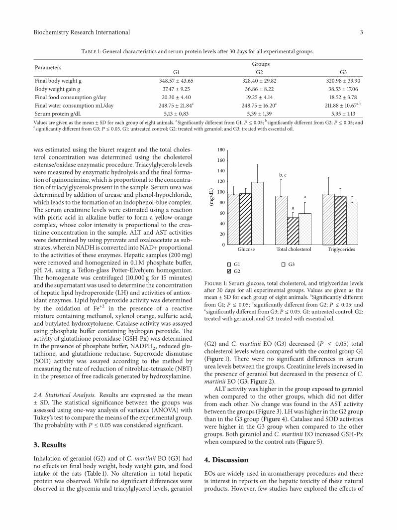
Table 1: General characteristics and serum protein levels after 30 days for all experimental groups.
Parameters GroupsG1 G2 G3
Final body weight g 348.57 ± 43.65 328.40 ± 29.82 320.98 ± 39.90Body weight gain g 37.47 ± 9.25 36.86 ± 8.22 38.53 ± 17.06Final food consumption g/day 20.30 ± 4.40 19.25 ± 4.14 18.52 ± 3.78Final water consumption mL/day 248.75 ± 21.84c 248.75 ± 16.20c 211.88 ± 10.67a,b
Serum protein g/dL 5,13 ± 0,83 5,39 ± 1,39 5,95 ± 1,13Values are given as the mean ± SD for each group of eight animals. aSignificantly different from G1; 𝑃 ≤ 0.05; bsignificantly different from G2; 𝑃 ≤ 0.05; andcsignificantly different from G3; 𝑃 ≤ 0.05. G1: untreated control; G2: treated with geraniol; and G3: treated with essential oil.
was estimated using the biuret reagent and the total choles-terol concentration was determined using the cholesterolesterase/oxidase enzymatic procedure. Triacylglycerols levelswere measured by enzymatic hydrolysis and the final forma-tion of quinoneimine, which is proportional to the concentra-tion of triacylglycerols present in the sample. Serum urea wasdetermined by addition of urease and phenol-hypochloride,which leads to the formation of an indophenol-blue complex.The serum creatinine levels were estimated using a reactionwith picric acid in alkaline buffer to form a yellow-orangecomplex, whose color intensity is proportional to the crea-tinine concentration in the sample. ALT and AST activitieswere determined by using pyruvate and oxaloacetate as sub-strates, whereinNADH is converted intoNAD+proportionalto the activities of these enzymes. Hepatic samples (200mg)were removed and homogenized in 0.1M phosphate buffer,pH 7.4, using a Teflon-glass Potter-Elvehjem homogenizer.The homogenate was centrifuged (10,000 g for 15 minutes)and the supernatant was used to determine the concentrationof hepatic lipid hydroperoxide (LH) and activities of antiox-idant enzymes. Lipid hydroperoxide activity was determinedby the oxidation of Fe+2 in the presence of a reactivemixture containing methanol, xylenol orange, sulfuric acid,and butylated hydroxytoluene. Catalase activity was assayedusing phosphate buffer containing hydrogen peroxide. Theactivity of glutathione peroxidase (GSH-Px) was determinedin the presence of phosphate buffer, NADPH
2, reduced glu-
tathione, and glutathione reductase. Superoxide dismutase(SOD) activity was assayed according to the method bymeasuring the rate of reduction of nitroblue-tetrazole (NBT)in the presence of free radicals generated by hydroxylamine.
2.4. Statistical Analysis. Results are expressed as the mean± SD. The statistical significance between the groups wasassessed using one-way analysis of variance (ANOVA) withTukey’s test to compare the means of the experimental group.The probability with 𝑃 ≤ 0.05 was considered significant.
3. Results
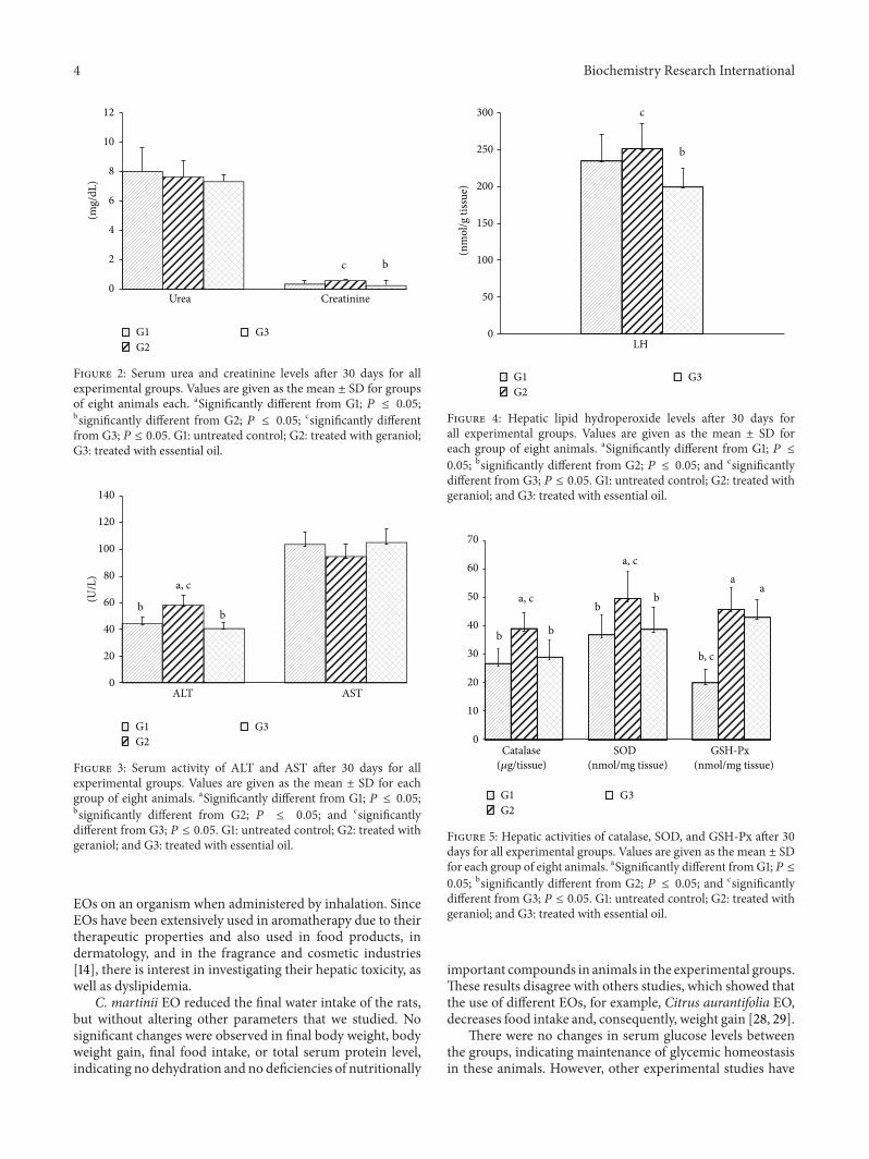
Inhalation of geraniol (G2) and of C. martinii EO (G3) hadno effects on final body weight, body weight gain, and foodintake of the rats (Table 1). No alteration in total hepaticprotein was observed. While no significant differences wereobserved in the glycemia and triacylglycerol levels, geraniol
0
20
40
60
80
100
120
140
160
180
(mg/
dL)
b, c
a
a
Glucose Total cholesterol Triglycerides
G1G2
G3
Figure 1: Serum glucose, total cholesterol, and triglycerides levelsafter 30 days for all experimental groups. Values are given as themean ± SD for each group of eight animals. aSignificantly differentfrom G1; 𝑃 ≤ 0.05; bsignificantly different from G2; 𝑃 ≤ 0.05; andcsignificantly different fromG3; 𝑃 ≤ 0.05. G1: untreated control; G2:treated with geraniol; and G3: treated with essential oil.
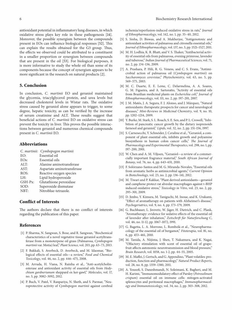
(G2) and C. martinii EO (G3) decreased (𝑃 ≤ 0.05) totalcholesterol levels when compared with the control group G1(Figure 1). There were no significant differences in serumurea levels between the groups. Creatinine levels increased inthe presence of geraniol but decreased in the presence of C.martinii EO (G3; Figure 2).
ALT activity was higher in the group exposed to geraniolwhen compared to the other groups, which did not differfrom each other. No change was found in the AST activitybetween the groups (Figure 3). LHwas higher in theG2 groupthan in the G3 group (Figure 4). Catalase and SOD activitieswere higher in the G3 group when compared to the othergroups. Both geraniol and C. martinii EO increased GSH-Pxwhen compared to the control rats (Figure 5).
4. Discussion
EOs are widely used in aromatherapy procedures and thereis interest in reports on the hepatic toxicity of these naturalproducts. However, few studies have explored the effects of
4 Biochemistry Research International
0
2
4
6
8
10
12
Urea Creatinine
c b
(mg/
dL)
G1G2
G3
Figure 2: Serum urea and creatinine levels after 30 days for allexperimental groups. Values are given as the mean ± SD for groupsof eight animals each. aSignificantly different from G1; 𝑃 ≤ 0.05;bsignificantly different from G2; 𝑃 ≤ 0.05; csignificantly differentfrom G3; 𝑃 ≤ 0.05. G1: untreated control; G2: treated with geraniol;G3: treated with essential oil.
0
20
40
60
80
100
120
140
(U/L
)
ALT AST
bb
a, c
G1G2
G3
Figure 3: Serum activity of ALT and AST after 30 days for allexperimental groups. Values are given as the mean ± SD for eachgroup of eight animals. aSignificantly different from G1; 𝑃 ≤ 0.05;bsignificantly different from G2; 𝑃 ≤ 0.05; and csignificantlydifferent from G3; 𝑃 ≤ 0.05. G1: untreated control; G2: treated withgeraniol; and G3: treated with essential oil.
EOs on an organism when administered by inhalation. SinceEOs have been extensively used in aromatherapy due to theirtherapeutic properties and also used in food products, indermatology, and in the fragrance and cosmetic industries[14], there is interest in investigating their hepatic toxicity, aswell as dyslipidemia.
C. martinii EO reduced the final water intake of the rats,but without altering other parameters that we studied. Nosignificant changes were observed in final body weight, bodyweight gain, final food intake, or total serum protein level,indicating no dehydration and no deficiencies of nutritionally
0
50
100
150
200
250
300
(nm
ol/g
tiss
ue)
LH
c
b
G1G2
G3
Figure 4: Hepatic lipid hydroperoxide levels after 30 days forall experimental groups. Values are given as the mean ± SD foreach group of eight animals. aSignificantly different from G1; 𝑃 ≤0.05; bsignificantly different from G2; 𝑃 ≤ 0.05; and csignificantlydifferent from G3; 𝑃 ≤ 0.05. G1: untreated control; G2: treated withgeraniol; and G3: treated with essential oil.
0
10
20
30
40
50
60
70
SOD(nmol/mg tissue) (nmol/mg tissue)
GSH-Px
b
a, c
b
b
a, c
b
b, c
aa
Catalase(𝜇g/tissue)
G1G2
G3
Figure 5: Hepatic activities of catalase, SOD, and GSH-Px after 30days for all experimental groups. Values are given as the mean ± SDfor each group of eight animals. aSignificantly different fromG1; 𝑃 ≤0.05; bsignificantly different from G2; 𝑃 ≤ 0.05; and csignificantlydifferent from G3; 𝑃 ≤ 0.05. G1: untreated control; G2: treated withgeraniol; and G3: treated with essential oil.
important compounds in animals in the experimental groups.These results disagree with others studies, which showed thatthe use of different EOs, for example, Citrus aurantifolia EO,decreases food intake and, consequently, weight gain [28, 29].
There were no changes in serum glucose levels betweenthe groups, indicating maintenance of glycemic homeostasisin these animals. However, other experimental studies have

Biochemistry Research International 5
demonstrated that C. martinii extracts exhibit antihyper-glycemic activities through inhibition of 𝛼-glucosidase underdiabetic conditions [30]. Assays with C. citratus aqueousextract (500mg/kg/day; via oral) showed that the mecha-nism by which the extract induced hypoglycemia could beattributed to increased insulin synthesis and secretion orincreased peripheral glucose utilization [31].
The inhalation of both geraniol (G2) andC. martinii EO(G3) reduced total cholesterol, but no changes in the serumtriacylglycerol concentration were observed. Our results arein agreement with the result of Adeneye and Agbaje [31] andBurke et al. [10], who observed hypocholesterolemic effectsusing an aqueous extract of Cymbopogon citratus by oraladministration. This reduction caused by EO can possibly beattributed to inhibition of 3-hydroxy-3-methylglutaryl CoAreductase, a key enzyme that regulates hepatic cholesterolsynthesis [32, 33], or by reduction in the expression of theseenzymes [34]. Yu et al. [35] demonstrated that geraniol inhib-ited the formation of mevalonate, a metabolic intermediatein the biosynthesis of cholesterol, in hepatomas. On the otherhand, the administration of the highest EO dose (100mg/kg)from Cymbopogon resulted in no change in total serumcholesterol [36].
Our results are also in agreement with Costa et al.[36] about total serum cholesterol; they reported that thebiochemical parameters did not change after treatment withCymbopogon but showed that there was a significant reduc-tion in it (F(4,27) = 3.061; 𝑃 ≤ 0.05) after the administrationof the highest EO dose (100mg/kg) by gavage over a periodof 21 days.
Since urea is formed in the liver and excreted by kidneys,estimation of this nitrogenous compound in the bloodstreamis important to estimate both hepatic and renal functions.Animals treated with geraniol and EO did not show alteredserum urea levels, suggesting a normal degree of proteincatabolism, which was confirmed by a normal concentrationof hepatic protein. Although there was no alteration in thelevels of serum urea in the G2 group, we cannot exclude theinvolvement of possible changes in the glomerular filtrationrate in these animals. In clinical practice, serum creatinine,a biomarker for renal failure, is used as an indicator of renalfunction [37]. Curiously, serum creatinine levels were higherin the G2 group and lower in the G3 group, suggestinga decrease in renal excretion and some degree of renalinsufficiency or early stages of kidney dysfunction when themajor compound of C. martinii EO was administrated alone.The animals treated with both products had a tendency tohave decreased levels of serum urea in our study. Serumcreatinine was higher in G2, which may indicate lowerrenal excretion since the creatinine production was relativelyconstant [38].
Plasmamembrane damage from some cells types, such ashepatic cells, is accompanied by release of cytosolic enzymesinto bloodstream, a phenomenon that always occurs underseveral pathophysiological conditions [39]. The aminotrans-ferases ALT and AST are used for diagnosis of hepatic injuryafter toxic agents exposure [40]. There was a significantincrease in serum ALT activity in animals that inhaledgeraniol (Figure 3). Since serum enzymatic activity of ALT is
often used as a biomarker of hepatic toxicity, we can assumethat there was some injury in hepatic tissue induced bygeraniol after 30 days.
ROS, such as superoxide anion (O2
−), hydroxyl rad-icals (OH−), and hydrogen peroxide (H
2O2), are formed
through mitochondrial respiration during normal cellularmetabolism. However, the cells have an enzymatic antioxi-dant defense system against ROS, but, under certain patho-logical conditions the excess formation of ROS results insuppression of antioxidant enzymes, the increase of ROS canoccur in this way leading to oxidative stress [41].
Lipid peroxidation is an important toxic event becauseinvolves the removal of hydrogen from fatty acid chainsmediated by ROS [42, 43] this way can lead to cell death andtissue damage.
The endogenous antioxidant enzyme includes superox-ide dismutase that catalyzes the dismutation of superoxideradicals [23]. Glutathione peroxidase catalyzes the reductionof hydrogen peroxidase to water through the oxidationof reduced glutathione. Catalase also participates in thisconversion [44].
Significantly high LH was observed in rats exposed togeraniol (G2), while the beneficial effect of C. martinii EOwas evidenced by the reduced LH in these animals. Thereductions observed in G3 can be attributed to a synergis-tic mechanism: a concomitant antioxidant action betweenother compounds, for example, linalool and𝛽-caryophyllene,present in theC.martinii EO that showed antioxidant activityin other researches [45, 46]. Since free radical scavengerability depends on the number of hydroxyl radicals in themolecule [43], the inhalation of the total C. martinii EOcontributed to the reduction in the formation of ROS.
Rats exposed to geraniol (G2) had higher catalase, SOD,and GSH-Px activities, indicating that antioxidant enzymeactivities were not sufficient to inhibit the ROS action and,consequently, the lipoperoxide generation in liver of theseanimals.
According to Koek et al. [47] the activity of antioxidantsenzymes is increased early in nonalcoholic steatohepatitisbut tends to decrease with progression of pathogenesis. Theactivity of the SOD and catalase did not change in theG3 group, while GSH-Px increased in these animals, whichshowed lower values for LH. Buch et al. [4] observed increasesin both SOD and catalase in the brains of rats treated withC. martini EO. Terpenoids, which are important componentsof EOs, lowered malondialdehyde levels and improved SODactivity in gastric mucosa [48]. Experimental data haveshown that terpenoids, which are main components of EOs,are responsible for their antioxidant action [49, 50].
Since lipid hydroperoxide has been widely studied asmarker of lipoperoxidation [51], a process that involvesremoval of hydrogen from fatty acids side chains by ROS,the result is referring to the mixture of compounds presentin C. martinii EO (G3) that was effective in controllingoxidative stress and, therefore, lipoperoxidation by reducingthe concentration of LH through a mechanism independentof the endogenous antioxidant enzymatic system.
In another study, geraniol reduced lipid peroxidationand inhibited the release of NO, indicating its possible
6 Biochemistry Research International
antioxidant potential in inflammatory lung diseases, in whichoxidative stress plays key role in these pathogenesis [14].Moreover, the possible synergism between the compoundspresent in EOs can influence biological responses [52]. Thiscan explain the results obtained for the G3 group. Thus,the effects we observed could be attributed to a constituentin a smaller proportion or synergism between compoundsthat are present in the oil [53]. For biological purposes, itis more informative to study the whole oil than some of itscomponents because the concept of synergism appears to bemore significant in the research on natural products [2].
5. Conclusion
In conclusion, C. martinii EO and geraniol maintainedthe glycemia, triacylglycerol protein, and urea levels butdecreased cholesterol levels in Wistar rats. The oxidativestress caused by geraniol alone appears to trigger, to somedegree, hepatic toxicity, as can be verified by the increaseof serum creatinine and ALT. These results suggest thatbeneficial actions of C. martinii EO on oxidative stress canprevent the toxicity in liver. This proves the possible interac-tions between geraniol and numerous chemical compoundspresent in C. martinii EO.
Abbreviations
C. martinii: Cymbopogon martiniiEO: Essential oilEOs: Essential oilsALT: Alanine aminotransferaseAST: Aspartate aminotransferaseROS: Reactive oxygen speciesLH: Lipid hydroperoxideGSH-Px: Glutathione peroxidaseSOD: Superoxide dismutaseNBT: Nitroblue-tetrazole.
Conflict of Interests
The authors declare that there is no conflict of interestsregarding the publication of this paper.
References
[1] P. Sharma, N. Sangwan, S. Bose, and R. Sangwan, “Biochemicalcharacteristics of a novel vegetative tissue geraniol acetyltrans-ferase from a monoterpene oil grass (Palmarosa, Cymbopogonmartinii var.Motia) leaf,” Plant Science, vol. 203, pp. 63–73, 2013.
[2] F. Bakkali, S. Averbeck, D. Averbeck, and M. Idaomar, “Bio-logical effects of essential oils—a review,” Food and ChemicalToxicology, vol. 46, no. 2, pp. 446–475, 2008.
[3] M. Arruda, H. Viana, N. Rainha et al., “Anti-acetylcholin-esterase and antioxidant activity of essential oils from Hedy-chium gardnerianum sheppard ex ker-gawl,” Molecules, vol. 17,no. 3, pp. 3082–3092, 2012.
[4] P. Buch, V. Patel, V. Ranpariya, N. Sheth, and S. Parmar, “Neu-roprotective activity of Cymbopogon martinii against cerebral
ischemia/reperfusion-induced oxidative stress in rats,” Journalof Ethnopharmacology, vol. 142, no. 1, pp. 35–40, 2012.
[5] S. Sinha, D. Biswas, and A. Mukherjee, “Antigenotoxic andantioxidant activities of palmarosa and citronella essential oils,”Journal of Ethnopharmacology, vol. 137, no. 3, pp. 1521–1527, 2011.
[6] M. H. Lodhia, K. R. Bhatt, and V. S.Thaker, “Antibacterial activ-ity of essential oils from palmarosa, evening primrose, lavenderand tuberose,” Indian Journal of Pharmaceutical Sciences, vol. 71,no. 2, pp. 134–136, 2009.
[7] A. Prashara, P. Hili, R. G. Veness, and C. S. Evans, “Antimi-crobial action of palmarosa oil (Cymbopogon martinii) onSaccharomyces cerevisiae,” Phytochemistry, vol. 63, no. 5, pp.569–575, 2003.
[8] M. C. Duarte, E. E. Leme, C. Delarmelina, A. A. Soares,G. M. Figueira, and A. Sartoratto, “Activity of essential oilsfrom Brazilian medicinal plants on Escherichia coli,” Journal ofEthnopharmacology, vol. 111, no. 2, pp. 197–201, 2007.
[9] J. M. Mates, J. A. Segura, F. J. Alonso, and J. Marquez, “Naturalantioxidants: therapeutic prospects for cancer and neurologicaldiseases,” Mini-Reviews in Medicinal Chemistry, vol. 9, no. 10,pp. 1202–1214, 2009.
[10] Y. Burke,M. Stark, S. L. Roach, S. E. Sen, andP. L. Crowell, “Inhi-bition of pancreatic cancer growth by the dietary isoprenoidsfarnesol and geraniol,” Lipids, vol. 32, no. 2, pp. 151–156, 1997.
[11] S. Carnesecchi, Y. Schneider, J. Ceraline et al., “Geraniol, a com-ponent of plant essential oils, inhibits growth and polyaminebiosynthesis in human colon cancer cells,” The Journal ofPharmacology and ExperimentalTherapeutics, vol. 298, no. 1, pp.197–200, 2001.
[12] W. Chen and A. M. Viljoen, “Geraniol—a review of a commer-cially important fragrance material,” South African Journal ofBotany, vol. 76, no. 4, pp. 643–651, 2010.
[13] F. Solorzano-Santos andM. G.Miranda-Novales, “Essential oilsfrom aromatic herbs as antimicrobial agents,” Current Opinionin Biotechnology, vol. 23, no. 2, pp. 136–141, 2012.
[14] M. Tiwari and P. Kakkar, “Plant derived antioxidants—geranioland camphene protect rat alveolar macrophages against t-BHPinduced oxidative stress,” Toxicology in Vitro, vol. 23, no. 2, pp.295–301, 2009.
[15] D. Jimbo, Y. Kimura, M. Taniguchi, M. Inoue, and K. Urakami,“Effect of aromatherapy on patients with Alzheimer’s disease,”Psychogeriatrics, vol. 9, no. 4, pp. 173–179, 2009.
[16] G. Buchbauer, L. Jirovetz, W. Jager, H. Dietrich, and C. Plank,“Aromatherapy: evidence for sedative effects of the essential oilof lavender after inhalation,” Zeitschrift fur Naturforschung C,vol. 46, no. 11-12, pp. 1067–1072, 1991.
[17] G. Bagetta, L. A. Morrone, L. Rombola et al., “Neuropharma-cology of the essential oil of bergamot,” Fitoterapia, vol. 81, no.6, pp. 453–461, 2010.
[18] M. Tanida, A. Niijima, J. Shen, T. Nakamura, and K. Nagai,“Olfactory stimulation with scent of essential oil of grape-fruit affects autonomic neurotransmission and blood pressure,”Brain Research, vol. 1058, no. 1-2, pp. 44–55, 2005.
[19] M. E.Maffei, J. Gertsch, andG.Appendino, “Plant volatiles: pro-duction, function and pharmacology,” Natural Product Reports,vol. 28, no. 8, pp. 1359–1380, 2011.
[20] A. Yousofi, S. Daneshmandi, N. Soleimani, K. Bagheri, and M.H. Karimi, “Immunomodulatory effect of Parsley (Petroselinumcrispum) essential oil on immune cells: mitogen-activatedsplenocytes and peritoneal macrophages,” Immunopharmacol-ogy and Immunotoxicology, vol. 34, no. 2, pp. 303–308, 2012.
Biochemistry Research International 7
[21] S. Inouye, T. Takizawa, and H. Yamaguchi, “Antibacterialactivity of essential oils and their major constituents againstrespiratory tract pathogens by gaseous contact,” Journal ofAntimicrobial Chemotherapy, vol. 47, no. 5, pp. 565–573, 2001.
[22] C. A. Burtis, E. R. Ashwood, and D. E. Bruns, Tietz: Fundamen-tos de Quımica Clınica, Elsevier Editora Ltda, 6th edition, 2008.
[23] J. S. Johansen, A. K.Harris, D. J. Rychly, andA. Ergul, “Oxidativestress and the use of antioxidants in diabetes: linking basicscience to clinical practice,” Cardiovascular Diabetology, vol. 4,article 5, 2005.
[24] J. M. Mates, J. A. Segura, F. J. Alonso, and J. Marquez,“Intracellular redox status and oxidative stress: implications forcell proliferation, apoptosis, and carcinogenesis,” Archives ofToxicology, vol. 82, no. 5, pp. 273–299, 2008.
[25] A.Wieckowska, A. J. McCullough, and A. E. Feldstein, “Nonin-vasive diagnosis and monitoring of nonalcoholic steatohepati-tis: present and future,” Hepatology, vol. 46, no. 2, pp. 582–589,2007.
[26] P. Muriel, “Role of free radicals in liver diseases,” HepatologyInternational, vol. 3, no. 4, pp. 526–536, 2009.
[27] R. N. de Almeida, S. C. Motta, C. D. B. Faturi, B. Catallani, andJ. R. Leite, “Anxiolytic-like effects of rose oil inhalation on theelevated plus-maze test in rats,” Pharmacology Biochemistry andBehavior, vol. 77, no. 2, pp. 361–364, 2004.
[28] A. Brenes and E. Roura, “Essential oils in poultry nutrition:main effects and modes of action,” Animal Feed Science andTechnology, vol. 158, no. 1-2, pp. 1–14, 2010.
[29] S. Asnaashari, A. Delazar, B. Habibi et al., “Essential oil fromCitrus aurantifolia prevents ketotifen-induced weight-gain inmice,”Phytotherapy Research, vol. 24, no. 12, pp. 1893–1897, 2010.
[30] V. Ghadyale, S. Takalikar, V. Haldavnekar, and A. Arvindekar,“Effective control of postprandial glucose level through inhi-bition of intestinal alpha glucosidase by Cymbopogon mar-tinii (Roxb.),” Evidence-Based Complementary and AlternativeMedicine, vol. 2012, Article ID 372909, 6 pages, 2012.
[31] A. A. Adeneye and E. O. Agbaje, “Hypoglycemic and hypolipi-demic effects of fresh leaf aqueous extract of Cymbopogoncitratus Stapf. in rats,” Journal of Ethnopharmacology, vol. 112,no. 3, pp. 440–444, 2007.
[32] P. L. Crowell, “Prevention and therapy of cancer by dietarymonoterpenes,” Journal of Nutrition, vol. 129, no. 3, pp. 775S–778S, 1999.
[33] P. Lu, M. L. Schrag, D. E. Slaughter, C. E. Raab, M. Shou,and A. D. Rodrigues, “Mechanism-based inhibition of humanliver microsomal cytochrome P450 1A2 by zileuton, A 5-lipoxygenase inhibitor,” Drug Metabolism and Disposition, vol.31, no. 11, pp. 1352–1360, 2003.
[34] S.-Y. Cho, H.-J. Jun, J. H. Lee, Y. Jia, K. H. Kim, and S.-J. Lee,“Linalool reduces the expression of 3-hydroxy-3-methylglutarylCoA reductase via sterol regulatory element binding protein-2-and ubiquitin-dependent mechanisms,” FEBS Letters, vol. 585,no. 20, pp. 3289–3296, 2011.
[35] S. G. Yu, L. A. Hildebrandt, and C. E. Elson, “Geraniol, aninhibitor of mevalonate biosynthesis, suppresses the growthof hepatomas and melanomas transplanted to rats and mice,”Journal of Nutrition, vol. 125, no. 11, pp. 2763–2767, 1995.
[36] C. Costa, L. T. Bidinotto, R. K. Takahira, D. M. F. Salvadori,L. F. Barbisan, and M. Costa, “Cholesterol reduction and lackof genotoxic or toxic effects in mice after repeated 21-day oralintake of lemongrass (Cymbopogon citratus) essential oil,” Foodand Chemical Toxicology, vol. 49, no. 9, pp. 2268–2272, 2011.
[37] R. D. Perrone, N. E. Madias, and A. S. Levey, “Serum creatinineas an index of renal function: new insights into old concepts,”Clinical Chemistry, vol. 38, no. 10, pp. 1933–1953, 1992.
[38] S. B. Heymsfield, C. Arteaga, C. M. McManus, J. Smith, and S.Moffitt, “Measurement of muscle mass in humans: validity ofthe 24-hour urinary creatinine method,”The American Journalof Clinical Nutrition, vol. 37, no. 3, pp. 478–494, 1983.
[39] R.-Z. Yang, S. Park, W. J. Reagan et al., “Alanine aminotrans-ferase isoenzymes: molecular cloning and quantitative analysisof tissue expression in rats and serum elevation in liver toxicity,”Hepatology, vol. 49, no. 2, pp. 598–607, 2009.
[40] J. Ozer, M. Ratner, M. Shaw, W. Bailey, and S. Schomaker, “Thecurrent state of serumbiomarkers of hepatotoxicity,”Toxicology,vol. 245, no. 3, pp. 194–205, 2008.
[41] B. Halliwell, “Antioxidants in human health and disease,”Annual Review of Nutrition, vol. 16, pp. 33–50, 1996.
[42] P. M. Abuja and R. Albertini, “Methods for monitoringoxidative stress, lipid peroxidation and oxidation resistance oflipoproteins,” Clinica Chimica Acta, vol. 306, no. 1-2, pp. 1–17,2001.
[43] L. A. Faine, H. G. Rodrigues, C. M. Galhardi et al., “Effects ofolive oil and its minor constituents on serum lipids, oxidativestress, and energy metabolism in cardiac muscle,” CanadianJournal of Physiology and Pharmacology, vol. 84, no. 2, pp. 239–245, 2006.
[44] J.-C. Preiser, “Oxidative stress,” Journal of Parenteral and EnteralNutrition, vol. 36, no. 2, pp. 147–154, 2012.
[45] S. Jana, K. Patra, S. Sarkar et al., “Antitumorigenic potential oflinalool is accompanied by modulation of oxidative stress: anin vivo study in sarcoma-180 solid tumor model,”Nutrition andCancer, vol. 66, pp. 835–848, 2014.
[46] M. A. Calleja, J. M. Vieites, T. Montero-Melendez et al., “Theantioxidant effect of beta-caryophyllene protects rat liver fromcarbon tetrachloride-induced fibrosis by inhibiting hepaticstellate cell activation,” British Journal of Nutrition, vol. 109, pp.394–401, 2013.
[47] G. H. Koek, P. R. Liedorp, and A. Bast, “The role of oxidativestress in non-alcoholic steatohepatitis,” Clinica Chimica Acta,vol. 412, no. 15-16, pp. 1297–1305, 2011.
[48] N. Rocha, G. de Oliveira, F. Y. de Araujo et al., “(-)-𝛼-Bisabolol-induced gastroprotection is associated with reduction in lipidperoxidation, superoxide dismutase activity and neutrophilmigration,” European Journal of Pharmaceutical Sciences, vol.44, no. 4, pp. 455–461, 2011.
[49] H. Fadel, F. Marx, A. El-Sawy, and A. El-Ghorab, “Effect ofextraction techniques on the chemical composition and antiox-idant activity of Eucalyptus camaldulensis var. brevirostris leafoils,”Zeitschrift fur Lebensmitteluntersuchung und -Forschung A,vol. 208, no. 3, pp. 212–216, 1999.
[50] J. Grassmann and G. Litwack, “Terpenoids as plant antioxi-dants,” Plant Hormones, vol. 72, pp. 505–535, 2005.
[51] K. Nageswari, R. Banerjee, andV. P.Menon, “Effect of saturated,𝜔-3 and 𝜔-6 polyunsaturated fatty acids on myocardial infarc-tion,”The Journal of Nutritional Biochemistry, vol. 10, no. 6, pp.338–344, 1999.
[52] J. Gershenzon and N. Dudareva, “The function of terpenenatural products in the natural world,”Nature Chemical Biology,vol. 3, no. 7, pp. 408–414, 2007.
[53] P. J. Houghton, M.-J. Howes, C. C. Lee, and G. Steventon, “Usesand abuses of in vitro tests in ethnopharmacology: visualizingan elephant,” Journal of Ethnopharmacology, vol. 110, no. 3, pp.391–400, 2007.

Submit your manuscripts athttp://www.hindawi.com
Hindawi Publishing Corporationhttp://www.hindawi.com Volume 2014
Anatomy Research International
PeptidesInternational Journal of
Hindawi Publishing Corporationhttp://www.hindawi.com Volume 2014
Hindawi Publishing Corporation http://www.hindawi.com
International Journal of
Volume 2014
Zoology
Hindawi Publishing Corporationhttp://www.hindawi.com Volume 2014
Molecular Biology International
GenomicsInternational Journal of
Hindawi Publishing Corporationhttp://www.hindawi.com Volume 2014
The Scientific World JournalHindawi Publishing Corporation http://www.hindawi.com Volume 2014
Hindawi Publishing Corporationhttp://www.hindawi.com Volume 2014
BioinformaticsAdvances in
Marine BiologyJournal of
Hindawi Publishing Corporationhttp://www.hindawi.com Volume 2014
Hindawi Publishing Corporationhttp://www.hindawi.com Volume 2014
Signal TransductionJournal of
Hindawi Publishing Corporationhttp://www.hindawi.com Volume 2014
BioMed Research International
Evolutionary BiologyInternational Journal of
Hindawi Publishing Corporationhttp://www.hindawi.com Volume 2014
Hindawi Publishing Corporationhttp://www.hindawi.com Volume 2014
Biochemistry Research International
ArchaeaHindawi Publishing Corporationhttp://www.hindawi.com Volume 2014
Hindawi Publishing Corporationhttp://www.hindawi.com Volume 2014
Genetics Research International
Hindawi Publishing Corporationhttp://www.hindawi.com Volume 2014
Advances in
Virolog y
Hindawi Publishing Corporationhttp://www.hindawi.com
Nucleic AcidsJournal of
Volume 2014
Stem CellsInternational
Hindawi Publishing Corporationhttp://www.hindawi.com Volume 2014
Hindawi Publishing Corporationhttp://www.hindawi.com Volume 2014
Enzyme Research
Hindawi Publishing Corporationhttp://www.hindawi.com Volume 2014
International Journal of
Microbiology